How could static telepathology improve diagnosis in...
7
177 How could static telepathology improve diagnosis in neuropathology? Myriam Remmelinck a , M. Beatriz S. Lopes b , Nathalie Nagy a , Sandrine Rorive a , Katja Rombaut a , Christine Decaestecker c,* , Robert Kiss c,** and Isabelle Salmon a,*** a Department of Pathology, Cliniques Universitaires de Bruxelles, Hôpital Erasme, Université Libre de Bruxelles, Brussels, Belgium b Division of Neuropathology, Department of Pathology, University of Virginia Health Sciences Center, Charlottesville, VA, USA c Laboratory of Histopathology, Faculty of Medicine, Université Libre de Bruxelles, Brussels, Belgium The present paper reports our experience with, and our opin- ion of static telepathology as applied to neuropathology by means of the PHAROS acquisition system and conventional telephone data transmission (modem). The classical proce- dure of expert consultation based on surface mailing of his- tological slides is routinely performed, especially in highly specialized fields of pathology. Telepathology is an easy means of sharing scientific expertise at international level and could thus improve diagnosis particularly in neuropathology, where certain tumor types are very rare and complex to di- agnose. Dynamic telepathology allows the referring patholo- gist to capture by himself images supporting their diagnosis. Using static telepathology the pathologist could be limited in diagnosis by problems in fields selection. We devoted a whole year to collecting all the technical parameters characterizing the use of digitized neuropatho- logical data files in order to investigate the feasibility of telepathology and the extent to which its use could improve diagnoses. Our results on a series of 38 histological brain ex- aminations illustrate how we successfully established an in- ternational connection between two departments of pathol- ogy in Belgium and the USA. The referring pathologists gave * This author is a Research Associate with the “Fonds National de la Recherche Scientifique” (FNRS), Belgium. ** This author is a Senior Research Associate with the “Fonds Na- tional de la Recherche Scientifique” (FNRS), Belgium. *** Corresponding author: Isabelle Salmon, M.D., Department of Pathology, Erasmus Hospital, Free University of Brussels, 808 route de Lennik, 1070 Brussels, Belgium. Tel.: +32 2 555 31 15; Fax: +32 2 555 47 90; E-mail: [email protected]. diagnoses in 35 cases and deferred only 3. Despite a time- consuming procedure for the telepathology session of a few cases, this tool provides easy access to expert diagnosis and real-time discussion, both of which are of considerable inter- est and offer significant improvements in neuropathology. Keywords: Static telepathology, neuropathology, data trans- mission, expert consultation 1. Introduction Communication and information, key words in our century, have played striking roles in recent develop- ments in pathology. In this context telepathology has been highly developed over the last 15 years, as indeed, is made clear by Kayser et al. in their well-documented book [11]. In fact, the various fields of activity covered by the terms are now so highly specialized that consensus dis- cussion of difficult cases is required to obtain expert diagnoses. In Erasmus hospital, where neurosurgery is particularly well developed, neuropathology, and es- pecially brain tumor classification, has become a very specialized subfield. In this field, an expert diagnostic panel has emerged as a very important means of in- creasing the accuracy of clinical diagnosis, especially in the field of pediatric tumors and stereotactic-guided biopsy. Since 1996 we have tried to replace the surface mail- ing of histological slides by static telepathology in the expert discussion of difficult cases. With the help of the Division of Neuropathology of the University of Vir- ginia Health Sciences Center, we decided to investi- gate the extent to which the use of telepathology could improve diagnosis. Furthermore, we devoted a whole year to collect all the technical parameters characteriz- ing the use of neuropathological files: the number and type of images per case, the time needed to set up a complete case file and the time of discussion and trans- mission. The present paper reports on these data, our experience, and our opinion of static telepathology as applied to neuropathology. Analytical Cellular Pathology 21 (2000) 177–182 ISSN 0921-8912 / $8.00 2000, IOS Press. All rights reserved
Transcript of How could static telepathology improve diagnosis in...

177
How could static telepathology improvediagnosis in neuropathology?
Myriam Remmelincka, M. Beatriz S. Lopesb,Nathalie Nagya, Sandrine Rorivea,Katja Rombauta, Christine Decaesteckerc,∗,Robert Kissc,∗∗ and Isabelle Salmona,∗∗∗
aDepartment of Pathology, Cliniques Universitairesde Bruxelles, Hôpital Erasme, Université Libre deBruxelles, Brussels, Belgiumb Division of Neuropathology, Department ofPathology, University of Virginia Health SciencesCenter, Charlottesville, VA, USAc Laboratory of Histopathology, Faculty of Medicine,Université Libre de Bruxelles, Brussels, Belgium
The present paper reports our experience with, and our opin-ion of static telepathology as applied to neuropathology bymeans of the PHAROS acquisition system and conventionaltelephone data transmission (modem). The classical proce-dure of expert consultation based on surface mailing of his-tological slides is routinely performed, especially in highlyspecialized fields of pathology. Telepathology is an easymeans of sharing scientific expertise at international level andcould thus improve diagnosis particularly in neuropathology,where certain tumor types are very rare and complex to di-agnose. Dynamic telepathology allows the referring patholo-gist to capture by himself images supporting their diagnosis.Using static telepathology the pathologist could be limited indiagnosis by problems in fields selection.
We devoted a whole year to collecting all the technicalparameters characterizing the use of digitized neuropatho-logical data files in order to investigate the feasibility oftelepathology and the extent to which its use could improvediagnoses. Our results on a series of 38 histological brain ex-aminations illustrate how we successfully established an in-ternational connection between two departments of pathol-ogy in Belgium and the USA. The referring pathologists gave
* This author is a Research Associate with the “Fonds National dela Recherche Scientifique” (FNRS), Belgium.
** This author is a Senior Research Associate with the “Fonds Na-tional de la Recherche Scientifique” (FNRS), Belgium.
*** Corresponding author: Isabelle Salmon, M.D., Department ofPathology, Erasmus Hospital, Free University of Brussels, 808 routede Lennik, 1070 Brussels, Belgium. Tel.: +32 2 555 31 15; Fax: +322 555 47 90; E-mail: [email protected].
diagnoses in 35 cases and deferred only 3. Despite a time-consuming procedure for the telepathology session of a fewcases, this tool provides easy access to expert diagnosis andreal-time discussion, both of which are of considerable inter-est and offer significant improvements in neuropathology.
Keywords: Static telepathology, neuropathology, data trans-mission, expert consultation
1. Introduction
Communication and information, key words in ourcentury, have played striking roles in recent develop-ments in pathology. In this context telepathology hasbeen highly developed over the last 15 years, as indeed,is made clear by Kayser et al. in their well-documentedbook [11].
In fact, the various fields of activity covered by theterms are now so highly specialized that consensus dis-cussion of difficult cases is required to obtain expertdiagnoses. In Erasmus hospital, where neurosurgery isparticularly well developed, neuropathology, and es-pecially brain tumor classification, has become a veryspecialized subfield. In this field, an expert diagnosticpanel has emerged as a very important means of in-creasing the accuracy of clinical diagnosis, especiallyin the field of pediatric tumors and stereotactic-guidedbiopsy.
Since 1996 we have tried to replace the surface mail-ing of histological slides by static telepathology in theexpert discussion of difficult cases. With the help of theDivision of Neuropathology of the University of Vir-ginia Health Sciences Center, we decided to investi-gate the extent to which the use of telepathology couldimprove diagnosis. Furthermore, we devoted a wholeyear to collect all the technical parameters characteriz-ing the use of neuropathological files: the number andtype of images per case, the time needed to set up acomplete case file and the time of discussion and trans-mission. The present paper reports on these data, ourexperience, and our opinion of static telepathology asapplied to neuropathology.
Analytical Cellular Pathology 21 (2000) 177–182ISSN 0921-8912 / $8.00 2000, IOS Press. All rights reserved

178 M. Remmelinck et al. / How could static telepathology improve diagnosis in neuropathology?
2. Materials and methods
In order to evaluate the real impact of telepathol-ogy in routine neuropathological diagnosis we under-took analysis of 38 neuropathological selected surgi-cal cases, raising difficulties in obtaining firm diagno-sis, with a view to the different technical parametersof acquisition and transmission of images, and also theability to perform diagnoses on transmitted images.
2.1. Technology used
The PHAROS system (version 1.52 VAMS, Zagreb,Croatia) is the acquisition and transmission softwarethat we used to establish a connection between the De-partment of Pathology of the Erasmus Hospital of theFree University of Brussels, ULB (Belgium) and theDivision of Neuropathology of the University of Vir-ginia (Charlottesville, USA). Both laboratories wereequipped with the same PHAROS system. The acqui-sition station was a 3CCD Sony XC.003P video cam-era (Japan) connected to an Olympus-BX 50 micro-scope (Japan) by a universal “C” mount (Olympus).The video camera was connected to a frame grabber(IMS Chroma) in a Pentium PC with 32Mb RAM and1 Gb of disk storage. A tape streamer allowed imagestorage and easy retrieval. Transmissions were affectedusing Bocamodem (v.34 and 28.800 kbps, USA) work-ing in conjunction with conventional telephone net-works.
2.2. Case file design
Whiteboard is Pharos’ basic system for image andtext interchange. Whiteboard can contain images, text,and graphical objects such as arrow. The first white-board of the transmitted file is related to clinical dataincluding the patient’s references, date of birth and sexas well as his/her clinical history, surgical report. Fol-lowing whiteboards consisted in histological imagescharacterizing the case.
The clinical history consisted of the symptoms re-ported chronologically, the computed tomography scanand the magnetic resonance imaging features describ-ing in detail the location and the single or multiple ap-pearance of the lesion, the tumor’s delineation and thepresence/absence of contrast enhancement. No digi-tized radiological images were transmitted even thoughthe system allows it.
The type of surgery, i.e., stereotactic biopsy, partialbiopsy or lobotomy, was reported with a macroscopicdescription of the surgical specimen when required.
In constituting the set of histological images wedecided to follow conventional histological examina-tion procedure: firstly, images from low- to high-powerfield of H-E (×40,×100,×200,×400) stained slideswere transmitted and secondly, if necessary for the di-agnoses, special staining and immunohistochemistryimages. Low-power field H-E images were taken in or-der to be able to appreciate the general appearance ofthe lesion (e.g., the surgical margins or the presenceof normal tissue around the lesion) with intermediate-power ones to describe the different appearance ofthe histological architecture and high-power ones toevaluate cytological features such as nuclear pleomor-phism, cytoplasm content, mitosis and so on. With re-spect to special staining and immunohistochemistry,annotations concerning the type of antibody or spe-cial staining were appended to each file. We used dy-namic arrows on the image to specify special immuno-histological or histological features. Images of pre-vious surgical examinations were included if neces-sary.
2.3. Communication procedure
Time schedule was defined for sending selected im-ages from the local to the referring pathologist. Thediscussion was made immediately after images trans-mission, for a few cases, after preliminary discussion,a second transfer of different histological area wasmade to obtain a final diagnosis. The PHAROS sys-tem allowed two way of communication, first by shar-ing whiteboard and secondly by using text-only chatoption. For discussion we have used the chat window.Images were stored after the session.
3. Results
3.1. Clinical data
The majority of the cases (34 out of 38) consistedof brain tumors in patients aged from 10 days to 65years. About 50% of the patients were younger than16. The 4 non-neoplastic lesions appeared in the con-text of infectious diseases, principally in immunocom-promised patients. Five of the 38 cases were stereotac-tic examinations and 33 were obtained through con-ventional surgery. No frozen section images or macro-scopic ones were transmitted.

M. Remmelinck et al. / How could static telepathology improve diagnosis in neuropathology? 179
3.2. Set-up of data file
Between 5 and 15 images were transmitted for eachcase. As we were able to include a maximum of 3 im-ages per whiteboard, each complete data file had be-tween 3 and 9 whiteboards, depending on case hetero-geneity. While only H-E images were transmitted for13 of the cases, immunohistostaining images were alsoprovided for 25. Cytological smear images were addedin 7 cases.
From a technical point of view, the PHAROS systemwas very easy to use, with a quick set-up procedure en-abling the files to be acquired (and transmitted, as ex-plained in the following section). All these stages werepassed through without any major technical problems.
We faced slight problems in image acquisition only:
– Our system used a universal “C” mount cannot beequipped with an ultra-wide field for optical rea-sons, and so allowed only 70% of the microscopefield to be covered by the camera. This caused adiscrepancy between the microscope field and theimage recorded.
– An absence of perfect focus adjustment betweenthe nose piece of the microscope and the cameracaused certain degree of annoyance.
– Our version of the PHAROS system had only alimited capacity for inserting clinical data due toan insufficient word-processing system.
– The time required to set-up a complete file ap-peared to be slightly time-consuming, varying asit did from 15 to 30 minutes according to the num-ber of images and the amount of clinical data.
3.3. File transmission
The communication procedure was well designedand very prompt for application.
– Using conventional telephone networks, Bocamo-dem was a source of very efficient transmission.In 1 year, only 2 interruptions obliged us to restarttransmission. The speed of the transmissions var-ied with the number and type of images sent, withthe transmission of each whiteboard taking from90 to 360 seconds (with an average of 150). In av-erage, a complete data file was transmitted in 15minutes.
3.4. Interactive expert diagnostic procedure anddiagnoses performed
The diagnostic procedure consisted of real-time dis-cussion on the basis of images seen simultaneously byboth teams of pathologists. After the individual exam-ination of the different images the discussion, using achat system, last from 5 to 15 minutes (mean= 8 min).During the discussion interactively usable indicatorsprojected on the images were used to underline specifichistological features.
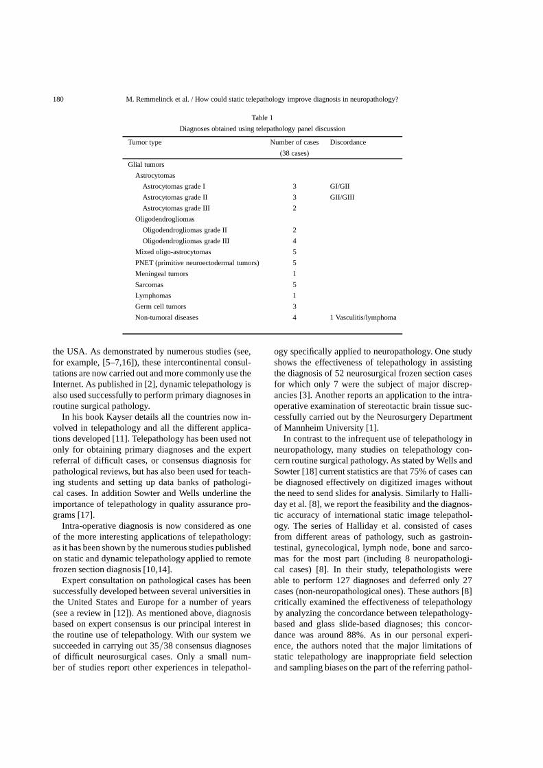
Table 1 illustrates the consensus diagnosis obtainedin 35 cases. For the remaining 3 cases, glass slides hadto be mailed. The first deferred case was a vasculiticlesion with a differential diagnosis of lymphoma; thesecond was a stereotactic specimen from a brain tumor,and the third was a pediatric tumor causing gradingdifficulties. The diagnoses obtained after telepathologydiscussion (35 cases) or slides review (3 cases) werecarried out according to the WHO Histological Clas-sification of Nervous System Tumors [13]. They in-cluded 19 glial tumors consisting of 8 astrocytomas(3 grade I, 3 grade II and 2 grade III tumors), 6 oligo-dendrogliomas (2 grade II and 4 grade III tumors)and 5 mixed oligo-astrocytomas. We also succeededin classifying 5 PNET tumors, 1 meningioma, 5 softtissue lesions, 1 lymphoma, 3 germ cell tumors and 4non-tumor lesions.
4. Discussion
Neuropathology, and particularly brain tumor di-agnosis, is becoming more and more specialized be-cause of the complexity of histological classificationsand the use of different markers originating from im-munohistochemistry and molecular genetics. Only afew pathologists in any country are experienced in thisarea, and requests for consultations are thus usual prac-tice in this field. Telepathology is an easy way of shar-ing scientific expertise at international level and couldthus improve diagnosis particularly in neuropathology,where certain tumor types are very rare and complexto diagnose.
Developments in videotechnology, high performan-ce PCs and progress in data transmission enable patho-logical diagnoses to be performed on high quality dig-itized images.
Our results illustrate how we successfully estab-lished an international connection between two de-partments of pathology, one in Belgium and one in
180 M. Remmelinck et al. / How could static telepathology improve diagnosis in neuropathology?
Table 1
Diagnoses obtained using telepathology panel discussion
Tumor type Number of cases Discordance
(38 cases)
Glial tumors
Astrocytomas
Astrocytomas grade I 3 GI/GII
Astrocytomas grade II 3 GII/GIII
Astrocytomas grade III 2
Oligodendrogliomas
Oligodendrogliomas grade II 2
Oligodendrogliomas grade III 4
Mixed oligo-astrocytomas 5
PNET (primitive neuroectodermal tumors) 5
Meningeal tumors 1
Sarcomas 5
Lymphomas 1
Germ cell tumors 3
Non-tumoral diseases 4 1 Vasculitis/lymphoma
the USA. As demonstrated by numerous studies (see,for example, [5–7,16]), these intercontinental consul-tations are now carried out and more commonly use theInternet. As published in [2], dynamic telepathology isalso used successfully to perform primary diagnoses inroutine surgical pathology.
In his book Kayser details all the countries now in-volved in telepathology and all the different applica-tions developed [11]. Telepathology has been used notonly for obtaining primary diagnoses and the expertreferral of difficult cases, or consensus diagnosis forpathological reviews, but has also been used for teach-ing students and setting up data banks of pathologi-cal cases. In addition Sowter and Wells underline theimportance of telepathology in quality assurance pro-grams [17].
Intra-operative diagnosis is now considered as oneof the more interesting applications of telepathology:as it has been shown by the numerous studies publishedon static and dynamic telepathology applied to remotefrozen section diagnosis [10,14].
Expert consultation on pathological cases has beensuccessfully developed between several universities inthe United States and Europe for a number of years(see a review in [12]). As mentioned above, diagnosisbased on expert consensus is our principal interest inthe routine use of telepathology. With our system wesucceeded in carrying out 35/38 consensus diagnosesof difficult neurosurgical cases. Only a small num-ber of studies report other experiences in telepathol-
ogy specifically applied to neuropathology. One studyshows the effectiveness of telepathology in assistingthe diagnosis of 52 neurosurgical frozen section casesfor which only 7 were the subject of major discrep-ancies [3]. Another reports an application to the intra-operative examination of stereotactic brain tissue suc-cessfully carried out by the Neurosurgery Departmentof Mannheim University [1].
In contrast to the infrequent use of telepathology inneuropathology, many studies on telepathology con-cern routine surgical pathology. As stated by Wells andSowter [18] current statistics are that 75% of cases canbe diagnosed effectively on digitized images withoutthe need to send slides for analysis. Similarly to Halli-day et al. [8], we report the feasibility and the diagnos-tic accuracy of international static image telepathol-ogy. The series of Halliday et al. consisted of casesfrom different areas of pathology, such as gastroin-testinal, gynecological, lymph node, bone and sarco-mas for the most part (including 8 neuropathologi-cal cases) [8]. In their study, telepathologists wereable to perform 127 diagnoses and deferred only 27cases (non-neuropathological ones). These authors [8]critically examined the effectiveness of telepathologyby analyzing the concordance between telepathology-based and glass slide-based diagnoses; this concor-dance was around 88%. As in our personal experi-ence, the authors noted that the major limitations ofstatic telepathology are inappropriate field selectionand sampling biases on the part of the referring pathol-

M. Remmelinck et al. / How could static telepathology improve diagnosis in neuropathology? 181
ogists. Another study also reports 87.5% diagnosticconcordance between CD-ROM digitized images andconventional glass sides; the discordances observedwere mostly related to image selection and/or qual-ity [19]. In fact, both the above studies and others em-phasize the importance in the selection of the histo-logical fields and the increase of diagnostic accuracywhen images are selected by expert pathologists [4,20]. These problems were not so significant in our ex-perience probably because both pathologists had theopportunity to work together in Charlottesville beforethe telepathology sessions, and thus used the samefield selection procedure and attached similar impor-tance to each diagnostic criterion. Dynamic telepathol-ogy avoid selection bias by enabling the interpretingpathologists to select histological fields themselves, inthe same way as using a conventional microscope [2].
Other common sources of diagnostic error, due todeficiencies in image quality were not encountered inour study. The slight technical problems connectedwith image acquisition reported under Results couldbe easily resolved by means of an ultra-wide field Cmount.
Our opinion is that the only serious problem withtelepathology concerns the time required for the com-plete procedure (file set-up, transmission and discus-sion), which may appear forbidding. However, as re-ported by Kayser [10], the classical consultative pro-cedure in pathology, as it is based on the mailing ofslides, is in fact also expensive and time-consuming.As shown under Results, the time taken for a completetelepathology session is at least 25 min, with a meantime of 40 min. Similar data are reported by Eusebiet al. [7], who used static telepathology between theUniversity of Bologna (Italy) and the Memorial Sloan-Kettering Cancer Center (USA).
In practice, our procedure for reducing the time re-quired for a telepathology session consisted of firsttransmitting the complete file (including all the imagesand clinical data). The real-time discussion was in factdone later to enable the referring pathologist to haveall the time required for the case analysis. If he/sheneeded more information, new images could be sendbefore the real time discussion. However, this seconddata transmission increased the time required for thecomplete procedure. We thus believe that it is impor-tant to set up a very complete data file directly, beforediscussion.
ISDN transmission is able to reduce the time nec-essary for telepathology consultation, as, proved bydifferent series in the literature. For instance, one of
the first studies on ISDN transmission published in1993 reports that the time required for a diagnosticsession in static telepathology was between 25 and35 min [15]. In transplantation pathology, Ito et al. de-veloped static telepathology with ISDN transmission,so enabling short sessions of on average 13 min [9].
In conclusion, the static telepathology system thatwe use provides us with accurate help in neuropatho-logical diagnosis. The procedure that we have devel-oped is easy to use, provides high-quality digitized im-ages and complete pathological files, including clinicaldata, which can also be used for training. One slightproblem encountered is its time-consuming nature, butthe system has the advantage of being cheaper than dy-namic telepathology. Telepathology provides easy ac-cess to expert diagnosis and real-time discussion, bothof which are of considerable interest and offer signifi-cant improvements in neuropathology.
References
[1] H.R. Abbasi, R. Weigel, C. Sommer, P. Schmiedek andM. Kiessling, Telepathology in neurosurgery,Stud. Health.Technol. Inform. 62 (1999), 1–7.
[2] U.A. Almagro, B.E. Dunn, H. Choi and D.L. Recla, Telepathol-ogy (letter to the editor),Am. J. Surg. Pathol.22 (1998), 1161–1163.
[3] R.L. Becker, Jr., C.S. Specht, R. Jones, M.E. Rueda Pedrazaand T.J. O’Leary, Use of remote video microscopy (telepathol-ogy) as an adjunct to neurosurgical frozen section consultation,Hum. Pathol.24 (1993), 909–911.
[4] P.W. Callas, K.O. Leslie, A.R. Mattia, D.L. Weaver, D. Cook,B. Travis, D.E. Stanley, L.A. Rogers, S.L. Mount, T.D. Trainer,M.A. Zarka and R.M. Belding, Diagnostic accuracy of a rurallive video telepathology system,Am. J. Surg. Pathol.21(1997),812–819.
[5] V. Della Mea, F. Puglisi, M. Bozanini, S. Forti, V. Amoroso,R. Visentin, P. Dalla Palma and C.A. Beltrami, Fine nee-dle aspiration cytology of the breast: a preliminary reporton telepathology through Internet multimedia electronic mail,Mod. Pathol.10 (1998), 636–641.
[6] M. Eusebi, L. Foschini, S. Grae and J. Rosai, Transcontinentalconsults in surgical pathology via the Internet,Hum. Pathol.28(1997), 13–16.
[7] M. Eusebi, L. Losi, S. Erde and J. Rosai, Telepathology: a pow-erful tool for improved communication among pathologists,Curr. Diagn. Pathol.4 (1997), 73–75.
[8] B.E. Halliday, A.K. Bhattacharyya, A.R. Graham, J.R. Davis,S.A. Leavitt, R.B. Nagle, W.J. Mc Laughlin, R.A. Rivas,R. Martinez, E.A. Krupinski and P.S. Weinstein, Diagnostic ac-curacy of an international static-imaging telepathology consul-tation service,Hum. Pathol.28 (1997), 17–21.
[9] H. Ito, H. Adachi, K. Taniyama, Y. Fukuda and K. Dohi,Telepathology is available for transplantation pathology, expe-rience in Japan using an integrated, low cost, and high qualitysystem,Mod. Pathol.7 (1994), 801–805.

182 M. Remmelinck et al. / How could static telepathology improve diagnosis in neuropathology?
[10] K. Kayser, P. Fritz, M. Drlicek and W. Rahn, Expert consulta-tion by use of telepathology: the Heidelberg experiences,Anal.Cell. Pathol.9 (1995), 53–60.
[11] K. Kayser, J. Szymas and R. Weinstein,Telepathology,Telecommunication, Electronic Education and Publication inPathology, Springer, Berlin, 1999.
[12] K. Kayser and G. Kayser, Basic aspect of and recent develop-ments in telepathology in Europe, with specific emphasis onquality assurance,Anal. Quant. Cytol. Histol.21 (1999), 319–328.
[13] P. Kleihues, B.C. Burger and B.W. Scheithauer,HistologicalTyping of Tumours of the Central Nervous System, 2nd edn,Springer-Verlag, Berlin, 1993.
[14] I. Nordrum, B. Engum, E. Rinde, A. Finseth, H. Ericsson,M. Kearney, H. Stalsberg and T.J. Eide, Remote frozen sec-tion service, a telepathology project in northern Norway,Hum.Pathol.22 (1991), 514–518.
[15] M. Oberholzer, H.R. Fischer, H. Christen, S. Gerber,M. Bruehlmann, M. Mihatsch, M. Famos, C. Winkler, P. Fehr,L. Bächtold and K. Kayser, Telepatholgogy with ISDN – a new
tool for image transfer in surgical pathology,Hum. Pathol.24(1993), 1078–1085.
[16] I. Petersen, G. Wolf, K. Roth and K. Schlüns, Telepathology bythe Internet,J. Pathol.191(2000), 8–14.
[17] C. Sowter and C.A. Wells, System requirements, the use oftelepathology in diagnostic pathology and its application toquality assurance programs,Curr. Diagn. Pathol.4 (1997), 65–72.
[18] C.A. Wells and C. Sowter, Telepathology: a diagnostic tool forthe millennium?,J. Pathol.191(2000), 1–7.
[19] D.S. Weinberg, F.A. Allaert, P. Dusserre, F. Drouot, B. Re-tailliau, W.R. Welch, J. Longtine, G. Brodsky, R. Folkerthand M. Doolittle, Telepathology diagnosis by means of digitalstill images, an international validation study,Hum. Pathol.27(1996), 111–118.
[20] M.H. Weinstein and J.I. Epstein, Telepathology diagnosis ofprostate needle biopsies,Hum. Pathol.28 (1997), 22–29.

Submit your manuscripts athttp://www.hindawi.com
Stem CellsInternational
Hindawi Publishing Corporationhttp://www.hindawi.com Volume 2014
Hindawi Publishing Corporationhttp://www.hindawi.com Volume 2014
MEDIATORSINFLAMMATION
of
Hindawi Publishing Corporationhttp://www.hindawi.com Volume 2014
Behavioural Neurology
EndocrinologyInternational Journal of
Hindawi Publishing Corporationhttp://www.hindawi.com Volume 2014
Hindawi Publishing Corporationhttp://www.hindawi.com Volume 2014
Disease Markers
Hindawi Publishing Corporationhttp://www.hindawi.com Volume 2014
BioMed Research International
OncologyJournal of
Hindawi Publishing Corporationhttp://www.hindawi.com Volume 2014
Hindawi Publishing Corporationhttp://www.hindawi.com Volume 2014
Oxidative Medicine and Cellular Longevity
Hindawi Publishing Corporationhttp://www.hindawi.com Volume 2014
PPAR Research
The Scientific World JournalHindawi Publishing Corporation http://www.hindawi.com Volume 2014
Immunology ResearchHindawi Publishing Corporationhttp://www.hindawi.com Volume 2014
Journal of
ObesityJournal of
Hindawi Publishing Corporationhttp://www.hindawi.com Volume 2014
Hindawi Publishing Corporationhttp://www.hindawi.com Volume 2014
Computational and Mathematical Methods in Medicine
OphthalmologyJournal of
Hindawi Publishing Corporationhttp://www.hindawi.com Volume 2014
Diabetes ResearchJournal of
Hindawi Publishing Corporationhttp://www.hindawi.com Volume 2014
Hindawi Publishing Corporationhttp://www.hindawi.com Volume 2014
Research and TreatmentAIDS
Hindawi Publishing Corporationhttp://www.hindawi.com Volume 2014
Gastroenterology Research and Practice
Hindawi Publishing Corporationhttp://www.hindawi.com Volume 2014
Parkinson’s Disease
Evidence-Based Complementary and Alternative Medicine
Volume 2014Hindawi Publishing Corporationhttp://www.hindawi.com