HOST RANGE, PHYLOGENETIC, AND PATHOGENIC DIVERSITY OF
102
1 HOST RANGE, PHYLOGENETIC, AND PATHOGENIC DIVERSITY OF Corynespora cassiicola (Berk. & Curt.) Wei By LINLEY JOY SMITH A DISSERTATION PRESENTED TO THE GRADUATE SCHOOL OF THE UNIVERSITY OF FLORIDA IN PARTIAL FULFILLMENT OF THE REQUIREMENTS FOR THE DEGREE OF DOCTOR OF PHILOSOPHY UNIVERSITY OF FLORIDA 2008
Transcript of HOST RANGE, PHYLOGENETIC, AND PATHOGENIC DIVERSITY OF

1
HOST RANGE, PHYLOGENETIC, AND PATHOGENIC DIVERSITY OF Corynespora cassiicola (Berk. & Curt.) Wei
By
LINLEY JOY SMITH
A DISSERTATION PRESENTED TO THE GRADUATE SCHOOL OF THE UNIVERSITY OF FLORIDA IN PARTIAL FULFILLMENT
OF THE REQUIREMENTS FOR THE DEGREE OF DOCTOR OF PHILOSOPHY
UNIVERSITY OF FLORIDA
2008

2
© 2008 Linley Joy Smith

3
To Peter, for making me laugh.

4
ACKNOWLEDGMENTS
Funding and support was made possible by the USDA Special Grant Program for Tropical
and Subtropical Agriculture Research, the University of Florida, IFAS, EREC, the Florida
Tomato Committee, the University of Guam, Guam Cooperative Extension, and the USDA IPM
3-D and Hatch funds.
I would like to thank Drs. Ken Pernezny, Pam Roberts, Jeffrey Rollins, and Jay Scott for
their support while serving on my supervisory committee. I would also like to express
appreciation to my major advisor, Dr. Lawrence Datnoff, for his commitment and help
throughout the course of my Ph.D. I would especially like to thank Dr. Robert Schlub for his
willingness to help in every step of the process and for his unwavering support, encouragement,
and friendship. Special thanks to my helpful coworkers in Guam, especially Roger Brown and
Lauren Gutierrez.
Most importantly, my heartfelt appreciation goes to my parents for their unconditional love
and support. Finally, I thank my husband for encouraging me to pursue this opportunity, an
ocean and continent away, for coming to Gainesville for me, and for keeping me smiling
throughout.
5
TABLE OF CONTENTS page
ACKNOWLEDGMENTS ...............................................................................................................4
LIST OF TABLES...........................................................................................................................6
LIST OF FIGURES .........................................................................................................................7
ABSTRACT.....................................................................................................................................8
CHAPTER
1 INDEX OF PLANT HOSTS OF Corynespora cassiicola .....................................................10
Introduction.............................................................................................................................10 Methods ..................................................................................................................................12
Literature Survey and Host Index....................................................................................12 Guam and Florida Surveys ..............................................................................................13
Results.....................................................................................................................................14 Discussion...............................................................................................................................16
2 GENETIC AND PATHOGENIC DIVERSITY OF CORYNESPORA CASSIICOLA ...........48
Introduction.............................................................................................................................48 Methods ..................................................................................................................................52
Collection and Solicitation of Fungal Isolates.................................................................52 Primer Development for Random Hypervariable Loci ...................................................54 Fungal Cultures and Extraction of Genomic DNA .........................................................55 Phylogenetic Analyses.....................................................................................................57 Pathogenicity Analyses ...................................................................................................59 Growth Rate Analyses.....................................................................................................60
Results.....................................................................................................................................61 Phylogenetic Analyses.....................................................................................................61 Pathogenicity Analyses ...................................................................................................65 Growth Rate Analyses.....................................................................................................66
Discussion...............................................................................................................................67
LIST OF REFERENCES...............................................................................................................90
BIOGRAPHICAL SKETCH .......................................................................................................102
6
LIST OF TABLES
Table page 1-1 Taxonomic grouping of Corynespora cassiicola host species. .........................................20
1-2 Occurrence and fungal-host interaction of Corynespora cassiicola identified during 2004-2005 Guam and Florida surveys...............................................................................21
2-1 Isolate designations, geographic location of isolation, host of isolation, phylogenetic lineage (PL), type of growth on associated host, and species of Corynespora used in the phylogenetic analyses. .................................................................................................72
2-2 Summary of sequence data from four loci used to confirm the phylogenetic lineage of Corynespora cassiicola isolates. ...................................................................................76
2-3 Pathogenicity profiles for 50 Corynespora cassiicola isolates..........................................77
2-4 Growth rate of Corynespora cassiicola isolates at 23°C. ..................................................79
2-5 Growth rate of Corynespora cassiicola isolates at 33°C. ..................................................81

7
LIST OF FIGURES
Figure page 1-1 Corynespora cassiicola isolate from Cucumis sativus ......................................................45
1-2 Various symptoms caused by Corynespora cassiicola on naturally infected leaves.........46
2-1 Fifty percent majority rule consensus tree-phylogram from Bayesian inference analysis of combined data from rDNA ITS, Cc-ga4, Cc-caa5, and Cc-act1 sequences. ..........................................................................................................................83
2-2 Fifty percent majority rule consensus tree-phylogram from Bayesian inference analysis of rDNA ITS locus. ............................................................................................84
2-3 Fifty percent majority rule consensus tree-phylogram from Bayesian inference analysis of the Cc-caa5 locus. ...........................................................................................85
2-4 Fifty percent majority rule consensus tree-phylogram from Bayesian inference analysis of the Cc-ga4 locus. .............................................................................................86
2-5 Fifty percent majority rule consensus tree-phylogram from Bayesian inference analysis of the Cc-act1 locus. . .........................................................................................87
2-6 UPGMA dendrogram of 50 Corynespora cassiicola isolates based on pathogenicity profiles on eight crop plants:..............................................................................................88
2-7 Demonstration of the C. cassiicola disease rating system.................................................89

8
Abstract of Dissertation Presented to the Graduate School of the University of Florida in Partial Fulfillment of the Requirements for the Degree of Doctor of Philosophy
HOST RANGE, PHYLOGENETIC, AND PATHOGENIC DIVERSITY OF Corynespora cassiicola (Berk. & Curt). Wei
By
Linley Joy Smith
August 2008
Chair: Lawrence E. Datnoff Major: Plant Pathology
The fungus Corynespora cassiicola (Berk. & Curt.) Wei is a pathogen, endophyte, and
saprophyte. It can be found growing on at least 530 plant species from 380 genera, primarily in
the tropics. Isolates from diverse hosts were collected or solicited from locations in American
Samoa, Brazil, Malaysia, Micronesia, and Florida, Mississippi, and Tennessee within the United
States. Outgroup taxa including C. citricola, C. melongenea, C. olivaceae, C. proliferata, C.
sesamum, and C. smithii were solicited from culture collections. A multilocus phylogenetic
analysis using 143 isolates was performed to investigate how genetic diversity correlates with
host-specificity, growth rate, and geographic distribution. Phylogenetic trees were congruent
from the rDNA ITS region, two random hypervariable loci (Cs caa5 and Cs ga4), and the actin
encoding locus CC act1, indicating asexual propagation. Fifty isolates had different
pathogenicity profiles when tested against eight known C. cassiicola hosts: basil, bean, cowpea,
cucumber, papaya, soybean, sweet potato, and tomato. Phylogenetic lineage correlated with
pathogenicity profiles, host originality, and growth rate, but not with geographic location.
Common fungal genotypes were widely distributed geographically indicating long distance and
global dispersal of clonal lineages. This research reveals an abundance of previously

9
unrecognized diversity within the species and provides evidence for redefining species
distinctions within Corynespora, which will aid in future disease control strategies.
10
CHAPTER 1 INDEX OF PLANT HOSTS OF CORYNESPORA CASSIICOLA
Introduction
Corynespora cassiicola (Berk. & Curt.) Wei has been commonly reported as a plant
pathogenic foliar fungus with a wide host range within tropical and subtropical areas (Holliday
1980; Farr et al. 1980; Romruensukharom et al. 2005). In addition to being a pathogen, on some
hosts C. cassiicola is also reported to grow as an endophyte or saprophyte (Collado 1999; Gond
et al. 2007; Promputtha et al. 2007; Suryanarayanan et al. 2002; Kingsland 1985; Hyde et al.
2001; Lee et al. 2004; Lumyong et al. 2003). Though the diseases attributed to C. cassiicola are
mainly foliar, it may also cause fruit, stem, and root diseases (Jones et al. 1991). The
generalization that individual C. cassiicola isolates have a wide host range is not supported by
the literature because host specific isolates, isolates pathogenic to select hosts, and weak
pathogens or secondary invaders of senescent tissue are known to exist (Onesirosan et al. 1974;
Cutrim and Silva et al. 2003; Kingsland 1985; Pereira et al. 2003). Rarely reported outside the
tropics and subtropics, there are occasional reports of the fungus from temperate regions,
particularly on soybean (Boosalis and Hamilton 1957; Malvick 2004; Raffel et al. 1999; Seaman
et al. 1965).
Disease symptoms attributed to C. cassiicola include necrosis, often with a surrounding
yellow halo (Pernezny and Simone 1993) due to the production of a host specific protein toxin,
cassiicolin (Barthe et al. 2007; Kurt 2004). With respect to foliage, young and mature leaves can
be affected, although the pathogen is more commonly associated with older leaves (Pernezny et
al. 2008). Substantial crop losses have been observed in many countries on numerous hosts:
southern United States on ornamentals (Alfieri et al. 1984, 1994; Chase 1981,1982, 1984, 1986,
1987, 1993; El-Gholl and Schubert 1990; El-Gholl et al. 1997; Miller and Alfieri 1973;

11
McRitchie and Miller 1973; Simone 2000, 2000), cucumber (Abul-Hayja et al. 1978; Blazquez
1967; Strandburg 1971), and tomato (Bliss et al. 1973; Blazquez 1972; Jones and Jones 1984;
Pernezny et al. 1996, 2002; Smith et al. 2006, Smith et al. 2008b); Midwestern United States on
soybean (Boosalis and Hamilton 1957), cowpea (Olive and Bain 1945) and sesame (Stone and
Jones 1960); India on ornamentals (Cheeran 1968; Mallaiah et al. 1981; Mehrotra 1987, 1997;
Silva et al. 2000; Singh et al. 1982), Hevea rubber trees (Atan and Hamid 2003; Silva et al.
1998), cotton (Lakshmanan et al. 1990), and weeds (Philip et al. 1972); Brazil on ornamentals
(Da Silva et al. 2005; Leite and Barreto 2000; Pohltronieri 2003), and weeds (Pereira et al.
2003); Philippines, Nigeria, and U.S. Virgin Islands on papaya (Quimio and Abilay 1979; Oluma
and Amuta 1999; Bird et al. 1966); and Micronesia and Asia on ornamentals (Florence and
Sharma 1987; Hasama et al. 1991), cucurbits (Yudin and Schlub 1998; Tsay and Kuo 1991),
tomato (Schlub and Yudin 2002), and pepper (Kwon et al. 2001).
Most regions report C. cassiicola diseases on only a few host species, despite the broad
host range of the fungus, prompting questions pertaining to isolate host specificity and
distribution. Addressing such questions will have implications for disease control and
quarantine. The host -specificity and severity of the fungus on Lantana camara in Brazil led to
the discovery of a new forma specialis, C. cassiicola f. sp. lantanae, and the use of the isolate as
a bioherbicide (Pereira et al. 2003). Based on the vast number of weeds that serve as hosts, and
past demonstration of host-specificity in some isolates, there is great potential for the discovery
of additional isolates useful for biological control. Further information on the fungal-host
interaction and host range of individual isolates will be useful in the study of disease epidemics.
The objective of this study was to compile a list of C. cassiicola hosts into a single
document, thereby aiding further research on the host range of individual isolates. Prior to this

12
study, the most complete host listing is in the fungal database of the ARS/USDA Systematic
Mycology and Microbiology Laboratory, which included 257 plant host species (Farr et al.
2008). This study will provide a more complete index for use by those engaged in phylogenetic
analysis of Corynespora spp. and in disease management. Awareness of the potential host range
of the fungal species is vital to the determination of the host-specificity of individual isolates.
The host range of individual isolates has direct implications for disease management, including
the identification of potential inoculum sources, recommendations for intercropping and crop
rotation, weed management, biological control candidacy, and isolate choice for resistance
breeding.
In order to obtain an estimate of the completeness of the list of hosts known to harbor C.
cassiicola, surveys were conducted to identify hosts in Guam and Florida. Guam is an ideal
location to discover new hosts due to its tropical climate, wet and dry seasons, and lack
heretofore of a Corynespora host survey (Schlub and Yudin 2002). Florida was included
because outbreaks of target spot on tomato caused by C. cassiicola are common and it represents
a subtropical environment located an ocean and a continent away from Guam.
Methods
Literature Survey and Host Index
An index of plant hosts of C. cassiicola was compiled from a search of world literature for
any reference regarding its presence on plant tissue. All plant-fungus associations were included
such as pathogenic, endophytic, and saprophytic. Resources included articles in refereed
journals, graduate student theses, books, and web-based resources such as annual reports,
production guides, and plant clinic lists. The final list of susceptible hosts of C. cassiicola was
compiled from the literature and personal observation from surveys in Florida and Guam.

13
All plant species, genera, and families were named and classified according to the USDA
Germplasm Resources Information Network (GRIN) taxonomy, which follows the APGII
system. In some cases, the host name given in the original citation was changed to be consistent
with GRIN taxonomy. In a few cases, neither the species cited nor a proper synonym was
identified using GRIN taxonomy and the species name was kept as originally cited. Only one
reference was provided per host, with emphasis on citing the first known report of that host. For
some hosts, the only reference that could be found was a website, and in those cases the website
is listed. The number of plant host species was conservatively determined by counting only
unique species within each genus. Genera with unidentified species (e.g. Crossandra spp.) were
counted only once when no other named species were present within that genus.
Guam and Florida Surveys
Surveys for the presence of C. cassiicola were conducted throughout Guam and Florida.
The Guam survey was conducted for one year beginning in January of 2004 and the Florida
survey was conducted for one year beginning in January of 2005. Survey areas focused on
roadsides, nurseries, and farms. During the course of the survey, leaves from plants with
characteristic C. cassiicola foliage disease symptoms were collected and placed in individual
plastic bags. Known hosts of C. cassiicola were sampled more intensely through the additional
collection of old and young asymptomatic leaves. An effort was made to sample from an equal
number of individual plants and unique plant species in Florida and Guam.
To induce sporulation, leaf tissue was placed abaxial side up in the moisture chamber for
10 days. Moisture chambers were created on the lab bench by placing 10 ml of sterilized
distilled water on a paper towel in a 150 mm petri plate. A plant species was identified as a host
of C. cassiicola if characteristic structures of the fungus developed within 10 days. An isolate
was labeled a pathogen if conidiophores arose from a necrotic spot and an endophyte if

14
conidiophores arose from healthy, green tissue. Petri plates were inspected under a dissecting
microscope daily for spores and conidiophores of C. cassiicola. Structures were confirmed
based on microscopic morphological features such as percurrent proliferation of the
conidiophores and pseudoseptation. Single spore isolates were obtained for long-term storage by
needle transfer of spores to antibiotic V8 agar agar slants (340 ml V8 juice, 660 ml water, 3 g
CaCO3, 17 g agar, 100 μg/ml ampicillin or kanamycin). Slants were left at room temperature
until colonies reached at least 5 cm in diameter, covered with autoclaved mineral oil, and stored
at 5o C until further study.
Results
Over 900 individual plants were surveyed in both Guam and Florida from 320 unique plant
species in Guam and 289 unique plant species in Florida. Compilation of Corynespora
cassiicola hosts from the literature and surveys conducted in Guam and Florida resulted in an
index of 530 plant species from 380 genera. The majority of index host species for C. cassiicola
are herbaceous Eudicotyledonae, but 52 Monocotyledonae, eight Magnoliids, five Filicopsida
(ferns), and one cycad are also represented. No hosts were found within the Anthocerotophyta
(hornworts), Bryophyta (mosses), Equisetopsida (horsetails, sphenophytes), Lycopsida
(lycophytes), or Marchantiomorpha (liverworts) (Table 1-1).
Hosts were found in two plant divisions: Filicopsida and Spermatopsida. The five hosts in
the Filicopsida include Arachniodes aristata (Davalliaceae), Athyrium niponicum
(Dryopteridaceae), Adiantum cuneatum (Pteridaceae), Davallia repens (Davalliaceae), and
Platycerium spp. (Pteridaceae). The plant division Spermatophyta (Cycadales, Magnoliidae,
Monocotolydonae, and Tricolpates) contains 99% of the host species (Table 1-1). There are
eight species from the Magnoliidae. Three species are from the Piperaceae (Piper betle, P.

15
hispidinervum, and Perperomia obtusifolia). Three species are from the Magnoliales in the
family Annonaceae (Annona reticulata, A. squamosa and Asimina triloba). Two species are
from the Laurales in the Hernandiaceae (Hernandia ovigera) and the Lauraceae (Ocotea
leucoxylon) (Table 1-2).
The 52 host species from the Monocotolydonae are from 16 families: Araceae (13
species), Poaceae (9 species), Arecaceae (7 species), Dioscoreaceae (5 species, all from the
genus Dioscorea), Orchidaceae (4 species), Agavaceae (3 species), Musaceae (2 species),
Alismataceae (1 species), Asparagaceae (1 species), Bromeliaceae (1 species), Commelinaceae
(1 species), Heliconiaceae (1 species), Hemerocallidaceae (1 species), Marantaceae (1 species),
Restonaceae (1 species), and Strelitziaceae (1 species), in decreasing order of host species
numbers.
The remaining 464 host species are Eudicots. Families that contain the largest number of
hosts include Fabaceae (70 species), Lamiaceae (33 species), Malvaceae (32 species),
Asteraceae (26 species), Apocynaceae (21 species), Acanthaceae (20 species), Euphorbiaceae
(20 species), Verbenaceae (17 species), Convolvulaceae (14 species), Cucurbitaceae (13
species), and Solanaceae (13 species), in decreasing order of host species numbers.
Between the two surveys, 91 new hosts species were identified, 87 of which were found in
the survey conducted on Guam. New hosts were found in 32 families, of which three families
had never been reported to harbor the fungus: Hernandiaceae, Moringaceae, and Mutingiaceae.
Ten new host species were found to harbor the fungus in the survey conducted in Florida
(Cerinthe major, Corchorus aestuans, Fatshedera lizei, Hibiscus rosa-sinensis, Jatropha spp.,
Salvia farinacea, Salvia microphylla, Salcia officinalis, Sida spinosa, and Stachytarpheta
16
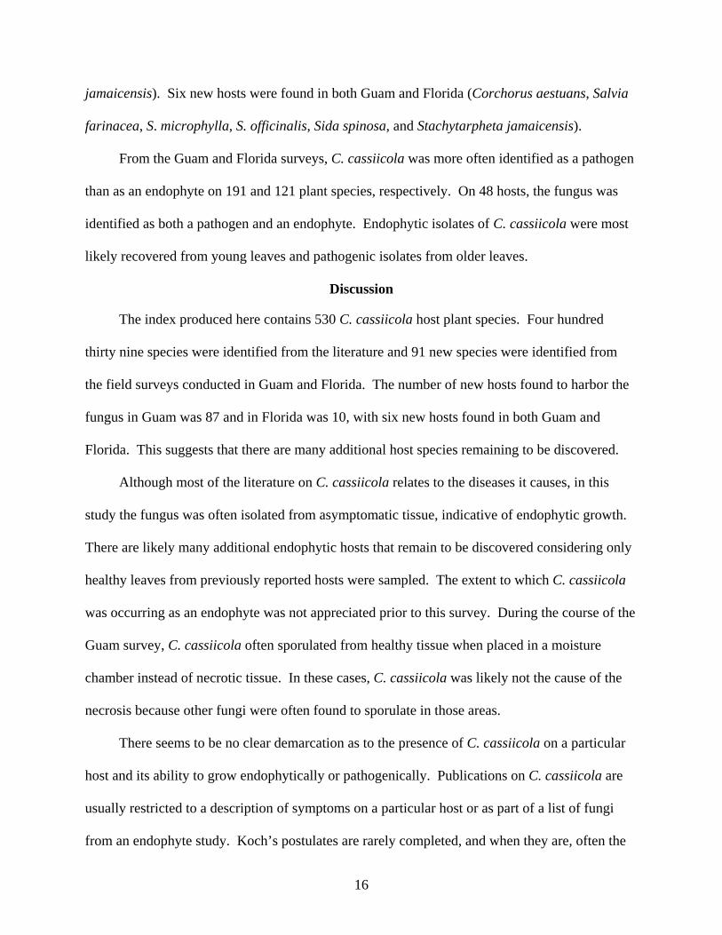
jamaicensis). Six new hosts were found in both Guam and Florida (Corchorus aestuans, Salvia
farinacea, S. microphylla, S. officinalis, Sida spinosa, and Stachytarpheta jamaicensis).
From the Guam and Florida surveys, C. cassiicola was more often identified as a pathogen
than as an endophyte on 191 and 121 plant species, respectively. On 48 hosts, the fungus was
identified as both a pathogen and an endophyte. Endophytic isolates of C. cassiicola were most
likely recovered from young leaves and pathogenic isolates from older leaves.
Discussion
The index produced here contains 530 C. cassiicola host plant species. Four hundred
thirty nine species were identified from the literature and 91 new species were identified from
the field surveys conducted in Guam and Florida. The number of new hosts found to harbor the
fungus in Guam was 87 and in Florida was 10, with six new hosts found in both Guam and
Florida. This suggests that there are many additional host species remaining to be discovered.
Although most of the literature on C. cassiicola relates to the diseases it causes, in this
study the fungus was often isolated from asymptomatic tissue, indicative of endophytic growth.
There are likely many additional endophytic hosts that remain to be discovered considering only
healthy leaves from previously reported hosts were sampled. The extent to which C. cassiicola
was occurring as an endophyte was not appreciated prior to this survey. During the course of the
Guam survey, C. cassiicola often sporulated from healthy tissue when placed in a moisture
chamber instead of necrotic tissue. In these cases, C. cassiicola was likely not the cause of the
necrosis because other fungi were often found to sporulate in those areas.
There seems to be no clear demarcation as to the presence of C. cassiicola on a particular
host and its ability to grow endophytically or pathogenically. Publications on C. cassiicola are
usually restricted to a description of symptoms on a particular host or as part of a list of fungi
from an endophyte study. Koch’s postulates are rarely completed, and when they are, often the
17
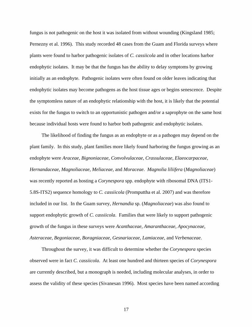
fungus is not pathogenic on the host it was isolated from without wounding (Kingsland 1985;
Pernezny et al. 1996). This study recorded 48 cases from the Guam and Florida surveys where
plants were found to harbor pathogenic isolates of C. cassiicola and in other locations harbor
endophytic isolates. It may be that the fungus has the ability to delay symptoms by growing
initially as an endophyte. Pathogenic isolates were often found on older leaves indicating that
endophytic isolates may become pathogens as the host tissue ages or begins senescence. Despite
the symptomless nature of an endophytic relationship with the host, it is likely that the potential
exists for the fungus to switch to an opportunistic pathogen and/or a saprophyte on the same host
because individual hosts were found to harbor both pathogenic and endophytic isolates.
The likelihood of finding the fungus as an endophyte or as a pathogen may depend on the
plant family. In this study, plant families more likely found harboring the fungus growing as an
endophyte were Araceae, Bignoniaceae, Convolvulaceae, Crassulaceae, Elaeocarpaceae,
Hernandaceae, Magnoliaceae, Meliaceae, and Moraceae. Magnolia liliifera (Magnoliaceae)
was recently reported as hosting a Corynespora spp. endophyte with ribosomal DNA (ITS1-
5.8S-ITS2) sequence homology to C. cassiicola (Promputtha et al. 2007) and was therefore
included in our list. In the Guam survey, Hernandia sp. (Magnoliaceae) was also found to
support endophytic growth of C. cassiicola. Families that were likely to support pathogenic
growth of the fungus in these surveys were Acanthaceae, Amaranthaceae, Apocynaceae,
Asteraceae, Begoniaceae, Boragniaceae, Gesnariaceae, Lamiaceae, and Verbenaceae.
Throughout the survey, it was difficult to determine whether the Corynespora species
observed were in fact C. cassiicola. At least one hundred and thirteen species of Corynespora
are currently described, but a monograph is needed, including molecular analyses, in order to
assess the validity of these species (Sivanesan 1996). Most species have been named according

18
to host identity, and only a few species have been described in culture. In addition, single
isolates exhibit considerable morphological plasticity that depends on humidity, light,
temperature, and substrate; therefore, morphological differences need to be compared with
molecular differences. Although the hosts included in this index are restricted to those reported
for C. cassiicola, some may actually be hosts of other Corynespora species due to
misidentification. Likewise, there may be hosts reported to harbor other species of Corynespora
that may, in fact, be harboring C. cassiicola because the morphological distinctions between
species are based on overlapping, variable, morphological characters. Phylogenetic analyses of
the isolates should help to clarify these issues.
Despite these complications, this is the first step taken to consolidate our knowledge of the
potential host range of C. cassiicola, which is vital for further studies of the biology of individual
isolates and ultimately in future studies of Corynespora species evolution. Although there is no
teleomorphic stage currently known for C. cassiicola, the Ascomycete species Corynesporasca
caryote and Pleomassaria swidae have unknown Corynespora species anamorphs (Sivanesan
1996; Tanaka et al. 2008). There is no evidence to suggest that C. cassiicola is reproducing
other than by asexual spores. However, evidence for sexual recombination needs to be tested
between isolates within and among host species. Insight into the evolutionary potential of the
fungus will lead to a better understanding of how to control its diseases (McDonald 2004).
The literature search and surveys elucidated several characteristics of C. cassiicola that
warrant further investigation: (1) the inability of some isolates recovered from symptomatic
tissue to re-infect the original hosts; (2) the ability to be endophytic, pathogenic, and saprophytic
on individual hosts; (3) the wide host range of the fungal species, yet restricted host ranges of
individual isolates; (4) the ability to grow on some members of a plant taxonomic group and not
19
others; (5) a lack of understanding of the diversity within the fungal species and how it relates to
host range; (6) the taxonomic validity of the 113 species of Corynespora considering the high
morphological plasticity of individual isolates. Future research should attempt to address these
issues and the organization of the plant hosts in a single publication will facilitate this.

20
Table 1-1. Taxonomic grouping of Corynespora cassiicola host species.
Plant Group
Number of Host Species in the Index
Number of Host Species Sampled in
Guam
Number of Host Species Sampled in
Florida Anthocerotphyta (hornworts) 0 2 3 Bryophyta (mosses) 0 5 2 Filicopsida (ferns) 5 14 21 Spermatopsida (seed plants) 525 299 263 Conifers 0 3 6 Cycads 1 4 5 Gnetales 0 2 1 Angiosperms 524 290 251 Magnoliids 8 6 4 Monocotyledons 52 61 38 Eudicots 464 223 209

21
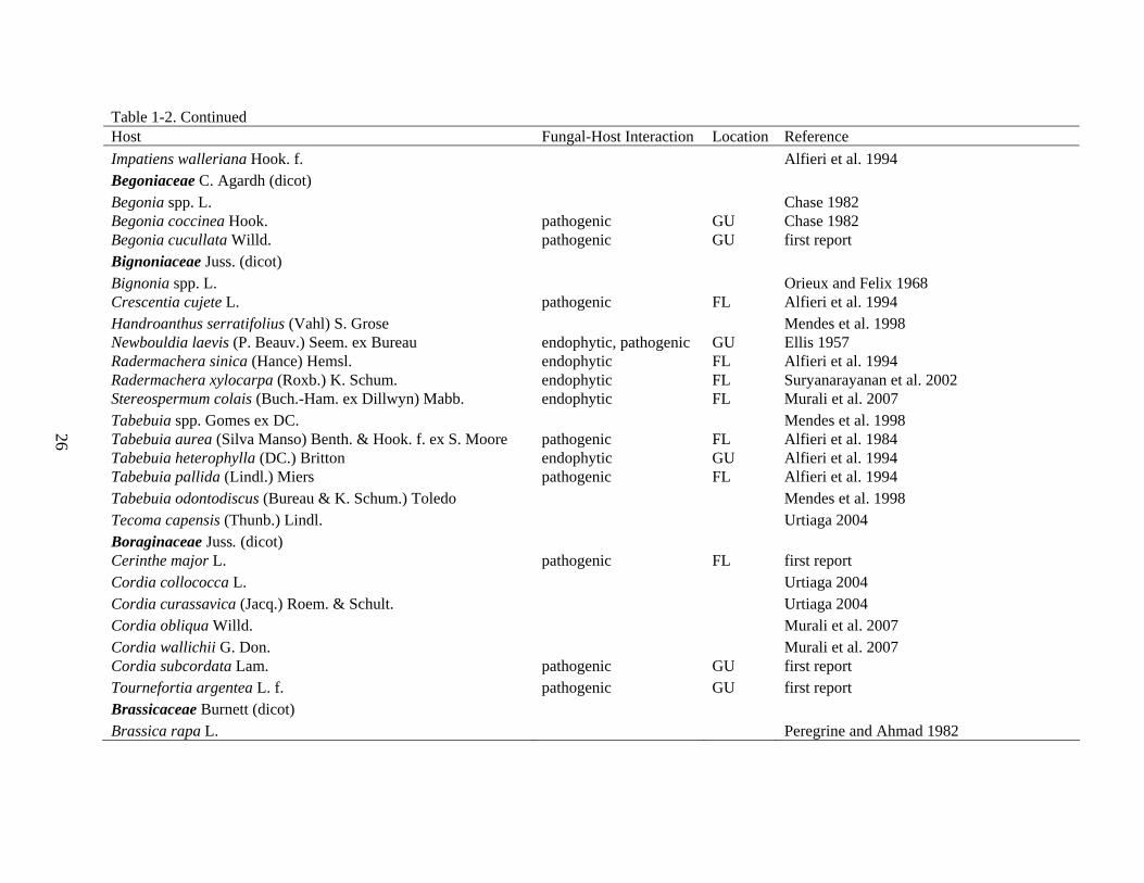
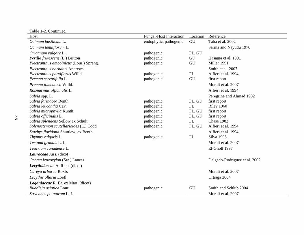
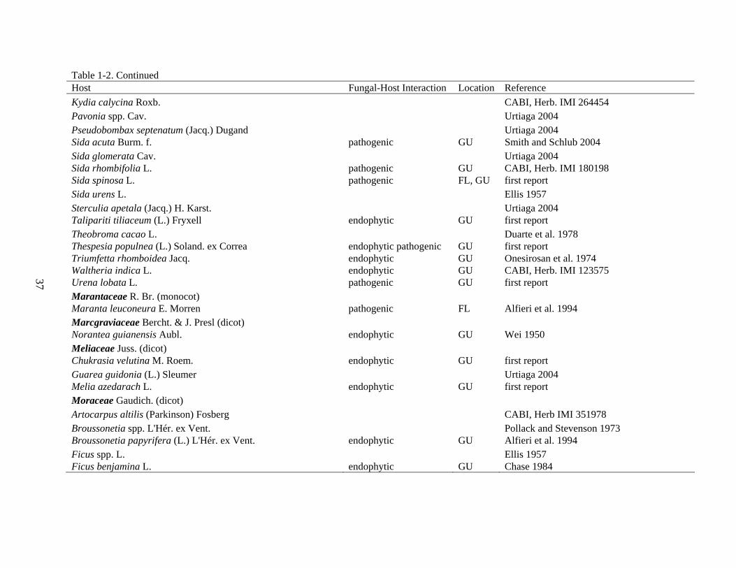
Table 1-2. Occurrence and fungal-host interaction of Corynespora cassiicola identified during 2004-2005 Guam and Florida surveys. Host Fungal-Host Interaction Location Reference Acanthaceae Juss. (dicot) Acanthus ilicifolius L. endophytic GU Sadaba et al. 1995 Aphelandra squarrosa Nees pathogenic FL, GU Chase 1982 Asystasia spp. Blume Alfieri et al. 1984 Asystasia gangetica (L.) T. Anders. pathogenic GU Alfieri et al. 1984 Crossandra spp. Salisb. pathogenic FL Alfieri et al. 1994 Eranthemum pulchellum Andrews pathogenic FL Alfieri et al. 1994 Fittonia spp. Coem. pathogenic FL Chase 1982 Fittonia albivenis (Lindl. ex hort. Veitch) Brummitt endophytic, pathogenic FL, GU Chase 1982 Hygrophila spp. R. Br. FL Alfieri et al. 1994 Justicia spp. L. Ellis 1957 Justicia brandegeeana Wasshausen & L.B. Sm. pathogenic FL, GU Alfieri et al. 1994 Justicia carnea Lindl. pathogenic GU Ellis 1957 Justicia ventricosa Wall. ex Hook. Zhuang 2001 Meisosperma oppositifolium endophytic GU Smith et al. 2007 Pachystachys coccinea (Aubl.) Nees Urtiaga 1986 Pachystachys lutea Nees pathogenic FL, GU Alfieri et al. 1994 Peristrophe spp. Nees Alfieri et al. 1994 Pseuderanthemum spp. Radlk. El-Gholl et al. 1997 Pseuderanthemum carruthersii (Seem.) Guillaumin pathogenic GU El-Gholl et al. 1997 Ruellia humboldtiana (Nees) Lindau endophytic, pathogenic FL Urtiaga 2004 Strobilanthes dyerianus M.T. Mast. pathogenic GU Coile and Dixon 1994 Thunbergia fragrans Roxb. Zhuang 2001 Warpuria clandestina Stapf. pathogenic GU Ellis 1957 Actinidiaceae Gilg & Werderm. (dicot) Actinidia chinensis Planch. Peregrine and Ahmad 1982 Adoxaceae E. Mey. (dicot) Viburnum spp. L. Alfieri et al. 1994 Viburnum odoratissimum Ker Gawl. endophytic FL, GU Alfieri et al. 1994

22
Table 1-2. Continued Host Fungal-Host Interaction Location Reference Agavaceae Dumort. (monocot) Agave sisalana Perrine Ellis 1957 Cordyline fruticosa (L.) Chev. endophytic GU Situmorang and Budimen 1984 Dracaena spp. Vand. ex L. Alfieri et al. 1984 Dracaena reflexa Lam. endophytic, pathogenic FL Alfieri et al. 1994 Alismataceae Vent. (monocot) Echinodorus spp. Rich. ex Engelm. Alfieri et al. 1994 Amaranthaceae Juss. (dicot) Achyranthes aspera L. CABI, Herb. IMI 191361 Alternanthera ficoidea (L.) P. Beauv. pathogenic GU first report Amaranthus spp. L. Alfieri et al. 1994 Amaranthus spinosus L. pathogenic FL, GU Alfieri et al. 1994 Amaranthus tricolor L. Peregrine and Ahmad 1982 Celosia argentea L. var. cristata (L.) Kuntze pathogenic GU first report Digera muricata (L.) Mart. Sarma and Nayudu 1970 Anacardiaceae R. Br. (dicot) Lannea coromandelica (Houtt.) Merr. CABI, Herb. IMI 266196 Mangifera indica L. Rajak and Pandey 1985 Schinus spp. L. endophytic, pathogenic FL Alfieri et al. 1984 Spondias purpurea L. pathogenic FL Freire 2005 Vernicia montana Lour. endophytic FL Ellis 1957 Annonaceae Juss. (dicot) Annona reticulata L. Peregrine and Ahmad 1982 Annona squamosa L. endophytic GU first report Asimina triloba (L.) Dunal CABI, Herb. IMI 364250 Apiaceae Lindl. (dicot) Foeniculum vulgare Mill. Peregrine and Ahmad 1982

23
Table 1-2. Continued Host Fungal-Host Interaction Location Reference Apocynaceae Juss. (dicot) Adenium obesum (Forssk.) Roem. & Schult. El-Gholl 1997 Allamanda spp. L. endophytic FL Alfieri et al. 1984 Allamanda cathartica L. pathogenic GU Alfieri et al. 1994 Alstonia scholaris (L.) R. Br. endophytic, pathogenic FL Suryanarayanan et al. 2002 Calotropis procera (Aiton) W. T. Aiton CABI, Herb. IMI 173980 Carissa spp. L. pathogenic FL Alfieri et al. 1994 Catharanthus roseus (L.) G. Don pathogenic FL, GU McGovern 1994 Conopharyngia longiflora (Benth.) Stapf Kranz 1963 Cryptolepis buchananii Schult. CABI, Herb. IMI 221003 Funastrum clausum (Jacq.) Schltr. Urtiaga 2004 Hoya spp. R. Br. pathogenic FL Alfieri et al. 1994 Mandevilla spp. Lindl. Alfieri et al. 1984 Mandevilla splendens (Hook. f.) Woodson pathogenic FL, GU Alfieri et al. 1994 Nerium oleander L. pathogenic FL Alfieri et al. 1994 Plumeria rubra L. forma acutifolia (Poir.) Woodson endophytic, pathogenic GU Ellis 1957 Rauvolfia serpentina (L.) Benth. ex Kurz CABI, Herb. IMI 122395 Tabernaemontana divaricata (L.) R. Br. ex Roem. & Schult. CABI, Herb. IMI 209321 Tabernaemontana sananho Ruiz & Pav. Urtiaga 2004 Tacazzea spp. Decne. Ellis 1957 Telosma cordata (Burm. f.) Merr. endophytic, pathogenic GU first report Thevetia peruviana (Pers.) K. Schum. CABI, Herb. IMI 231448 Trachelospermum jasminoides (Lindl.) Lem. pathogenic FL Alfieri et al. 1984 Vinca spp. L. Alfieri et al. 1994 Aquifoliaceae Bercht. & J. Presl (dicot) Ilex vomitoria Sol. ex Aiton endophytic FL Alfieri et al. 1994 Araceae Juss. (monocot) Aglaonema spp. Schott pathogenic FL Alfieri et al. 1994 Alocasia macrorrhizos (L.) G. Don endophytic, pathogenic GU Mercado et al. 1997 Amorphophallus paeoniifolius (Dennst.) Nicolson Puzari and Saikia 1981

24
Table 1-2. Continued Host Fungal-Host Interaction Location Reference Anthurium spp. Schott pathogenic Alfieri et al. 1994 Anthurium andraeanum Linden ex André pathogenic GU Alfieri et al. 1994 Anubias afzelii Schott El-Gholl 1997 Caladium bicolor (Aiton) Vent. endophytic, pathogenic GU first report Colocasia esculenta (L.) Schott endophytic GU Onesirosan et al. 1974 Dieffenbachia spp. Schott endophytic FL Alfieri et al. 1994 Epipremnum pinnatum (L.) Engl. pathogenic FL Alfieri et al. 1984 Philodendron bipinnatifidum Schott ex Endl. endophytic GU first report Syngonium podophyllum Schott pathogenic GU Coile and Dixon 1994 Xanthosoma sagittifolium (L.) Schott endophytic GU Ellis 1957 Zantedeschia spp. Spreng. Raabe et al. 1981 Zantedeschia aethiopica (L.) Spreng. Raabe et al. 1981 Araliaceae Juss. (dicot) Fatshedera spp. Guillaumin Alfieri et al. 1984 Fatshedera lizei (hort. ex Cochet) Guillaumin endophytic FL first report Polyscias balfouriana L.H.Bailey Alfieri et al. 1984 Polyscias fruticosa (L.) Harms pathogenic FL Alfieri et al. 1994 Polyscias scutellaria (Burm. f.) Fosberg pathogenic GU first report Arecaceae Bercht. & J. Presl (monocot) Attalea butyracea (Mutis ex L. f.) Wess. Boer Urtiaga 2004 Calyptronoma plumeriana (Mart.) Lourteig Delgado-Rodriguez and Mena-Portales 2004 Cocos nucifera L. CABI, Herb. IMI 317357 Dypsis lutescens (H. Wendl.) Beentje & J. Dransf. endophytic, pathogenic FL Alfieri et al. 1994 Elaeis guineensis Jacq. Ellis 1957 Licuala ramsayi (Mueler) Domin. Shivas and Alcorn 1996 Rhopalostylis sapida H. Wendl and Drude McKenzie et al. 2004 Asparagaceae Juss. (monocot) Asparagus officinalis L. Urtiaga 2004 Asteraceae Bercht. & J. Presl (dicot) Ageratum conyzoides L. pathogenic GU Smith and Schlub 2004

25
Table 1-2. Continued Host Fungal-Host Interaction Location Reference Aspilia africana (Pers.) C. D. Adams pathogenic Onesirosan et al. 1974 Bidens spp. L. Alfieri et al. 1984 Bidens alba (L.) DC. pathogenic FL, GU Alfieri et al. 1984 Calyptocarpus vialis Less. pathogenic GU Smith and Schlub 2004 Chromolaena odorata (L.) R. M. King & H. Rob. pathogenic GU CABI, Herb. IMI 147913 Chrysanthemum spp. L. endophytic FL Turner 1971 Chrysanthemum indicum L. Peregrine and Ahmad 1982 Elephantopus mollis Kunth endophytic GU first report Elephantopus scaber L. CABI, Herb. IMI 199985 Elephantopus tomentosus L. Zhuang 2001 Emilia sonchifolia (L.) DC pathogenic GU McKenzie 1990 Gaillardia aristata Pursh pathogenic GU Ellis 1957 Lactuca sativa L. pathogenic GU Ellis 1957 Liatris spp. Gaertn. ex Schreb. endophytic, pathogenic FL Alfieri et al. 1994 Melanthera biflora (L.) Wild Ellis 1957 Mikania micrantha Kunth pathogenic GU Smith et al. 2007 Pseudelephantopus spicatus (B. Juss. ex Aubl.) C. F. Baker endophytic GU first report Pseudogynoxys chenopodioides (Kunth) Cabrera endophytic, pathogenic FL Alfieri et al. 1994 Sphagneticola trilobata (L.) Pruski endophytic GU Alfieri et al. 1994 Symphyotrichum novi-belgii (L.) G. L. Nesom Dixon 1997 Synedrella nodiflora (L.) Gaertn. pathogenic GU Onesirosan et al. 1974 Tithonia rotundifolia (Mill.) S. F. Blake Wei 1950 Tridax procumbens L. pathogenic GU first report Verbesina turbacensis Kunth Urtiaga 2004 Vernonia cinerea (L.) Less. pathogenic GU Cutrim and Silva 2003 Zinnia violacea Cav. Urtiaga 2004 Balsaminaceae A. Rich. (dicot) Impatiens balsamina L. pathogenic GU Wei, 1950 Impatiens noli-tangere L. pathogenic FL CABI, Herb. IMI 124564 Impatiens sultanii Hook. f. Urtiaga 2004
26
Table 1-2. Continued Host Fungal-Host Interaction Location Reference Impatiens walleriana Hook. f. Alfieri et al. 1994 Begoniaceae C. Agardh (dicot) Begonia spp. L. Chase 1982 Begonia coccinea Hook. pathogenic GU Chase 1982 Begonia cucullata Willd. pathogenic GU first report Bignoniaceae Juss. (dicot) Bignonia spp. L. Orieux and Felix 1968 Crescentia cujete L. pathogenic FL Alfieri et al. 1994 Handroanthus serratifolius (Vahl) S. Grose Mendes et al. 1998 Newbouldia laevis (P. Beauv.) Seem. ex Bureau endophytic, pathogenic GU Ellis 1957 Radermachera sinica (Hance) Hemsl. endophytic FL Alfieri et al. 1994 Radermachera xylocarpa (Roxb.) K. Schum. endophytic FL Suryanarayanan et al. 2002 Stereospermum colais (Buch.-Ham. ex Dillwyn) Mabb. endophytic FL Murali et al. 2007 Tabebuia spp. Gomes ex DC. Mendes et al. 1998 Tabebuia aurea (Silva Manso) Benth. & Hook. f. ex S. Moore pathogenic FL Alfieri et al. 1984 Tabebuia heterophylla (DC.) Britton endophytic GU Alfieri et al. 1994 Tabebuia pallida (Lindl.) Miers pathogenic FL Alfieri et al. 1994 Tabebuia odontodiscus (Bureau & K. Schum.) Toledo Mendes et al. 1998 Tecoma capensis (Thunb.) Lindl. Urtiaga 2004 Boraginaceae Juss. (dicot) Cerinthe major L. pathogenic FL first report Cordia collococca L. Urtiaga 2004 Cordia curassavica (Jacq.) Roem. & Schult. Urtiaga 2004 Cordia obliqua Willd. Murali et al. 2007 Cordia wallichii G. Don. Murali et al. 2007 Cordia subcordata Lam. pathogenic GU first report Tournefortia argentea L. f. pathogenic GU first report Brassicaceae Burnett (dicot) Brassica rapa L. Peregrine and Ahmad 1982

27
Table 1-2. Continued Host Fungal-Host Interaction Location Reference Bromeliaceae Juss. (monocot) Ananas comosus (L.) Merr. Blazquez 1968 Burseraceae Kunth (dicot) Bursera simaruba (L.) Sarg. endophytic, pathogenic FL Alfieri et al. 1994 Canarium album (Lour.) Raeusch. Zhang and Ji 2005 Cannabaceae Martinov (dicot) Trema micrantha (L.) Blume Arnold 1986 Trema orientalis (L.) Blume CABI, Herb. IMI 256125 Capparaceae Juss. (dicot) Capparis spp. L. CABI, Herb. IMI 259297 Caprifoliaceae Juss. (dicot) Lonicera japonica Thunb. endophytic FL Alfieri et al. 1984 Lonicera sempervirens L. Alfieri et al. 1994 Caricaceae Dumort. (dicot) Carica papaya L. pathogenic FL, GU Beaver 1981 Vasconcellea cauliflora (Jacq.) A. DC. Urtiaga 2004 Vasconcellea pubescens A. DC. Johnston 1960 Celastraceae R. (dicot) Celastrus paniculatus Willd. CABI, Herb. IMI 302698 Elaeodendron glaucum (Rottb.) Pers. Murali et al. 2007 Euonymus spp. L. Alfieri et al. 1994 Salacia senegalensis (Lam.) DC. Ellis 1957 Combretaceae R. Br. (dicot) Anogeissus latifolia (Roxb. ex DC.) Wall. ex Guill. & Perr. Suryanarayanan et al. 2002 Terminalia arjuna (Roxb. ex DC.) Wight & Arn. CABI, Herb. IMI 302839 Terminalia catappa L. endophytic GU first report Terminalia crenulata Roth. Murali et al. 2007 Terminalia elliptica Willd. Suryanarayanan et al. 2002

28
Table 1-2. Continued Host Fungal-Host Interaction Location Reference Commelinaceae Mirb. (monocot) Commelina benghalensis L. pathogenic GU Cutrim and Silva 2003 Convolvulaceae Juss. (dicot) Evolvulus glomeratus Nees & Mart. endophytic GU Alfieri et al.1994 Ipomoea alba L. endophytic, pathogenic GU McKenzie 1990 Ipomoea aquatica Forssk. endophytic GU McKenzie 1990 Ipomoea batatas (L.) Lam. endophytic, pathogenic FL, GU Silva et al. 2003 Ipomoea indica (Burm.) Merr. endophytic, pathogenic GU first report Ipomoea littoralis (L.) Blume endophytic, pathogenic GU first report Ipomoea obscura (L.) Ker Gawl. endophytic, pathogenic GU Smith and Schlub 2004 Ipomoea pes-caprae (L.) R. Br. endophytic GU Hawaiian Ecosystems at Risk (HEAR) 2008 Ipomoea triloba L. endophytic, pathogenic GU Smith and Schlub 2004 Lepistemon spp. Blume Onesirosan et al. 1974 Merremia aegyptia (L.) Urb. endophytic, pathogenic GU first report Merremia peltata (L.) Merr. endophytic, pathogenic GU first report Operculina turpethum (L.) Silva Manso GU first report Stictocardia tiliifolia (Desr.) Hallier f. endophytic GU first report Cornaceae Bercht. & J. Presl (dicot) Alangium chinense (Lour.) Harms Guo 1992 Cornus florida L. Alfieri et al. 1994 Crassulaceae J. St.-Hil. (dicot) Crassula ovata (Mill.) Druce Alfieri et al. 1994 Kalanchoe spp. Adans. endophytic FL Alfieri et al. 1994 Kalanchoe pinnata (Lam.) Pers. endophytic GU first report Kalanchoe thyrsiflora Harv. endophytic, pathogenic GU first report Sedum spp. L. Chase 1982 Cucurbitaceae Juss. (dicot) Citrullus lanatus (Thunb.) Matsum. & Nakai pathogenic GU Sobers 1966 Coccinia grandis (L.) Voigt endophytic, pathogenic GU Philip et al. 1972 Cucumis anguria L. endophytic GU Cutrim and Silva 2003

29
Table 1-2. Continued Host Fungal-Host Interaction Location Reference Cucumis melo L. endophytic, pathogenic GU Ellis and Holliday 1971 Cucumis sativus L. pathogenic FL, GU Wei 1950 Cucurbita spp. L. Grand 1985 Cucurbita maxima Duchesne Williams and Liu 1976 Cucurbita moschata Duchesne Minter et al. 2001 Cucurbita pepo L. pathogenic GU Cutrim and Silva 2003 Lagenaria siceraria (Molina) Standl. endophytic, pathogenic GU Ellis 1957 Luffa acutangula (L.) Roxb. endophytic, pathogenic GU Onesirosan et al. 1974 Luffa aegyptiaca Mill. Onesirosan et al. 1974 Momordica charantia L. pathogenic GU Alfieri et al. 1994 Sechium edule (Jacq.) Sw. endophytic, pathogenic FL, GU Alfieri et al. 1984 Davalliaceae M. R. Schomb. (dicot) Arachniodes aristata (G. Forst.) Tindale endophytic, pathogenic GU Anderson and Dixon 2004 Davallia spp. Sm. Alfieri et al. 1994 Davallia repens (L. f.) Kuhn pathogenic GU Alfieri et al. 1994 Dioscoreaceae R. Br. (monocot) Dioscorea alata L. CABI, IMI 229871 Dioscorea bulbifera L. endophytic, pathogenic GU Onesirosan et al. 1974 Dioscorea cayenensis Lam. CABI IMI 83832 Dioscorea esculenta (Lour.) Burkill endophytic, pathogenic GU Onesirosan et al. 1974 Dioscorea pentaphylla L. Peregrine and Ahmad 1982 Dryopteridaceae Herter (fern) Athyrium niponicum (Mett.) Hance endophytic GU El-Gholl 1997 Ebenaceae Gürke (dicot) Diospyros montana Roxb. Murali et al. 2007 Elaeocarpaceae Juss. ex DC. (dicot) Elaeocarpus joga Merr. endophytic GU first report Elaeocarpus tuberculatus Roxb. Suryanarayanan et al. 2002 Muntingia calabura L. endophytic GU first report

30
Table 1-2. Continued Host Fungal-Host Interaction Location Reference Ericaceae Juss. (dicot) Oxydendrum arboreum (L.) DC. Alfieri et al. 1994 Rhododendron spp. L. Alfieri et al. 1984 Rhododendron canescens (Michx.) Sweet Rhododendron obtusum (Lindl.) Planch. endophytic, pathogenic FL Ellis and Holliday 1971 Vaccinium corymbosum L. pathogenic FL Hongn et al. 2007 Erythroxylaceae Kunth (dicot) Erythroxylum monogynum Roxb. Murali et al. 2007 Euphorbiaceae Juss. (dicot) Acalypha macrostachya Jacq. Urtiaga 2004 Bridelia ferruginea Benth. Ellis 1957 Chamaesyce hirta (L.) Millsp. pathogenic GU first report Codiaeum variegatum (L.) A. Juss. endophytic, pathogenic GU CABI, IMI 179212 Cnidoscolus aconitifolius (Mill.) I. M. Johnst. Peregrine and Ahmad 1982 Croton bonplandianus Baill. endophytic, pathogenic FL, GU Sarma and Nayudu 1970 Croton fragrans Kunth. Urtiaga 2004 Drypetes alba Poit. Mercado 1984 Euphorbia spp. L. Ellis 1957 Euphorbia cyathophora Murray endophytic, pathogenic GU Barreto and Evans 1998 Euphorbia pulcherrima Willd. ex Klotzsch pathogenic FL Chase 1986 Euphorbia milii Des Moulins pathogenic GU Smith et al. 2007 Givotia rottleriformis Griff. Murali et al. 2007 Hevea brasiliensis (Willd. ex A. Juss.) Müll. Arg. Silva et al. 1995 Hura crepitans L. Urtiaga 1986 Jatropha spp. L. pathogenic FL first report Jatropha gossypiifolia L. pathogenic GU Smith et al. 2007 Manihot spp. Mill. Malvick 2004 Manihot carthagenensis (Jacq.) Müll. Arg. Onesirosan et al. 1974 Manihot esculenta Crantz endophytic, pathogenic GU Ellis 1957

31
Table 1-2. Continued Host Fungal-Host Interaction Location Reference Phyllanthus amarus Schumach. & Thonn. endophytic, pathogenic GU Mathiyazhagan et al. 2004 Phyllanthus emblica L. Prakash and Garg 2007 Tragia spp. L. Ellis 1957 Fabaceae Lindl. (dicot) Acacia spp. Mill. Situmorang and Budimen 1984 Acacia auriculiformis A. Cunn. ex Benth. endophytic, pathogenic GU first report Afzelia africana Sm. ex Pers. Dade 1940 Albizia lebbeck (L.) Benth. endophytic GU first report Albizia zygia (DC.) J. F. Macbr. Ellis 1957 Alysicarpus vaginalis (L.) DC. endophytic GU first report Arachis hypogaea L. Vyas et al. 1985 Bauhinia spp. L. Alfieri et al. 1994 Bauhinia galpinii N. E. Br. pathogenic GU Smith and Schlub 2004 Bauhinia purpurea L. pathogenic FL, GU Ellis 1957 Bauhinia racemosa Lam. Suryanarayanan et al. 2002 Butea monosperma (Lam.) Taub. Murali et al. 2007 Caesalpinia granadillo Pittier Urtiaga 2004 Cajanus cajan (L.) Millsp. Lenné 1990 Calopogonium mucunoides Desv. pathogenic GU Onesirosan et al. 1974 Cassia fistula L. endophytic GU Suryanarayanan et al. 2002 Clitoria ternatea L. pathogenic GU first report Crotalaria goreensis Guill. & Perr. Hyde and Alcorn 1993 Crotalaria juncea L. GU Wei 1950 Crotalaria micans Link Shaw 1984 Crotalaria pallida Aiton Turner 1971 Crotalaria retusa L. endophytic, pathogenic GU first report Crotalaria spectabilis Roth Malvick 2004 Cyamopsis tetragonoloba (L.) Taub. Spencer 1962 Dalbergia spp. L. f. Ellis 1957

32
Table 1-2. Continued Host Fungal-Host Interaction Location Reference Dalbergia latifolia Roxb. endophytic Suryanarayanan et al. 2002 Dalbergia lanceolaria L. f. Murali et al. 2007 Delonix regia (Bojer ex Hook.) Raf. CABI, Herb. IMI 314022 Desmodium spp. Desv. Lenné, 1990 Desmodium incanum DC. pathogenic GU Smith and Schlub 2004 Desmodium tortuosum (Sw.) DC. pathogenic GU Smith and Schlub 2004 Desmodium triflorum (L.) DC. pathogenic GU Smith and Schlub 2004 Erythrina spp. L. Delgado-Rodriguez et al. 2002 Gliricidia sepium (Jacq.) Kunth ex Walp. Boa and Lenné 1994 Glycine max (L.) Merr. pathogenic FL, GU Olive et al. 1945 Glycine soja Siebold & Zucc. Lenné 1990 Hymenaea courbaril L. Urtiaga 2004 Lens culinaris Medik. Khare 1991 Lupinus albus L. Sobers 1966 Lupinus angustifolius L. Sobers 1966 Lupinus luteus L. Sobers 1966 Lupinus pilosus L. Malvick 2004 Macrolobium spp. Schreb. Kranz 1963 Macroptilium atropurpureum (Moc. & Sessé ex DC.) Urban pathogenic GU first report Macroptilium lathyroides (L.) Urban pathogenic GU Smith and Schlub 2004 Mimosa diplotricha C. Wright Silva 1995 Mimosa pudica L. endophytic, pathogenic GU Smith and Schlub 2004 Mucuna pruriens (L.) DC. Sobers 1966 Phaseolus lunatus L. Malvick 2004 Phaseolus vulgaris L. pathogenic GU Wei 1950 Pisum sativum L. pathogenic GU first report Pithecellobium dulce (Roxb.) Benth. pathogenic GU first report Psophocarpus tetragonolobus (L.) DC. Ellis 1957 Pterocarpus indicus Willd. Situmorang and Budimen 1984

33
Table 1-2. Continued Host Fungal-Host Interaction Location Reference Pueraria montana (Lour.) Merr. Peregrine and Ahmad 1982 Ricinus communis L. Spencer and Walters 1968 Saraca indica L. CABI, Herb. IMI 210811 Senna alata (L.) Roxb. Wei 1950 Senna occidentalis (L.) Link pathogenic GU first report Senna surattensis (Burm. f.) H. S. Irwin & Barneby pathogenic GU first report Senna tora (L.) Roxb. Situmorang and Budimen 1984 Sesamum indicum L. endophytic GU Wei 1950 Spathodea campanulata P. Beauv. pathogenic GU Smith et al. 2007 Teramnus labialis (L. f.) Spreng. pathogenic GU Smith et al. 2007 Trifolium repens L. Cho and Shin 2004 Trigonella foenum-graecum L. Komaraiah and Reddy 1986 Tylosema esculentum (Burch.) A. Schreib. Alfieri et al. 1994 Vicia spp. L. Alfieri et al. 1984 Vigna mungo (L.) Hepper Gowda et al. 2001 Vigna radiata (L.) R. Wilczek Malvick 2004 Vigna unguiculata (L.) Walp. subsp. sesquipedalis (L.) Verdc. pathogenic GU Seaman et al. 1965 Vigna umbellata (Thunb.) Ohwi & H. Ohashi Peregrine and Ahmad 1982 Wisteria sinensis (Sims) DC. endophytic FL Alfieri et al. 1984 Fagaceae Dumort. (dicot) Quercus ilex L. Collado et al. 1999 Gesneriaceae Rich. & Juss. (dicot) Aeschynanthus longicaulis Wall. ex R. Br. Chase 1982 Aeschynanthus radicans Jack pathogenic GU Chase 1982 Columnea spp. L. Chase 1982 Episcia cupreata (Hook.) Hanst. pathogenic FL Alfieri et al. 1994 Gloxinia perennis (L.) Fritsch Brooks 2002 Nematanthus spp. Schrad. Chase 1982 Saintpaulia ionantha H. Wendl. pathogenic GU Smith et al. 2007

34
Table 1-2. Continued Host Fungal-Host Interaction Location Reference Sinningia speciosa (Lodd. et al.) Hiern pathogenic FL Alfieri et al. 1994 Streptocarpus spp. Lindl. Alfieri et al. 1994 Streptocarpus rexii (Bowie ex Hook.) Lindl. pathogenic FL, GU Alfieri et al. 1994 Heliconiaceae Nakai (monocot) Heliconia caribaea Lam. Urtiaga 2004 Hemerocallidaceae R. Br. (monocot) Hemerocallis spp. L. Peregrine and Ahmad 1982 Hernandiaceae Blume (dicot) Hernandia spp. L. endophytic GU first report Hernandia ovigera L. endophytic GU first report Hydrangeaceae Dumort. (dicot) Hydrangea spp. L. Alfieri et al. 1984 Hydrangea macrophylla (Thunb.) Ser. pathogenic FL Sobers 1966 Lamiaceae Martinov (dicot) Ajuga spp. L. Alfieri et al. 1984 Ajuga reptans L. pathogenic FL Alfieri et al. 1984 Anisochilus carnosus (L. f.) Wall. ex Benth. CABI, Herb. IMI 151008 Coleus barbatus (Andrews) Benth. pathogenic FL, GU Fernandes and Barreto 2003 Congea tomentosa Roxb. Peregrine and Ahmad 1982 Clerodendrum inerme (L.) Gaertn. Ahmad 1969 Clerodendrum infortunatum L. CABI, Herb. IMI 112265 Clerodendrum speciosissimum Van Geert ex C. Morren Urtiaga 1986 Hyptis suaveolens (L.) Poit. endophytic GU Smith et al. 2007 Leucas aspera (Willd.) Link Sarma and Nayudu 1970 Mentha arvensis L. endophytic, pathogenic GU Cheeran 1968 Mentha ×piperita L. Williams and Liu 1976 Moluccella spp. L. Alfieri et al. 1984 Moluccella laevis L. Alfieri et al. 1984 Monarda punctata L. Alfieri et al. 1994
35
Table 1-2. Continued Host Fungal-Host Interaction Location Reference Ocimum basilicum L. endophytic, pathogenic GU Taba et al. 2002 Ocimum tenuiflorum L. Sarma and Nayudu 1970 Origanum vulgare L. pathogenic FL, GU Perilla frutescens (L.) Britton pathogenic GU Hasama et al. 1991 Plectranthus amboinicus (Lour.) Spreng. pathogenic GU Miller 1991 Plectranthus barbatus Andrews Smith et al. 2007 Plectranthus parviflorus Willd. pathogenic FL Alfieri et al. 1994 Premna serratifolia L. pathogenic GU first report Premna tomentosa Willd. Murali et al. 2007 Rosmarinus officinalis L. Alfieri et al. 1994 Salvia spp. L. Peregrine and Ahmad 1982 Salvia farinacea Benth. pathogenic FL, GU first report Salvia leucantha Cav. pathogenic FL Riley 1960 Salvia microphylla Kunth pathogenic FL, GU first report Salvia officinalis L. pathogenic FL, GU first report Salvia splendens Sellow ex Schult. pathogenic FL Chase 1982 Solenostemon scutellarioides (L.) Codd pathogenic FL, GU Alfieri et al. 1994 Stachys floridana Shuttlew. ex Benth. Alfieri et al. 1994 Thymus vulgaris L. pathogenic FL Silva 1995 Tectona grandis L. f. Murali et al. 2007 Teucrium canadense L. El-Gholl 1997 Lauraceae Juss. (dicot) Ocotea leucoxylon (Sw.) Laness. Delgado-Rodriguez et al. 2002 Lecythidaceae A. Rich. (dicot) Careya arborea Roxb. Murali et al. 2007 Lecythis ollaria Loefl. Urtiaga 2004 Loganiaceae R. Br. ex Mart. (dicot) Buddleja asiatica Lour. pathogenic GU Smith and Schlub 2004 Strychnos potatorum L. f. Murali et al. 2007

36
Table 1-2. Continued Host Fungal-Host Interaction Location Reference Lythraceae J. St.-Hil. (dicot) Lagerstroemia indica L. pathogenic FL Alfieri et al. 1994 Lagerstroemia microcarpa Wight Murali et al. 2007 Lagerstroemia parviflora Roxb. Murali et al. 2007 Pemphis acidula Forst. & Forst. endophytic GU first report Magnoliaceae Juss. (dicot) Magnolia champaca (L.) Baill. ex Pierre CABI, Herb. IMI 254407 Magnolia liliifera (L.) Baill. endophytic FL Promputtha et al. 2007 Malpighiaceae Juss. (dicot) Malpighia glabra L. Poltronieri et al. 2003 Malvaceae Juss. (dicot) Abelmoschus esculentus (L.) Moench pathogenic GU Wei 1950 Abutilon theophrasti Medik. endophytic, pathogenic GU Spencer and Walters 1969 Ceiba pentandra (L.) Gaertn. endophytic GU Mehrotra 1989 Ceiba speciosa (A. St.-Hil.) Ravenna Ferreira 1989 Corchorus aestuans L. pathogenic FL, GU Smith and Schlub 2004 Corchorus capsularis L. pathogenic GU Wei 1950 Corchorus olitorius L. endophytic, pathogenic GU Ellis 1957 Desplatsia spp. Bocq. Ellis 1957 Durio zibethinus L. Williams and Liu 1976 Gossypium barbadense L. endophytic, pathogenic GU Jones 1961 Gossypium hirsutum L. Jones 1961 Grewia tiliifolia Vahl Suryanarayanan et al. 2002 Helicteres isora L. Murali et al. 2007 Hibiscus spp. L. Urtiaga 2004 Hibiscus cannabinus L. Shaw 1984 Hibiscus mutabilis L. Kwon and Park 2003 Hibiscus rosa-sinensis L. endophytic FL first report Hibiscus sabdariffa L. endophytic GU Wei 1950
37
Table 1-2. Continued Host Fungal-Host Interaction Location Reference Kydia calycina Roxb. CABI, Herb. IMI 264454 Pavonia spp. Cav. Urtiaga 2004 Pseudobombax septenatum (Jacq.) Dugand Urtiaga 2004 Sida acuta Burm. f. pathogenic GU Smith and Schlub 2004 Sida glomerata Cav. Urtiaga 2004 Sida rhombifolia L. pathogenic GU CABI, Herb. IMI 180198 Sida spinosa L. pathogenic FL, GU first report Sida urens L. Ellis 1957 Sterculia apetala (Jacq.) H. Karst. Urtiaga 2004 Talipariti tiliaceum (L.) Fryxell endophytic GU first report Theobroma cacao L. Duarte et al. 1978 Thespesia populnea (L.) Soland. ex Correa endophytic pathogenic GU first report Triumfetta rhomboidea Jacq. endophytic GU Onesirosan et al. 1974 Waltheria indica L. endophytic GU CABI, Herb. IMI 123575 Urena lobata L. pathogenic GU first report Marantaceae R. Br. (monocot) Maranta leuconeura E. Morren pathogenic FL Alfieri et al. 1994 Marcgraviaceae Bercht. & J. Presl (dicot) Norantea guianensis Aubl. endophytic GU Wei 1950 Meliaceae Juss. (dicot) Chukrasia velutina M. Roem. endophytic GU first report Guarea guidonia (L.) Sleumer Urtiaga 2004 Melia azedarach L. endophytic GU first report Moraceae Gaudich. (dicot) Artocarpus altilis (Parkinson) Fosberg CABI, Herb IMI 351978 Broussonetia spp. L'Hér. ex Vent. Pollack and Stevenson 1973 Broussonetia papyrifera (L.) L'Hér. ex Vent. endophytic GU Alfieri et al. 1994 Ficus spp. L. Ellis 1957 Ficus benjamina L. endophytic GU Chase 1984

38
Table 1-2. Continued Host Fungal-Host Interaction Location Reference Ficus elastica Roxb. ex Hornem. endophytic GU Chase 1987 Ficus exasperata Vahl Onesirosan et al. 1974 Ficus hispida L. f. CABI, Herb IMI 311137 Ficus lyrata Warb. endophytic FL Alfieri et al. 1994 Ficus racemosa L. Gilson 2002 Ficus religiosa L. CABI, Herb. IMI 217075 Moringaceae Martinov (dicot) Moringa oleifera Lam. endophytic GU Smith et al. 2007 Muntingiaceae C. Bayer et al. (dicot) Muntingia calabura L. pathogenic GU first report Musaceae Juss. (monocot) Musa ×sapientum L. Blazquez 1968 Musa acuminata Colla Lumyong et al. 2003 Myrsinaceae R. Br. (dicot) Ardisia foetida Willd. Urtiaga 2004 Myrtaceae Juss. (dicot) Eucalyptus spp. L'Hér. Eucalyptus grandis W. Hill ex Maiden C.M.I. No. 303 Eucalyptus tereticornis Sm. Vittal and Dorai 1994 Eugenia uniflora L. pathogenic GU CABI, Herb IMI 99533 Psidium guajava L. Alfieri et al. 1984 Syzygium aromaticum (L.) Merr. & L. M. Perry Saikia and Sarbhoy 1981 Syzygium cumini (L.) Skeels pathogenic GU Sarbhoy et al. 1971 Syzygium jambos (L.) Alston pathogenic GU Smith et al. 2007 Nyctaginaceae Juss. (dicot) Bougainvillea spectabilis Willd. endophytic GU first report Mirabilis jalapa L. CABI, Herb IMI 259283 Nymphaeaceae Salisb. (dicot) Nymphaea ampla (Salisb.) DC. Urtiaga 2004

39
Table 1-2. Continued Host Fungal-Host Interaction Location Reference Nyssaceae Juss. ex Dumort. (dicot) Nyssa spp. L. Alfieri et al. 1994 Oleaceae Hoffmanns. & Link (dicot) Chionanthus retusus Lindl. & Paxton Alfieri et al. 1994 Jasminum spp. L. Alfieri et al. 1984 Jasminum laurifolium Roxb. forma nitidum (Skan) P. S. Green Alfieri et al. 1994 Jasminum multiflorum (Burm. f.) Andrews Alfieri et al. 1994 Jasminum sambac (L.) Aiton CABI Herb. IMI 111858 Jasminum simplicifolium G. Forst. pathogenic FL Alfieri et al. 1994 Ligustrum lucidum W. T. Aiton Alfieri et al. 1994 Ligustrum japonicum Thunb. Alfieri et al. 1994 Ligustrum sinense Lour. endophytic GU Alfieri et al. 1994 Orchidaceae Juss. (monocot) Cattleya spp. Lindl. Simone 2000 Dendrobium spp. Sw. Alfieri et al. 1994 Phalaenopsis spp. Blume Alfieri et al. 1994 Vanilla planifolia Andrews Urtiaga 2004 Passifloraceae Juss. ex Roussel (dicot) Passiflora spp. L. Pernezny and Simone 1993 Passiflora edulis Sims endophytic FL Alfieri et al. 1994 Passiflora foetida L. pathogenic GU Smith et al. 2007 Passiflora suberosa L. endophytic GU first report Pedaliaceae R. Br. (dicot) Josephinia imperatricis Vent. Hyde and Alcorn 1993 Martynia annua L. CABI, Herb IMI 264260 Sesamum indicum L. Riley 1960 Piperaceae Giseke (dicot) Piper betle L. endophytic, pathogenic GU Acharya et al. 2003

40
Table 1-2. Continued Host Fungal-Host Interaction Location Reference Piper hispidinervum C. DC. Poltronieri et al. 2003 Peperomia obtusifolia (L.) A. Dietr. pathogenic FL Chase 1982 Poaceae Barnhart (monocot) Arundinaria pygmaea (Miq.) Asch. & Graebn. LSU Ag Center 2008 Bambusa vulgaris Schrad. ex J. C. Wendl. endophytic GU first report Dendrocalamus spp. Nees Lu et al. 2000 Oryza sativa L. CABI, Herb IMI 280017 Ottochloa nodosa (Kunth) Dandy Situmorang and Budimen 1984 Panicum repens L. Situmorang and Budimen 1984 Pennisetum glaucum (L.) R. Br. Lenné 1990 Megathyrsus maximus (Jacq.) B. K. Simon & S. W. L. Jacobs endophytic GU Smith and Schlub 2004 Sorghum bicolor (L.) Moench Mendes et al. 1998 Polypodiaceae Bercht. & J. Presl (fern) Platycerium spp. Desv. pathogenic FL Alfieri et al. 1994 Polygonaceae Juss. (dicot) Coccoloba fallax Lindau Urtiaga 2004 Pteridaceae E. D. M. Kirchn. (fern) Adiantum spp. L. Situmorang and Budimen 1984 Adiantum tenerum Sw. pathogenic FL Alfieri et al. 1984 Restionaceae R. Br. (monocot) Ischyrolepis subverticillata Steud. Lee et al. 2004 Rhamnaceae Juss. (dicot) Colubrina retusa (Pittier) Cowan Urtiaga 2004 Ziziphus cyclocardia S.F. Blake pathogenic FL Urtiaga 2004 Ziziphus mauritiana Lam. pathogenic GU first report Ziziphus xylopyrus (Retz.) Willd. Murali et al. 2007 Rosaceae Juss. (dicot) Malus pumila Mill. CABI, Herb IMI 284207 Pyrus communis L. Alfieri et al. 1984

41
Table 1-2. Continued Host Fungal-Host Interaction Location Reference Rubiaceae Juss. (dicot) Guettarda speciosa L. endophytic GU first report Ixora coccinea L. CABI, Herb. IMI 129296 Ixora nigricans R. Br. ex Wt. & Am. Murali et al. 2007 Morinda citrifolia L. endophytic GU first report Nauclea diderrichii (De Wild.) Merr. CABI, Herb. IMI 126192 Pentas lanceolata (Forssk.) Deflers pathogenic GU first report Spermacoce spp. L. Situmorang and Budimen 1984 Rutaceae Juss. (dicot) Aegle marmelos Gond et al. 2007 Naringi crenulata (Roxb.) Nicolson Murali et al. 2007 Salicaceae Mirb. (dicot) Casearia decandra Jacq. Urtiaga 2004 Sapindaceae Juss. (dicot) Acer negundo L. El-Gholl 1997 Acer rubrum L. Alfieri et al. 1994 Cupaniopsis anacardioides (A. Rich.) Radlk. Alfieri et al. 1994 Dodonaea viscosa Jacq. Singh et al. 1982 Litchi chinensis Sonn. Matayba scrobiculata (Kunth) Radlk. Urtiaga 2004 Saxifragaceae Juss. (dicot) Saxifraga stolonifera Curtis El-Gholl 1997 Tolmiea spp. Torr. & A. Gray Alfieri et al. 1984 Tolmiea menziesii (Pursh) Torr. & Gray pathogenic FL Alfieri et al. 1984 Scrophulariaceae Juss. (dicot) Alectra sessiliflora (Vahl) Kuntze Urtiaga 2004 Antirrhinum majus L. Alfieri et al. 1994 Buchnera americana L. pathogenic GU Smith and Schlub 2004 Digitalis spp. L. Alfieri et al. 1994

42
Table 1-2. Continued Host Fungal-Host Interaction Location Reference Paulownia spp. Siebold & Zucc. Mehrotra 1997 Paulownia tomentosa (Thunb.) Steud. endophytic GU Mehrotra 1997 Russelia equisetiformis Schltdl. & Cham. endophytic, pathogenic FL, GU Alfieri et al. 1984 Simaroubaceae DC. (dicot) Ailanthus excelsa Roxb. CABI, Herb IMI 337615 Solanaceae Juss. (dicot) Capsicum annuum L. endophytic GU Kwon et al. 2001 Capsicum frutescens L. Pernezny and Simone 1993 Nicotiana glutinosa L. Tsay and Kuo 1991 Nicotiana tabacum L. pathogenic GU Fajola and Alasoadura 1973 Petunia ×hybrida hort. ex E. Vilm. pathogenic GU Alfieri et al. 1994 Petunia integrifolia (Hook.) Schinz & Thell. Peregrine and Ahmad 1982 Solanum erianthum D. Don Shaw 1984 Solanum lycopersicum L. pathogenic FL, GU Wei 1950 Solanum melongena L. endophytic GU Onesirosan et al. 1974 Solanum nigrum L. endophytic FL, GU Sarma and Nayudu 1971 Solanum torvum Sw. endophytic GU Onesirosan et al. 1974 Solanum tuberosum L. Peregrine and Ahmad 1982 Solanum viarum Dunal Casady 1994 Strelitziaceae Hutch. (monocot) Strelitzia spp. Aiton Alfieri et al. 1994 Strelitzia reginae Aiton pathogenic FL, GU Alfieri et al. 1994 Theaceae Mirb. (dicot) Camellia sinensis (L.) Kuntze endophytic GU El-Gholl et al. 1997 Turneraceae Kunth ex DC. (dicot) Turnera ulmifolia L. Urtiaga 2004 Urticaceae Juss. (dicot) Boehmeria nivea (L.) Gaudich. Cecropia peltata L. Minter et al. 2001

43
Table 1-2. Continued Host Fungal-Host Interaction Location Reference Cecropia schreberiana Miq. Minter et al. 2001 Laportea aestuans (L.) Chew Alfieri et al. 1994 Pilea spp. Lindl. Chase 1982 Pilea cadierei Gagnep. & Guillaumin pathogenic GU Alfieri et al. 1994 Pilea microphylla (L.) Liebm. pathogenic GU Smith and Schlub 2004 Pilea nummulariifolia (Sw.) Weddell pathogenic FL, GU Alfieri et al. 1994 Verbenaceae J. St.-Hil. (dicot) Callicarpa americana L. Alfieri et al. 1994 Citharexylum spinosum L. El-Gholl 1997 Clerodendrum buchananii (Roxb.) Walp. pathogenic GU first report Clerodendrum paniculatum L. pathogenic FL Ellis 1957 Clerodendrum quadriloculare (Blanco) Merr. pathogenic GU first report Clerodendrum thomsoniae Balf. pathogenic FL Daughtrey 2000 Gmelina arborea Roxb. endophytic GU Florence and Sharma 1987 Lantana camara L. pathogenic FL, GU Pereira et al. 2003 Petrea spp. L. Ellis 1957 Stachytarpheta angustifolia (Mill.) Vahl. pathogenic GU Ellis 1957 Stachytarpheta cayennensis (Rich.) Vahl pathogenic GU McKenzi 1990 Stachytarpheta jamaicensis (L.) Vahl pathogenic FL, GU Smith and Schlub 2004 Vitex agnus-castus L. Alfieri et al. 1994 Vitex negundo L. CABI, Herb. IMI 244917 Vitex parviflora Juss. pathogenic GU Smith and Schlub 2004 Vitex pinnata L. Ellis 1957 Vitex trifolia L. pathogenic GU McKenzie 1996 Vitaceae Juss. (dicot) Cissus spp. L. Alfieri et al. 1994 Cissus alata Jacq. Alfieri et al. 1994 Tetrastigma voinierianum (Baltet) Pierre ex Gagnep. Alfieri et al. 1994 Vitis spp. L. Alfieri et al. 1994

44
Table 1-2. Continued Host Fungal-Host Interaction Location Reference Zamiaceae Horan. (gymnosperm) Encephalartos spp. Lehm. Alfieri et al. 1994
Host plants are listed alphabetically by family (in bold). Each species is followed by the first known reported reference. Fungal-host interaction refers to the endophytic or pathogenic nature of the fungus and was only reported for hosts that were collected during the Guam (GU) and Florida (FL) surveys. Location refers to whether the plant species was found as a host of C. cassiicola in FL or GU. Forty of the hosts were found on the CABI online database website (http://194.203.77.76/herbIMI/DisplayResults.asp?strName=Corynespora+cassiicola CABI Databases: Herb. IMI records for Fungus: Corynespora cassiicola).

45
Figure 1-1. Corynespora cassiicola isolate from Cucumis sativus A) sporulating on naturally
infected leaf tissue after 24 hours in the moisture chamber, B) germinating spore on water agar, and C) growing on V8 agar after single spore isolation (images are not shown to scale).
46
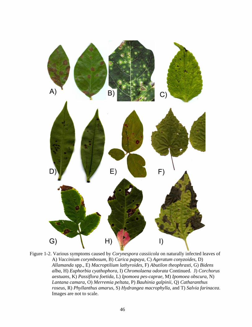
Figure 1-2. Various symptoms caused by Corynespora cassiicola on naturally infected leaves of
A) Vaccinium corymbosum, B) Carica papaya, C) Ageratum conyzoides, D) Allamanda spp., E) Macroptilium lathyroides, F) Abutilon theophrasti, G) Bidens alba, H) Euphorbia cyathophora, I) Chromolaena odorata Continued. J) Corchorus aestuans, K) Passiflora foetida, L) Ipomoea pes-caprae, M) Ipomoea obscura, N) Lantana camara, O) Merremia peltata, P) Bauhinia galpinii, Q) Catharanthus roseus, R) Phyllanthus amarus, S) Hydrangea macrophylla, and T) Salvia farinacea. Images are not to scale.

47
Figure 1-2. Continued.

48
CHAPTER 2 GENETIC AND PATHOGENIC DIVERSITY OF CORYNESPORA CASSIICOLA
Introduction
Target spot, caused by the fungal pathogen Corynespora cassiicola (Berk. & Curt.) Wei, is
common in the tropics, subtropics, and greenhouses (Chase 1987). C. cassiicola is reported to
infect 530 plant species from 380 genera, including monocots, dicots, ferns, and one cycad
(Chapter 1, this dissertation). Isolate characterization is needed to determine which hosts might
serve as sources of inoculum for target spot of tomato species and other hosts since there is much
variability concerning the host range of individual isolates. Some isolates show pathogenicity to
a wide range of hosts, whereas others exhibit host specificity, and some are only pathogenic
when associated with wounding (Chase 1982; Cutrim and Silva 2003; Kingsland 1985;
Onesirosan et al. 1973, 1974; Pereira et al. 2003; Poltronieri et al. 2003; Seaman et al. 1965;
Smith and Schlub 2004; Smith and Schlub 2005; Spencer and Walters 1969; Volin and
Pohronezny 1989). At least two races of the fungus have been distinguished based on their
differential pathogenicity response on soybean and cowpea (Olive and Bain 1945; Spencer and
Walters 1969). However, isolates from soybean, sesame, cowpea and cotton in Mississippi were
alike in pathogenicity (Jones 1961). A more extensive study found eight different pathogenicity
profiles among 28 isolates from soybean in Mexico, cucumber in Florida, and diverse hosts in
Nigeria (Onesirosan et al. 1974). Furukawa et al. (2008) found that an isolate from Salvia
splendens was not pathogenic to cucumber, green pepper or hydrangea; however isolates from
these hosts were pathogenic to Salvia splendens. Furukawa et al. (2008), therefore,
demonstrated that isolates with different pathogenicity profiles can be found on the same host.
Since the 1960’s, a leaf and fruit spot disease of tomato caused by C. cassiicola has
become increasingly serious in tropical countries worldwide (Jones and Jones 1984). It was first

49
reported in Florida in 1972 and has since become one of state’s most damaging foliage and fruit
diseases (Blazquez 1972; Pernezny et al. 1993, 1996, 2000, 2002). Under warm, humid,
conditions the disease leads to heavy defoliation and significant losses in yield (Volin and
Pohronezny 1989). Currently, there are no resistant tomato cultivars available, although
resistance found in PI 120265 (Lycopersicon esculentum) and PI 11215 (L. pimpinellifolium) and
was controlled by a single recessive gene (Bliss et al. 1973). Understanding the genetic and
pathogenic diversity of the pathogen and its distribution is vital to isolate selection for resistance
screening.
Kingsland (1985) compared three isolates from tomato, cucumber and papaya debris and
found that tomato and cucumber were susceptible to all isolates, but the isolate from papaya
debris was not pathogenic on papaya, indicating that it was possibly growing as a saprophyte. In
many studies, isolates were found to be non-pathogenic on the hosts from which they were
isolated, further indicating that C. cassiicola can grow as a saprophyte (Chase 1982; Kingsland
1985; Onesirosan et al. 1974; Hyde et al. 2001; Lee et al. 2004). Other studies show that isolates
are only secondary invaders, or invaders of senescent tissue. Isolates from the ornamental hosts
Aeschynanthus pulcher (lipstick vine), Aphelandra squarrosa (zebra plant), azalea and
hydrangea were pathogenic on all hosts in cross-pathogenicity trials when wounded; however,
only A. pulcher was susceptible without wounding (Chase 1982).
Silva et al. (1998) compared pathogenicity of 16 isolates from rubber trees in Sri Lanka
and five isolates from diverse hosts in Australia. Papaya isolates from Australia were pathogenic
to tomato and rubber, but not cowpea and eggplant. Mimosa and thyme isolates from Australia
were pathogenic to eggplant, rubber, and tomato, but not cowpea. Isolates from Sri Lanka
50
collected from different rubber clones were either pathogenic to all hosts (cowpea, eggplant,
rubber, and tomato), or pathogenic to all hosts but eggplant.
The host specificity and severity of the fungus on Lantana camara in Brazil has led to the
discovery that C. cassiicola may be useful as a bioherbicide (Pereira et al. 2003). Based on the
vast number of weeds that serve as hosts of the fungus, there is great potential for the discovery
of several more isolates useful for biological control of weeds. Considering the wide variation in
isolate pathogenicity that has been previously reported, additional studies are needed to further
understand the host range of individual isolates from different hosts and locations.
Prior research on the genetic characterization of C. cassiicola is limited to restriction
fragment length polymorphism (RFLP) of ITS rDNA and random amplified polymorphic DNA
(RAPD) studies. No variation between five isolates of C. cassiicola collected from mimosa,
papaya, and thyme in Australia was found based on RFLP of ITS (Silva et al. 1995). Silva et al.
(1995) concluded that RFLP of the ITS regions of rDNA can be used to distinguish between
Corynespora and the morphologically similar genus Helminthosporium, but not different isolates
of C. cassiicola. However, the three isolates from papaya had identical RAPD patterns, growth
rate, isolate color, and pathogenicity profiles, which were different from the isolates from
mimosa and thyme, indicating an ongoing process of host specialization on papaya (Silva et al.
1995).
RAPD analyses from 27 isolates collected from Hevea brasiliensis, in Sri Lanka revealed
correlations between host location, host genotype, isolate morphology, and isolate pathogenicity
(Silva et al. 1998). Silva et al. (1998) concluded that a progenitor strain may have been spread in
India by distribution of live plant material. Prior outbreaks of the disease on the susceptible

51
rubber clone RRIC 103 in other countries, and the sudden appearance and severity of target spot
on the same clone in Sri Lanka in 1985, is evidence for such dissemination.
Silva et al. (2003) characterized 42 isolates from bitter gourd, cocoa, manihot, papaya,
rubber, sweet potato, tomato, and wing-bean from various regions in India based on RAPD
analyses. RAPD groups did not correlate with geographic origin, but isolates obtained from
rubber clone RRIC 103 grouped together. This strain might be responsible for several recent
outbreaks on this clone. In addition, all but one of the isolates from rubber clone RRIC 110
clustered in 2 RAPD groups, which may identify the strain that caused the outbreak on this clone
in 1995. Silva et al. (2003) concluded that correlation of RAPD groups with pathogenicity was
needed to help develop resistant clones against all pathogenic isolates.
Atan and Hamid (2003) characterized nine C. cassiicola isolates from Hevea brasiliensis
in Malaysia using RAPD of genomic DNA and RFLP of amplified ITS regions. RFLP analyses
with three restriction enzymes yielded monomorphic patterns. However, isolate OPEN 1 from
clone RRIM 2020 had a distinct RFLP pattern from the other eight isolates after digestion with
HaeIII. RAPD results indicated the presence of at least two genetically distinct races that infect
rubber. Seven isolates pathogenic to clones RRIM 600, RRIM 2009, and two unidentified
rubber clones were molecularly similar and identified as Race 1. The remaining two isolates,
both pathogenic on clone RRIM 2020, had identical banding patterns and were considered Race
2.
Unfortunately, the majority of the diversity assessments are limited to rubber isolates from
Malaysia and Sri Lanka and are based on RAPD techniques, which is problematic with respect to
repeatability and homology assessment (Isabel et al. 1999). In addition, all the RFLP studies
used the ITS rDNA region which has minimal variation among isolates (Silva et al. 1995, 1998).

52
Investigations into the genetic variation among C. cassiicola isolates using more reliable
molecular methods and more diverse isolates are needed.
In this study, we collected and solicited 143 isolates from diverse hosts and locations. To
test whether C. cassiicola is panmictic throughout its range, allelic genealogies were constructed
from four loci including the rDNA ITS region, two random hypervariable loci, Cc caa5 and Cc
ga4, and the single copy actin-encoding nuclear gene, Cc act1. Fifty of these isolates were spray
inoculated on seedlings of eight crop plants to test pathogenicity profiles. Correlations among an
isolate’s pathogenicity profile, its host of origin, and genotype were investigated. The purpose of
this research is to gain knowledge of the diversity within the species C. cassiicola because of its
implications for resistance breeding and disease management of target spot of basil, bean,
cowpea, cucumber, papaya, soybean, sweet potato, tomato, and potentially other crops.
Methods
Collection and Solicitation of Fungal Isolates
C. cassiicola isolates were collected from diverse plant hosts during 5-day collecting trips
to locations in the Pacific: American Samoa (AS), Hawaii (HI), Palau (PW), Pohnepei (PH),
Saipan (SN), and Yap (YP) in the summer of 2005. More extensive surveys were conducted to
collect the fungus in Florida (FL) and Guam (GU) between 2004-2006 (see Chapter 1).
Farms, nurseries, and roadsides were surveyed for plants with target spot symptoms. First,
second, and third priority was given to crops, weeds, and naturalized or indigenous hosts of C.
cassiicola, respectively. Symptomatic leaves were put into individual plastic bags in the field
and later placed abaxial side up in petri dishes with moistened paper towels in a laboratory.
After 24 hours in the moisture chamber, petri plates were placed under the dissecting microscope
and suspected spores and conidiophores of C. cassiicola were confirmed microscopically.

53
Single spores were captured at the end of a teasing needle and transferred to antibiotic V8
agar (340 ml V8 juice, 660 ml water, 3g CaCO3, 17g agar, 100 μg/ml Ampicillin or Kanamycin)
slants, left at room temperature until the colony reached at least 5 cm in diameter, whereby it was
covered with autoclaved mineral oil, and stored at 5o C until further study. Sporulation from
non-symptomatic leaf material was noted, possibly indicating non-pathogenic growth.
To obtain globally diverse isolates, individual researchers in Brazil (BZ), Malaysia (MY),
Mississippi (MS), and Tennessee (TN) were solicited for additional C. cassiicola cultures.
Isolates from BZ on lantana (JMP216), papaya (DOA16b), soybean (RWB321) and tomato
(JMP217) came from Alvaro Almeida, EMBRAPA. Isolates CBPP, CLN 16 and CSB1 2 were
received from MY off of rubber from Dr. Safiah Atan, Malaysian Rubber Board. Isolate TN13-3
was received from Nashville, TN on greenhouse African violet from Justin S. Clark, University
of Tennessee. Isolate MS01 was received from MS on greenhouse tomato leaves from David
Ingram, Central MS Research and Extension Center.
Isolates of different species were also solicited from culture collections to serve as
outgroups. Cultures from Commonwealth Agricultural Bureaux International (CABI) in the
United Kingdom included C. smithii IMI 5649b and C. citricola IMI 211585. Cultures from
National Institute of Agrobiological Sciences (NIAS) in Japan included C. citricola MAFF No.
425231, C. melongenea MAFF No. 712045, and C. sesamum MAFF No. 305095. Cultures from
Centraalbureau voor Schimmelcultures (CBS) in the Netherlands included C. proliferata CBS
112393, C. citricola CBS 169.77, and C. olivaceae CBS 291.74. Cultures were single-spored
after they were received. A complete list of isolates used in these studies, along with the plant
host, geographic location and the type of association with the host plant (endophytic or
pathogenic growth) can be found in Table 2-1.

54
Primer Development for Random Hypervariable Loci
Three C. cassiicola isolates from long-term storage (FL31, GU112, and PW56) were
chosen based on unique host and location. A small piece of mycelium from the monosporic
cultures was extracted with tweezers and placed onto a V8 agar plate. The isolates were grown
under constant fluorescent light for 7 days. Aerial mycelium was scraped from the agar surface,
placed in 1.5 ml microcentrifuge tubes, lyophilized overnight, and then frozen in liquid nitrogen.
Genomic DNA was purified using the DNeasy plant Mini Kit (Quiagen, Inc.) according to the
manufacturer’s specifications.
Genomic DNA combined from all three isolates was digested with the Sau 3AI restriction
enzyme (7.2 μl of DNA from each of the three isolates; 2.5 μl 10X buffer; 1.0 μl 10 U/μl Sau 3A
I enzyme; incubated at 37oC for 2 hours). The digested genomic DNA was fractionated to
remove fragments less than 400 bp using a Chroma Spin® column (Chroma Spin + TE – 400,
Clonetech Laboratories, Inc.) according to the manufacturer’s specifications. The digested
fractionated DNA was quantified and ligated to Sau 3AI linkers and incubated at 16oC overnight.
Excess linkers were removed using the same Chroma® Spin column as above. The linker-
ligated fragments were PCR amplified using SauL-A primers and a program consisting of initial
denaturation for 3 min at 94oC, followed by 25 cycles of 94oC for 1 min, 68oC for 1 min, and
72oC for 2 min, and a final amplification at 72oC for 10 min.
The amplified genomic PCR library (composed of 400-1500 bp fragments) was enriched
for fragments containing two different microsatellite repeats, (CAA)n and (GA)n. The denatured
genomic PCR library was hybridized to the following biotinylated oligoprobes:
[5’(CAA)15TATAAGATA-Biotin] and [5’(GA)15TATAAGATA-Biotin] (Tepnel Lifecodes
Corporation) and incubated at 48oC overnight. The PCR fragments that hybridized to the repeat
55
probes were captured and eluted using two VECTREX® Avidin D matrix columns (cat. No. A-
2020, Vector Laboratories, Burlingame, CA) according to the manufacturers specifications. The
two mixtures containing genomic fragments enriched for the (CAA)n tri-repeat and the (GA)n di-
repeat were PCR amplified using SauL-A primers following the same PCR conditions as above.
PCR products from the amplification of the enriched microsatellite library were ligated
into a plasmid vector (pCR 2.1-TOPO® vector; Invitrogen, Inc.) and transformed into E. coli
(One ShotTM TOP 10 Cells, Invitrogen, Inc.) using the TOPO TA Cloning Kit® (Invitrogen, Inc.)
according to the manufacturers instructions. Transformed colonies were lifted and crosslinked
onto nylon membranes in an UV chamber (GS Gene LinkerTM, Bio-Rad Laboratories, Inc.,
Hercules, CA) using the “optimal crosslink” program.
Nylon membranes were hybridized with alkaline phosphatase-labeled repeat probes
((CAA)n and (GA)n) and the Quick-LightTM hybridization Kit (Tepnel Lifecodes Corp.)
according to the manufacturers recommendation. Colonies containing plasmids that tested
positive for inserts with repeats were sequenced in one direction. Primers were designed to
amplify 300-500 base pair fragments flanking low repeat number (<10) sequences using Primer3
(v. 0.4.0). Sequences with repeats of less than 10 were likely to be non-variable microsatellite
loci, but may contain polymorphic flanking sequences. Sequences were screened for
polymorphisms using five isolates from different hosts and locations. Primers that amplified the
Cc ga4 and Cc caa5 loci were chosen for further study because they amplified sequences with
relatively high levels of polymorphism (>5%).
Fungal Cultures and Extraction of Genomic DNA
Genomic DNA from 143 isolates (Table 2-2) in long-term storage was purified and
amplified using Extract-N-Amp™ (Sigma-Aldrich) according to the manufacturer’s
specifications.

56
The following primers were used for PCR amplification: ITS1 and ITS4 (White et al.
1990) for the internal transcribed spacer region, including the 5.8 rRNA coding region; ACT-
512F and ACT-783R (Carbone and Kohn 1999) for the single copy nuclear actin locus Cc act1;
GA4-F (5’-CCT GCT CCG ACT TTG TTG AG-3’) and GA4-R (5’-GTC TGG GAG CAG CAA
AGA CT-3’) for the random hypervariable Cc ga4 locus; CAA5-F (5’-GTC CAC AAG TGG
AAC CTC GT-3’) and CAA5-R (5’-CCT CGT CTG CCA GTT CTT CT-3’) for the random
hypervariable Cc caa5 locus. “Hot-start” PCR was performed with a MyCyclerTM thermocycler
(BioRad) with a program consisting of initial denaturation for 3 min at 94oC, followed by 30
cycles of 30 sec at 94oC, 30 sec at 58oC, and 30 sec at 72oC, and a final cycle of 5 min at 72oC
for the ITS, Cc ga4, and Cc caa5 loci. For the Cc act1 locus, the program was identical except
for an annealing temperature of 61oC. PCR products were purified using the QIA quick PCR
purification Kit (QIAGEN Inc.) according to the manufacturer’s instructions. The purified
products were then quantified on 1% ethidium bromide-stained agarose gels. Sequencing of the
DNA samples was done at the University of Florida DNA Sequencing Core Laboratory using
ABI Prism BigDye Terminator cycle sequencing protocols (part number 4303153) developed by
Applied Biosystems (Perkin-Elmer Corp., Foster City, CA). The excess dye-labeled terminators
were removed using MultiScreen® 96-well filtration system (Millipore, Bedford, MA, USA).
The purified extension products were dried in SpeedVac® (ThermoSavant, Holbrook, NY, USA)
and then suspended in Hi-di formamide. Sequencing reactions were performed using POP-7
sieving matrix on 50-cm capillaries in an ABI Prism® 3130 Genetic Analyzer (Applied
Biosystems, Foster City, CA, USA) and were analyzed by ABI Sequencing Analysis software v.
5.2 and KB Basecaller.

57
Phylogenetic Analyses
Four loci (rDNA ITS, Cc caa5, Cc ga4, and Cc act1) from 143 isolates were sequenced.
Forward and reverse sequences from each PCR product were concatenated in SequencherTM 4.8
and trimmed to include only bases sequenced in both directions. Samples with ambiguities were
sent for re-sequencing. Multiple alignments from each locus were executed separately with
Clustal X (1.83.1) and the alignments were inspected and adjusted manually using MacClade
4.08 OS X (Maddison and Maddison 2005). Data from ITS rDNA, Cc ga4, Cc caa5, and Cc
act1 loci were partitioned to facilitate different permutations of combined analysis. A partition-
homogeneity test (incongruence length-difference test or ILD) was implemented to evaluate the
homogeneity of different data partition subsets using PAUP* v4.0b10 (Swafford 2002). The test
implemented 1,000 replicates (heuristic search; random simple sequence additions; TBR; max-
trees = 1,000). Comparisons were evaluated using a threshold of p < 0.001 and were made
between all data partitions.
With the ILD test indicating the combinability of all molecular data, neighbor joining (NJ)
and maximum parsimony (MP) analyses were conducted for each data partition and the
combined data set using PAUP* (Swafford 2002). C. smithii IMI 5649b, C. citricola IMI
211585, C. proliferata CBS 112393, C. citricola CBS 169.77, and C. olivaceae CBS 291.74
were defined as outgroups. Cultures from National Institute of Agrobiological Sciences (NIAS)
in Japan (C. citricola MAFF No. 425231, C. melongenea MAFF No. 712045, and C. sesamum
MAFF No. 305095) were not included as outgroups because they grouped with C. cassiicola
isolates in phylogenetic analyses (see Results below). For the NJ analyses, default settings were
used except ties were broken randomly by initial seed.
Due to long computational time, MP analyses were conducted in the following manner.
An initial heuristic search was conducted with one random addition replicate, TBR (tree-

58
bisection-reconnection) branch swapping, and the MulTrees option (saving all optimal trees) in
effect. A second heuristic search was conducted using 1000 random addition replicates with the
above settings and saving no more than 10 trees with a score greater than or equal to the best tree
score from the first replicate in the previous analysis. In all analyses, gaps were treated as
missing data. Strict consensus trees were generated from analyses with multiple equally
parsimonious trees. For all MP analyses, statistical support for nodes was estimated using
maximum parsimony bootstrap (BS) replicates (Felsenstein 1985). For the combined data set,
BS estimates were obtained using 1,000 replicates, each with 100 random taxon addition
replicates and saving no more than 1,500 trees per bootstrap replicate, TBR branch swapping and
the MulTrees option in effect.
All data were also analyzed by Bayesian inference (BI) methods with MrBayes v3.1.2
(Huelsenbeck and Ronquist 2001; Ronquist and Huelsenbeck 2003). An appropriate model of
evolution (under the AIC criterion) was selected for each data partition using the program
Modeltest v3.4 (Posada and Crandall 1998). All Bayesian analyses (individual loci and
combined data) were conducted while retaining the appropriate model for each data partition.
Markov Chain Monte Carlo was implemented with four heated chains and trees were sampled
every 1,000th generation for one million generations. The first 25 percent of the total number of
generations was discarded as burn-in. A 50 percent majority rule consensus tree was generated
from the remaining trees, in which the percentage of nodes recovered represented their posterior
probability (PP).
Congruent nodes resulting from the NJ, MP, and BI analyses of the combined molecular
data was used to assign isolates to a phylogenetic lineage (PL). Only isolates that fell within
clades of high support (BS value >70 and PP value > 95) were assigned to a PL.

59
Pathogenicity Analyses
Fifty out of the 143 Corynespora isolates were used for pathogenicity profiling (Table 2-3)
on eight crop plants. Isolates originally isolated from crop plants and from all phylogenetic
lineages were chosen. Each isolate was spray-inoculated onto four replicate plants of eight-
week-old: basil ‘Italian Large Leaf’ (Ba), bean ‘Bush Kentucky Wonder’ (Be), cowpea
‘California black-eye’ (Co), cucumber ‘Straight 8’ (Cu), soybean ‘AG00901’ (So), and tomato
‘Rutgers’ (To) seedlings; 8-week-old sweet potato ‘Beauregard’ (Sw) cuttings; and 12-week-old
papaya ‘HI Sunrise’ (Pa) seedlings. Cultivars were chosen based on their known susceptibility
in the survey regions.
To increase colony sporulation for inoculum preparation, aerial mycelium from 10-day-old
V8 agar plates was gently scraped with a glass cover slip to flatten mycelium and then placed
under constant cool-white fluorescent light (Onesirosan et al. 1975). Three days later, the
surface of the agar was scraped with a glass cover slip and the resulting mycelia was blended in
200 ml sterile distilled water for two seconds and filtered through three layers of cheesecloth.
Spores were counted under a hemacytometer and the concentration was adjusted to 20,000
spores/ml. One drop of Tween® 20 per 100 ml was added to the inoculum. Plants were sprayed
with the spore suspension until leaf run off (about 500 ml), making sure that both leaf surfaces
were fully covered. Plants were kept on a mist bench to maintain constant leaf moisture 3 days
prior to inoculation and for the remainder of the experiment. Plants were rated 7 days after
inoculation using the rating system developed by Onesirosan et al. (1973): (0) symptomless, no
lesions on leaves or stems; (1) non pathogenic hypersensitive response, a few to many non-
expanding pinpoint lesions; (2) moderately virulent, many expanding lesions, some coalescing,
but not resulting in blight; (3) highly virulent, lesions spreading to form large areas of dead tissue
resulting in a blighting effect. Incidence (I), defined as the number of plants showing symptoms

60
(with ratings of 1, 2, or 3), and severity (S), defined as the average rating for all symptomatic
plants, were recorded. The experiment was repeated.
Each isolate was assigned a pathogenicity profile (PP), which is a list of susceptible hosts.
Hosts were considered susceptible if at least one of the replicates from the two experiments (total
of eight plants) received a rating of 2 or 3. PPs were converted to a binary character matrix so
that each isolate received a zero (non-pathogenic, all reps with ratings of 0 or 1) or a one
(pathogenic, at least one rep with a rating of 2 or 3) for each host. Unweighted pair group
method with arithmetic mean (UPGMA) trees were constructed from the binary matrix and
internal support for nodes was estimated using bootstrap analyses with 1,000 reps and a UPGMA
algorithm. The tree topology was visually compared to the PL designation of each isolate tested
(Figure 2-6). PPs were also visually mapped on the four-locus combined BI phylogenetic tree
(Figure 2-1).
Growth Rate Analyses
Seventy-seven isolates were tested for growth rate at two temperatures (23 C and 33 C). A
small piece of aerial mycelium was extracted from the monosporic cultures in long-term storage
with tweezers and placed onto a V8 agar plate. After 5 days, the 77 colonies had grown beyond
the mineral oil and six 4 mm agar plugs were cut from actively growing mycelium at the colony
edge. A single plug was placed in the center of six V8 agar plates. Three replicate plates of each
isolate were immediately placed in growth chambers at 23 C and 33 C under 12 hours of
alternating fluorescent light (ca. 25 lux) and dark.
The average of two colony diameters at 90 degrees from each other was recorded at 48, 72,
96, 120, 144, and 166 hours. Average colony diameter was plotted against time and a line of
best fit was generated for each replicate. The slope of the line of best fit (R2>0.98) was used to
compare variation within reps to variation between isolates in SAS® Statistical Software
61
(Version 8, 1999). The experiment was repeated with five isolates with no statistically
significant variation (data not shown). A correlation between isolate growth rate and
phylogenetic lineage was tested using SAS statistical software.
Results
Phylogenetic Analyses
The General Time Reversible model (GTR + I + Γ) was selected by Modeltest for each of
the four gene partitions. The corresponding model for each locus was applied to all BI analyses
and the combined dataset was partitioned. The final combined dataset contains 2,136 aligned
characters used for analyses. Tree topologies resulting from NJ, MP, and BI analyses recovered
essentially the same well-supported nodes. The analyses reveal four major phylogenetic lineages
(PL) with high statistical support (BS value >70 and PP value > 95) (Figure 2-1).
All major PLs contain isolates from diverse locations, indicating their global dispersal.
PL1 contains a distinct clade with high statistical support (designated PL1.1) containing only
isolates collected from papaya from around the world indicating specialization on this host.
PL1.2 contains two isolates from Stachytarpheta jamaicensis, collected from Guam and Palau,
indicating potential specialization on this host. This supports pathogenicity studies showing
isolate specificity to this host (Smith and Schlub 2005). Isolates from diverse hosts are present
in PL1 including crops (basil, bitter melon, eggplant, cowpea, cucumber, oregano, pumpkin,
rubber, soybean, sweet potato, watermelon), ornamentals (Buddleja, Catharanthus, Codiaeum,
Coleus, Episcia, and Tabebouia), and weeds (Bidens, Buchnera, Clerodendrum, Commelina,
Lantana, Macroptilium, Meisosperma, Vitex). Tomato isolates are missing from PL1, indicating
that isolates in this lineage may not be pathogenic to tomato.
Isolates in PL2 are also globally distributed and include crops (cucumber, rubber, sweet
potato), ornamentals (African violet, Allamanda, Catharanthus, Pilea), and weeds (Piper, Pilea).

62
There is also a lack of tomato isolates in PL2 indicating that isolates from this lineage may be
nonpathogenic on this host. Though PL2 was highly supported (BS and PP values of 100), its
sister relationship to PL1, PL3, and PL4 remains unresolved.
Globally distributed isolates from PL3 include crops (basil, bitter melon, cucumber,
pumpkin, soybean, tomato), ornamentals (Bauhinia, Moringa, Pachystachys, Plectranthus,
Saintpaulia), and weeds (Acanthus, Asystasia, Calopogonium, Coccinia, Euphorbia, Luffa,
Passiflora, Teramnus). These are hosts that may harbor isolates pathogenic to tomato. PL5 and
PL6 group with PL3 with low support (MPBS value of 60). PL5 contain C. cassiicola isolates
from African violet in Guam and Tennessee that are very similar in sequence, especially at the
Cc-caa5 locus, indicating specialization on this host. African violet isolates from Saipan and
Yap are found in PL3. PL6 is highly supported and contains isolates from Brazil on Coleus,
Palau on cowpea, and Saipan on Asystasia.
The majority of tomato isolates group in PL4 from diverse locations including American
Samoa, Brazil, Florida, Guam, Mississippi, Palau, and Saipan. These twelve tomato isolates also
group with isolates from crops (bean, cassava, cucumber, sweet potato), ornamentals (Bauhinia,
Cassia, Coleus, Eugenia, Ficus, Jatropha, Salvia, Syzygium), and common weeds
(Calopogonium, Calyptocarpus, Chromolaena, Euphorbia, Hyptus, Lantana, Mikania,
Spathodea), which are likely inoculum sources for the initiation of disease on tomato.
The rDNA ITS region (Figure 2-2) is composed of 1,013 characters, 400 of which are an
insertion in the outgroup taxa C. smithii. Of the 612 remaining characters, 141 are variable and
107 are informative. The rDNA ITS sequences reveal the misidentification of three outgroup
taxa from the NIAS culture collection. C. sesamum 305095, C. citricola 425231, and C.
melongenea 712045 should be reclassified as C. cassiicola based on rDNA ITS sequences.

63
When the outgroup taxa C. citricola, C. olivaceae, C. proliferata, and C. smithii are removed
from the analyses, the rDNA ITS sequences of C. cassiicola contain only three informative
characters out of 584 bases. Two of these characters separate the isolates into three distinct
phylogenetic lineages that correlate with the PLs in the combined analysis. These two characters
are base pair 158 (C or T), and base pair 497 (A or G) of the C. cassiicola rDNA ITS alignment.
Three haplotypes are represented by these two characters: CA, CG, and TG (no haplotype TA);
all isolates with haplotype CA group in PL4, isolates with haplotype TG group in PL1, isolates
with haplotype CG group in PL2, PL3, PL5 and PL6. CG is also the ancestral haplotype, present
in all outgroups except for C. proliferata (haplotype CA). The third informative character in the
rDNA ITS sequences of C. cassiicola is base pair 123, which is a T in the majority of isolates,
but a C in isolates PW101 (PL5), RWB321 (PL5), SN64 (PL5.1), and TN13-3 (PL6). It is this
character (bp 123) that caused the polymorphic band pattern observed by Atan and Hamid (2003)
in their RFLP analysis of the rDNA ITS region of rubber isolates using HaeIII (recognition
sequence GGCC).
The sister relationships between the phylogenetic lineages remain unresolved in the
analyses of the individual loci and in the combined analyses. In addition, the ITS rDNA region
was the only locus that showed good support for C. citricola, C. olivaceae, C. proliferata, and C.
smithii as sister taxa to the ingroup of C. cassiicola isolates. The phylogenetic placement of the
outgroup taxa was not well supported in the combined analyses, or the GA4 locus. The CAA5
locus showed support for C. olivaceae, C. proliferata, and C. smithii as basal to PL1, PL2, PL3,
PL5, and PL6, but PL4 and C. citricola fell basal to that group. The apparent paraphyly of C.
cassiicola at the Cc-caa5 locus may be a result of character variation that occurred at this locus

64
before the species evolved. Additional loci containing characters that reveal the sister
relationships of the different PLs are needed in future analyses.
The Cc-caa5 locus (Figure 2-3) reveals similar tree topologies to the combined analyses
with high support for the four major PLs. Differences at this locus in PL1 include the lack of
PL1.1 that distinguishes papaya isolates from other isolates in PL1 in the combined analyses. In
addition, PL1.2, which includes two rubber isolates from Malaysia, has low support. GU70 and
SN59, isolates basal to PL1 in the combined analyses, groups with other isolates in PL1 at this
locus. The Cc-caa5 locus shows strong support for PL2 with the same nine isolates as in the
combined analysis. The Cc-caa5 locus does not resolve PL3.1 or PL3.3 as distinct from PL3,
although the five isolates in PL3.2 group together with strong support. This locus does not
distinguish isolates FL2920, GU120, GU136 as distinct from other isolates in PL4. PL5 and
PL5.1 isolates are group basal to PL3, but with low support. PL6, which includes African violet
isolates from Guam and Tennessee, group with isolate NIAS 712045 with high support. Isolates
FL50 (Hydrangea macrophylla) and FL51 (Vaccinium corymbosum) are unresolved at this locus
as well as in combined analyses.
The Cc-ga4 locus (Figure 2-4) highly supports PL1, PL2, PL4, and PL6, although the
sister relationships between the PLs are unresolved. Isolates in PL3 form a clade with low
support. The Cc-ga4 locus did reveal a shared haplotype between papaya isolates with a point
mutation from an A to a G at base 74. C. cassiicola isolates are not monophyletic at this locus
because the outgroups C. proliferata, C. olivaceae, and C. smithii fall basal to PL1, PL2, and
PL6 with low support. This may be a result of character variation before speciation, a high
incidence of homoplasious characters, or the convergent evolution of specific adaptations.

65
The Cc-act1 locus could not be amplified in the outgroup taxa C. citricola, C. proliferata,
and C. smithii, perhaps due to mutations in the primer annealing site. The locus did amplify in
C. olivaceae and shows high variation from C. cassiicola isolates (Figure 2-5). There is good
support for PL3, PL2, and PL4 at this locus, although only marginal support for PL1 (BS value
of 68). Again, the sister relationships between the PLs are unresolved. The Cc-act1 locus also
reveals a shared haplotype between papaya isolates with a point mutation from an A to a G at
base 229.
Pathogenicity Analyses
As a result of screening fifty isolates for pathogenicity on eight index hosts, 16 unique
pathogenicity profiles (PP) were developed (Table 2-3). The most common PP was CuTo,
followed by Pa and CuSwTo. Cucumber was the most susceptible host, with all isolates
producing symptoms and an average severity rating of 2.3. Tomato was also highly susceptible
with 49 out of 50 isolates showing symptoms with an average severity rating of 1.8. Even
though only eight isolates were pathogenic on papaya, the average severity rating was 2.1
indicating that pathogenic isolates were highly virulent. Isolates pathogenic to basil, bean,
cowpea, soybean and sweet potato were less virulent on these hosts with average severity ratings
less than 1.5.
There was a strong correlation between PP and PL (Figure 2-6). Seven out of ten isolates
with PP CuTo were from PL4 and all isolates with PP CuSwTo and BeCuSwTo were from PL4.
In PL4, all isolates but SN37 were highly virulent on tomato (average severity ratings ranging
from 2.5 to 3) and all isolates but GU28 were pathogenic to cucumber (average severity ratings
ranging from 1.3 to 3). In addition, the only isolates pathogenic to bean were from PL4,
although these five isolates were weakly virulent (average severity ratings ranging from 1.3 to
1.9).

66
In PL3, all isolates were strongly pathogenic to cucumber (average severity ratings ranging
from 2.3 to 2.8) and six out of seven isolates were strongly pathogenic to tomato (average
severity ratings ranging from 2.5 to 3). Four out of the six isolates also were pathogenic to basil
and all isolates with PP BaCuTo were from PL3.
Pathogenicity profile CuSw was unique to isolates from PL2 and all isolates tested from
PL2 had this profile. In addition, isolates collected in the field from papaya in PL1.1 were
specific to papaya in pathogenicity studies, although all isolates were weakly virulent on
cucumber with average severity ratings of 1.3 or less. All isolates from PL1 were pathogenic to
cucumber with average severity ratings ranging from 1.3 to 3. Nine out of the 13 isolates from
PL1 were pathogenic to cowpea and seven were pathogenic to basil. The only other host
susceptible to isolates from PL1 was soybean, which was only weakly susceptible when
inoculated with isolate PW87.
Growth Rate Analyses
The null hypotheses of no growth rate differences among isolates, phylogenetic lineage,
and temperatures were rejected (P<0.0001), while the null hypotheses of no growth rate
differences among repetitions was accepted with a probability of 0.7546. The 77 isolates tested
all grew faster at 23 C than 33 C. At 23 C, average isolate growth rate (average of three
repetitions) was between 0.1479 and 0.474 with an overall mean of 0.3855 (Table 2-4). At 33 C,
average isolate growth rate was between 0.1382 and 0.4153, with an overall mean of 0.2958
(Table 2-5). At 23 C, there were 29 significantly different growth rates and at 33 C there were
39 significantly different growth rates. Among the fastest growing isolates at both temperatures
were FL37, GU90, GU99, AS67, and HI01. The slowest growing isolates were very different at
the two temperatures. Slow growing isolates at 23 C were PH01, JMP216a, GU120, FL15, and

67
DOA16b. Slow growing isolates at 33 C were AS119, AS117, JMP217, AS49, GU120, and
GU112.
Though there were not enough replicates to test for interactions among effects, growth rate
alone correlated with location, phylogenetic lineage, and with host. Isolates from Oahu and
Palau grew the fastest at both temperatures. Isolates from Brazil, Florida and Malaysa tended to
have slower growth rates at both temperatures. Surprisingly, American Samoan isolates grew
proportionately much faster at 23 C than 33 C, despite its tropical climate. Isolates from PL6
and PL1 grew the fastest at both temperatures. Isolates from PL2 and PL4 grew the slowest at
23 C and isolates from PL5 and PL3 grew the slowest at 33 C. All isolates from Clerodendrum,
Commelina, Ficus, Macroptilium, pumpkin, and Stachytarpheta were fast growing at both
temperatures. In addition, isolates from Allamanda, Coleus, eggplant, Lantana, and tomato
isolates had slower growth rates at both temperatures.
Discussion
The current study presents the first robust, global phylogeny of the species Corynespora
cassiicola. Based on sequence data from four unique loci, there is evidence for high genetic
diversity within the species. The highly clonal nature of C. cassiicola is demonstrated in the
congruence of the phylogenetic trees from distinct loci. All loci distinguish four major clonal
lineages within C. cassiicola. The low level of sequence variation at the rDNA ITS region
within the species relative to other Corynespora species suggests that these lineages are in fact
clonal populations, rather than taxonomically distinct species.
As reported previously, the pattern of distribution of the diversity within the species
correlates with the host (Smith et al. 2008a). Identical haplotypes are widely distributed
geographically. The lack of correlation between phylogenetic data and location provides
evidence for the recent global dispersal of isolates from all four phylogenetic lineages. In

68
addition, geographically diverse isolates from the same host plant shared identical haplotypes,
potentially indicating host specialization. For example, isolates collected from tomato in Brazil,
Florida, Guam, Mississippi, Palau, and Saipan had identical haplotypes at all four loci. Isolates
collected from Lantana in Florida and Brazil were also identical at all four loci. Isolates
collected from African violet in Guam and Tennessee were unique from all other isolates and
nearly identical to each other. Perhaps the most compelling evidence for host specialization is
the shared identical sequences of all isolates collected from papaya from very diverse locations.
Tomato isolates from diverse locations including North and South America and the Pacific
Islands are found in only two of the five major phylogenetic lineages (PL3 and PL4). Isolates
from other hosts that fall into these same PLs are likely pathogenic to tomato and may serve as
source hosts or alternative hosts for target spot of tomato. Tomato isolates are genetically similar
to isolates from common crops (basil, bean, bitter melon, cassava, cucumber, papaya, pumpkin,
soybean, sweet potato), weeds (Acanthus, Calopogonium, Calyptocarpus, Chromolaena,
Coccinia, Euphorbia, Lantana, Macroptilium, Mikania, Momordica, Passiflora, and Teramnus),
and ornamentals (Asystasia, Bauhinia, Cassia, Coleus, Eugenia, Euphorbia, Ficus, Hyptus,
Jatropha, Luffa, Moringa, Pachystachys, Plectranthus, Saintpaulia, Salvia, Spathodea, and
Syzygium). Based strictly on these data, control of target spot should involve isolation of tomato
fields from these plant species, when possible.
Pathogenicity testing, in addition to phylogenetics, should be used to determine which
hosts might serve as sources of inoculum for the initiation of target spot of tomato. There are at
least sixteen unique pathogenicity profiles within C. cassiicola on the eight crop plants that were
tested. Isolates from the same lineages show similar but not identical profiles (Figure 2-1 and
Figure 2-6). For example, all but two isolates in PL3 and PL4 are pathogenic to tomato, and

69
isolates from all other lineages are nonpathogenic to tomato. All isolates pathogenic to basil are
from PL1 and PL3, but not all isolates in these clades are pathogenic to basil. Interestingly, the
majority of isolates, excluding the isolates collected from papaya, were pathogenic on cucumber.
Though there were no isolates collected from tomato that grouped in PL1 and PL2, isolates from
these lineages produced a hypersensitive response on tomato, showing pinpoint lesions that were
given a disease rating of one.
These data are similar to pathogenicity tests using 18 C. cassiicola isolates from Nigeria,
the Southern U.S., and Mexico (Onesirosan et al. 1973) in that both studies found isolates
specific to papaya and cucumber. Likewise, both studies found that isolates pathogenic to
tomato also were likely to be pathogenic on several other hosts. The number of isolates screened
compared to the number of unique pathogenicity profiles in both studies indicates that gains and
losses of pathogenicity are common.
Growth rate at different temperatures has provided evidence for isolates adapted to tropical
and temperate environments. Using an isolate collected from tomato in Florida, Pernezny et al.
(2000) found the best colony growth occurred at 32C, whereas Sobers (1966) reported an
optimum growth rate at 24C for Florida isolates collected from hydrangea and azalea. Jones and
Jones (1984) report higher disease severity on tomato inoculated and maintained at temperatures
between 20-23 C. In this study, two temperature extremes (23 C and 33 C) were chosen in
attempt to discern between isolates adapted to temperate and tropical climates. Though the
majority of isolates were collected from tropical climates, all isolates grew faster at 23 C than 33
C. Growth rate also strongly correlated with phylogenetic lineage. Isolates from PL2 and PL4
may be more adapted to warmer temperatures, and isolates from PL5 and PL3 might be more
adapted to cooler temperatures. Such physiological traits, including growth rate and
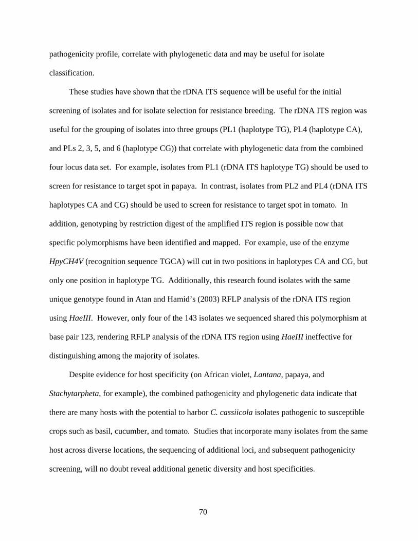
70
pathogenicity profile, correlate with phylogenetic data and may be useful for isolate
classification.
These studies have shown that the rDNA ITS sequence will be useful for the initial
screening of isolates and for isolate selection for resistance breeding. The rDNA ITS region was
useful for the grouping of isolates into three groups (PL1 (haplotype TG), PL4 (haplotype CA),
and PLs 2, 3, 5, and 6 (haplotype CG)) that correlate with phylogenetic data from the combined
four locus data set. For example, isolates from PL1 (rDNA ITS haplotype TG) should be used to
screen for resistance to target spot in papaya. In contrast, isolates from PL2 and PL4 (rDNA ITS
haplotypes CA and CG) should be used to screen for resistance to target spot in tomato. In
addition, genotyping by restriction digest of the amplified ITS region is possible now that
specific polymorphisms have been identified and mapped. For example, use of the enzyme
HpyCH4V (recognition sequence TGCA) will cut in two positions in haplotypes CA and CG, but
only one position in haplotype TG. Additionally, this research found isolates with the same
unique genotype found in Atan and Hamid’s (2003) RFLP analysis of the rDNA ITS region
using HaeIII. However, only four of the 143 isolates we sequenced shared this polymorphism at
base pair 123, rendering RFLP analysis of the rDNA ITS region using HaeIII ineffective for
distinguishing among the majority of isolates.
Despite evidence for host specificity (on African violet, Lantana, papaya, and
Stachytarpheta, for example), the combined pathogenicity and phylogenetic data indicate that
there are many hosts with the potential to harbor C. cassiicola isolates pathogenic to susceptible
crops such as basil, cucumber, and tomato. Studies that incorporate many isolates from the same
host across diverse locations, the sequencing of additional loci, and subsequent pathogenicity
screening, will no doubt reveal additional genetic diversity and host specificities.

71
It is hoped that this research will aid others in unraveling the many complexities that
remain to be discovered with respect to C. cassiicola and its disease development in the field.
For example, more studies are needed to explain why C. cassiicola is rare in Hawaii on all
cultivated crops except basil, if there are isolates adapted to tropical and temperate climates, and
how isolate genotype and pathogenicity profiles are correlated using more diverse isolates and
hosts.

72
Table 2-1. Isolate designations, geographic location of isolation, host of isolation, phylogenetic lineage (PL), type of growth on associated host, and species of Corynespora used in the phylogenetic analyses.
Isolate ID Location Host PL Growth Species CABI 211585 New Zealand Poncirus trifoliatus O endophytic C. citricola CBS 169.77 New Zealand Poncirus trifoliatus O endophytic C. citricola NIAS 425231 Japan Ocimum basilicum 1 pathogen C. citricola NIAS 712045 Japan Solanum melongenea ? pathogenic C. melongenae CBS 291.74 Netherlands Tilia spp. O saprophyte C. olivacea CBS 112393 Italy Fagus sylvatica O endophytic C. proliferata NIAS 305095 Japan Sesamum indicum 1 pathogenic C. sesamum CABI 5649b England Fagus sylvatica O saprophyte C. smithii AS49 Amer. Samoa Solanum lycopersicum 3 pathogenic C. cassiicola AS50 Amer. Samoa Solanum lycopersicum 3 pathogenic C. cassiicola AS54 Amer. Samoa Vigna unguiculata 1 saprophyte C. cassiicola AS58 Amer. Samoa Vigna unguiculata 1 saprophyte C. cassiicola AS65 Amer. Samoa Solanum melongenea 6 saprophyte C. cassiicola AS67 Amer. Samoa Commelina benghalensis 1 pathogenic C. cassiicola AS71 Amer. Samoa Cucurbita pepo 1 saprophyte C. cassiicola AS78 Amer. Samoa Ocimum basilicum 1 pathogenic C. cassiicola AS80 Amer. Samoa Ocimum basilicum 3.1 pathogenic C. cassiicola AS81 Amer. Samoa Clerodendrum quadriloculare 1 pathogenic C. cassiicola AS92 Amer. Samoa Cucumis sativus 6 pathogenic C. cassiicola AS98 Amer. Samoa Cucumis sativus 1 pathogenic C. cassiicola AS117 Amer. Samoa Carica papaya fruit 3.1 saprophyte C. cassiicola AS119 Amer. Samoa Cucurbita pepo 3.1 saprophyte C. cassiicola DOA16b Brazil Carica papaya 1.1 pathogenic C. cassiicola JMP216a Brazil Lantana camara 6 pathogenic C. cassiicola JMP217 Brazil Solanum lycopersicum 6 pathogenic C. cassiicola JMP218 Brazil Glycine max 1 pathogenic C. cassiicola RWB321 Brazil Coleus barbatus 4 pathogenic C. cassiicola FL09 FL, USA Lantana camara 6 pathogenic C. cassiicola FL11 FL, USA Carica papaya 1.1 pathogenic C. cassiicola FL12 FL, USA Solanum lycopersicum 6 pathogenic C. cassiicola FL15 FL, USA Salvia farinacea 6 pathogenic C. cassiicola FL21 FL, USA Bauhinia galpinii 6 pathogenic C. cassiicola FL34 FL, USA Tabebouia pallida 1 pathogenic C. cassiicola FL36 FL, USA Catharanthus roseus 2 pathogenic C. cassiicola FL37 FL, USA Clerodendrum paniculatum 1 pathogenic C. cassiicola FL50 FL, USA Hydrangea macrophylla ? pathogenic C. cassiicola FL51 FL, USA Vaccinium corymbosum ? pathogenic C. cassiicola FL62 FL, USA Coleus barbatus 1 pathogenic C. cassiicola FL757 FL, USA Origanum vulgare 1 pathogenic C. cassiicola FL2920 FL, USA Solanum lycopersicum 6 pathogenic C. cassiicola MS31 MS, USA Solanum lycopersicum 6 pathogenic C. cassiicola TN3-3 TN, USA Saintpaulia ionantha 5 pathogenic C. cassiicola GU01 Guam Cassia fistula 6 saprophyte C. cassiicola

73
Table 2-1. Continued. Isolate ID Location Host PL Growth Species GU06 Guam Hyptus suarelens 6 endophytic C. cassiicola GU08 Guam Lantana camara 1 pathogenic C. cassiicola GU10 Guam Codiaeum variegatum 1 endophytic C. cassiicola GU11 Guam Citrullus vulgaris 1 saprophyte C. cassiicola GU12 Guam Calopogonium mucunoides 6 pathogenic C. cassiicola GU14 Guam Calyptocarpus vialis 6 pathogenic C. cassiicola GU16 Guam Asystasia gangetica 3 pathogenic C. cassiicola GU21 Guam Buddleja asiatica 1 pathogenic C. cassiicola GU23 Guam Ipomoea batatas 6 endophytic C. cassiicola GU25 Guam Buchnera floridana 1 pathogenic C. cassiicola GU28 Guam Solanum lycopersicum 6 pathogenic C. cassiicola GU32 Guam Euphorbia heterophylla 3 endophytic C. cassiicola GU38 Guam Allamanda cathartica 2 pathogenic C. cassiicola GU41 Guam Eugenia uniflora 6 endophytic C. cassiicola GU42 Guam Bidens alba 1 pathogenic C. cassiicola GU44 Guam Jatropha curcas 6 endophytic C. cassiicola GU49 Guam Syzygium jambos 6 endophytic C. cassiicola GU51 Guam Meisosperma oppositifolium 1 endophytic C. cassiicola GU55 Guam Calopogonium mucunoides 3.1 pathogenic C. cassiicola GU65 Guam Passiflora foetida 3 endophytic C. cassiicola GU68 Guam Moringa oleifera 3 endophytic C. cassiicola GU70 Guam Solanum melongenea 1.3 endophytic C. cassiicola GU79 Guam Acanthus ilicifolius 3 endophytic C. cassiicola GU83 Guam Euphorbia heterophylla 6 endophytic C. cassiicola GU90 Guam Stachytarpheta jamaicensis 1 pathogenic C. cassiicola GU92 Guam Carica papaya 1.1 pathogenic C. cassiicola GU93 Guam Capsicum annum 1 endophytic C. cassiicola GU98 Guam Spathodea campanulata 6 pathogenic C. cassiicola GU99 Guam Saintpaulia ionantha 5 pathogenic C. cassiicola GU101 Guam Euphorbia milii 6 saprophyte C. cassiicola GU102 Guam Phaseolus vulgaris 6 saprophyte C. cassiicola GU103 Guam Pilea nummulariifolia 2 endophytic C. cassiicola GU104 Guam Macroptilium atropurpureum 1 pathogenic C. cassiicola GU107 Guam Mikania micrantha 6 pathogenic C. cassiicola GU109 Guam Bauhinia galpinii 3 pathogenic C. cassiicola GU110 Guam Plectranthus ambionicus 3 pathogenic C. cassiicola GU111 Guam Manihot esculenta 6 endophytic C. cassiicola GU112 Guam Glycine max 3 endophytic C. cassiicola GU114 Guam Teramnus labialis 3 endophytic C. cassiicola GU115 Guam Vitex parviflora 1 pathogenic C. cassiicola GU120 Guam Coleus barbatus 6 pathogenic C. cassiicola GU128 Guam Solanum lycopersicum 6 pathogenic C. cassiicola GU136 Guam Ficus benjamani 6.1 endophytic C. cassiicola HI01 Oahu, Hawaii Ocimum basilicum 1 pathogenic C. cassiicola CBPP Malaysia Hevea brasiliensis clone unk. 1.2 pathogenic C. cassiicola CLN16 Malaysia Hevea brasiliensis RRIM 2020 1.2 pathogenic C. cassiicola

74
Table 2-1. Continued. Isolate ID Location Host PL Growth Species CSB12 Malaysia Hevea brasiliensis RRIM 725 2 pathogenic C. cassiicola GU136 Guam Ficus benjamani 6.1 endophytic C. cassiicola HI01 Oahu, Hawaii Ocimum basilicum 1 pathogenic C. cassiicola CBPP Malaysia Hevea brasiliensis clone unk. 1.2 pathogenic C. cassiicola CLN16 Malaysia Hevea brasiliensis RRIM 2020 1.2 pathogenic C. cassiicola CSB12 Malaysia Hevea brasiliensis RRIM 725 2 pathogenic C. cassiicola PH01 Pohnpei Carica papaya 1.1 endophytic C. cassiicola PW01 Palau Carica papaya 1.1 pathogenic C. cassiicola PW12 Palau Carica papaya 1.1 pathogenic C. cassiicola PW17 Palau Carica papaya 1.1 pathogenic C. cassiicola PW20 Palau Carica papaya 1.1 pathogenic C. cassiicola PW25 Palau Carica papaya 1.1 pathogenic C. cassiicola PW27 Palau Carica papaya 1.1 pathogenic C. cassiicola PW34 Palau Carica papaya 1.1 pathogenic C. cassiicola PW37 Palau Carica papaya 1.1 pathogenic C. cassiicola PW38 Palau Carica papaya 1.1 pathogenic C. cassiicola PW43 Palau Carica papaya 1.1 pathogenic C. cassiicola PW48 Palau Carica papaya 1.1 pathogenic C. cassiicola PW53 Palau Carica papaya 1.1 pathogenic C. cassiicola PW56 Palau Carica papaya 1.1 pathogenic C. cassiicola PW57 Palau Solanum lycopersicum 6 pathogenic C. cassiicola PW63 Palau Solanum lycopersicum 6 pathogenic C. cassiicola PW69 Palau Piper betle 2 endophytic C. cassiicola PW79 Palau Pilea microphylla 2 pathogenic C. cassiicola PW80 Palau Saintpaulia ionantha 1 pathogenic C. cassiicola PW83 Palau Saintpaulia ionantha 1 pathogenic C. cassiicola PW87 Palau Cucumis sativus 1 pathogenic C. cassiicola PW89 Palau Chromolaena odorata 6 endophytic C. cassiicola PW91 Palau Luffa acutangula 1 endophytic C. cassiicola PW92 Palau Catharanthus roseus 1 pathogenic C. cassiicola PW94 Palau Stachytarpheta jamaicensis 1 pathogenic C. cassiicola PW99 Palau Momordica charantia 3 pathogenic C. cassiicola PW101 Palau Vigna unguiculata 4 saprophyte C. cassiicola SN03 Saipan Momordica charantia 1 pathogenic C. cassiicola SN05 Saipan Ipomoea batatas 1 pathogenic C. cassiicola SN06 Saipan Luffa acutangula 3.1 endophytic C. cassiicola SN07 Saipan Carica papaya 1.1 endophytic C. cassiicola SN18 Saipan Carica papaya 1.1 pathogenic C. cassiicola SN24 Saipan Solanum lycopersicum 6 pathogenic C. cassiicola SN27 Saipan Solanum lycopersicum 6 pathogenic C. cassiicola SN30 Saipan Solanum lycopersicum 6 pathogenic C. cassiicola SN37 Saipan Vigna unguiculata 6 saprophyte C. cassiicola SN40 Saipan Cucumis sativus 6 pathogenic C. cassiicola SN43 Saipan Saintpaulia ionantha 3 pathogenic C. cassiicola SN48 Saipan Coccinia grandis 3 endophytic C. cassiicola SN53 Saipan Carica papaya 1.1 pathogenic C. cassiicola
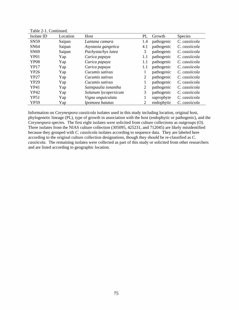
75
Table 2-1. Continued. Isolate ID Location Host PL Growth Species SN59 Saipan Lantana camara 1.4 pathogenic C. cassiicola SN64 Saipan Asystasia gangetica 4.1 pathogenic C. cassiicola SN69 Saipan Pachystachys lutea 3 pathogenic C. cassiicola YP01 Yap Carica papaya 1.1 pathogenic C. cassiicola YP08 Yap Carica papaya 1.1 pathogenic C. cassiicola YP17 Yap Carica papaya 1.1 pathogenic C. cassiicola YP26 Yap Cucumis sativus 1 pathogenic C. cassiicola YP27 Yap Cucumis sativus 2 pathogenic C. cassiicola YP29 Yap Cucumis sativus 1 pathogenic C. cassiicola YP41 Yap Saintpaulia ionantha 2 pathogenic C. cassiicola YP42 Yap Solanum lycopersicum 3 pathogenic C. cassiicola YP51 Yap Vigna unguiculata 1 saprophyte C. cassiicola YP59 Yap Ipomoea batatas 2 endophytic C. cassiicola
Information on Corynespora cassiicola isolates used in this study including location, original host, phylogenetic lineage (PL), type of growth in association with the host (endophytic or pathogenic), and the Corynespora species. The first eight isolates were solicited from culture collections as outgroups (O). Three isolates from the NIAS culture collection (305095, 425231, and 712045) are likely misidentified because they grouped with C. cassiicola isolates according to sequence data. They are labeled here according to the original culture collection designations, though they should be re-classified as C. cassiicola. The remaining isolates were collected as part of this study or solicited from other researchers and are listed according to geographic location.

76
Table 2-2. Summary of sequence data from four loci used to confirm the phylogenetic lineage of
Corynespora cassiicola isolates. Locus Total Variable Informative Tree Score No. MP Trees
Combineda 2136 248 174 330 7430rDNA ITS 1013 135 100 158 9990rDNA ITSb 584 4 3 4 1Cc-ga4 414 31 25 40 9530Cc-ga4b 414 28 25 36 9560Cc-caa5 366 38 34 52 40Cc-caa5b 366 37 32 46 12Cc-act1 343 44 15 49 11Cc-act1b 343 16 15 17 4a Combined loci: rDNA ITS, Cc-ga4, Cc-caa5, and Cc-act1. b Locus analyzed with only C. cassiicola taxa represented (no outgroups).

77
Table 2-3. Pathogenicity profiles for 50 Corynespora cassiicola isolates. Path Pro PL Isolate Host Ba Be Co Cu Pa So Sw To
I S I S I S I S I S I S I S I S BaCoCu 1 AS78 Ba 5 2.6 0 - 4 1.8 6 2.3 0 - 0 - 1 1 2 1 BaCoCu 1 AS58 Co 6 2.3 0 - 3 2.3 6 3 0 - 0 - 0 - 7 1 BaCoCu 1 YP29 Cu 7 2.1 0 - 3 1.7 8 3 0 - 0 - 0 - 0 - BaCoCu 1 AS71 Pu 8 2.1 0 - 2 2 7 1.3 0 - 1 1 0 - 2 1
BaCoCuSo 1 PW87 Cu 5 2.2 0 - 5 1.6 8 2.5 0 - 2 1.5 0 - 7 1 BaCu 1 HI01 Ba 4 2.5 0 - 0 0 7 3 0 - 0 - 1 1 8 1 BaCu 1 SN05 Sw 7 1.9 1 1 0 0 6 2.7 0 - 0 - 0 - 2 1
BaCuTo 3 AS50 To 5 2.2 0 - 2 1 6 2.7 0 - 1 1 2 1 8 3 BaCuTo 3 YP42 To 6 2.3 0 - 0 - 7 2.6 0 - 0 - 0 - 8 2.6 BaCuTo 3.1 AS80 Ba 5 1.8 0 - 0 - 8 2.5 0 - 1 1 0 - 8 2.8 BaCuTo 3.1 AS117 Sap 7 1.9 0 - 2 1 8 2.8 0 - 2 1 8 1 7 2.9
BeCoCuSw 4 SN37 Co 7 1 5 1.6 5 1.4 8 2.8 0 - 2 1 3 1.3 8 1 BeCuSwTo 4 JMP217 To 5 1 7 1.9 0 - 8 2.9 0 - 0 - 1 2 8 2.5 BeCuSwTo 4 GU102 Be 0 - 7 1.3 0 - 8 2.6 0 - 0 - 2 1.5 8 2.5 BeCuSwTo 4 SN40 Cu 0 - 6 1.3 0 - 8 2.5 0 - 0 - 1 2 8 2.6
BeCuTo 4 PW57 To 0 - 6 1.7 0 - 8 2.1 0 - 2 1 0 - 8 3 CoCu 1 AS98 Cu 0 - 5 1 6 1.8 8 2.6 0 - 0 - 0 - 7 1 CoCu 1 YP26 Cu 0 - 7 1 4 1.5 7 2.4 0 - 0 - 8 1 8 1 CoCu 1 JMP218 So 0 - 0 - 7 2.1 8 3 0 - 0 - 7 1 7 1 CoCu 1 GU08 La 0 - 0 - 3 1.7 7 3 0 - 0 - 0 - 8 1
Cu 1 AS54 Co 0 - 1 1 0 - 8 3 0 - 1 1 8 1 1 1 Cu 1 YP51 Co 0 - 0 - 0 - 8 3 0 - 0 - 7 1 1 1
CuPa 1.1 DOA16b Pa 7 1 0 - 0 - 8 1.1 8 2.3 0 - 8 1 1 1 CuPa 1.1 FL11 Pa 0 - 5 1 0 - 8 1.3 7 2.6 0 - 0 - 8 1 CuPa 1.1 PH01 Pa 1 1 1 1 0 - 7 1.1 6 1.8 0 - 0 - 8 1 CuSo 3 GU112 So 0 - 0 - 5 1 8 2.8 0 - 1 2 1 1 2 1 CuSw 2 YP27 Cu 1 1 0 - 2 1 6 2.8 0 - 0 - 5 1.4 2 1 CuSw 2 YP59 Sw 2 1 0 - 1 1 7 2.6 0 - 0 - 6 1.4 7 1 CuSw 2 SN59 La 1 1 5 1 0 - 8 2.8 0 - 0 - 7 1.3 7 1
CuSwTo 4 PW63 To 4 1 0 - 7 1 8 2.6 0 - 0 - 1 2 8 3 CuSwTo 4 SN24 To 0 - 0 - 0 - 8 2.5 0 - 0 - 2 1.5 8 2.6 CuSwTo 4 SN27 To 5 1 0 - 0 - 8 3 0 - 0 - 5 1.2 8 2.9
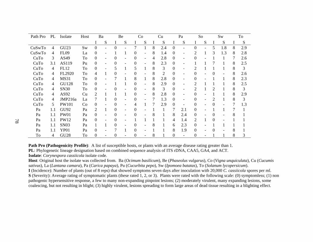
78
Path Pro PL Isolate Host Ba Be Co Cu Pa So Sw To
I S I S I S I S I S I S I S I S CuSwTo 4 GU23 Sw 0 - 0 - 7 1 8 2.4 0 - 0 - 5 1.8 8 2.9 CuSwTo 4 FL09 La 0 - 1 1 0 - 8 1.4 0 - 2 1 3 1.3 8 2.8
CuTo 3 AS49 To 0 - 0 - 0 - 4 2.8 0 - 0 - 1 1 7 2.6 CuTo 3.1 AS119 Pu 0 - 0 - 0 - 8 2.3 0 - 1 1 7 1 8 2.5 CuTo 4 FL12 To 0 - 5 1 5 1 8 3 0 - 2 1 1 1 8 3 CuTo 4 FL2920 To 4 1 0 - 0 - 8 2 0 - 0 - 0 - 8 2.6 CuTo 4 MS31 To 0 - 7 1 8 1 8 2.8 0 - 0 - 1 1 8 2.3 CuTo 4 GU128 To 0 - 1 1 0 - 8 2.9 0 - 2 1 1 1 8 2.5 CuTo 4 SN30 To 0 - 0 - 0 - 8 3 0 - 2 1 2 1 8 3 CuTo 4 AS92 Cu 2 1 1 1 0 - 8 2.8 0 - 0 - 1 1 8 2.9 CuTo 4 JMP216a La 7 1 0 - 0 - 7 1.3 0 - 0 - 2 1 8 3 CuTo 5 PW101 Co 0 - 0 - 4 1 7 2.9 0 - 0 - 0 - 7 1.3
Pa 1.1 GU92 Pa 2 1 0 - 0 - 1 1 7 2.1 0 - 1 1 7 1 Pa 1.1 PW01 Pa 0 - 0 - 0 - 8 1 8 2.4 0 - 0 - 8 1 Pa 1.1 PW12 Pa 0 - 0 - 1 1 1 1 4 1.4 2 1 0 - 1 1 Pa 1.1 SN03 Pa 1 1 0 - 0 - 8 1 6 2.3 0 - 1 1 1 1 Pa 1.1 YP01 Pa 0 - 7 1 0 - 1 1 8 1.9 0 - 0 - 8 1 To 4 GU28 To 0 - 0 - 0 - 8 1 0 - 0 - 1 1 8 3
Path Pro (Pathogenicity Profile): A list of susceptible hosts, or plants with an average disease rating greater than 1. PL: Phylogenetic lineage designation based on combined sequence analysis of ITS rDNA, CAA5, GA4, and ACT. Isolate: Corynespora cassiicola isolate code. Host: Original host the isolate was collected from. Ba (Ocimum basilicum), Be (Phaseolus vulgarus), Co (Vigna unquiculata), Cu (Cucumis sativus), La (Lantana camara), Pa (Carica papaya), Pu (Cucurbita pepo), Sw (Ipomoea batatas), To (Solanum lycopersicum). I (Incidence): Number of plants (out of 8 reps) that showed symptoms seven days after inoculation with 20,000 C. cassiicola spores per ml. S (Severity): Average rating of symptomatic plants (these rated 1, 2, or 3). Plants were rated with the following scale: (0) symptomless; (1) non pathogenic hypersensitive response, a few to many non-expanding pinpoint lesions; (2) moderately virulent, many expanding lesions, some coalescing, but not resulting in blight; (3) highly virulent, lesions spreading to form large areas of dead tissue resulting in a blighting effect.

79
Table 2-4. Growth rate of Corynespora cassiicola isolates at 23°C. Iso. ID PL Location Host Avg GR LSD
GU99 6 Guam Saintpaulia 0.4743 a AS81 1 Samoa Clerodendron 0.4639 ab GU90 1 Guam Stachytarpheta 0.4528 abc HI01 1 Oahu Basil 0.4521 abcd PW94 1 Palau Stachytarpheta 0.4521 abcd FL37 1 Florida Clerodendron 0.4514 abcd GU104 1 Guam Macroptilium 0.4507 abcde AS67 1 Samoa Commelina 0.4479 bcde AS54 1 Samoa Bean 0.4444 bcdef GU08 1 Guam Lantana 0.4438 bcdef AS71 1 Samoa Pumpkin 0.4410 bcdef SN03 1 Saipan Bitter melon 0.4389 cdef YP26 1 Yap Cucumber 0.4375 cdef GU136 4 Guam Ficus 0.4375 cdef PW80 1 Palau Saintpaulia 0.4375 cdef SN05 1 Saipan SwPotato 0.4375 cdef AS58 1 Samoa Bean 0.4368 cdef YP29 1 Yap Cucumber 0.4361 cdef YP51 1 Yap Bean 0.4326 cdef SN37 4 Saipan Bean 0.4313 cdef GU115 1 Guam Vitex 0.4278 defg PW92 1 Palau Catharanthus 0.4264 efg AS78 1 Samoa Basil 0.4229 fgh GU21 1 Guam Buddleja 0.4215 fgh AS80 3 Samoa Basil 0.4202 fgh PW91 1 Palau Luffa 0.4202 fgh AS50 3 Samoa Tomato 0.4063 ghi FL34 1 Florida Tabebouia 0.4055 ghi YP08 1 Yap Papaya 0.4000 hij PW79 2 Palau Pilea 0.3951 ijk SN59 1 Saipan Lantana 0.3951 ijk RWB321 5 Brazil Coleus 0.3945 ijk JMP218 1 Brazil Soybean 0.3924 ijkl CSB12 2 Malaysia Rubber 0.3917 ijklm SN06 3 Saipan Luffa 0.3903 ijklmn PW37 1 Palau Papaya 0.3896 ijklmn FL2920 4 Florida Tomato 0.3882 ijklmn SN07 1 Saipan Papaya 0.3868 ijklmno PW101 5 Palau Bean 0.3833 ijklmno PW99 3 Palau Bitter melon 0.3833 ijklmno SN64 5 Saipan Asystasia 0.3822 ijklmno GU109 3 Guam Bauhinia 0.3820 ijklmno AS98 1 Samoa Cucumber 0.3819 ijklmno CLN16 1 Malaysia Rubber 0.3778 jklmnop PW89 4 Palau Chromolaena 0.3778 jklmnop YP59 2 Yap SwPotato 0.3778 jklmnop

80
Table 2-4. Continued. Iso. ID PL Location Host Avg GR LSD
AS49 3 Samoa Tomato 0.3771 jklmnopq GU107 4 Guam Mikania 0.3729 klmnopqr CBPP 1 Malaysia Rubber 0.3695 lmnopqrs AS119 3 Samoa Papaya 0.3687 lmnopqrst YP42 3 Yap Tomato 0.3674 mnopqrstu YP01 1 Yap Papaya 0.3667 nopqrstu AS92 4 Samoa Cucumber 0.3632 opqrstu GU112 3 Guam Bean 0.3632 opqrstu FL12 4 Florida Tomato 0.3556 pqrstuv GU98 4 Guam Spathodea 0.3549 pqrstuv GU92 1 Guam Papaya 0.3529 qrstuvw FL09 4 Florida Lantana 0.3521 rstuvw YP17 1 Yap Papaya 0.3521 rstuvw GU102 4 Guam Bean 0.3507 rstuvwx GU38 2 Guam Allamanda 0.3507 rstuvwx PW57 4 Palau Tomato 0.3500 rstuvwx YP41 2 Yap Saintpaulia 0.3480 stuvwxy AS117 3 Samoa Papaya 0.3458 stuvwxy SN40 4 Saipan Cucumber 0.3444 tuvwxy AS65 4 Saipan Eggplant 0.3437 uvwxy PW01 1 Palau Papaya 0.3368 vwxyz GU41 4 Guam Eugenia 0.3361 vwxyz YP27 2 Yap Cucumber 0.3340 vwxyz FL36 2 Florida Catharanthus 0.3312 vwxyz JMP217 4 Brazil Tomato 0.3299 wxyz SN24 4 Saipan Tomato 0.3264 xyz DOA16b 1 Brazil Papaya 0.3250 yz FL15 4 Florida Salvia 0.3146 z GU120 4 Guam Coleus 0.2792 A JMP216a 4 Brazil Lantana 0.2535 B PH01 1 Pohnpei Papaya 0.1479 C
PL: Phylogenetic Lineage based on sequence data from 4 loci. Avg GR: Average slope (growth rate) of three replicate plates. LSD: Average slope (growth rate) values followed by different letters are significantly different from one another according to least significant difference test (P<0.05).

81
Table 2-5. Growth rate of Corynespora cassiicola isolates at 33°C. Iso. ID PL Location Host Avg GR LSD AS71 1 Samoa Pumpkin 0.4153 a FL37 1 Florida Clerodendron 0.3972 b AS78 1 Samoa Basil 0.3965 b SN37 4 Saipan Bean 0.3917 bc PW92 1 Palau Catharanthus 0.3813 bcd SN03 1 Saipan Bitter melon 0.3799 bcd GU90 1 Guam Stachytarpheta 0.3778 cd GU99 6 Guam Saintpaulia 0.3771 cde AS67 1 Samoa Commelina 0.3764 cde PW79 2 Palau Pilea 0.3722 def PW80 1 Palau Saintpaulia 0.3715 defg HI01 1 Oahu Basil 0.3680 defgh GU136 4 Guam Ficus 0.3674 defghi YP51 1 Yap Bean 0.3653 defghi SN05 1 Saipan SwPotato 0.3597 efghij GU115 1 Guam Vitex 0.3577 fghij GU21 1 Guam Buddleja 0.3576 fghij YP26 1 Yap Cucumber 0.3569 fghij YP29 1 Yap Cucumber 0.3548 fghij AS54 1 Samoa Bean 0.3542 ghijk AS58 1 Samoa Bean 0.3542 ghijk GU104 1 Guam Macroptilium 0.3542 ghijk PW91 1 Palau Luffa 0.3514 hijkl GU08 1 Guam Lantana 0.3500 ijklm AS98 1 Samoa Cucumber 0.3451 jklmno SN07 1 Saipan Papaya 0.3368 klmno YP08 1 Yap Papaya 0.3354 lmno AS92 4 Samoa Cucumber 0.3327 mnop PW94 1 Palau Stachytarpheta 0.3292 nopq JMP218 1 Brazil Soybean 0.3285 nopq GU98 4 Guam Spathodea 0.3278 nopq PW89 4 Palau Chromolaena 0.3278 nopq YP17 1 Yap Papaya 0.3236 opqr PW37 1 Palau Papaya 0.3224 opqr GU107 4 Guam Mikania 0.3215 opqr PH01 1 Pohnpei Papaya 0.3174 pqrs DOA16b 1 Brazil Papaya 0.3132 qrst GU102 4 Guam Bean 0.3125 qrstu PW01 1 Palau Papaya 0.3063 rstuv GU92 1 Guam Papaya 0.3028 stuv YP01 1 Yap Papaya 0.3014 stuv PW57 4 Palau Tomato 0.2993 tuvw GU41 4 Guam Eugenia 0.2959 tuvwx AS81 1 Samoa Clerodendron 0.2952 uvwx SN40 4 Saipan Cucumber 0.2951 uvwx PW101 5 Palau Bean 0.2951 uvwx
82
Table 2-5. Continued. Iso. ID PL Location Host Avg GR LSD
YP41 2 Yap Saintpaulia 0.2903 vwxy FL2920 4 Florida Tomato 0.2889 vwxy SN64 5 Saipan Asystasia 0.2829 wxyz CLN16 1 Malaysia Rubber 0.2792 xyz CSB12 2 Malaysia Rubber 0.2771 yz CBPP 1 Malaysia Rubber 0.2736 yz YP59 2 Yap SwPotato 0.2688 zA FL15 4 Florida Salvia 0.2686 zA YP27 2 Yap Cucumber 0.2667 zA FL36 2 Florida Catharanthus 0.2521 AB PW99 3 Palau Bitter melon 0.2486 BC GU109 3 Guam Bauhinia 0.2438 BC SN24 4 Saipan Tomato 0.2431 BC FL12 4 Florida Tomato 0.2326 CD AS80 3 Samoa Basil 0.2313 CD GU38 2 Guam Allamanda 0.2312 CD SN06 3 Saipan Luffa 0.2250 DE RWB321 5 Brazil Coleus 0.2188 DE FL09 4 Florida Lantana 0.2097 EF AS65 4 Saipan Eggplant 0.2076 EF AS50 3 Samoa Tomato 0.1993 FG YP42 3 Yap Tomato 0.1938 GFH SN59 1 Saipan Lantana 0.1875 GHI FL34 1 Florida Tabebouia 0.1763 HIJ JMP216a 4 Brazil Lantana 0.1750 IJ GU112 3 Guam Bean 0.1709 IJK GU120 4 Guam Coleus 0.1688 JKL AS49 3 Samoa Tomato 0.1660 JKL JMP217 4 Brazil Tomato 0.1549 KLM AS117 3 Samoa Papaya 0.1521 LM AS119 3 Samoa Papaya 0.1382 M
PL: Phylogenetic Lineage based on sequence data from 4 loci. Avg GR: Average slope (growth rate) of three replicate plates. LSD: Average slope (growth rate) values followed by different letters are significantly different from one another according to least significant difference test (P<0.05).

83
Figure 2-1. Fifty percent majority rule consensus tree-phylogram from Bayesian inference
analysis of combined data from rDNA ITS, Cc-ga4, Cc-caa5, and Cc-act1 sequences. Numbers above branches indicate maximum parsimony bootstrap > 70% and numbers below branches indicate posterior probability values > 0.90. Pathogenicity profiles on eight crop plants: basil (Ba), bean (Be), cowpea (Co), cucumber (Cu), papaya (Pa), soybean (So), sweet potato (Sw), and tomato (To), and phylogenetic lineage (PL) are indicated.
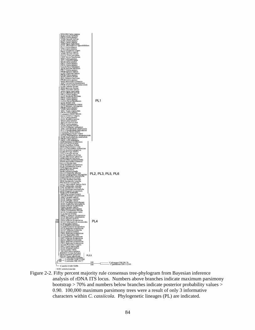
84
Figure 2-2. Fifty percent majority rule consensus tree-phylogram from Bayesian inference
analysis of rDNA ITS locus. Numbers above branches indicate maximum parsimony bootstrap > 70% and numbers below branches indicate posterior probability values > 0.90. 100,000 maximum parsimony trees were a result of only 3 informative characters within C. cassiicola. Phylogenetic lineages (PL) are indicated.

85
Figure 2-3. Fifty percent majority rule consensus tree-phylogram from Bayesian inference
analysis of the Cc-caa5 locus. Numbers above branches indicate maximum parsimony bootstrap > 70% and numbers below branches indicate posterior probability values > 0.90. Phylogenetic lineages (PL) are indicated.

86
Figure 2-4. Fifty percent majority rule consensus tree-phylogram from Bayesian inference
analysis of the Cc-ga4 locus. Numbers above branches indicate maximum parsimony bootstrap > 70% and numbers below branches indicate posterior probability values > 0.90. Phylogenetic lineages (PL) are indicated.

87
Figure 2-5. Fifty percent majority rule consensus tree-phylogram from Bayesian inference
analysis of the Cc-act1 locus. Numbers above branches indicate maximum parsimony bootstrap > 70% and numbers below branches indicate posterior probability values > 0.90. Phylogenetic lineages (PL) are indicated.

88
Figure 2-6. UPGMA dendrogram of 50 Corynespora cassiicola isolates based on pathogenicity
profiles on eight crop plants: basil (Ba), bean (Be), cowpea (Co), cucumber (Cu), papaya (Pa), soybean (So), sweet potato (Sw), tomato (To). Isolates are labeled with their phylogenetic lineage (PL) designation to demonstrate that isolates from the same PL cluster together. Statistical support for nodes by 1,000 UPGMA Bootstrap repetitions is indicated.

89
Figure 2-7. Demonstration of the C. cassiicola disease rating system. Symptoms on A) basil, B)
bean, C) cowpea, and D) tomato plants seven days after inoculation with different isolates of Corynespora cassiicola. Plants were rated with the following scale: (0) symptomless; (1) non pathogenic hypersensitive response, a few to many non-expanding pinpoint lesions; (2) moderately virulent, many expanding lesions, some coalescing, but not resulting in blight; (3) highly virulent, lesions spreading to form large areas of dead tissue resulting in a blighting effect.

90
LIST OF REFERENCES
Abul-Hayja ZP, Williams H, Peterson CE (1978) Inheritance of resistance to anthracnose and target leaf spot in cucumbers. Plant Dis Rep 62:43-45
Acharya B, Mishra SK, Acharya A, Mohapatra KB, and Das, AK (2003) Bioassay of culture
metabolites of Corynespora cassiicola (Berk. Curt.) Wei on the detached plant parts of betelvine (Piper betle L.). Orissa J Hortic 31:8-9
Ahmad S (1969) Fungi of West Pakistan. Biological Society of Pakistan Monograph 5(Sup.1):1-
110 Alfieri SA Jr, Langdon KR, Wehlburg C, Kimbrough JW (1984) Index of Plant Diseases in
Florida. Florida Dept. of Agriculture and Consumer Sciences, Div. Of Plant Industry. Bull. No. 11 (Revised), pp 389
Alfieri SA Jr, Langdon KR, Kimbrough JW, El-Gholl NE, Wehlburg C (1994) Diseases and
Disorders of Plants in Florida. Florida Department of Agriculture and Consumer Services, pp 1114
Anderson PJ, Dixon WN (2004) Florida Department of Agriculture and Consumer Services Plant
Pathology Section, Ornamentals, Foliage Plants, Tri-ology, Vol. 43, No. 1 Arnold GRW (1986) Lista de Hongos Fitopatogenos de Cuba. Ministerio de Cultura Editorial
Cientifico-Tecnica, 207 pp Atan S, Hamid NH (2003) Differentiating races of Corynespora cassiicola using RAPD and
internal transcribed spacer markers. J Rub Res 6:58-64 Barreto RW, Evans HC (1998) Fungal pathogens of Euphorbia heterophylla and E. hirta in
Brazil and their potential as weed biocontrol agents. Mycopathologia 141:21-36 Barthe P, Pujade-Renauld V, Breton F, Gargani D, Thai R, Roumestand C, de Lamotte F (2007)
Structural analysis of cassiicolin, a host-selective protein toxin from Corynespora cassiicola. J Mol Biol 367:89-101
Beaver RG (1981) Guam Agricultural Experiment Station Annual Report, 36 pp Bird J, Krochmal A, Zentmyer G, Adsuar J (1966) Fungus diseases of papaya in the U.S. Virgin
Islands. J Agric Univ Puer Rico 50:186-200 Blazquez CH (1967) Corynespora leaf spot of cucumber. Fla Agric Exp Stn J Ser No 2858, pp
177-182 Blazquez CH (1968) Corynespora cassiicola on bananas. Phytopathology 52:1347 (Abstr.) Blazquez CH (1972) Target spot of tomato. Plant Dis Rep 56:243-245

91
Bliss FA, Onesirosan PT, Arny DC (1973) Inheritance of resistance in tomato to target leaf spot.
Phytopathology 63:837-840 Boa E, Lenné J (1994) Diseases of Nitrogen Fixing Trees in Developing Countries. An
annotated list. Natural Resources Inst., Kent, United Kingdom, pp 82 Boosalis MG, Hamilton RI (1957) Root and stem rot of soybean caused by Corynespora
cassiicola. Plant Dis Rep 41:8:696-698 Brooks F (2002) List of plant diseases in American Samoa. Land Grant Technical Report No. 38.
50 CABI Databases (2008, July 21). Herb. IMI records for Fungus: Corynespora cassiicola.
Retrieved July 21, 2008 from: http://194.203.77.76/herbIMI/DisplayResults.asp?strName=Corynespora+cassiicola
Carbone I, Kohn LM (1999) A method for designing primer sets for speciation studies in
filamentous ascomycetes. Mycologia 91:553-556 Casady W (1994) Florida Department of Agriculture and Consumer Services, P94-5328, Tri-
ology, Vol. 33, No.6, Nancy C. Coile Managing Editor. Retrieved May 12, 2008 from: http://www.doacs.state.fl.us/pi/enpp/94-11&12all.htm
Chase AR (1981) Comparison of Myrothecium sp. and Corynespora cassiicola leaf spots of two
cultivars of Aphelandra squarrosa. Proc Fla State Hortic Soc 94:115-116 Chase AR (1982) Corynespora leaf spot of Aeschynanthus pulcher and related plants. Plant Dis
66:739-740 Chase AR (1984) Leaf spot disease of Ficus benjamina caused by Corynespora cassiicola. Plant
Dis 68:251 Chase AR (1986) Corynespora bract spot of Euphorbia pulcherrima in Florida. Plant Dis
70:1074 Chase AR (1987) Compendium of Ornamental Foliage Plant Diseases. American
Phytopathological Society Press, St. Paul, 92 pp Chase AR (1993) Corynespora leaf spot and stem rot of Salvias. CFREC-Apopka Research
Report, RH-93-12 Cheeran A (1968) Leaf and stem blight of Japanese Mentha caused by Corynespora cassiicola.
Agric Res J Karala 6:141

92
Cho WD, Shin HD (2004) List of Plant Diseases in Korea. Fourth ed. Korean Society of Plant Pathology, 779 pp
Coile NC, Dixon WN (1994) Florida Department of Agriculture and Consumer Services Plant
Pathology Section, Ornamentals, Foliage Plants, Tri-ology, Vol. 33, No. 5 Collado J, Platas G, Gonzalez I, and Pelaez F (1999) Geographical and seasonal influences on
the distribution of fungal endophytes in Quercus ilex. New Phytol 144:525-532 Cutrim FA, Silva GS (2003) Pathogenicity of Corynespora cassiicola to different plant species.
Fitopatol Brasil 28:193-194 Da Silva JL, Soares DJ, Barreto RW (2005) Eye-spot of Rudbeckia laciniata caused by
Corynespora cassiicola in Brazil. Brit Soc Plant Pathol, New Dis Rep No. 12 Dade HA (1940) A revised list of Gold Coast fungi and plant diseases. XXIX. Bull. Misc.
Inform. Kew 6:205-247 Daughtrey M (2000) Diseases of bleeding heart (Clerodendrum thomsoniae Balf.). APSnet:
Common names of plant diseases. Plant Pathology Online. http://www.apsnet.org/online/common/names/bleedhrt.asp
Delgado-Rodriguez G, Mena-Portales J (2004) Hifomicetos (hongos anamorficos) de la reserva
ecologica "alturas de banao" (Cuba). Bol Soc Micol Madrid 28:115-124 Delgado-Rodriguez G, Mena-Portales J, Calduch M, Decock C (2002) Hyphomycetes (hongos
mitosporicos) del area protegida mil cumbres, Cuba Occidental. Cryptog Mycol 23:277-293 Dixon WN (1997) Florida Department of Agriculture and Consumer Services Plant Pathology
Section, Ornamentals, Foliage Plants, Tri-ology, Vol. 36, No 2 Duarte MLR, Albuquerque FC, Prabhu AS (1978) A new leaf disease of cacao plants
(Theobroma cacao) caused by the fungus Corynespora cassiicola. Fitopatol Brasil 3:259-265 El-Gholl NE, Schubert TS (1990) Corynespora leaf spot of Tabebuia. Fla Dept Agric &
Consumer Serv. Division of Plant Industry. Plant Pathol Circ No 328 El-Gholl NE, Schubert TS, Coile NC (1997) Diseases and disorders of plants in Florida. Bulletin
No. 14 Supplement No 1. Fla Dept of Agric and Cons Serv, pp 90-91 Ellis MB (1957) Some species of Corynespora. Mycological Papers 65:1-15 Ellis MB, Holliday P (1971) Corynespora cassiicola (Berk. & Curt.) Wei. CMI Descriptions of
Fungi and Bacteria No 31, Sheet 303

93
Fajola AO, Alasoadura SO (1973) Corynespora leaf spot, a new disease of tobacco (Nicotiana tabacum). Plant Dis Rep 57:375-378
Farr DF, Rossman AY, Palm ME, McCray EB. Fungal Databases, Systematic Mycology and
Microbiology Laboratory, ARS, USDA. Retrieved May 24, 2008, from http://nt.ars-grin.gov/fungaldatabases/
Felsenstein J (1985) Confidence limit on phylogenies: an approach using the bootstrap.
Evolution; International Journal of Organic Evolution 39:783-791 Fernandes RC, Barreto RW (2003) Corynespora cassiicola causing leaf spots on Coleus
barbatus. Plant Pathol 52:786 Ferreira FA (1989) Principais Doencas Florestais no Brasil. Patologia Florestal. Vicosa. MG –
Minas Gerais, 570 pp Florence EJM, Sharma JK (1987) Corynespora cassiicola: a new leaf pathogen for Gmelina
arborea in India. J Trop For 3:181-182 Freire FCO (2005) An updated list of plant fungi from Ceará state (Brazil) – I Hyphomycetes.
Revista Ciência Agronômica 36:364-370 Furukawa T, Ushiyama K, and Kishi K (2008) Corynespora leaf spot of scarlet sage caused by
Corynespora cassiicola. J Gen Plant Pathol 74:117-119 Gond SK, Verma VC, Kumar A, Kumar V, Kharwar RN (2007) Study of endophytic fungal
community from different parts of Aegle marmelos Correae (Rutaceae) from Varanasi (India). World J Microbiol Biotechnol 23:1371-1375
Gowda CLL, Ramakrishna A, Rupela OP, Wani SP (2001) Legumes in Rice-Based Cropping
Systems in Tropical Asia. Andhra Pradesh, India, pp 11-25 Grand LF (1985) North Carolina Plant Disease Index. North Carolina Agric Res Serv Techn Bull
240:1-157 Guo YL (1992) Foliicolous hyphomycetes of Guniujiang in Anhui Province II. Mycosystema
5:109-112 Hasama W, Morita S, Kato T (1991) Corynespora leaf spot of Perilla caused by Corynespora
cassiicola. Annals Phytopathol Soc Jap 57:732-736 Hawaiian Ecosystems at Risk (HEAR). (2008, July 11). Pathogens of Plants of Hawaii,
Corynespora cassiicola. Retrieved July 11, 2008, from: http://www.hear.org/pph/pathogens/1065.htm

94
Holliday P (1980) Fungus Diseases of Tropical Crops. Cambridge University Press. Cambridge, UK
Hongn S, Ramallo A, Baino O, Ramallo JC (2007) First Report of Target Spot of Vaccinium
corymbosum caused by Corynespora cassiicola. Plant Dis 91:771 Huelsenbeck JP, Ronquist F (2001) MrBayes: Bayesian inference of phylogeny. Bioinformatics
(Oxford, England) 17:754-755 Hyde KD, Alcorn JL (1993) Some disease-associated microorganisms on plants of Cape York
Peninsula and Torres Strait Islands. Australas Plant Pathol 22: 73-83 Hyde KD, McKenzie EHC, Dalisay TU (2001) Saprobic fungi on bamboo culms. Fungal Divers
7:35-48 Isabel N, Beaulieu J, Theriault P, Bousquet J (1999) Direct evidence for biased gene diversity
estimates from dominant random amplified polymorphic DNA (RAPD) fingerprints. Molecular Ecology 8:477-483
Johnston A (1960) A supplement to a host list of plant diseases in Malaya. Mycol Pap 77:1-30 Jones JP (1961) A leaf spot of cotton caused by Corynespora cassiicola. Phytopathology 51:305-
308 Jones JP, and Jones JB (1984) Target spot of tomato: epidemiology, and control. Proc Fla State
Hortic Soc 97:216-218 Jones JB, Jones JP, Stall RE, Zitter TA (1991) Compendium of Tomato Diseases. APS Press, St.
Paul, MN, 100 pp Khare MN (1991) Lentil diseases with special reference to seed quality. Indian J Mycol Plant
Pathol 21:1-13 Kingsland GC (1985) Pathogenicity and epidemiology of Corynespora cassiicola in the
Republic of the Seychelles. Acta Hortic (ISHS) 153:229-230 Komaraiah M, Reddy SM (1986) Production of cellulases by Corynespora cassiicola Wei, a
seed borne fungus of methi. Acta Botan Ind 14:133-138 Kranz J (1963) Fungi collected in the Republic of Guinea, Collections from the Kindia area in
1962. Sydowia 17:174-185 Kurt S (2004) Host-specific toxin production by the tomato target leaf spot pathogen
Corynespora cassiicola. Turk J Agric and Fores 28:389-395

95
Kwon JH, Kang SW, Kim JS, Park CS (2001) First report of Corynespora leaf spot in pepper caused by Corynespora cassiicola in Korea. Plant Pathol J 17:180-183
Kwon JH, Park CS (2003) Leaf spot of cotton rose caused by Corynespora cassiicola in Korea.
Mycobiology 31:57-59 Lakshmanan P, Jeyarajan R, Vidhyasekaran P (1990) A boll rot of cotton caused by Corynespora
cassiicola in Tamil Nadu, India. Phytoparasitica 18:171-174 Lee S, Melnik V, Taylor JE, Crous PW (2004) Diversity of saprobic hyphomycetes on
Proteaceae and Restionaceae from South Africa. Fung Diversity 17:91-114 Leite RS, Barreto RW (2000) Petal spotting of hydrangea flowers caused by Corynespora
cassiicola: old pathogen new disease. Mycologist 14:80-83 Lenné JM (1990) World List of Fungal Diseases of Tropical Pasture Species. Phytopathol Pap
31:1-162 LSU Ag Center (2008, July 21). Common Diseases of Ornamental Plants. Retrieved July 21,
2008, from: http://www.lsuagcenter.com/NR/rdonlyres/F512A031-FDF9-46C6-AEE2-B800409C9FDF/42824/ornamentals1.PDF
Lu B, Hyde KD, Ho WH, Tsui KM, Taylor JE, Wong KM, Yanna, Zhou D (2000) Checklist of
Hong Kong Fungi. Fungal Diversity Press, Hong Kong, 207 pp Lumyong P, Photita W, McKenzie EHC, Hyde KD, Lumyong S (2003) Saprobic fungi on dead
wild banana. Mycotaxon 85:345-346 Maddison DR, Maddison WP (2005) MacClade 4, version 4.08. Sunderland: Sinuar Associates Mallaiah KV, Vijayalakshmi M, Rao AS (1981) New records of some foliar diseases. Indian
Phytopathology 34: 247 Malvick D (2004) Fungus foliage diseases of soybeans. University of Illinois Extension. Report
on Plant Disease No. 503 Mathiyazhagan S, Kavitha K, Nakkeeran S, Chandrasekar G, Manian K, Renukadevi P,
Krishnamoorthy AS, Fernando WGD (2004) PGPR mediated management of stem blight of Phyllanthus amarus (Schum and Thonn) caused by Corynespora cassiicola (Berk and Curt) Wei. Arch Phytopathol Plant Prot 37:183-199
McDonald BA (2004) Population genetics of plant pathogens. The Plant Health Instructor ,
doi:10.1094/PHI-A-2004-0524-01 McGovern RJ (1994) Target spot of Catharanthus roseus caused by Corynespora cassiicola.
Plant Dis 78:830

96
McKenzie EHC, Buchanan PK, Johnston PR (2004) Check-list of fungi on nikau palm
(Rhopalostylis sapida and R. baueri var. cheesemanii) in New Zealand. N Z J Bot 42:335–355
McKenzie EHC (1990) The fungi, bacteria and pathogenic algae of the Republic of Palau.
Technical Paper No. 198. South Pacific Commission, Noumea, New Caledonia, 41 pp McKenzie EHC (1996) Fungi, bacteria and pathogenic algae on plants in American Samoa.
Technical Paper No. 206. South Pacific Commission, Noumea, New Caledonia McRitchie JJ, Miller JW (1973) Corynespora leaf spot of zebra plant. Proc Fla State Hort Soc
86:389-390 Mehrotra MD (1987) Corynespora cassiicola leaf spot of Ceiba pentandra and its control in the
nursery. Ind For 115:905-909 Mehrotra MD (1997) Diseases of Paulownia and their management. Indian Forester 123:66-72 Mendes MAS, da Silva VL, Dianese JC, Ferreira MASV, Santos CEN, Gomes Neto E, Urben
AF, Castro C (1998) Fungos em Plantas no Brasil. Brasília. Empresa Brasileira de Pesquisa Agropecuária, 555 pp
Mercado SÁ (1984) Hifomicetes Demaciaceos de Sierra del Rosario, Cuba. Editorial Academica,
Havana, 181 pp Mercado SÁ, Holubová-Jechová V, Mena Portales J (1997) Hifomicetes Demaciáceos de Cuba
Enteroblásticos. Museo Regionale di Scienze Naturali 23:388 Miller JW (1991) Bureau of Plant Pathology. Tri-ology Techn. Rep. Div. Plant Indust, Florida
30:1-2 Miller JW, Alfieri SA Jr (1973) Leaf spot of Ligustrum sinense caused by Corynespora
cassiicola. Phytopathology 63:445-446 Minter DW, Rodríguez Hernández M, Mena Portales J (2001) Fungi of the Caribbean: an
annotated checklist. PDMS Publishing, 946 pp Murali TS, Suryanarayanan TS, Venkatesan G (2007) Fungal endophyte communities in two
tropical forests of southern India: diversity and host affiliation. Mycol Progr 6:191-199 Olive LS, Bain DC (1945) A leafspot of cowpea and soybean caused by an undescribed species
of Helminthosporium. Phytopathology 35:822-831 Oluma HOA, Amuta EU (1999) Corynespora cassiicola leaf spot of pawpaw (Carica papaya L.)
in Nigeria. Mycopathologia 145:23-27

97
Onesirosan PT, Arny DC, Durbin RD (1975) Increasing sporulation of Corynespora cassiicola.
Mycopathologia 55:121-123 Onesirosan PT, Arny DC, Durbin RD (1974) Host specificity of Nigerian and North American
isolates of Corynespora cassiicola. Phytopathology 64:1364-1367 Onesirosan PT, Arny DC, Durbin RD (1973) Target Spot of Tomato Incited by Corynespora
cassiicola (Berk. & Curt.) Wei. Ph. D. Thesis, University of Wisconsin, Madison, 93 pp Orieux L, Felix S (1968) List of plant diseases in Mauritius. Phytopathol Pap 7:1-48 Peregrine WTH, Ahmad KB (1982) Brunei: A first annotated list of plant diseases and associated
organisms. Phytopathol Pap 27:1-87 Pereira JM, Barreto RW, Ellison CA, Maffia LA (2003) Corynespora cassiicola f. sp. lantanae:
a potential biocontrol agent from Brazil for Lantana camara. BiolControl 26:21-31 Pernezny K, Simone GW (1993) Target spot of several vegetable crops. PP-39, A series of the
Plant Pathology Department, Fla Coop Ext Ser, IFAS, Univ of Fla Pernezny K, Datnoff LE, Mueller T, Collins J (1996) Losses in fresh-market tomato production
in Florida due to target spot and bacterial spot and the benefits of protectant fungicides. Plant Dis 80:559-563
Pernezny K, Datnoff LE, Rutherford B, Carroll A (2000) Relationship of temperature to growth,
sporulation, and infection of tomato by the target spot fungus. Florida Tomato Committee Tomato Research Report for 2000, pp 16-19
Pernezny K, Stoffella P, Collins J, Carroll A, Beaney A (2002) Control of target spot of tomato
with fungicides, systemic acquired resistance activators, and a biocontrol agent. Plant Prot Sci 38:81-88
Pernezny KL, Datnoff LE, Smith LJ, Schlub RL (2008) An overview of target spot of tomato
caused by Corynespora cassiicola. Acta Hort xxx: Second International Symposium on Tomato Diseases (accepted)
Philip S, Ramakrishnan CK, Menon MR (1972) Leaf blight of Coccinia indica (Wight & Arn.)
caused by Corynespora cassiicola. Agric Res J Kerala 10:196 Pollack FG, Stevenson JA (1973) A fungal pathogen of Broussonetia papyrifera collected by
George Washington Carver. Plant Dis Rep 57:296-298 Poltronieri LS, Duarte MLR, Alfenas AC, Trindade DR, Albuquerque FC (2003) Three new
pathogens infecting Antilles Cherry in the state of Para. Fitopatol Brasil 28:424-426

98
Posada D, Crandall KA (1998) Modeltest: testing the model of DNA substitution. Bioinformatics 14:817-81
Prakash O, Garg N (2007) A new report of Corynespora casiicola, causing black rot of aonla
seedlings. J Mycol Plant Pathol 37:120-121 Promputtha I, Lumyong A, Dhanasekaran V, McKenzie EHC, Hyde KD, Jeewon R (2007) A
phylogenetic evaluation of whether endophytes become saprotrophs at host senescence. Micro Ecol 53:579-590
Puzari KC, Saikia UN (1981) Amorphophallus campanulatus, a new host of Corynespora
cassiicola. Indian Phytopathol 34:537-538 Quimio RH, Abilay LE (1979) Note: Corynespora disease of papaya in the Philippines. Philipp
Phytopathology 15:158-161 Raabe RD, Conners IL, Martinez AP (1981) Checklist of plant diseases in Hawaii. College of
Tropical Agriculture and Human Resources, University of Hawaii. Information Text Series No. 22. Hawaii Inst Trop Agric Human Resources, 313 pp
Raffel SJ, Kazmar ER, Winberg R, Oplinger ES, Handelsman J, Goodman RM, Grau CR (1999)
First report of root rot of soybeans caused by Corynespora cassiicola in Wisconsin. Plant Disease 83:696
Rajak RC, Pandey AK (1985) Fungi from Jabalpur-II. Indian J Mycol Plant Pathol 15:186-194 Riley EA (1960) A revised list of plant diseases in Tanganyika Territory. Mycol Pap 75:1-42 Romruensukharom P, Tragoonrung S, Vanavichit A, Toojinda T (2005) Genetic variability of
Corynespora cassiicola populations in Thailand. J Rub Res 8:38-49 Ronquist F, Huelsenbeck JP (2003) MrBayes3: Bayesian phylogenetic inference under mixed
models. Bioinformatics 19:1572-1574 Sadaba RB, Vrijmoed LLP, Jones EBG, Hodgkiss IJ (1995) Observations on vertical distribution
of fungi associated with standing senescent Acanthus ilicifolius stems at Mai Po Mangrove, Hong Kong. Hydrobiologia 295:119-126
Saikia UN, Sarbhoy AK (1981) Corynespora leaf spot of Eugenia caryophyllata. Indian
Phytopathol 34:401-402 Sarbhoy AK, Lal G, Varshney JL (1971) Fungi of India. Navyug Traders, New Delhi, 148 pp Sarma YR, Nayudu MV (1970) Corynespora leaf spot of Brinjal. Proc Indian Acad Sci
B(LXXIV):92-97

99
Schlub RL, Yudin L (2002) Eggplant, pepper, and tomato production guide for Guam. Guam Cooperative Extension Publication, 188 pp
Seaman WL, Shoemaker RA, Peterson EA (1965) Pathogenicity of Corynespora cassiicola on
soybean. Can J Bot 43:1461-1469 Shaw DE (1984) Microorganisms in Papua New Guinea. Dept. Primary Ind., Res Bull 33:1344
Shivas RG, Alcorn JL (1996) A checklist of plant pathogenic and other microfungi in the
rainforests of the wet tropics of northern Queensland. Australas Mycol 25:158-173 Silva WPK, Deverall BJ, Lyon BR (1995) RFLP and RAPD analyses in the identification and
differentiation of isolates of the leaf spot fungus Corynespora cassiicola. Austral J Bot 43:609-618
Silva WPK, Deverall BJ, Lyon BR (1998) Molecular, physiological and pathological
characterization of Corynespora leaf spot from rubber plantations in Sri Lanka. Plant Pathol 47:267-277
Silva WPK, Karunanayake EH, Wijesundera RLC, Priyanka UMS (2003) Genetic variation in
Corynespora cassiicola: a possible relationship between host origin and virulence. Mycol Res 107:567-571
Silva WPK, Wijesundera RLC, Karunanayake EH, Jayasinghe CK, Priyanka UMS (2000) New
hosts of Corynespora cassiicola in Sri Lanka. Plant Dis 84:202 Simone GW (2000) Diseases of Cattleya Lindl. spp. APSnet: Common Names of Plant Diseases.
http://www.apsnet.org/online/common/names/cattleya.asp Simone GW (2000) Diseases of Pointsettia (Euphorbia pulcherrina). APSnet: Common Names
of Plant Diseases. http://www.apsnet.org/online/common/names/poinsett.asp Singh KP, Shukla RS, Kumar S, Hussain A (1982) A leaf-spot disease of Dodonaea viscosa
caused by Corynespora cassiicola in India. Ind Phytopathol 35:325 Situmorang A, Budiman A (1984) Corynespora cassiicola (Berk. And Curt.) Wei, penyebab
penyakit gugur duan pada karet. Kumpulan Makalah Lokakarya Karet. PNP/PTP Wilayah 1 dan P4TM, Medan
Sivanesan A (1996) Corynesporasca caryote gen. et sp. nov. with a Corynespora anamorph, and
the family Corynesporascaceae. Mycol Res 100:783-788 Smith LJ, Datnoff LE, Rollins JA, Pernezny KL, Scott JW, Schlub RL (2008a) High genetic
diversity within Corynespora cassiicola based on multilocus sequence data, pathogenicity, and growth rate. Acta Hort xxx: Second International Symposium on Tomato Diseases (accepted)

100
Smith LJ, Datnoff LE, Pernezny KL, Rollins J, Schlub RL (2008b) Phylogenetic analyses of
diverse Corynespora cassiicola isolates indicate an evolutionary correlation with host not geography. 9th European Conference on Fungal Genetics Meeting Abstracts
Smith LJ, Datnoff LE, Rollins JA, Pernezny KL, Schlub RL (2007) Phylogenetic analysis of
Corynespora isolates from diverse hosts and locations. Phytopathology 97:S109 Smith LJ, Datnoff LE, Pernezny KL, Roberts PD, Rollins JA, Schlub RL, Scott JW (2006)
Characterization and host-range of the tomato target spot fungus, Corynespora cassiicola and resistance of tomato cultivars. Florida Tomato Committee, Tomato Research Report for 2004-2005, pp 14-20
Smith LJ, Schlub RL (2005) Foliar fungi on weeds of Guam and the potential for Corynespora
cassiicola as a bioherbicide for Stachytarpheta jamaicensis. Phytopathology 95:S93 Smith LJ, Schlub RL (2004) Host range of Corynespora cassiicola and its occurrence on weeds,
ornamentals and crops of Guam. Phytopathology 92:S77 Sobers EK (1966) A leaf spot disease of azalea and hydrangea caused by Corynespora
cassiicola. Phytopathology 59:455-457 Spencer JA (1962) Study of variations in Corynespora cassiicola (Berk. & Curt.) Wei. M. S.
Thesis, University of Arkansas, Fayetteville, 31 pp Spencer JA, Walters HJ (1969) Variations in certain isolates of Corynespora cassiicola.
Phytopathology 59:58-60 Stone WJ, Jones JP (1960) Corynespora blight of sesame. Phytopathology 50:263-266 Strandberg JO (1971) Evaluation of cucumber varieties for resistance to Corynespora cassiicola.
Plant Dis Rep 55:142-144 Suryanarayanan TS, Murali TS, Venkatesan G (2002) Occurrence and distribution of fungal
endophytes in tropical forests across a rainfall gradient. Can J Bot 80:818-826 Swofford DL (2002) PAUP*. Phylogenetic analysis using parsimony (*and other methods).
Version4. Sunderland: Sinauer Associates Taba S, Ooshiro A, Takaesu K (2002) Black stem and root rot of basil Ocimum basilicum L.
caused by Corynespora citricola. Ann Phytopathol Soc Japan 68:43-45 Tanaka K, Yasuyoshi O, Hatakeyama S, Harada Y, Barr ME (2008) Pleosporales in Japan (5):
Pleomassaria, Asteromassaria, and Splanchnonema. Mycoscience 46:248-260
101
Tsay JG, Kuo CH (1991) The occurrence of Corynespora blight of cucumber in Taiwan. Plant Prot Bull 33:227-229
Turner GJ (1971) Fungi and Plant Disease in Sarawak. Phytopathol Pap 13:1-55 Urtiaga R (1986) Indice de enfermedades en plantas de Venezuela y Cuba. Impresos en
Impresos Nuevo Siglo. SRL, Barquisimeto, Venezuela, 202 pp Urtiaga R (2004) Indice de enfermedades en plantas de Venezuela y Cuba, 2nd Ed, 301 pp Vittal BPR, Dorai M (1995) Studies on litter fungi VIII. Quantitative studies of the mycoflora
colonizing Eucalyptus tereticornis Sm. Litter. Kavaka 22/23: 35-41 Volin RB, Pohronezny K (1989) Severe spotting of fresh market tomato fruit incited by
Corynespora cassiicola after storm-related injury. Plant Dis 73:1018-1019 Vyas SC, Shastry PP, Shukla BN, Varma RK (1985) Two new leaf blight diseases of groundnut.
Plant Prot Bull 33:121-122 Wei CT (1950) Notes on Corynespora. Mycol Papers 34:1-10 White TJ, Bruns T, Lee S, Taylor J (1990) Amplification and direct sequencing of fungal
ribosomal RNA genes for phylogenetics. PCR Protocols: A Guide to Methods and Applications. Innis MA, Gelfand DH, Sninsky JJ, White TJ (ed.), Academic Press, Inc, New York, NY, ch 38, pp 315-322
Williams TH, Liu PSW (1976) A host list of plant diseases in Sabah, Malaysia. Phytopathol Pap
19:1-67 Yudin L, Schlub RL (1998) Guam Cucurbit Guide. Guam Cooperative Extension Publication, 64
pp Zhang XG, Ji M (2005) Taxonomic studies of Corynespora from Yunnan, China. Mycotaxon
92:425-429 Zhuang WY (2001) Higher Fungi of Tropical China. Mycotaxon, Ltd., Ithaca, NY, pp 485

102
BIOGRAPHICAL SKETCH
I was born in Pompano Beach, FL to Janis Therrell and Kenneth Wayne Smith on October
15, 1977. I have an older sister, Allison, and two younger brothers, Scott and Reid. Our family
moved to Baltimore, MD when I was nine and I attended Baltimore Friends School, where my
father was head of the Middle School. Though I have always loved biology and gardening, my
interest in agriculture took off in high school when I attended Maine Coast Semester, a small
school for students in their junior year located on a coastal farm in Wiscasset, Maine.
I received my B. A. at Colorado College in 2000 with a major in Biology, while fostering
my interest in farming through summer jobs at nurseries, CSA’s, and internships. My
sophomore year in college, I traveled abroad to East Africa through The School for Field Studies
where I learned the importance of economic value in conservation by focusing on wildlife
ranching, national parks, and medicinal plant use as case studies.
In 2002, I received my Masters Degree from West Virginia University in Plant Pathology
as part of the Organic Farm Project by studying the effect of intercropping on diseases caused by
Alternaria solani and Meloidogyne incognita. I then spent two years in Micronesia on the island
of Guam as a Research Assistant documenting pathogens of agronomically important weeds and
working in the diagnostic clinic. It was in Guam where I first became aware of Corynespora
cassiicola as an agent of disease. An opportunity presented itself to continue the work begun on
this pathogen at the University of Florida in the Fall of 2004. At the University of Florida, I
became well trained in Phylogenetics and this has become my specialty. I plan to continue
studying fungal systematics beginning in September, 2008 as a post-doc with the USDA in
Beltsville, MD.