Hormones lecture h01
27
Hormones Bio-signaling Dr. Aga Syed Sameer CSIR Lecturer (Demonstrator) Department of Biochemistry, Medical College, Sher-I-Kashmir Institute of Medical Sciences, Bemina, Srinagar, Kashmir, 190010. India. First MBBS Lecture No: H 01 Time : 10:00am Dated: 05/03/2015
-
Upload
aga-syed-sameer -
Category
Science
-
view
50 -
download
1
Transcript of Hormones lecture h01
HormonesBio-signaling
Dr. Aga Syed SameerCSIR Lecturer (Demonstrator)Department of Biochemistry,Medical College,Sher-I-Kashmir Institute of Medical Sciences, Bemina, Srinagar, Kashmir, 190010. India.
First MBBS
Lecture No: H 01
Time : 10:00am
Dated: 05/03/2015

References:
1. Biochemistry – Lehninger’s, Nelson & Cox
2. Cell biology – Gerald Karp
3. Molecular Biology of Cell – Bruce Alberts’
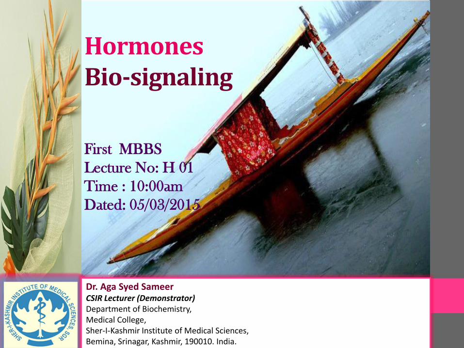
Cellular Communication
• Cells usually communicate with each other through “Extracellular Messenger Molecules”
• Extracellular messengers can travel
• “Short distance” and “Stimulate” cells that are in close proximity to the origin of the message
• “Throughout the Body”, potentially stimulating cells that are far away from the source
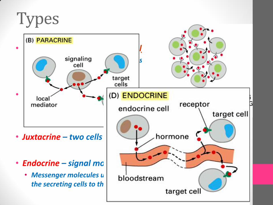
Types
• Autocrine – cell elaborates a self-addressed message• Cells producing a messenger expresses receptors on its own surface
to respond to the messenger
• Paracrine – local communication mechanism• Messenger molecules travel only short distances through the
extracellular space to cell that are in close proximity of the secreting cell
• Juxtacrine – two cells - direct contact
• Endocrine – signal molecule →blood → target cell• Messenger molecules use the bloodstream for their transport from
the secreting cells to the target ones
Hormones• Organic substances that are produced and secreted
by “Endocrine glands” in minute quantity to regulate the various physiological and metabolic functions of body
• The action of hormones is regulated by many factors:
• Rate of secretion/synthesis in endocrine organs
• Specified transport system in plasma
• Hormone specific receptors in target cells/organs
• Rate/mechanism of degradation of hormoneswithin liver/kidneys

HormonesProperties Group I Group II
Types Steroids, Triiodothyroxine, Retinoids
Polypeptides, Proteins, Catecholamines
Solubility Lipophilic Hydrophilic
Transport Proteins
Yes No
Plasma Half Life Long (h→days) Short (min)
Receptor Intracellular Plasmamembrane
Mediator Hormone Receptor Complex
cAMP, cGMP, Ca2+, Kinasecacades

Polypeptides
Steroids
Amino acid derivatives
Insulin
glucagon
somatotropin
FSH
LH
vasopressin
Oxytocin
thyrotropin
ACTH
Estrogen
testosterone
cortisol
Aldosterone
corticosterone
Progesterone
Epinephrine
norepinephrine
dopamine
Thyroxine, T3 and T4
Melatonin
Serotonin
Hormones

Hormones
Substances that fool the responder into
thinking a hormone has bound are call agonists
Substances that prevent the binding of the
natural hormone and do not elicit a response from
the receptor are called antagonists
Receptors: Specialized glycoproteins which are expressed on the surface of target cells with which hormone interacts to mediate its action
Ligands: A substance which can combine with the receptor to form ligand- receptor complex and in-turn evokes cellular response
All hormones interact with target cells by
first binding to specific receptors located either on
the plasma membrane or as a cytosolic protein
The receptor for hormones must be linked
to a component that is able to respond to the
binding of hormone that in turn elicits the response
Hormones
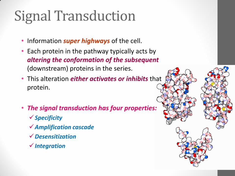
Signal Transduction
• Information super highways of the cell.
• Each protein in the pathway typically acts by altering the conformation of the subsequent (downstream) proteins in the series.
• This alteration either activates or inhibits that protein.
• The signal transduction has four properties:
Specificity
Amplification cascade
Desensitization
Integration

Signal Transduction
The series of events and components that
take part in transmitting a hormonal signal to a
the interior of the cell
Membrane or cytosolic Receptor
Signal Initiator
Target molecule
Signal mediator/effector
Action
Signal Transduction

nucleus
cytoplasm
plasma membraneDNA
mRNA protein
steroid hormone blood
proteincarrier
2
3
5
4
S
S
S
1
S
Action of Lipid (steroid) hormones
receptor protein
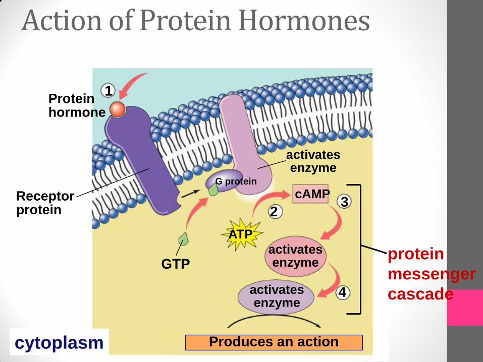
Action of Protein Hormones
3
4
GTPactivatesenzyme
activatesenzyme
activatesenzyme
Receptorprotein
cAMP
Proteinhormone
ATP
1
2
cytoplasm Produces an action
protein
messenger
cascade
G protein

The 2012 Nobel Prize in Chemistry was awarded jointly to
Robert J. Lefkowitz (l) and Brian K. Kobilka
for
studies of G-protein-coupled receptors

• They are the receptor named just because they interact with G Protein’s.
• Also referred to as “Serpentine Receptor’s” as they contain seven trans-membrane helices (7TM)
• Largest known receptor family – Constitutes > 1% of the human genome.
• Comprises receptors for a diverse array of molecules: neurotransmitters, odorants, lipids, neuropeptides, large glycoprotein hormones
G Protein Coupled Receptor’s (GPCR’s)
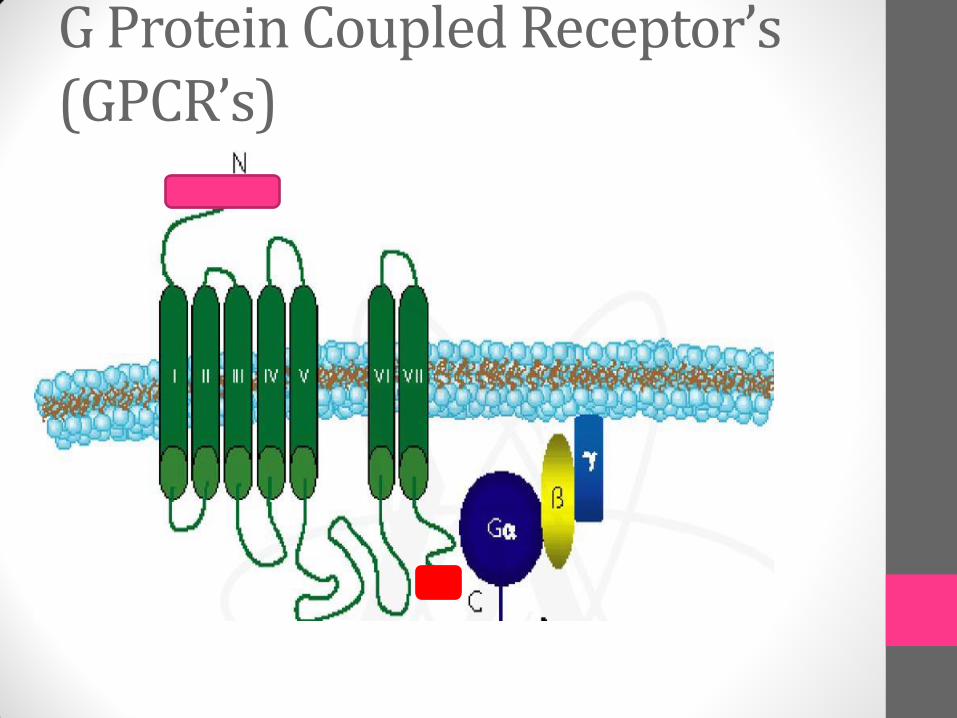
G Protein Coupled Receptor’s (GPCR’s)

• Their Structure consists of:
Amino- Terminal: Present on the outside of the cell
Seven α helices: traversing the plasma membrane & connected by loops at varying length
Carboxyl-Terminal: Present on Inside of the cell
• Ligand binding site: three loops that are on outer surface of the cell
• Docking site: three loops that are on inner surface/ cytoplasmic side of the cell; provide site for binding of intracellular protein – G protein’s
G Protein Coupled Receptor’s (GPCR’s)
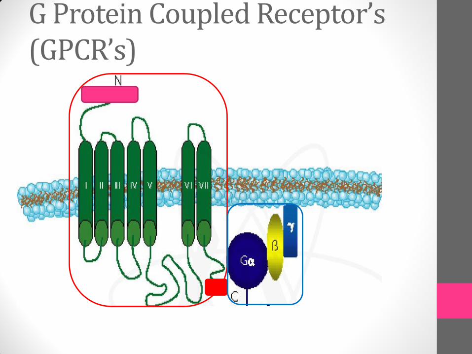
G Protein Coupled Receptor’s (GPCR’s)

• They are named so because they bind to guanine nucleotide as prosthetic group
GDP – Inactive form
GTP – Active form
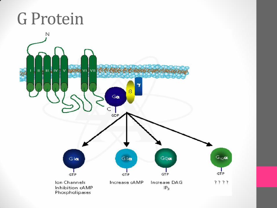
• Hetero-trimeric in nature : Three different polypeptide subunits – α, β & γ
• Held at plasma membrane by lipid chains that are covalently attached to the α & γ
G Protein

• The guanine nucleotide binding site is present on Gα subunit:
In GDP bound conformation: Gα subunit has high affinity for the Gβγ; hence they remain together as trimer on cell surface
In GTP bound conformation: Gα subunit has low affinity for the Gβγ; leading to its dissociation from complex
• The two conformations are inter-convertible via activation by GPCR’s; which cause GDP-GTP switching on Gα subunit of trimer.
• Each dissociated Gα subunit in turn is free to activate an effector protein – Like “Adenylyl Cyclase”
G Protein

G Protein
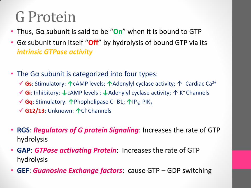
• Thus, Gα subunit is said to be “On” when it is bound to GTP
• Gα subunit turn itself “Off” by hydrolysis of bound GTP via its intrinsic GTPase activity
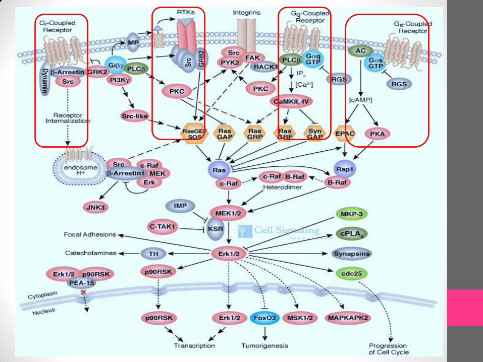
• The Gα subunit is categorized into four types: Gs: Stimulatory: ↑cAMP levels; ↑Adenylyl cyclase activity; ↑ Cardiac Ca2+
Gi: Inhibitory: ↓cAMP levels ; ↓Adenylyl cyclase activity; ↑ K+ Channels
Gq: Stimulatory: ↑Phopholipase C- B1; ↑IP3; PIK3
G12/13: Unknown: ↑Cl- Channels
• RGS: Regulators of G protein Signaling: Increases the rate of GTP hydrolysis
• GAP: GTPase activating Protein: Increases the rate of GTP hydrolysis
• GEF: Guanosine Exchange factors: cause GTP – GDP switching
G Protein
G Protein

The G Protein Cycle
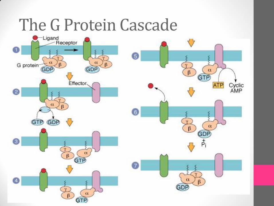
The G Protein Cascade