Hogyan változtatta meg a dióda lézer a retinoblastomás...
30
Intraocular tumors
Transcript of Hogyan változtatta meg a dióda lézer a retinoblastomás...

Intraocular tumors

25 ago, if we found a choroideal melanoma
in an eye, the eye had to be enucleated.
Pigmented tumor in an eye
(melanoma chorioideae)
An eye surgically removed because of
choroideal melanoma, the eye is cut in half

Since 1986 intraocular tumors are treated with Ruthenium applicators at the Department of
Ophthalmology, University of Debrecen.
Ophthalmic applicators used for
the radiotherapy of intraocular tumors
Applicator on the eye,
isodose curves
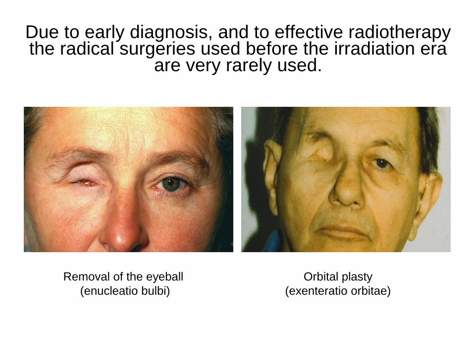
Due to early diagnosis, and to effective radiotherapy the radical surgeries used before the irradiation era
are very rarely used.
Removal of the eyeball
(enucleatio bulbi)
Orbital plasty
(exenteratio orbitae)

The outer surface of the applicators is covered by a
radioprotective layer, which is missing on the inner surface.
Radiotransmitting area on the inner surface

Different applicators are used for the irradiation of tumors with different size
and different localization.
The applicators are sutured on the surface of the eye,
and removed after sufficient irradiaton dose is achieved.
Implantation of anterior
segment applicator Implantation of posterior
segment applicator

Planning of the irradiation

A prominent, half spere shaped intraocular tumor,
turns into a flat scar following irradiation.
Melanoma before irradation The same tumor 1 year
after the irrradiation

Malignant melanomas can be differentiated from benign nevi
with fluorescein-angiography with very high precision.
Color fundusphoto Angiographic appearence
of the same fundus

With ultrasonography the size (prominence) and the inner
structure (reflectivity) of intraocular tumors are examined.
Different tumors have different ultrasonographic appearence.

By transpupillar and by transscleral illumination of the
eyeball (diaphanoscopy) the localization of the tumors
in the eye can be determined.
Strate and bent fiberoptic light sources
Transpupillar diaphanoscopy Transscleral diaphanoscopy

K.J., a 72 years old female patient, who formerly lost her left
eye in an accident, received irradiation with an applicator on
the other eye because of choroideal melanoma in the year of
1986. She was the first patient we treated with this method.
Right after irradiation Three months later

After irradiation the melanoma gradually turns into a flat scar in
6 to 12 months, during an after this period the patients have to
be regularly controlled for five years.
A melanoma right after irradiation Three months following irradiation
In half a year most of
the tumor cicatrized
Flat pigmented scar developed
in the place of the former tumor

Besides choroideal melanoma seen
in adults, the second most frequent
malignant intraocular tumor is retino-
blastoma, that we see in newborns or
in small children.

Based on clinical appearence four types of retinoblastoma are known:
Intraretinal Endophytic
Exophytic Peripherial

Different size retinoblastomas are treated differently
Small retinoblastomas Medium size retinoblastomas
Big retinoblastomas Those that fill up the eye,
and cause retina detachment

Unilateral retinoblastoma
Unilateral retinoblastomas are usually
diagnosed in older children (not new-
borns), in advanced stage when enuc-
leation is the only possible therapy. In
cases with small tumors local therapy
is also possible, but this occurs very ra-
rely. Unilateral retinoblastomas are not
treated with systemic cytostatic drugs!
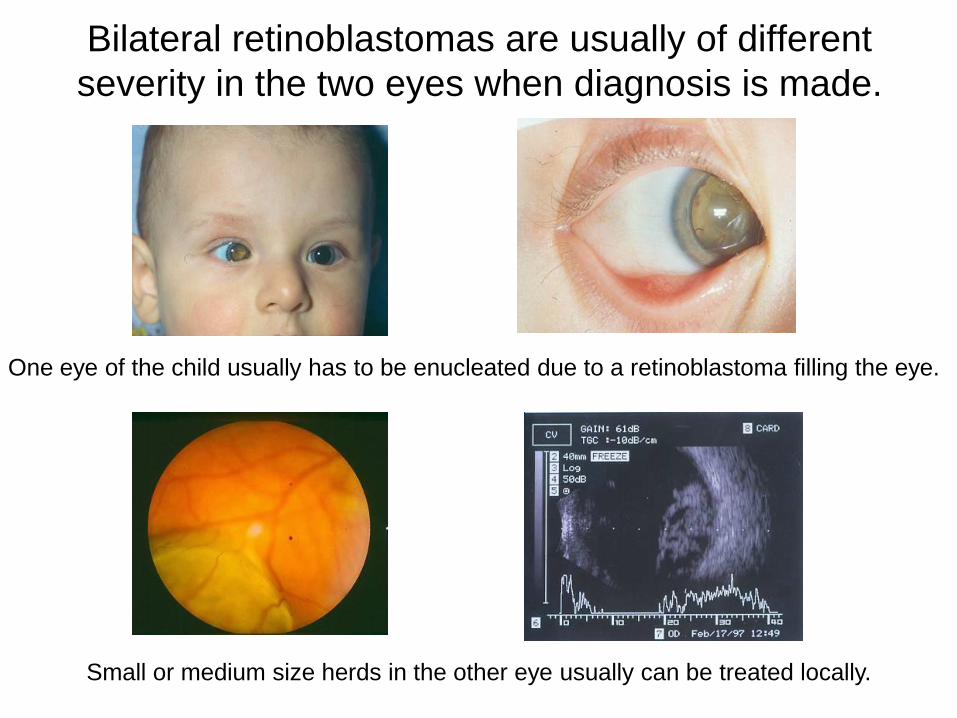
Bilateral retinoblastomas are usually of different
severity in the two eyes when diagnosis is made.
One eye of the child usually has to be enucleated due to a retinoblastoma filling the eye.
Small or medium size herds in the other eye usually can be treated locally.

In the last 15 years local treatment was almost always Ruthenium
applicator treatment (in some cases transscleral cryoapplication).
One month after irradiation Three months after irradiation
Six months after irradiation More than one herd can be
treated with one applicator

Transpupillary thermotherapy (TTT) performed with a diode
laser started a new era in the treatment of intraocular tumors.

The benefits of transpupillary thermotherapy
• Small tumors can be treated with TTT alone.
• TTT can be a good additive treatment to irradiation in case
of medium size tumors.
• In some big tumor cases with sandwich therapy enuclea-
tion can be avoided.
• TTT is more selective, causes less damage in the eye than
radiotherapy .

Children with bilateral retinoblastomas get
additional cytostatic treatment, most of
them at the Department of Pediatric
Hemato-Oncology, Institute of Pediatrics,
University of Debrecen. Children coming
from other parts of the country at other
Pediatric Oncological Centers in Hungary.

The CT scans of one of our first bilateral retinoblastoma
patients, who is presently 12 years old, and goes to normal
school
Before treatment: in the right eye a big,
in the left egye a small retinoblastoma,
with juxtapapillary localization.
Four years following the enucleation
of the right eye, and the irradiation of
the left eye. Note the normal appear-
ance of the left eye, without relapse.

Molecular genetics of retinoblastoma
• Retinoblastoma (Rb) gene is in
the 13q14 chromosomal re-gion.
• The Rb is a tumor suppressor gene, that palys a role in the regulation of the mitotic cell cycle.
• Retinoblastoma develops if the two allels of the retinoblastoma gene are lost or suffer mutation (Knudson’s ”two hits hypothe-sis”)
13q14

Genetic counselling in retinoblastoma

Retinal angioma treated with Ruthenium applicator
(angiomatosis retinae, von Hippel-Lindau disease)
Retinal angioma with feeding
vessels right after irradiation
Cicatrized angioma, closed feeding
vessels, half year following irradiation

By irradiation of metastatic tumors in the eye the quality of
life of patients with multiple metastais can be improved.
Intraocular metastasis of prostata
carcinoma right after irradiation Flat scar in the level of the sclera
one month following irradiation

We published our first 15 years’ results in 2005 in the journal
„Magyar Onkológia”, on patients whose follow up period was at
least 5 years at that time.
The distribution of intraocular tumors, according
to diagnosis, treated with Ruthenium applicators
at our Department between 1986 and 1999

On this table those patients are shown who were treated in the fol-lowing 5 years, and whose follow up period in at least 5 years now. In this period small tumors were already treated with diode laser.
The distribution of intraocular tumors, according
to diagnosis, treated with Ruthenium applicators
at our Department between 2000 and 2005