
HMS_1313
48
1 Dental Head and Neck Anatomy September 2004 I NSIDE T HIS M ANUAL Lab 1 Skull 2 Neck I and II 3 Face 4 Cranial cavity 5 Parotid, t emporal and infratempo ral reg ions 6 Prevertebral region, pharynx, larynx, nasal cavities 7 Mouth, tongue, palate Lab 1 THE SKULL Objectives Be able to identify all major bones of the skull: frontal, temporal, occipital, parietal, maxillary, mandible (also individual parts), in both skulls and basic radiographic films. Realize the presence of bony sinuses which may not be directly visualized on the intact skull, but may be seen in radiographs. Know all major foramina through which pass cranial nerves, blood vessels, spinal cord. External bony landmarks: Examine your skull and find the following landmarks, using any atlas as a guide: 1. Frontal bone 2. Parietal bones 3. Occipital bone 4. Temporal bone with mastoid process 5. Zygomatic arch 6. Maxilla 7. Mandible Suture lines are located where the individual bones are joined. Identify the following: 1. Coronal suture-separating the frontal and parietal bones 2. Lambdoid suture-separating the occipital and parietal bones See skull figures at the end of the manual to help locate the structures, or use one of your anatomy atlases.
-
Upload
siddharth-dhanaraj -
Category
Documents
-
view
217 -
download
0
Transcript of HMS_1313

8/17/2019 HMS_1313
http://slidepdf.com/reader/full/hms1313 1/48
1
Dental Head and Neck Anatomy
September 2004
I N S I D E T H I S M A N U A L
Lab 1 Skull
2 Neck I and II
3 Face
4 Cranial cavity
5 Parotid, temporal and infratemporal regions
6 Prevertebral region, pharynx, larynx, nasalcavities
7 Mouth, tongue, palate
Lab 1 THE SKULL
ObjectivesBe able to identify all major bones of the skull: frontal, temporal,
occipital, parietal, maxillary, mandible (also individual parts), in bothskulls and basic radiographic films.
Realize the presence of bony sinuses which may not be directlyvisualized on the intact skull, but may be seen in radiographs.
Know all major foramina through which pass cranial nerves, blood
vessels, spinal cord.
External bony landmarks:Examine your skull and find the following landmarks, using any atlas as aguide:
1. Frontal bone
2. Parietal bones3. Occipital bone
4. Temporal bone with mastoid process
5. Zygomatic arch6. Maxilla
7. Mandible
Suture lines are located where the individual bones are joined. Identify thefollowing:
1. Coronal suture-separating the frontal and parietal bones
2. Lambdoid suture-separating the occipital and parietal bones
See skull figure
at the end of the
manual to helplocate thestructures, oruse one of youranatomy
atlases.

8/17/2019 HMS_1313
http://slidepdf.com/reader/full/hms1313 2/48
2
3. Sagittal suture- separating the two parietal bones4. Bregma- the point where the coronal and sagittal sutures meet
5. Lambda- the point where the sagittal and lambdoid sutures meet
Bones of the face
The face is formed mainly by the maxilla, the zygomatic bone, and the mandible. Abovethe orbits is the frontal bone.
A. Maxilla- is continuous across the midline under the nose and forms part of the medial
wall and floor of the orbit. It joins laterally with the zygomatic bone. Below the inferior
margin of the orbit is the infraorbital foramen through which pass the infraorbital
nerve and blood vessels. The upper row of teeth are embedded in the alveolar processes
of the maxilla. Superior to the teeth on the interior of the bone is the maxillary sinus, an
air-filled cavity, which has a small opening which connects it with the nasal cavity.Each maxilla contains 16 maxillary teeth forming the upper, or maxillary, dental
arch: on each side, a central and a lateral incisor, a canine, two premolars, and
three molars. The canine root forms a prominence on the maxilla known as thecanine eminence. Medial to the canine eminence and superior to the two incisors
is the incisive fossa, and lateral to the canine eminence is the canine fossa.
B. Zygomatic bone (cheek bone)-has frontal, maxillary, and temporal processes (whereit joins the zygomatic arch of the temporal bone). It forms the lateral third of the orbit.
C. Mandible- consists of a horizontal body and two vertically oriented rami. It has an
alveolar process that surrounds and supports the roots of the lower teeth.
Find the coronoid and condylar processes- the temporal muscle ( a powerful closer of
the jaw) attaches to the coronoid process.
The condyle articulates with the mandibular fossa. The condyle has a head and a neck-the head is covered with cartilage and is contained in the temporomandibular joint.
On the medial side of the mandible is the mandibular foramen, the opening of themandibular canal, through which the inferior alveolar nerve and vessels pass to supply
the lower teeth. A small bony projection, the lingula, just above the mandibular foramen,
serves as the attachment of the sphenomandibular ligament. The mental foramen is
the opening of the mandibular canal at the chin through which the terminal end of theinferior alveolar nerve passes and becomes the mental nerve.
The mandible contains 16 teeth on the mandibular dental arch; each side has a
central and lateral incisor, a canine, two premolars, and three molars.

8/17/2019 HMS_1313
http://slidepdf.com/reader/full/hms1313 3/48
3
Interior of the skullRemove the top of the skull and examine the interior. It is divided into three fossae- the
anterior, middle, and posterior cranial fossae.
A. The anterior cranial fossa is bordered by the middle cranial fossa at the posterior borders ofthe right and left wings of the sphenoid bone and the anterior margin of the chiasmatic groove.
Identify the 3 bones which form the anterior cranial fossa: the sphenoid bone, crista galli and
cribriform plate of ethmoid bone and orbital plate of frontal bone (which forms the roof of
the orbit).
B. The middle cranial fossa is composed of 2 bones- the sphenoid bone and the temporal bone.
On each side are a series of foramina for passage of cranial nerves and vessels which you willsee later in the course.
1. Superior orbital fissure- cranial nerves III, IV, V1, VI, superior ophthalmic vein.2. Foramen rotundum- nerve V2
3. Foramen ovale- nerve V3, accessory meningeal artery
4. Foramen spinosum- middle meningeal artery
Find the following bony landmarks-
1. Hypophyseal fossa- for the pituitary gland
2. Optic canal - for the passage of the optic nerve and ophthalmic artery, withsympathetic fibers
3. Dorsum sellae - the back edge of the sella turcica (between the two posterior clinoid
processes)4. Carotid groove - for the carotid artery (which runs over the top of the foramen
lacerum)
5. Foramen lacerum - normally covered over by cartilage6. Sella turcica-Turkish saddle (contains the hypophyseal fossa with the pituitary gland)
7. Clinoid processes (4) - above the body of the sphenoid (two anterior and two
posterior)
C. Identify the posterior cranial fossa and note its borders. It is composed primarily of the
occipital bone, bordered laterally by the temporal bones. At the base of the fossa is the
foramen magnum through which the base of the brainstem becomes continuous with the spinalcord. Other important structures passing through the foramen magnum include the vertebral
arteries and the anterior and posterior spinal arteries. Identify the grooves in the bone at the
location of venous sinuses: the sigmoid sinus, transverse sinus, and petrosal sinuses.
Identify the following openings:
1. Internal acoustic meatus- nerves VII, VIII, labyrinthine artery to cochlea
2. Jugular foramen- nerves IX, X, XI, jugular vein3. Hypoglossal canal- nerve XII

8/17/2019 HMS_1313
http://slidepdf.com/reader/full/hms1313 4/48
4
Inferior aspect of the skull
The base of the skull can be divided into anterior, middle, and posterior regions. Theanterior region extends as far back as the hard palate, the middle region ends at the
anteriormost edge of the foramen magnum, and the posterior region includes the posteriorremainder of the skull.
A. The anterior area:
The anterior region contains the superior alveolar, or maxillary, arch, a U-shaped
ridge of bone which supports the 16 maxillary teeth.The hard palate is formed by parts of the maxillary bone and the horizontal process of the
palatine bones. An incisive foramen is seen anteriorly at the midline through which passes
the nasopalatine nerve (V2) and artery.Posteriorly on the palate are the greater and lesser palatine foramina which transmit the
greater and lesser palatine nerves and vessels to the hard and soft palate, respectively.
B. The middle area:This portion of the base of the skull is composed of parts of the sphenoid, palatine,
temporal, vomer, and occipital bones. The lateral and medial pterygoid plates of the
sphenoid bone project inferiorly; the lateral and medial pterygoid muscles (muscles ofmastication) arise from the lateral and medial sides of the lateral pterygoid plate.
The nasal cavities open posteriorly as the choanae and are separated by the nasal
septum, a midline bone whose posterior aspect is called the vomer.
The occipital bone meets the petrous portion of the temporal bone, which contains
the carotid canal through which the internal carotid artery enters the cranial cavity. The
petrous portion of the temporal bone joins anteriorly with the sphenoid forming a groove
which becomes the cartilaginous part of the Eustachian tube located anterior to the carotidcanal. Lateral to this is the foramen spinosum for the middle meningeal artery, and anterior
and medial is the foramen ovale, for the mandibular division of the trigeminal nerve.
C. The posterior area:
This region contains the foramen magnum, the occipital condyles, condylar
(hypoglossal) canals, styloid and mastoid processes. The occipital condyle on either side of the foramen magnum articulates with the atlas of the vertebral column. The condylar canals
(hypoglossal canals) allow passage of the hypoglossal nerve (XII). Find the jugular
foramen which you saw on the interior of the skull. Just lateral to it is the small
stylomastoid foramen through which cranial nerve VII leaves the skull.

8/17/2019 HMS_1313
http://slidepdf.com/reader/full/hms1313 5/48
5
Introduction to the Neck
The neck has three compartments: a median visceral compartment, continuousinferiorly with the superior mediastinum in the thorax; and two posterior triangles,one on each side, continuous inferolaterally with the axilla. Thesternocleidomastoid and scalene muscles form boundaries between thesecompartments.Dissect: Make a median vertical incision in the skin, from the chin, to the sternalmanubrium. From the upper end of the incision, make a transverse cut along the lower borderof the mandible to a point behind the lobule of the pinna. Reflect the skin flaps laterally as far as possible At that time you may see the thin platysma muscle, situated in the superficial fasciaof the neck. Reflect the platysma upwards, taking care not to damage the external jugular vein
Clean the sternocleidomastoid muscle as it extendsobliquely from the mastoid processes behind the pinna tothe sternal manubrium and medial third of the clavicle.Observe the external jugular vein as it crosses thesternocleidomastoid obliquely on each side on its way to join the subclavian vein.. Notice the size of the external jugular vein in different cadavers.
Grant’s 10th
, fig. 8.3,
8.5;
Clemente’s 4th
, fig 693,
699;
Netter’s 2nd
, fig. 22, 26
Fascial layers of the neckThere are several connective tissue, or fascial layers of the neck: onesuperficial layer and three main deep layers. The superficial layerof fascia forms a thin layer that covers the platysma muscle. Thedeep cervical fascia is composed of an investing layer, the visceral,layer, and the vertebral layer.The visceral fascia divides into two parts. The part surrounding theesophagus and trachea is also called the visceral fascia. The partsurrounding the pharyngeal constrictors is called the pharyngeal
Lab 2- NECK I
Objectives
Know the boundaries and contents of the triangles of the neck, especiallythe submandibular triangle.Know the superficial and deep nerves of the neck and what is innervated.Know the branches of the external carotid artery.Know the relationship of longitudinal structures in the neck to eachother.Know the fascial planes of the neck.
Grant’s
10th
, fig. ;
Clemente’s
4th
, fig 704

8/17/2019 HMS_1313
http://slidepdf.com/reader/full/hms1313 6/48
6
fascia..The vertebral fascia is subdivided into prevertebral and vertebral. Theprevertebral fascia attaches between the anterior tubercles of the transversevertebral processes with intermediate attachments to the body of the vertebrae. Theremaining vertebral fascia attaches between the anterior tubercle of the transverse
process and the spinous process. A subdivision of the prevertebral fascia is the alarfascia which attaches to the anterior tubercles of the cervical vertebrae. The carotidsheath is formed by contributions from the three fascial layers, i.e., deep investingfascia, prevertebral and visceral fasciae.
The retropharyngeal space is posterior to the pharyngeal and visceral fasciae andanterior to the alar fascia. The retropharyngeal space is continuous inferiorly to theposterior mediastinum.Between the alar and vertebral fasciae is the "danger space", which is continuous to
Posterior Triangle
The posterior triangle is the area of the neck bordered by theposterior margin of the sternocleidomastoid muscle, theanterior margin of the trapezius muscle and the intermediatethird of the clavicle (removed earlier). The deep (investing)fascia of the neck forms a tough roof for this region; the fasciaextends across the posterior triangle from the posterior border
of the sternocleidomastoid to the anterior border of thetrapezius.
Dissect: Thespinal accessory nerve lies below the deep fascia inthe posterior triangle. The nerve crosses the triangle obliquely, appearingat the mid-point of the posterior border of the sternocleidomastoidmuscle, then passing out of view under the edge of the trapezius muscleclose to the lower third of its anterior border. Note the position of thenerve in your atlas and its innervation of the sternocleidomastoid and thetrapezius muscles. What are the actions of these muscles? Becausethe nerve frequently lies very deep and can be difficult to locate, please
wait and identify it later after reflecting the sternocleidomastoid.Examine the greater auricular nerve, which runs on the
sternocleidomastoid, parallel and posterior to the external jugular veintowards the angle of the mandible. The nerve supplies the back of theauricle and the skin from the angle of the mandible to the mastoid process. Look for the transverse cervical nerve which runstransversely across the middle of the sternocleidomastoid, and suppliesthe skin of the anterior triangle of the neck. These two nerves maycontribute innervation to the vestibule of the mouth, and occasionally
Grant’s 10t
, fig 8.3,
8.5;
Clemente’s 4th
,
fig. 693, 704-5;
Netter’s 2nd
, fig.
22- 3, 30.
Grant’s 10t , fig.
8.5
Netter’s 2nd
, fig.26- 7
Clemente’s 4th
, fig.
-

8/17/2019 HMS_1313
http://slidepdf.com/reader/full/hms1313 7/48
7
Posterior
triangle Anterior
triangle
Clavicle
Cut the external jugular vein in half and reflect the superior halftowards the head and the inferior half downwards. Separate thesternocleidomastoid muscle from the surrounding fascia and cutit in half transversely, about 5cm above the sternum, reflecting onehalf superiorly and the other inferiorly.
You should now see the posterior (inferior) belly of theomohyoid muscle which passes deep to the sternocleidomastoid
muscle. The omohyoid muscle is attached to the hyoid bone in theanterior part of the neck, runs inferiorly and laterally under thesternocleidomastoid muscle, and appears in the posterior triangleimmediately above the clavicle, and running parallel to it.
“Omo”refers to “shoulder”; the omohyoid attaches inferiorly to thesuperior border of the scapula.
Follow the omohyoid muscle from the hyoid bone down to the floor ofthe posterior triangle. If you have not yet identified the spinalaccessory nerve innervating the sternocleidomastoid and trapezius
muscles, try to do so now. The spinal accessory should be seenentering the deep surfaces of the sternocleidomastorid and trapeziusmuscles. Do not spend too much time looking for the nerve, but ask
for help from an instructor if you don’t see it.
Grant’s 10t
, fig.
8.3, 8.7-8
Netter’s 2
nd
, fig. 22,27-8
Clemente’s 4th
, fig.
697, 700-1

8/17/2019 HMS_1313
http://slidepdf.com/reader/full/hms1313 8/48
8
Anterior Triangle
Observe: The anterior triangles are enclosed laterally bythe two sternocleidomastoid muscles; superiorly, by thelower border of the mandible; and medially by theanterior midline of the neck. This area contains thevisceral compartment of the neck (containing the larynx,esophagus, and trachea) which freely communicates withthe mediastinal compartment of the thorax.
Identify by Palpation: Before beginning your dissectionof the anterior triangle, identify the following landmarks,
both on yourself and your cadaver. Palpate the softtissues of the floor of the mouth, starting under the lower
border of the mandible at the chin, and continuingposteriorly. Deep in the tissues just below the floor of themouth, the hyoid bone can be located through firm, butgentle palpation between your index finger and thumb.Note that the bone seems to disappear when you swallow;it is actually elevated and drawn posteriorly duringswallowing.
Inferior to the hyoid bone is the upper border of thethyroid cartilage, a bilaminar structure with acharacteristic median notch along its superior border. Inthe midline, just inferior to the notch, is the pronouncedthyroid prominence (Adam's apple). The thyroidcartilage also moves up and down during swallowing.Continue to follow the angle of the thyroid cartilage
below the prominence until you can feel the space whichseparates the thyroid cartilage from the cricoid cartilage;the crico-thyroid ligament fills this space between thecricoid and thyroid cartilages. You can feel changes in theheight of this space as you change the pitch of your voiceover a wide range. Emergency cricothyrotomy can bedone by piercing this ligament and introducing a tube toform an artificial airway. Tracheotomy tubes will beprovided during class which you should use to try this
Grant’s 10t , fig.
8.3- .4
Netter’s 2nd
, fig.22
Clemente’s 4th
,
fig. 692- 5
Grant’s 10t ,
fig. 8.23, 8.46
Netter’s 2nd
,fig. 68, 71
Clemente’s 4th
,
fig. 711-713,695

8/17/2019 HMS_1313
http://slidepdf.com/reader/full/hms1313 9/48
9
Grant’s 10t
,fig. 8.26
Netter’s 2nd
,
fig. 24, 68Clemente’s 4th,
fig. 706
Grant’s 10t
,fig. 8.3
Netter’s 2nd,
fig. 23
Clemente’s 4th
,
fig. 693, 700,706-708
procedure. The classical tracheotomy is performed moreinferiorly, by removing tracheal rings under controlledsurgical conditions. During this procedure the surgeonmust be careful to avoid injury to the arch of the aorta orthe brachiocephalic artery.
Inferior to the cricoid cartilage, palpate the rings of thetrachea; the isthmus of the thyroid gland lies over the 2nd and3rd rings.
Subdvisions of the Anterior Triangle
It is customary to subdivide the anterior triangle into twotriangles by drawing an imaginary line in the medianvertical plane. Structures in the neck are symmetrical on
each side of the line. Below the chin there is a singlesubmental triangle which is limited on each side by theanterior bellies of the digastric muscles. The floor of thesubmental triangle consists of the mylohyoid muscles,fused in the midline, and extending from the mandible tothe hyoid bone. The mylohyoid muscles also represent thefloor of the oral cavity when approached from that space.
A. Muscular Triangle
Dissect: The space below the hyoid bone is subdivided intotwo muscular triangles containing the strap musclesarranged in two layers. The superficial layer on each sideconsists of the sternohyoid medially, and the anterior(superior) belly of the omohyoid laterally. The deeper layerconsists of the sternothyroid and thyrohyoid muscles ; toexpose these deep muscles transect the sternohyoid at its middle,
and reflect the cut ends. The strap muscles cover the thyroid gland ; they are innervated by the cervical plexus nerves (C1 -
C3) through the ansa cervicalis.

8/17/2019 HMS_1313
http://slidepdf.com/reader/full/hms1313 10/48
10
Submandibilar tr.
Submental tr.
Carotid tr.
Muscular tr.
Sternocleidomastoid
Occipital tr.
CN XI
Supraclavicular triangle
B. Carotid Triangle
Dissect: This small, bilateral triangle is bounded by theanterior belly of the omohyoid, the sternocleidomastoid, andthe posterior belly of the digastric muscles. Major structureslocated within this triangle include internal ande xternal
carotid arteries, hypoglossal nerve, and internal jugular vein.
C. Digastric or Submandibular Triangle
Dissect: Also bilateral, this triangle is bounded by theanterior and posterior bellies of the digastric muscle, and thelower border of the mandible. It is occupied by thesubmandibular salivary gland which is tightly covered by
the deep fascia of the neck, and tucked superiorly under the
Grant’s 10t
,
fig. Table 8.2,8.13, 8.12B
Netter’s 2nd
, fig
23-4, 26-7Clemente’s 4th,
fig. 701,702,
706,707
Grant’s 10t
, fig.
8.3, 8.12B, 8.13Netter’s 2nd, fig.
26-7
Clemente’s 4th
,fig. 693, 709, 724

8/17/2019 HMS_1313
http://slidepdf.com/reader/full/hms1313 11/48
11
inferior border of the mandible. Remove the fascia to exposethe gland; raise the gland with your finger to locate the acialartery embedded in a deep groove in the gland's posteriorsurface. Follow the artery as it loops, from deep to superficial,around the inferior border of the mandible to enter the face.The facial vein, running superficial to the artery, appears atthe lower border of the mandible, just lateral to the artery.Identify the facial artery and vein as they cross the lowerborder of the mandible.
The mylohyoid and hyoglossus muscles can be seendeep in the digastric triangle. Detach the anterior bellies of thedigastric muscles from the mandible and expose the mylohyoidmuscles arising from the deep surface of the mandiblebilaterally, and converging on the median raphe which
extends from the chin to the hyoid bone. The posterior borderof the mylohyoid muscle is free; note that the hypoglossalnerve and the duct of the submandibular gland pass
posteriorly around the margin of the muscle to gain access tothe oral cavity. The mylohyoid muscles raise the floor of themouth and the hyoid bone during swallowing; together withthe anterior bellies of the digastric muscles, they areinnervated by the trigeminal (5th ) cranial nerve. Thehyoglossus muscle, one of the muscles of the tongue,is
innervated by the hypoglossal (12th ) cranial nerve.
NECK II
Deep Dissection of the Neck
The deep structures of the neck can best be
examined after the sternocleidomastoid muscles have been reflected. This should have already been done.
Carotid SheathDissect: Examine the contents of the carotid
sheath, deep to the sternocleidomastoid muscle. The sheath
Grant’s 10t
, fig.
8.3, 8.12B, 8.13Netter’s 2nd, fig.
26-7
Clemente’s 4th
,fig. 693, 709, 724
Grant’s 10t , fig.
816-20
Netter’s 2nd
, fig.
47, 62-5
Clemente’s 4th,fig. 724-7, 751

8/17/2019 HMS_1313
http://slidepdf.com/reader/full/hms1313 12/48
12
surrounds the internal jugular vein laterally; the commoncarotid artery medially; and the vagus nerve posteriorly, in the
plane between the vessels. The ansa cervicalis, a loopconnecting cervical nerves C2 and C3, usually lies anterior to thesheath; try to locate this plexus of nerves. The ansa carries fibers
from the cervical plexus to innervate the strap muscles. (See next page for more details on the ansa cervicalis.)
Carefully remove the connective tissue of the carotidsheath to expose the division at the level of the upper border of thethyroid cartilage of the common carotid artery into internal andexternal carotid arteries. Observe the carotid sinus, a dilationof the upper end of the common carotid artery where receptors inthe wall of the vessel detect changes in blood pressure.
Clean the medial side of the external carotid artery to locatethe following branches:
• superior thyroid artery, descends along the anterior border of thethyroid gland and supplies the infrahyoid muscles of the neck and thesuperior half of the thyroid gland, and, via the superior laryngealartery, supplies the superior half of the larynx and the cricothyroidmuscle
• lingual artery, arches over the greater cornu of the hyoid bone, passing deep to the hyoglossus muscle, and supplies structuresof the mouth, such as the tongue, mucous membranes, and glands.
• facial artery, ascends in the plane deep to the posterior belly of thedigastric muscle, crossing over the edge of the mandible., and generally supplies the facial region.
The internal jugular vein, lateral and parallel to thecommon carotid artery, receives the common facial, lingual, andsuperior and middle thyroid veins.
The hypoglossal nerve appears in the carotid triangle
deep to the posterior belly of the digastric muscle, between theinternal jugular vein and internal carotid artery. The nerve then follows a looping path across the superficial surfaces of theinternal and external carotid arteries (eventually passing deep tothe mylohyoid muscle to enter the oral cavity). You may see thesuperior root of the ansa cervicalis, descendens hypoglossi,
Grant’s 10t
, fig.
8.13Netter’s 2nd, fig.
62-5
Clemente’s 4th
,fig. 700-2
Grant’s 10t , fig.
8.12B-.13
Netter’s 2nd, fig.63
Clemente’s 4th,
fig. 702
Grant’s 10t ,
fig. 8.12B-.13;Netter’s 2
nd,
fig. 65
Clemente’s
4th
, fig. 701

8/17/2019 HMS_1313
http://slidepdf.com/reader/full/hms1313 13/48

8/17/2019 HMS_1313
http://slidepdf.com/reader/full/hms1313 14/48
14
Thyroid Gland
Dissect: Cut the strap muscles transverselyand reflect them from the surface of the thyroid gland to expose thetwo lobes and isthmus of the gland. The lobes extend superiorly tothe oblique line of the lamina of the thyroid cartilage, and inferiorlyto the level of the sixth tracheal ring. The trachea and esophagusare adjacent to the medial surface of each lobe.
Expose the recurrent laryngeal nerve as it runs superiorlyin the gutter between the lateral surfaces of the trachea andesophagus. Superior to the trachea and esophagus,the lateral lobesof the thyroid gland are adjacent to the cricoid and thyroidcartilages of the larynx, and to the inferior constrictor muscles of
the pharynx. The posterior border of the gland is related to thecarotid sheath. The isthmus of the gland, connecting the twolateral lobes, overlies the 2nd, 3rd and 4th tracheal rings; inferiorto the isthmus the trachea is subcutaneous, covered only by looseconnective tissue and the inferior thyroid veins. A tracheotomy isusually performed inferior to the isthmus of the gland; however, itis sometimes necessary to split the isthmus to gain access to thetrachea.
The parathyroid glands are embedded in the connectivetissue on the posterior surface of the thyroid gland, usually two oneach side superiorly and inferiorly. In the cadaver they are difficultto distinguish grossly from thyroid tissue. Do not spend timelooking for them.
---------------------------------------------------------------COMPLETE THE REMAINDER OF THE DISSECTION
DURING THE NECK LAB OF THE HUMAN BODYCOURSE:
Posterior Triangle (continued)

8/17/2019 HMS_1313
http://slidepdf.com/reader/full/hms1313 15/48
15
Grant’s 10t ,
fig. 8.26,
8.30-.31Netter’s 2nd,
fig. 68-70
Clemente’s
4
th
, fig. 703,715
For better exposure of structures in the inferior portion ofthe triangle remove the intermediate third of the clavicle. Dissectaway the fascial floor of the triangle and notice the roots of thebrachial plexus as they pass between scalenus anterior andscalenus medius muscles. Find the phrenic nerve ; it arises
from C4 spinal cord segment (with contributions from C3 and
C5), then
descends obliquely over the anterior surface of the scalenusanterior muscle, from its lateral border superiorly, to its medialborder inferiorly, deep to the investing fascia. Dissect under the
posterior edge of the inferior portion of the sternocleidomastoidmuscle to find the nerve.
The subclavian artery arches upward and then laterally, posterior to the sternoclavicular joint, then passes posterior to the
scalenus anterior muscle.The subclavian vein (the continuation of the axillary
vein) is not seen in the posterior triangle. The vein crosses the 1strib anterior to the scalenus anterior muscle, and posterior to themedial end of the clavicle and the tendon of thesternocleidomastoid muscle; thus, it is outside of the boundaries othe posterior triangle. After receiving the external jugular vein,the subclavian vein turns inferiorly to enter the mediastinum.The subclavian vein can be reached from below the clavicle for
introduction of a central venous catheter.
Root of the Neck
Realize: In the dissection of the thorax, the apicesof the pleurae were found to extend into the root of theneck. They are, however, separated from the viscera of theneck by the suprapleural membrane, a rigid fibrousseptum which prevents displacement of neck viscera
during respiration.
The key structure at the root of the neck is thescalenus anterior muscle, arising from the transverseprocesses of the cervical vertebrae, then descendingobliquely to insert at the scalene tubercle of the first rib.

8/17/2019 HMS_1313
http://slidepdf.com/reader/full/hms1313 16/48
16
The aim of the following dissection is to conceptuallymerge the root of the neck with the superior mediastinum.To accomplish this it is beneficial to remove the remainingpart of the sternal manubrium, together with the attachedmedial end of the clavicles, leaving in situ the first rib andthe scalenus anterior muscles.
Dissect: Cut the costal cartilages of the first rib as closeas possible to the manubrium, making sure that your cut is medialto the scalenus anterior muscle attachment. Free the internalthoracic arteries and phrenic nerves from the back of themanubrium and preserve them. Free the inferior thyroid veins and
preserve them by pushing the left brachiocephalic vein posteriorly.
Remove en mass the manubrium, clavicles and the inferior
portion of the strap muscles. Examine the structures in the root ofthe neck from superficial to deep, then follow each structure fromthe neck into the thorax.
The subclavian vein crosses in front of the scalenusanterior muscle, joining with the internal jugular vein medial tothe muscle to form the brachiocephalic vein.
Identify the phrenic nerve coursing obliquely over the
anterior surface of the scalenus anterior muscle deep to its fascia;this path carries the descending nerve across the muscle from itslateral border superiorly, to its medial border inferiorly. Continueto follow the nerve inferiorly, between the subclavian artery andsubclavian vein, until it reaches the superior mediastinum.
The subclavian artery arches superiorly behind thescalenus anterior, a relationship which provides the theoreticalbasis to divide the artery into three parts. The first part of the
artery, medial to the scalenus anterior muscle, has the followingbranches:
• vertebral artery, which ascends through the foramina transversaria ofthe upper six cervical vertebrae

8/17/2019 HMS_1313
http://slidepdf.com/reader/full/hms1313 17/48
17
• internal thoracic artery
• thyrocervical trunk, gives rise to the inferior thyroid artery whichmakes an upward loop deep to the carotid sheath before it descendstoward the deep surface of the thyroid gland. Close to the glandbranches of the artery are intermingled with branches of the recurrentlaryngeal nerve.
The branches of the second part of the subclavian artery,located posteriorly to the scalenus anterior muscle, include thesuperior intercostal artery which descends posterior to the apexof the pleura to supply the first two intercostal spaces.
The third part of the subclavian artery is lateral to theanterior scalene muscle; the subclavian artery becomes the axillaryartery as it crosses the lateral border of the first rib.
Vagus Nerve
Dissect: Follow the vagus nerve from the neck into thethorax; it passes between the common carotid and subclavianarteries to reach the superior mediastinum where it can be foundwith the arch of the aorta on its left side, and the trachea on itsright side. Review the path of the right recurrent laryngeal nerveas it hooks around the subclavian artery in the root of the neck,
then courses superiorly in the groove lateral to the trachea andesophagus.

8/17/2019 HMS_1313
http://slidepdf.com/reader/full/hms1313 18/48
18
Lab 3- THE FACE
Objectives:Know the major muscles of the face, especially the oral musclesand their functions; know the nerves and arteries of the face.Know the course of the parotid duct and where it enters the oralcavity.Be able to locate the pterygomandibular raphe in your cadaver aswell as in yourself. Know its importance as a landmark for dentalanesthesia.
Observe:The muscles of facial expression are subcutaneous and innervated bythe facial nerve (cranial nerve VII), while the trigeminal nerve (cranialnerve V) supplies sensation over the face and innervates the muscles ofmastication. Review the following bony structures on a skull:
• frontal bone• maxilla• zygomatic bone• zygomatic arch• mandible identify its body, ramus, angle, condyle, and
coronoid process.• temporomandibular joint between the head of mandible
and a fossa on the tem oral bone.
Dissect: Make the following incisions:1. In the midline, from vertex to chin,encircling the mouth at the margin of the lips.2. Start at the nasion and encircle the orbitalmargins.3. From the vertex, in front of the ear, downto a point just behind the angle of themandible.
First reflect the skin between eyebrows andvertex. Notice that the skin is closely adherentto the thick and tough subcutaneous fascia.
Leave this fascia intact. The skin of the face isthin, but there may be a considerable amountof subcutaneous fat. Reflect the skin carefully,trying not to damage the thin facial muscles just under the surface. Reflect the skin of the face downward and parallel to the inferiorborder of the mandible.

8/17/2019 HMS_1313
http://slidepdf.com/reader/full/hms1313 19/48
19Facial Nerve, Vessels, and Related Structures
The platysma reaches down over the clavicle and mayhave been seen during the neck dissection. Cut it along themandible and reflect it toward the angle of the mouth.
Identify the masseter muscle which extends from thezygomatic arch to the ramus of the mandible. Identify theparotid duct crossing the lateral aspect of the masseter muscle
about 2-2.5 cm inferior to the zygomatic arch. It is empty andflattened and pierces the buccinator (the principal muscle ofthe cheek) at the anterior border of the masseter. Trace theparotid duct and note the position where it penetrates the buccinator muscle. The parotid duct transmits salivary fluidfrom the parotid gland and enters the oral cavity near theupper second molar tooth.
Facial nerve.After emerging from the base of the skull, the facial
nerve turns anteriorly and runs through the parotid gland,where it divides into the branches (temporal, zygomatic, buccal, mandibular, cervical) which innervate the muscles ofthe face.
The facial nerve is the ONLY motor supply to the facial muscles, and
is also sensory to the taste buds on the anterior two-thirds of the
tongue, as well as providing parasympathetic innervation to the
submandibular and sublingual glands. Bell’s Palsy is a common
problem of the facial nerve, usually temporary, which causes loss of
function of the nerve.
Dissect: To find the facial nerve branches, proceed as follows: Follow the parotid duct posteriorly to the edge of the parotid gland, and lift the anterior border of the gland fromthe masseteric fascia to find the white flattened branches ofthe facial nerve . The highest of the radiating branches, thetemporal branch, crosses the zygomatic bone. The lowestbranch, the cervical branch, runs below the mandible andinnervates the platysma. Look above the parotid duct for the zygomatic branch of the facial nerve.Define the anterior border of the masseter . Anterior to the masseteris a buccal fat pad which must be removed in order to expose the
underlying buccinator muscle. Note that two different nerves enterthe substance of the buccinator: the buccal branch of the facial nerve(VII, motor), and the buccal branch of the trigeminal nerve (V,sensory). The buccal branch of V penetrates the buccinator to provide sensation to the mucosa inside the mouth, but also branchesoutside the buccinator to provide sensation to the cheek in that region.The buccinator muscle ori inates on the maxilla and mandible (the
Grant’s 10th
, fig.
7.13;
Clemente’s 4th
, pl.
463;
Netter’s 2nd
, fig 20,
21.
Grant’s 10t
, fig.
7.10;
Clemente’s 4th
, pl.
466;
Netter’s 2nd
, fig 17.
Grant’s 10t
, fig. 7.12;
Clemente’s 4th
, pl.
469;
Netter’s 2nd, fig. 19.

8/17/2019 HMS_1313
http://slidepdf.com/reader/full/hms1313 20/48
20
Grant’s 10th
, fig.
7.8;
Clemente’s 4th
, pl.
466-7.
ridge of the pterygomandibular raphe can be found inside the mouth; it isan important landmark to use for anesthetic injections of the inferior alveolar nerve.
Facial artery and vein.On yourself, palpate the pulse of the facial artery, the main
artery of the face, which crosses the mandible at the anterior border
of the masseter. The facial vein lies posterior to the artery. Findthese vessels on the cadaver and trace them to the medial angle ofthe eye.
The facial vein provides the major venous drainage of the face. It hasimportant connections with the cavernous sinus (inside the cranialcavity) through the superior and inferior ophthalmic veins, and withthe pterygoid plexus (in the infratemporal fossa) through the deepfacial vein. Since it has no valves, blood containing potentialinfection or clots can pass from the facial region to the inside of thecranial cavity.
Muscles of the mouth.
The muscles of the mouth are important for moving the lipsor changing the shape of the mouth, and we will concentrate onthese. Think about movements of the oral muscles and forces whichthey can generate relative to dentures or orthodontures. Refer to anatlas to define other muscles of the face if there is time.
Dissect: Look carefully for fine muscle fasciculi since the facialmuscles may not be well defined.
1. Orbicularis oris , the important sphincter muscle of themouth.
2. Depressor anguli oris depresses the corner of the mouth3. Zygomaticus major descends from the zygomatic bone to the
corner of the mouth, draws angle of mouth upward and backward4. Levator labii superioris from infraorbital margin to upper
lip, elevates upper lip5. Risorius , horizontal muscle which pulls corners of the mouth
laterally, for smiling
The lower lip is often involved in cancer, often because of sunexposure or irritation from pipe smoking. Cancer cells from thecentral region of the lip, the floor of the mouth, and the tongue tips read to submental l m h nodes. Cancer cells from the lateral
Grant’s 10th
, fig.
7.13;
Clemente’s 4th
, pl.
462;
Netter’s 2nd, fig
20, 21.

8/17/2019 HMS_1313
http://slidepdf.com/reader/full/hms1313 21/48
21
Orbital region
Dissect: The circular fibers of the orbicularis oculi. Thisconsists of two portions: a thick orbital part surrounding theorbital margin which is responsible for the tight closure of the eye,
and a thin palpebral portion in the eyelids involved in theblinking of the eye. Do not spend time dissecting the palpebral portion unless you have extra time.The levator palpebrae superioris is the muscle which opens theeye by lifting the eye lid. (It is innervated by the oculomotornerve, III.)
Sensory nerves of the face
Dissect: Examine the sensory nerves of the face which arederived from the three divisions of the trigeminal nerve:
Supraorbital nerve (a branch of the ophthalmic division , V1)The supraorbital nerve emerges from under the frontalis musclewhich may have been cut in order to remove the top of the skull.Reflect the remaining frontalis muscle downward over the eyes to find the nerve emerging from the supraorbital foramen, or notch.
Infraorbital nerve (a branch of the maxillary division, V2)With a probe, loosen the tissue deep to the levator labii superiorismuscle. Carefully cut the muscle horizontally close to theinfraorbital margin. Reflect the muscle downward, exposing the
nerve.
Mental nerve (a branch of the mandibular division, V3) Make one midline incision through the thickness of the lower lipand a second incision downward from the corner of the mouth (onone side of the body only). Turn down the flap of tissue and look for the nerve exiting from the mental foramen next to the boneand about 3 cm from the midline. If your cadaver is edentulous,compare the difference in relative location of its mental foramenand that of other cadavers still possessing teeth. Find the body
with the largest deviation in location of the mental foramen. If your cadaver is NOT edentulous, examine those of your fellowstudents which are.
Grant’s 10th
, fig.
7.9;
Clemente’s 4th
,
pl. 462;
Netter’s 2nd
, fig
20, 21.
Grant’s 10t
, fig.
7.6;
Clemente’s 4th
, pl.
466;

8/17/2019 HMS_1313
http://slidepdf.com/reader/full/hms1313 22/48
22
Grant’s 10
t
,fig. 7.18A
Netter’s 2nd
,
fig. 94
Clemente’s
4th
, fig. 765-6
Grant’s 10t
,
fig. 7.18-.21Netter’s 2nd,
fig. 96-7
Clemente’s4th, fig. 765-7
Grant’s 10
t
,fig. 7.22-.23
Netter’s 2nd
,
fig. 7, 112Clemente’s
4th
, fig. 777-
80
Lab 4- Cranial cavity
Objectives:Know the meningeal layers and their arterial supply,
location of cranial dural sinuses, and cavernous sinuses.Know the connections of the dural sinuses and theircommunication outside the skull, especially between the
cavernous sinus and the facial veins.
Be able to locate each of the 12 cranial nerves inside theskull after the brain has been removed and know through
which foramena they exit the skull. Know the general
function of each cranial nerve.Know the components of the circle of Willis.
Dissect: This dissection will have been initiated for you by a sawcut around the calvaria (skull cap) at a level just superior to the bony
orbital margin anteriorly, and the superior nuchal line posteriorly. Use
a chisel to carefully lever the bone free from the underlying dura. The
dura performs the function of periosteal lining for the interior of the
calvaria, therefore it may not be possible to separate the tightly
adherent dura from bone at this time if the bone cut has also been
carried completely through the dura.
Observe the superior sagittal venous sinus formed by the dura
in the midline on the underside of the calvaria. If the dura is intact over
the brain, cut through it about 1/2 inch lateral to the sinus on either
side. Next, with scissors, cut the dura mater at the same level as the
circular bone cut. Detach the falx cerebri from the crista galli and
reflect it posteriorly. You are now ready to remove the brain.
With the fingers of one hand under the frontal lobes of the
cerebrum, gently lift the brain and examine the floor of the anterior
cranial fossa. Two thin tracts of nervous tissue with slightly bulbous
endings, lying alongside the crista galli are the olfactory tracts.
Identify and sever the remaining cranial nerves as you gently lift
the brain out of the cranial cavity. The large optic nerves diverge from
their partial fusion at the optic chiasm to exit the cranial cavity through
the optic canals; cut these nerves between the optic chiasm and their
entrance into the optic canals.
Continue posteriorly and identify the internal carotid arteries as
they penetrate the dura and course towards the arterial circle of Willis
which is located on the base of the brain; cut the two internal carotid
arteries.

8/17/2019 HMS_1313
http://slidepdf.com/reader/full/hms1313 23/48
23
Just lateral to the internal carotid arteries identify the rather larger
oculomotor nerves , and still slightly more laterally the thinner trochlear
nerves; cut these nerves on both sides. Also in this general region, identify
the single midline infundibulum of the pituitary gland which connects the
gland to the hypothalamic region of the brain. If this delicate structure has
not already been torn, cut it.
The large trigeminal nerves exit the brainstem at the level of the
ons, then enter the dura high in the anterior wall of the posterior cranial
ossa. In their subdural course, they run along either side of a median bony
ridge in the middle cranial fossa, the body of the sphenoid bone. Locate
these nerves, and cut them.
Identify the tentorium cerebelli. This dural sheet is firmly attached
to the lateral walls of the calvarium, then anteriorly at the boundary
between the middle and posterior cranial fossae (the petrous portion of the
temporal bone on both sides),and lastly, to the posterior clinoid processes of the sphenoid bone near the midline . A midline opening in the "tent-like"
tentorium allows passage of the brainstem . Carefully cut through the bony
attachments of the tentorium and expose the cerebellum and caudal portion
of the brainstem.
Sever the attachments of the remaining cranial nerves and cut the
brainstem transversely as far caudally as possible.
Identify the two vertebral arteries as they enter the cranial cavity
through the foramen magnum; cut these arteries just before their union at
the lower border of the pons, forming the basilar artery.
Remove the hemispheres and brainstem in one piece. Examine the
ollowing cranial nerves where they exit from the brainstem and where they
enter the dura:
• abducens nerve, piercing the dura covering the posterior surface of
the clivus
• facial nerve and auditory-vestibular (vestibulocochlear) nerve,
entering the internal acoustic meatus of the posterior cranial fossa,
located in the posterior wall of the shelf-like petrous portion of the
temporal bone
• glossopharyngeal, vagus and accessory nerves , all three nerves utilize the jugular foramen to leave the cranial cavity. The spinal component of the
accessory nerve exits the upper part of the spinal cord, then ascends through the foramen
magnum to enter the cranial
Grant’s 10t
,fig. 6.11
Netter’s 2nd,
fig. 130-133Clemente’s
th -
Grant’s 10t
,fig. 7.22-.23
Netter’s 2nd
,
fig. 98
Clemente’s4th
, fig. 773,
Grant’s 10t ,
fig. 6.11
Netter’s 2nd
,fig. 399
Clemente’sth
Grant’s 10t
,
fig. 7.23
Netter’s 2nd,
fig. 98
Clemente’s
4th
, fig.777
Grant’s 10t ,
fig. 7.23, 7.4
Netter’s 2nd
,fig. 98, 7
Clemente’s
4th
, fig.777, 780

8/17/2019 HMS_1313
http://slidepdf.com/reader/full/hms1313 24/48
24
cavity. For a very short distance, it joins the cranial component of the
accessory nerve to exit the cranial cavity through the jugular
foramen. The two components again separate, and the spinalaccessory nerve runs independently through the posterior triangle of
the neck (note: the CRANIAL portion of the accessory nerve joins
the vagus nerve and is distributed with that nerve's branches; some
authors consider the cranial portion of the accessory to be part of thevagus).
• hypoglossal nerve, formed from a series of rootlets arising in the
medulla, this nerve exits the skull via the hypoglossal canal, located
at the base of the skull next to the articular condyles for the jointwith the first cervical vertebra.
The Neurosciences program in year II has requested that we preserve
the brains form the cadavers for use in that course next fall. Please
follow the instructions for removal of the brain, or ask an instructor
to assist you.At the end of the lab period, place the brain from your cadaver into the
bucket of fixative provided in each room, unless it is inadequately
presreved.
After you have attempted to identify the cranial nerves of the posterior fossa
with the brain and brainstem in place, remove the brain and brainstem from
the cadaver in the following manner:
• Place several fingers of one hand under the frontal lobe of the brain andlift it off the floor of the cranial cavity until you can see the inclined
surface of the clivus upon which the brainstem rests. (This is onlypossible if you have previously slit open the tentorium cerebelli on eachside.)
• Use your other hand to slide a scalpel down the surface of the clivus tothe level of the foramen magnum, or slightly more distal into thevertebral canal if possible.
• Use a side-to-side movement of the scalpel to transect the spinal cord.
• Remove the scalpel and lift the brain and brainstem out of the cranialcavity. Use the scalpel to section any remaining attachments of cranial
nerves, blood vessels, etc. as you lift the brain out of the cavity. Put thebrain aside for any additional study.
Meninges and Dural Venous Sinuses
Realize: When the brain is removed some of the meningeal
layers remain attached to the bone of the cranial cavity, while other layers
are retained on the surface of the brain. The dura mater usually remainsattached to the bone of the cranial cavity, while the pia mater and
arachnoid are retained on the surface of the brain.
Grant’s 10t
, fig.
7.18;
Clemente’s 4th
,
fig. 765;
Netter’s 2nd
, fig.
96.

8/17/2019 HMS_1313
http://slidepdf.com/reader/full/hms1313 25/48
25
Inferiorsagittal
Superiorsagittal
Great cerebral vein
Tentoriumcerebelli
Sigmoid
Int. jug. v.Transverse
Straight Straight
Superior sagittal
TransverseTentorium
Opthalmic vv.
Cavernous
Inferior petrosal
Sigmoid
Superiorpetrosal
Dural sinuses and veins
The dura mater consists of two fused layers: the endosteum of the
cranial cavity and the dura mater proper. The two layers are fused, except incertain locations where they form the walls of the dural venous sinuses. These
sinuses receive blood from the brain, but they also connect with superficial
veins of the scalp and face and veins draining other regions of the head (orbit).
The dural sinuses are lined by a single layer of endothelium as are
ordinary veins; the sinuses do not have a muscular wall. The outer layer of the dura,
functioning as endosteum, is attached to the bone; the inner layer of the dura, on the
other hand, forms folds which act as partitions between different parts of the brain,
acting to check undesirable movement of the brain within the cranial cavity. These
folds include:
• falx cerebri, in the vertical plane between the two cerebralhemispheres
• tentorium cerebelli, in the horizontal plane between cerebrum
and cerebellum• falx cerebelli, in the vertical plane between the two lobes of thecerebellum.
Examine: Refer to your atlas to review in the dried skull and cadaver the course of the
Grant’s 10t
,
fig. 7.19, 7.23;
Clemente’s 4th,
fig. 767;
Netter’s 2nd
,
fig. 97-8.
Grant’s 10th
,
fig. 6.11;
Clemente’s 4th
,
fig. 399;
Netter’s 2nd
,
fig. 18, 20.

8/17/2019 HMS_1313
http://slidepdf.com/reader/full/hms1313 26/48
26
• superior sagittal sinus, within the upper border of falx cerebri; itends posteriorly by joining the transverse sinus of either the rightor left side, or by draining into the confluence of the sinuses
• inferior sagittal sinus, contained within the lower border of the
falx cerebri; it drains into the straight sinus
• cavernous sinus, located on either side of the hypophyseal fossa(sella turcica), the two cavernous sinuses are connected byintercavernous sinuses across the midline. Each cavernous sinusdrains the superior ophthalmic veins, and receives emissaryveins from the infratemporal fossa; these connections make itvulnerable to thrombosis resulting from ascending infection.The cavernous sinus drains into the superior petrosal sinus
• superior and inferior petrosal sinuses, located along themargins of the petrous part of the temporal bone; they drain intothe sigmoid sinuses
• transverse sinuses, one on either side, occupying the peripheralmargin of the tentorium cerebelli. Usually the right sinusreceives the superior sagittal sinus, and the left receives thestraight sinus which runs along the line of attachment of the falxcerebri to the tentorium cerebelli. The transverse sinus of eachside turns inferiorly to run in the posterior cranial fossa as thesigmoid sinus
• sigmoid sinus, an "S" shaped sinus which leaves a deepimpression on the interior surface of the temporal bone; allvenous sinuses ultimately drain into the sigmoid sinuses. Eachsigmoid sinus passes through a jugular foramen where it iscontinuous with the internal jugular vein. The relations of thesigmoid sinuses are extremely important; they groove themastoid part of the temporal bone where they are relatedlaterally to the mastoid air cells and middle ear cavity, notuncommonly the sites of infection. The sigmond sinuses receiveemissary veins via the mastoid foramen and the posteriorcondylar canal, connecting them to the scalp and occipitalvenous plexus.
Dissect: Slit open the superior sagittal sinus and examine its interior. Look for the
arachnoid granulations, the site of return of the cerebrospinal fluid from the subarachnoid
space to the venous system. Also look for lateral evaginations of the endothelial lining of the
sinus, the lateral lacunae, where veins from the cortical region of the brain open into the
Grant’s 10t
,
fig. 7.19;
Clemente’s 4th
,
fig. 767-8;
Netter’s 2nd
,
fig. 97-8.
Grant’s 10t
,
fig. 7.19,7.40;
Clemente’s 4th
,
fig. 770;
Netter’s 2nd
,
fig. 98.
Grant’s 10th
,
fig. 7.19, 7.23;
Clemente’s 4th
,
fig. 767-8;
Netter’s 2nd
,
fig. 97-8.
Grant’s 10t ,fig. 7.15;
Clemente’s 4th
,
fig. 767-8;
Netter’s 2nd
,
fig. 92.
Grant’s 10t
,
fig. 7.18, .19 ;
Clemente’s 4th
,
fig.766,765 ;
Netter’s 2nd
,
fig. 94,96.

8/17/2019 HMS_1313
http://slidepdf.com/reader/full/hms1313 27/48
27
(4th cranial) and the ophthalmic division ( first division) of the trigeminal (5th)
cranial nerve in the lateral wall of the sinus. The internal carotid artery runs through the
cavity of the sinus, together with the abducens (6th cranial) nerve ; the nerve is below the
artery, and both are separated from the blood by the endothelial lining of the sinus.
Thetrigeminal ganglion is lodged in a depression on the apex of the petrous portion of the temporal bone, sheltered within a pocket of dura mater,the cavum trigeminale. Slit open the cavum and follow the three divisions ofthe trigeminal nerve (ophthalmic, maxillary, andmandibular ) as they leavethe ganglion, then course to their exit from the cranial cavity through thesuperior orbital fissure, foramen rotundum and foramen ovale ,respectively.
Examine the anterior, middle ,and posterior cranial fossae in both the
cadaver and dried skull, and review the cranial nerves and the location of theirbony exits from the cranial cavity Note that the entrance of the nerves into thedura lining the cranial cavity may be some distance away from their actualentrance into their bony passageways.
Lastly, remove the pituitary gland from its fossa and note itsrelationship to the optic chiasm ; tumors of the gland may press upwards uponthe chiasm, creating visual field disturbances.
Examine: Utilize an atlas and dried skull to review the followinglandmarks and relate them to your cadaver.
Anterior Cranial Fossa
The anterior cranial fossa is superior and anterior to the middle cranial fossa. It is formed by the orbital plate of the frontal bones and the lesserwing of the sphenoid bones, together separating the orbit from the frontallobe of the brain. In the midline observe the crista galli, cribiform plate ofthe ethmoid bones, and the anterior part of the body of the sphenoid bone.The olfactory bulbs, resting on the cribiform plates, receive the olfactory nerves from the mucosa of the nasal fossae.
Middle Cranial Fossa
This fossa consists of a median region, containing the hypophyseal fossa, and two lateral regions which house the temporal poles of the brain.On each side the superior orbital fissure connects the fossa to the orbitanteriorly, transmitting nerves and veins to and from the orbit. The petrous
Grant’s 10t
,
fig. 7.23, 7.39
Clemente’s 4th
fig.770,773;Netter’s 2
nd,
fig. 98.
Grant’s 10t
,
fig. 7.23,7.4;
Clemente’s 4th
fig.777, 779;
Netter’s 2nd
,
fig. 98.
Grant’s 10th
,
fig. 7.41;
Clemente’s 4th
fig.770;
Netter’s 2nd
,
fig.98, 114.
Grant’s 10th
,
fig. 7.4;
Clemente’s 4th
fig.780;
Netter’s 2nd,
fig. 6.
Grant’s 10t
,
fig. 7.4;
Clemente’s 4th
fig.780;
Netter’s 2nd
,
fig. 6.
Grant’s 10t
,
fig. 7.17;
Clemente’s 4th
fig. 766, 777;
Netter’s 2nd
,
fig. 95.

8/17/2019 HMS_1313
http://slidepdf.com/reader/full/hms1313 28/48
28
nerve, respectively. The oramen lacerum is closed inferiorly bycartilage in the living, transmiting only an emissary vein; however, whenviewed from the cranial cavity, the opening of the carotid canal can be seen inthe posterior wall of this "false foramen."
The median ridge of bone in the middle cranial fossa is largelycomposed of the body of the sphenoid bone; it is marked by the following:
• optic canal• hypophyseal fossa• anterior and posterior clinoid processes• dorsum sellae.
Pia-Arachnoid
Realize: The arachnoid is a delicate membrane, loosely coveringthe surface of the brain, and bridging over the sulci intervening between adjacent gyri. The arteries of the brain run in thesubarachnoid space, bathed in cerebrospinal fluid; therefore,subarachnoid hemorrhages can be detected early by examination ofthe cerebrospinal fluid. The pia mater intimately covers the outersurface of the brain, dipping down into the dividing fissures and sulci.
Arteries and Veins of the Brain
Realize: Veins of the brain are mostly cortical, and drain into theneighboring dural venous sinuses. Veins from the interior of the braindrain into the great cerebral vein of Galen, which empties into thestraight sinus posterior to the splenium of the corpus callosum.
The arterial supply of the brain comes from two main sources:• two vertebral arteries and• two internal carotid arteries.
The vertebral artery, a branch of the first part of the subclavianartery, ascends through the foramina transversaria of the upper sixcervical vertebrae, then enters the cranial cavity through the foramenmagnum. The two vertebral arteries unite at the lower border of thepons to form a single basilar artery. The latter divides opposite theupper border of the pons into two posterior cerebral arteries. Theposterior cerebral artery winds around the brainstem, supplying thetentorial surface of the hemisphere and the occipital lobe; it isconnected to the internal carotid by the posterior communicating
Grant’s 10t
,
fig. 7.4;
Clemente’s 4th
,
fig. 780;
Netter’s 2nd
,
fig. 6-7.
Grant’s 10t
,
fig. 7.18 ;
Clemente’s 4th
,
fig.765 ;
Netter’s 2nd
,
fig. 96.
Grant’s 10th
,
fig. 7.21;
Clemente’s 4th
,
fig.767 ;
Netter’s 2nd
,
fig. 96, 103
Grant’s 10t
,
fig. 7.36 ;
Clemente’s4
th, fig.775-6;
Netter’s 2nd
,
fig. 130-33.

8/17/2019 HMS_1313
http://slidepdf.com/reader/full/hms1313 29/48
29
the sinus, gives rise to the ophthalmic artery, then divides at the base of the brain into anterior and middle cerebral arteries.
The anterior cerebral artery passes forward and medially, to the
median fissure of the brain where it is distributed to the medial surfaceof the cerebral hemisphere, except for the occipital lobe. It also suppliesthe orbital surface of the brain, and a small strip of the lateral surface.The two anterior cerebral arteries are connected by the anteriorcommunicating artery.
The middle cerebral artery enters the lateral fissure of the brain,supplying the cortex of the insula and lateral surface of the brain, exceptfor the occipital lobe, and a strip next to the superior border which issupplied by the anterior cerebral. Therefore, the middle cerebral arterysupplies that portion of cortex which controls the opposite side of the body, except for regions controlling the movements of the leg and foot,which are supplied by the anterior cerebral artery.
Posterior Cranial Fossa
Examine: This space houses the cerebellum and the caudal portion of thebrainstem (pons and medulla, collectively called the hindbrain). Thetentorium cerebelli separates the contents of the fossa from the occipital lobesof the cerebral hemispheres. The foramen magnum lies in the midline, just posterior to the basilar part of the occipital bone. The jugular foramen,
hypoglossal canal, and internal acoustic meatusare easily identifiable.Identify the grooves for the transverse andsigmoid sinuses , just posterior to the lateral margin of the petrous portion of the temporal bone. Inthis location, the sigmoid sinus is related laterally to the mastoid air cells.
In the cadaver, reveiw the blood supply of the dura mater as follows:
• middle meningeal artery, a branch of the maxillary artery, this vessel passes through the foramen spinosum, pierces the outer layer of thedura and proceeds in the plane between the two dural layers, dividinginto anterior and posterior branches. The anterior branch of the middlemeningeal artery is clinically important as it can be cut by a depressed
fracture of the bone,s near the pterion, a common cause of extraduralhemorrhage
The circle of Willis is a system of anastomotic arteriesconnecting the internal carotid and vertebral arteries; it consists of theposterior cerebral arteries (from the basilar arteries), posteriorcommunicating arteries, internal carotid arteries, anterior cerebralarteries, and anterior communicating artery(s). It is located at the baseof the brain in the interpeduncular cistern, an expanded region of the
Grant’s 10t
,
fig. 7.4, 7.23 ;Clemente’s 4th
fig. 779-80;
Netter’s 2nd
,
fig. 6-7, 98.

8/17/2019 HMS_1313
http://slidepdf.com/reader/full/hms1313 30/48
30
Middle Ear and Temporal Bone
It is not necessary to spend a lot of time examining the structuresof the temporal bone, but it is instructive to appreciate its organizationand main features. Within the mass of the temporal bone are canals for
the passage of nerves (for example, internal auditory meatus), bloodvessels (carotid canal), or fluid-filled membranes (of the cochlea andsemicircular canals). The middle ear is an air space located within thetemporal bone; the middle ear is connected to the nasopharynx by theEustachian, or auditory tube. You will be examining the nasopharyngealopening of the Eustachian tube later during labs 6 and 7. What would bethe consequence of closing the Eustachian tube connection to the middleear?
Dissect: Use a medium-sized chisel to cut away the superior surface of one
temporal bone. Hold the chisel at the opening of the internal auditory meatus and parallel to its plane and to the top of the bone. Have a partner hold the temporalbone steady while you hit the chisel with a mallet. Ask an instructor for help getting started. The cut should expose the cut edges of the cochlea,semicircular canals, mastoid air cells, and the internal auditory meatus.The middle ear cavity should also be visible with the three middle ear bones, themalleus, incus, and stapes. Identify the tympanic membrane to which themalleus is attached on the lateral wall of the middle ear. The auditory tubeopening is found on the anterior wall of the middle ear.
Grant’s 10t
,
fig. 7.17, .18 ;
Clemente’s
4th
, fig. 777;
Netter’s 2nd,fig. 95.

8/17/2019 HMS_1313
http://slidepdf.com/reader/full/hms1313 31/48
31
Lab 5 PAROTID, TEMPORAL , ANDINFRATEMPORAL REGIONS
Objectives:Be able to identify parts of the skull in the temporal region and
mandible.Know the muscles, arteries and nerves in the region of theparotid.Know function and destination of the facial nerve branchesemerging from the parotid gland.Know the venous routes to the interior of the skull.Know the organization of the muscles of mastication (temporal,masseter, medial and lateral pterygoids) and their actions.Know the organization of the temporomandibular joint and itsmovements.Know the limits and contents of the infratemporal fossa,
including its arterial supply. Know the organization and courseof the trigeminal nerve branches to upper and lower teeth.
Skull review
Review on a skull the following landmarks:temporal bone- styloid process, mastoid process, external
acoustic meatus, mandibular fossa (for the head of the mandible),articular tubercle
mandible- head, neck, angle, ramus and mandibular notchbase of skull (exterior)- stylomastoid foramen (foramen throughwhich VII exits the skull)
zygomatic arch- composed of the zygomatic process of thetemporal bone and the temporal process of the zygomatic bone
temporal fossa- space formed by several bony components:parietal and frontal bones, squamous part of temporal bone and greaterwing of sphenoid bone
Parotid gland
Dissect : The parotid gland should be cut away carefully as much as possible toexpose, embedded in it, the trunk of the facial nerve, the terminal part of theexternal carotid artery, and the retromandibular vein. To find the facial trunk,clean the sternocleidomastoid up to the mastoid process. Reflect it and push the

8/17/2019 HMS_1313
http://slidepdf.com/reader/full/hms1313 32/48
32
posterior belly of the digastric and the stylohyoid.Notice the proximity of the external ear canal to the parotidgland. Painful swelling of the parotid gland (as in mumps)typically pushes the earlobe up and outward.
Observe: After emerging from the stylomastoid foramen the facialnerve gives rise first to a small posterior auricular branch, and thento a branch which descends to supply the stylohyoid muscle andposterior belly of the digastric. Don't spend much time looking forthese small branches. The trunk of the facial then subdivides intotwo trunks which give rise to the 5 terminal branches seen in aprevious dissection. The retromandibular vein is formed in theregion of the root of the zygomatic arch by the union of thesuperficial temporal and middle temporal veins.
Ascending from the carotid triangle of the neck deep to the
stylohyoid and posterior belly of the digastric, the external carotidartery becomes embedded in the deepest part of the parotid glandwhere it is crossed externally by the facial nerve. It ascends behindthe posterior border of the ramus of the mandible and divides intomaxillary and superficial temporal arteries. The maxillary arterywill be seen in more detail after the mandible has been removed.
The auriculotemporal nerve is a cutaneous nerve derivedfrom the mandibular division of the trigeminal. It emerges from behind the condyle of the mandible and is distributed over the skinof the auricle and greater part of the temporal region.
Temporal and Masseteric Regions
Dissect : Remove the remains of the masseteric fascia and clean themasseter. Detach the posterior third of the muscle from the zygomatic arch.In order to reflect the masseter downward with its bone of origin proceed as follows: Pass a probe or closed forceps deep to the zygomatic arch to protectthe underlying soft structures. Saw obliquely through the zygomatic boneas far forward and backward as possible. Turn down the section of the
zygomatic arch together with the attached masseter muscle.Detach the muscle ibers o the masseter rom the ramus and the coronoid
Clemente’s
4th
,
figs.935,936
Grant’s 10t
,
fig. 7.8 ;
Clemente’s
4th, fig.731.
Grant’s 10th
,
fig. 7.65 ;
Clemente’s
4th
, figs,.749,
750 ;
Netter’s 2nd
,
fig.35 .

8/17/2019 HMS_1313
http://slidepdf.com/reader/full/hms1313 33/48
33
Z. ARCH
MANDIBLE
Observe: Now with the temporal fascia exposed, note that thetemporal muscle arises partly from the fascia, and inserts into thecoronoid process of the mandible.
Infratemporal Region
The infratemporal fossa contains muscles of mastication (themedial and lateral pterygoids), branches of the mandibular nerve(V3), and the maxillary vessels. The lateral wall of the fossa is theramus of the mandible, which will need to be removed in order to
gain access to the space.Observe: Begin by reviewing the following bony landmarks ona skull:1. On the inner aspect of the mandible:
coronoid processlingula for the attachment of the sphenomandibular
ligamentmandibular foramen for the inferior alveolar nerve and
vesselsmylohyoid groove, for the nerve and vessels to the
mylohyoid and anterior belly of the digastric
2. In the infratemporal fossa (after removing the mandible from theskull):
lateral pterygoid plate of sphenoid bonepterygopalatine fossa, a cleft for passage of nerves and blood
vessels
Grant’s 10th
,
fig. 7.62 ;
Clemente’s 4th
,
figs.750-754 ;
Netter’s 2nd
,
fig. 9.
8/17/2019 HMS_1313
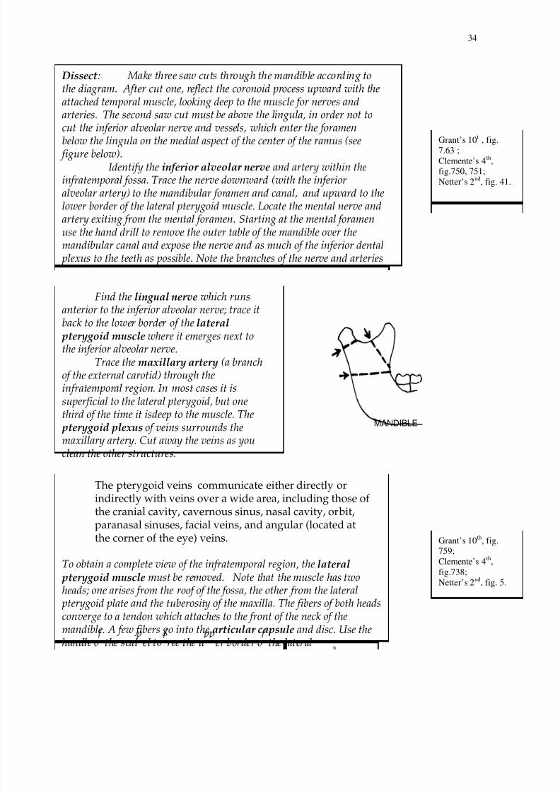
http://slidepdf.com/reader/full/hms1313 34/48

8/17/2019 HMS_1313
http://slidepdf.com/reader/full/hms1313 35/48
35
pterygoid from the roof. Then define the lower border of the muscle byinserting the handle between the lateral and medial pterygoids., where theinferior alveolar and lingual nerves emerge. Free the muscle from the lateral pterygoid plate. Finally, sever the muscle close to its insertion into the neck ofthe mandible and into the articular disc and remove the muscle completely.
Now clean the medial pterygoid muscle. It arises from two heads, one from the maxillary tuberosity and palatine bone, the other from the pterygoid fossa. The muscle runs downward and laterally to be inserted into the medialside of the ramus of the mandible.
Now examine the structures in the infratemporal fossa in detail:inferior alveolar and lingual nerves, branches of the trigeminal,
carrying sensation generally from the mandibular teeth and tongue,respectively
chorda tympani , a branch of the facial nerve carrying taste fibers
from the tongue; it joins the lingual nerve from behindbuccal nerve, its branches pierce the buccinator to supply the
buccal mucosa with sensory fibersmaxillary artery, identify two branches, the inferior alveolar
artery and the middle meningeal arterymuscular branches of maxillary a. to muscles of mastication
(most of these have probably been torn or cut)posterior superior alveolar nerve and artery, pass through
foramena on the maxilla. The nerve originates from the main trunk ofthe maxillary nerve just before it enters the infraorbital canal. One
branch of the nerve may remain external on the bone, continuingdownward to innervate the buccal gingiva in the maxillary molar regionand nearby facial mucosal surfaces, and another enters the posteriorsuperior alveolar canal to travel through the maxillary sinus andinnervate its mucous membrane. This branch continues on to providesensation to the maxillary first, second, and third molars.
branches in the infraorbital canal - these cannot be seen now, but are the origin of the middle and anterior superior alveolar nerves.
The middle superior alveolar nerve (MSA) provides sensation to thetwo maxillary premolars and possibly the mesiobuccal root of the first
molar as well as the periodontal tissues, buccal soft tissue, and bone inthe premolar region. When this nerve is absent (about 30-50% ofindividuals), these areas are supplied by branches of the anterior
Grant’s 10th
,
fig. 7.65 ;
Clemente’s 4th
,
fig.750, 751.
Clemente’s
4th
,fig. 738.

8/17/2019 HMS_1313
http://slidepdf.com/reader/full/hms1313 36/48
36
and lateral incisors and the canine as well as sensory innervationto periodontal tissues, mucous membranes of these teeth, and the buccal bone. The ASA nerve communicates with the MSA andgives off a small nasal branch that innervates the anterior part ofthe nasal cavity along with branches of the pterygopalatinenerves.
Temporomandibular Joint (TMJ)
Although the superior portion of the mandibular ramus has been removed, the head and neck of the mandible are still intact. TheTMJ is a diarthrodial joint where the condyle of the mandiblearticulates with the mandibular fossa of the temporal bone. Thecapsule of the joint is thickened laterally to form the
temporomandibular, or lateral, ligament. Identify thesphenomandibular ligament, which is a thin fibrous band runningfrom the lingula of the mandible to the spine of the sphenoid.Manipulate the head of the mandible to verify the types of movementspermitted at this joint: hinge movements, protraction and retraction.
Dissect : Enter the point of the scalpel into the mandibular fossa close to thebone, and open the upper cavity of the joint. Remove the articular disctogether with the head of the mandible and note the following:
1. The insertion of the severed lateral pterygoid. The remains of themuscle are attached to the neck and articular disc.
2. Open the lower cavity of the joint by cutting the discanteroposteriorly.
There are two types of movements in this joint, one on eachside of the disc. In the lower cavit sim le hin e movements between
Grant’s 10th
,
fig. 7.58A ;
Clemente’s
4th
, figs.740-
744;
Netter’s 2nd
,
fig. 11.

8/17/2019 HMS_1313
http://slidepdf.com/reader/full/hms1313 37/48
37
Lab 6 PREVERTEBRAL REGION, PHARYNX,LARYNX, AND NASAL CAVITIES
Objectives:Be able to identify structures located around the pharynx(nerves, arteries).Know the fascial spaces of the neck.Know the pharyngeal constrictor muscles and theirattachments.Know the structures of the nasal cavity and the openings whichconnect the nasal fossae to the different sinuses.Know the arterial supply and innervation to the nasal mucosa,and the contents and location of the pterygopalatine fossa.
Know the muscles and nerves involved in opening and closingthe larynx.
The major goal of the following dissection is the separation of theanterior part of the skull, face, pharynx, and larynx from the posteriorpart of the skull which articulates with the vertebral column. Onlythen is it possible to complete a thorough study of the visceralcompartment of the neck.
Dissect: Refer to the following illustration of the cranial floor tounderstand the placement of the saw and chisel cuts necessary for separation
of the anterior and posterior portions of the cranium.
IAM
JUG. FOR.
FOR. MAGNUM
When using the chisel, be careful to direct the chisel edge so as

8/17/2019 HMS_1313
http://slidepdf.com/reader/full/hms1313 38/48
38
Start by cutting along the suture line between the body of thesphenoid bone and the basilar portion of the occipital bone.Continue the cut laterally on each side, keeping just posterior to the
jugular foramen, (jug. for.) until you reach the suture between thetemporal bone and the occipital bone. Stay anterior to the f oramenmagnum ( for. magnum).With a saw, cut the two sides of the skullvertically, posterior to the mastoid process. Use a chisel and hammer tocautiously separate the two parts of the skull ; use a scalpel to cut the soft parts (skin and muscle) along a vertical plane passing just behind themastoid process (be careful to stay behind the neurovascular bundleof the neck and the sympathetic trunks). Reflect the anterior portionof the cranium and attached neck viscera onto the anterior thoracic wall.The plane of separation should lie in the retropharyngeal space. The
prevertebral musculature should remain attached to that portion of theskull still articulated with the vertebral column, and the neurovascularbundles of the neck should remain attached to the posterior surface of theviscera of the neck.It is now possible to examine the posterior aspect of the viscera of theneck.
Lateral Pharyngeal Spaces
Dissect: Follow the internal carotid artery, internal jugular vein,
and vagus nerve (10th cranial) to the base of the skull. Identify thethree ganglia of the sympathetic trunk. The superior ganglion is large,approximately one inch in length; the middle ganglion is usually smalland may be absent; the inferior ganglion is usually fused with the firstthoracic ganglion, forming the stellate or cervico-thoracic ganglion.Thespinal ac cessory nerve (11th cranial) lies immediately lateral tothe vagus at the base of the skull; it runs to the sternocleidomastoidmuscle, either through the interval between the internal jugular vein and
internal carotid artery or by coursing lateral to the vein. The
hypoglossal nerve (12th cranial) can be followed from the digastric
triangle toward the base of the skull ; it loops across the surface of internaland external carotid arteries, close to the accessory nerve. The glossopharyngeal nerve (9th cranial) is very small; it lies deep in the
Grant’s 10th
,
fig. 8.36;
Clemente’s 4th
,
pl. 892;
Netter’s 2nd
,
fig. 65.

8/17/2019 HMS_1313
http://slidepdf.com/reader/full/hms1313 39/48
39
Superior pharyngeal
constrictor
Middle pharyngeal
constrictor
Inferior pharyngeal constrictor
Esophagus
Thyroid cartilage
Hyoid bone
Cricoid cartilage
Mandible
Lateral pterygoid
plate
Buccinator
Pharynx
Dissect: Clean the posterior surface of the pharyngeal constrictormuscles and observe the pharyngeal plexus of nerves. The plexusderives its fibers from three sources:
• pharyngeal branches of the vagus nerve, provide motor fibers to the constrictors of the pharynx• glossopharyngeal branches, are sensory to the mucosallining of the pharynx• sympathetic branches, are vasoconstrictor.
The constrictors of the pharynx (superior, middle, andinferior) are incomplete anteriorly, and overlap each otherposteriorly. The muscular part of the pharynx is, therefore a "U"shaped tube completed anteriorly by attachment of the constrictormuscles to the maxilla, mandible, hyoid bone, and thyroid andcricoid cartilages. The muscles of the two sides are insertedposteriorly into a median raphe which extends superiorly from thepharyngeal tubercle, a point on the base of the skull in the midline between the nasal cavity and the foramen magnum, to theesophagus inferiorly. Open the pharynx by making a vertical cut inthe constrictors along the median raphe.Observe the anatomical features of each part of the pharynx as
Grant’s 10th
,
fig. 8.36 ;Clemente’s 4
th,
fig. 892 ;
Netter’s 2nd
,
fig. 56 .
Grant’s 10t ,fig. 8.34-.36;
Clemente’s 4th
,fig. 888, 891;
Netter’s 2nd
, fig
61-62.

8/17/2019 HMS_1313
http://slidepdf.com/reader/full/hms1313 40/48
40
follows:
• laryngopharynx, the portion of the pharynx locatedposterior to the larynx, it merges with the esophagus anteriorto the 6th cervical vertebra. Its anterior wall is marked by theleaf-like epiglottis, situated posterior to the tongue. Theepiglottis guards the obliquely positioned aperture of thelarynx. Extending inferiorly along each side of the laryngealaperture are the piriform fossae. Two branches of the vagus,the internal and recurrent laryngeal nerves, pierce the walls ofthe laryngopharynx to innervate the larynx. These nerves liedeep to the mucosa in the piriform fossae and will beexamined later.
• oropharynx, the part of the pharynx posterior to the oral
cavity; it can be shut off from the nasopharynx by the elevationof the soft palate, an action normally occurring duringswallowing of food or fluid with the contraction of the levatorveli palatini muscle. In the oropharynx, observe thepalatoglossal and palatopharyngeal folds, (overlying thepalatoglossal and palatopharyngeal muscles); the palatinetonsil is located in the depressed area between the two folds.Although these tonsils are large and bulging in children, inadults the site of the tonsil is marked by crypts and no bulgingshould be expected. The bed of the tonsil is related to thesuperior constrictor muscle, and deeper still to the facialartery. The anterior wall of the oropharynx is the posteriorportion of the tongue.
Think about the nasopharynx as described here, but realizethat most of it will be more visible after you have cut the facein the sagittal plane as outlined in the section “Nasal Fossae”to follow. Proceed to make the sagittal cut and thencontinue with the nasopharynx examination.• nasopharynx, located posterior to the two nasal fossae andseptum, and extending to the base of the skull superiorly.
Anteriorly it communicates with the nasal fossae via thechoanae (apertures) which are separated from one another bythe posterior margin of the nasal septum. The roof of thenasopharynx in children contains aggregations of lymphoidtissue, the nasopharyngeal tonsil. The openings of theauditory tubes are the main features of the nasopharynx; eachopening produces a tubal elevation opposite the posterior end
Grant’s 10
th
,fig. 8.37,
8.50;
Clemente’s
4th
, figs.
893,898;
Netter’s 2nd
,
fig. 60-1,70.
Grant’s 10th
,
fig. 8.39-.40,
8.43;
Clemente’s 4th
,
figs. 830;
Netter’s 2nd
,
fig. 54,58 .

8/17/2019 HMS_1313
http://slidepdf.com/reader/full/hms1313 41/48
41
of the inferior concha of the nasal fossae. Transmission ofinfection from the nasopharynx to the middle ear along theauditory tube is not uncommon, especially in children; thus,oitis media and mastoiditis can be complications of a
common cold.
Nasal FossaeDissect: Use a saw to divide the face and cranium in the median(sagittal) plane, then examine the cut surfaces. The two nasal fossae areseparated by a median septum which is cartilaginous anteriorly andbony posteriorly. The septum is covered on both sides by the mucousmembrane. The upper part of the mucosa is olfactory; the central processes of the olfactory neurons pass through the cribiform plate toenter the anterior cranial fossa where they join the olfactory bulbs
beneath the frontal lobes of the cerebrum.
The lateral wall of the nasal cavity presents three bonyelevations, the inferior , middle, and superior conchae. Underneatheach concha is a groove, or meatus which can be examined when theconcha is removed or dislocated. Opening into the meatuses are the paranasal air sinuses, bony cavities lined by mucous membranewhich is continuous with the mucosa of the nasal cavity. Inflammationof the nasal mucosa is usually accompanied by inflammation of the paranasal sinus mucosa.
The spheno-ethmoid recess, a groove above the superiorconcha, receives the opening of the sphenoid sinus. Thesuperiormeatus, the most superior region of the nasal cavity, receives theopening of the posterior ethmoid sinuses.
Carefully remove the middle concha and note a semicirculardepression, the hiatus semilunaris ; above it is an elevation, the bullaethmoidalis. Entrance into the maxillary sinus is through a large,rounded opening in the middle of the hiatus semilunaris; the high position of the opening in the sinus wall relative to the level of the sinus
floor results in inadequate drainage of inflamed sinus mucosa.
The frontal sinus opens into the anterior part of the meatus.Pass a probe from the cavity of the sinus through the opening; it will
Grant’s 10
th
, fig.8.37-.40 ;
Clemente’s 4th
,
figs.
831,832,890,893;
Netter’s 2nd
, fig.
57-61.
Grant’s 10t ,
fig. 7.82-.88 ;
Clemente’s 4th
,
fig. 826-32,
837-41;
Netter’s 2nd
,
fig. 32-4,42-4 .
Grant’s 10th
,
fig. 7.82-
.84,7.87-.88 ;
Clemente’s 4th
,
fig. 826, 832 ;
Netter’s 2nd
,
fig. 32-3 .

8/17/2019 HMS_1313
http://slidepdf.com/reader/full/hms1313 42/48
42
emerge just in front of the hiatus semilunaris.
The anterior and middle ethmoid sinuses open onto oraround the bulla ethmoidalis, an elevation of the bone marking theunderlying ethmoid sinuses.
The nasolacrimal duct opens into the anterior part of theinferior meatus. This duct drains the tears produced by the lacrimal gland inside the orbit.
MAXILLARY SINUS
Examine the interior of the maxillary sinus by breaking through theanterolateral wall of the sinus above the premolar teeth; enlarge theopening.Note the size of the sinus and the location of the sinus floor relative to
the medial opening into the nasal cavity. The floor is located above theroots of the molar teeth and may extend anteriorly to the premolar teeth,and more rarely to the canine tooth. Look for any bumps on the floorover the roots of the teeth. Examine the mucous membrane of the sinusand its thickness. There is some variability from one body to another, sobe sure to examine some of the other cadavers in the lab. Use your handdrill to carefully remove the bone around fine superior alveolar nervebranches.
The dental plexus of the superior alveolar nerve islocated between the floor of the maxillary sinus and theapices of the teeth. The dental plexus innervates both theteeth and the lower portion of the sinus, so inflammationin either region will give rise to referred pain in the otherarea.
LarynxThe thyroid cartilage consists of two laminae, united
anteriorly at an angle (laryngeal prominence, or Adam's apple), butopen posteriorly. Anteriorly, the thyroid cartilage has a mediannotch on its upper border. The thyrohyoid and sternothyroidmuscles attach on the lateral surface of each lamina. The cricoid
cartilage resembles a "signet ring", with the narrow portion of thering situated anteriorly. The cricothyroid ligament connects cricoid
Clemente’s 4t
,
fig. 863.
Grant’s 10th
,
fig. 8.46-.47;
Clemente’s 4th
,
fig. 899-907;
Netter’s 2nd
,
fig. 71.

8/17/2019 HMS_1313
http://slidepdf.com/reader/full/hms1313 43/48
43
and thyroid cartilages.The arytenoid cartilages are two small pyramidal cartilages
whose bases articulate with the large posterior lamina of the cricoidcartilage.
The epiglottis is a leaf-like structure fixed to the posterioraspect of the thyroid angle, the hyoid bone, and back of the tongue.It functions as a lid which deflects materials which might otherwiseenter the laryngeal aperture.
Dissect: Make a shallow median cut in the mucous membrane over theback of the cricoid cartilage and carefully remove the mucous membrane ofthe laryngopharynx. Dissect free the recurrent laryngeal nerve whichenters the larynx at the lower border of the inferior constrictor muscle, and passes superiorly and laterally, deep to the mucous membrane of the piriform fossa, to innervate the laryngeal musculature.
Realize : On the upper border of the cricoid cartilage are locatedarytenoid cartilages on each side of the midline. Each arytenoidcartilage is a 3-sided pyramid with its base resting on the cricoidcartilage.The arytenoid base is triangular; the forward-pointing apex,providing attachment to the vocal ligament, is called the vocalprocess; the prominent laterally projecting angle, or muscularprocess, provides attachment for lateral and posterior cricoarytenoidmuscles which rotate the arytenoids around vertical axes, therebychanging the tension on the vocal ligament during speech.
Fracture the thyroid lamina by pushing it laterally, and disarticulatethe joint between its inferior horn and the cricoid ring. With the scalpel, cutthe larynx posteriorly along the midline, and open it by fracturing itscartilages. Locate two pairs of anteroposterior folds:• vestibular folds, the superior pair of folds which contains thevestibular ligaments.• vocal folds, the inferior pair of folds which contains the vocalligaments and the vocal portion of the thyroarytenoid muscle
The glottis is the region of the larynx at the level of the vocal folds:
it consists of the rima glottidis (the space between the vocal folds) and the
Grant’s 10
t
,fig. 8.56;
Clemente’s 4th
,
fig. 908-10;
Netter’s 2nd
,
fig. 57,72.

8/17/2019 HMS_1313
http://slidepdf.com/reader/full/hms1313 44/48
44
Lab 7 ORAL CAVITY
Objectives:Know the bones, nerves, and arteries of the oral cavityand palate.
Know the location and function of the Eustachian tube.Know the limits of the vestibule and the oral cavityproper.Know what structures can be palpated around themouth, both inside and outside.Know where to locate the major nerves for anesthesiainside the oral cavity (nerves from both maxillary andmandibular trunks of the trigeminal).Know the spaces of the face and neck which allowpassage of infection.Be able to identify major structures (bones, arteries andnerves, etc.) in cross-sectional drawings of the headthrough the occlusal plane.Know the location of the pterygomandibular raphe inyourself and your cadaver, and its relationship toinjections of the inferior alveolar nerve.
See the list of structures at the end of this labdescription. Before you leave the lab today,find your tutor and demonstrate thestructures on your cadavers. (All students
should participate in the identification.)
Mouth and TongueObserve: The oral cavity consists of the vestibule
and oral cavity proper. The vestibule is limited externally bythe cheeks and lips and is separated from the oral cavity proper by the teeth and gingiva. An important anatomical feature ofthe vestibule is the orifice of the duct of the parotid gland,situated at the apex of a slight elevation or papilla opposite theupper 2nd molar tooth. Pass a probe through the parotidpapilla and note the course of the duct.
The walls of the oral cavity proper are: laterally andanteriorly, teeth and gingiva; superiorly, hard and soft palates.Inferiorly, the oral cavity is limited by the mylohyoid muscle:the sublingual gland lies under the mucous membraneadjacent to the base of the tongue.

8/17/2019 HMS_1313
http://slidepdf.com/reader/full/hms1313 45/48
45
Numerous glands are located under the mucousmembranes of the oral cavity, such as labial glands on the insideof the lips, and the palatine glands under the hard palate.
Floor of the mouth:
In the bisected head of the cadaver, examine the musclesof the floor of the mouth and the tongue; the mylohoid,geniohyoid, and the large fan-shaped genioglossus.
Dissect : In order to expose the sublingual gland, make a thin cut in themucous membrane just medial to the mandible, and lateral and inferiorto the tongue and retract the tongue medially to separate the tongue from the mylohyoid muscle. (Do not extend the cut posterior to thesecond molar.) Now you will be able to expose the sublingual gland,submandibular gland and duct, lingual and hypoglossal nerves, andextrinsic muscles of the tongue.
Identify:Sublingual gland- identify several short ducts which open at theplica sublingualis.Submandibular duct- runs diagonally across the medial aspectof the sublingual gland. Find the papilla of the duct lateral to thefrenulum linguae, then follow the duct posteriorly to thesubmandibular gland.Lingual nerve- find the nerve behind the last molar running between the ramus of the mandible and the medial pterygoidand trace it forward into the floor of the mouth. It should befollowed downward across the muscles of the tongue deep to thesublingual gland where its terminal branches originate. These aresensory nerves to the anterior 2/3 of the tongue. It also carriesthe taste fibers originating from the chorda tympani (of the VIInerve) for taste on the anterior 2/3 of the tongue.Submandibular ganglion- In the region of the 3rd molar look forthe submandibular ganglion which is suspended from the lingualnerve by several short branches.
Dissect:
From the lateral side, identify the mylohyoid muscle and detach it from the hyoid bone; reflect it superiorly to expose the hyoglossusmuscle. Identify the hypoglossal and lingual nerves from this side.Notice that the hypoglossal nerve is situated between thesubmandibular gland and hyoglossus muscle, inferior to the lingualnerve. Follow it to the tongue musculature. Locate the lingual arterymedial to the hyoglossus muscle and follow it to the tongue.
Grant’s 10t
, fig.7.71,7.78;
Clemente’s 4th,
fig. 890;Netter’s 2
nd, fig.
53,57.
Grant’s 10t , fig.
7.78;
Clemente’s 4th
,
fig. 850, 855;Netter’s 2nd, fig.
41,53.

8/17/2019 HMS_1313
http://slidepdf.com/reader/full/hms1313 46/48
46
TongueThe tongue has a tip, body, and base. On its superior, or palatinesurface observe the V-shaped terminal sulcus that separates the body from the base. In front of the terminal sulcus are 8 to 12
circumvallate papillae; behind the sulcus, the surface of thetongue appears irregularly folded due to the presence of bumpsand depressions representing accumulations of lymphoid tissue,collectively called the lingual tonsil.Note that the lingual tonsil, palatine tonsils, and lymphoidnodules in the thickness of the soft palate form a complete ring oflymphoid tissue around the oropharyngeal isthmus.
In a similar fashion, the nasopharyngeal tonsil in thepharyngeal vault, lymphoid tissue near the opening of theauditory tube (tubal tonsil), and lymphoid nodules in thethickness of the soft palate, all contribute to a ring of lymphoid
tissue around the choanae (the opening of the nasal cavity intothe nasopharynx).
Look for the following features on the tongue of thecadaver, as well as on your own:
• Sulcus terminalis- divides the anterior two thirds andthe posterior one third
• Foramen cecum- at the site of the embryonicthyroglossal duct that was attached to the developingthyroid gland
• Fungiform papillae- mushroom-shaped papillae that
appear to be red spots in your tongue• Filiform papillae- sensitive to touch
• Circumvallate papillae- located anterior to the sulcusterminalis
• Glossoepiglottic fold- running from the tongue to theepiglottis, with the valleculae located on either side
• Lingual tonsils- lymphoid follicles on the posteriorthird of the tongue
Palate
Review on a skull:The base of the nasal cavity is formed by the bony palate,
which is composed of, anteriorly, the palatine process of themaxilla, and, posteriorly, the horizontal plates of the palatinebones. Behind the incisor teeth at the midline of the hard palate
Grant’s 10t
,
fig. 7.83;Clemente’s 4th,
fig. 860;
Netter’s 2nd
, fig.
52.

8/17/2019 HMS_1313
http://slidepdf.com/reader/full/hms1313 47/48
47
is the incisive foramen through which the thin nasopalatine nerveexits. Medial to the third molar tooth, locate the greater palatineforamen, which is the opening of the greater palatine canal.Identify the vertical part of the palatine bone which forms part ofthe lateral wall of the nasal cavity. Examine the skull from the
lateral side. Locate the pterygopalatine fossa between thepterygoid process, maxilla, palatine bone, and sphenoid bone. Itssphenopalatine (pterygopalatine) foramen is formed where thenotch on the superior border of the palatine bone meets thesphenoid bone and is a passageway for nerves and blood vesselsfrom the pterygopalatine fossa into the nasal cavity.
Realize: The palate on your cadaver has anteriorly, a hard portion,and posteriorly, a soft portion. Palatine glands, small mucousglands, are present over the palate, with small ducts open to thesurface.
Dissect: Find the approximate location of the greater palatine foramen with a dissecting needle. Posterior to it cut through the mucousmembrane of the hard palate and peel it off anteriorly to expose the greater palatine nerve and vessels. Next, follow the greater palatinecanal superiorly from the greater palatine foramen to observe the greater palatine nerve and artery within the canal and the sphenopalatine ganglion at the opening of the sphenopalatine foramen. To find thecanal, insert a 1 1/4 inch needle probe into the greater palatine foramen.Then, using a probe, or small bone rongeurs, open the medial wall of thecanal to expose its nerve and vessels. Follow the nerve upwards to thesphenopalatine (pterygopalatine) ganglion which lies deep in the pterygopalatine fossa.Examine your cadaver from the lateral side. Identify:
1. Maxillary artery- giving off the greater palatine artery andsphenopalatine artery
2. Maxillary nerve- Passing from the foramen rotundum to theinferior orbital fissure , giving off posterior superior alveolar nervebranches which enter the maxillary bone to innervate upper teeth
3. Sphenopalatine (pterygopalatine) ganglion- connected tothe maxillary nerve by 2 short branches. The ganglion contains
postganglionic parasympathetic cell bodies whose axons follow branches
Grant’s 10
t
, fig.7.111, 7.105;
Clemente’s 4th
,
figs. 833-836;Netter’s 2nd, fig.
35-38.
Grant’s 10t , fig.
8.64-8.65;
Clemente’s 4th
,fig. 845,846;
Netter’s 2nd, fig.
49,54,58-59.

8/17/2019 HMS_1313
http://slidepdf.com/reader/full/hms1313 48/48
48
of the maxillary nerve to innervate the lacrimal gland and small glands of the nasal cavity, pharynx, and palate.
Palatine tonsilThe palatine tonsils sit on either side of the oropharynx
extending partway into the soft palate. As mentionedpreviously, each tonsil is situated in the depression betweenthe palatoglossal and palatopharyngeal arches covering themuscles, although they are seldom present in our cadavers.
Pharyngeal wallDissect:Remove the mucous membrane from the soft palate, lateral pharyngeal wall and nasopharynx to expose the palatoglossal and palatopharyngeal muscles. The palatopharyngeus and thestylopharyngeus form the longitudinal portions of the pharynx.
Demonstrate the following structures in the region of the oralcavity to your tutor:
• Lin ual arter


