HÅKAN NILSSON RESILIENT APPLIANCE THERAPY OF - MUEP
89
HÅKAN NILSSON RESILIENT APPLIANCE THERAPY OF TEMPOROMANDIBULAR DISORDERS Subdiagnoses, sense of coherence and treatment outcome Swedish Dental Journal, Supplement 206, 2010 SWEDISH DENTAL JOURNAL, SUPPLEMENT 206. DOCTORAL DISSERTATION IN ODONTOLOGY.
Transcript of HÅKAN NILSSON RESILIENT APPLIANCE THERAPY OF - MUEP

HÅKAN NILSSONRESILIENT APPLIANCE THERAPY OF TEMPOROMANDIBULAR DISORDERSSubdiagnoses, sense of coherence and treatment outcome
Swedish Dental Journal, Supplement 206, 2010SW
ED
ISH
DE
NTA
L JO
UR
NA
L, S
UP
PL
EM
EN
T 2
06
. DO
CT
OR
AL
DIS
SE
RTA
TIO
N IN
OD
ON
TO
LOG
Y.


R E S I L I E N T A P P L I A N C E T H E R A P Y O F T E M P O R O M A N D I B U L A R D I S O R D E R S
S U B D I A G N O S E S , S E N S E O F C O H E R E N C E A N D T R E A T M E N T O U T C O M E

Swedish Dental Journal, Supplement 206, 2010
© Copyright Håkan Nilsson, 2010
Photo: Håkan Nilsson
ISBN 91-7104-311-X
ISSN 0348-6672
Holmbergs, Malmö 2010

HÅKAN NILSSON RESILIENT APPLIANCE THERAPY OF TEMPOROMANDIBULAR DISORDERS
Subdiagnoses, sense of coherence and treatment outcome
Departments of Stomatognathic Physiology and Oral and Maxillofacial
Radiology, Faculty of Odontology, Malmö University, Malmö, Sweden
Department of Stomatognathic Physiology, Specialist Dental Centre, Kalmar County Hospital,
Kalmar County Council, Kalmar, Sweden 2010

The summary of this publication is also available at www.mah.se/muepPlease visit!

CONTENTS
ABSTRACT ..................................................................... 9POPULÄRVETENSKAPLIG SAMMANFATTNING .................. 11PREFACE ..................................................................... 13ABBREVIATIONS ........................................................... 15INTRODUCTION ........................................................... 17
Oral health and temporomandibular disorders (TMD) ................17Diagnosing TMD ...................................................................21Psychosocial factors, sense of coherence (SoC) and general health ................................................................24TMD management .................................................................26Evaluating treatment outcome .................................................29
OBJECTIVES ................................................................. 33HYPOTHESES ............................................................... 35MATERIALS AND METHODS ........................................... 37
Patients (I-IV) .........................................................................37Procedures (I-IV) ....................................................................39
Diagnosing and imaging (II) ...............................................39Experimental Methods (III-IV) ..............................................42Treatment Method (III-IV) ....................................................44SoC, grade of depression, non-specific physical symptoms and general health (IIII) .......................................45
Statistical methods (I-IV) ..........................................................46Ethical considerations ............................................................47
RESULTS ...................................................................... 49Paper I and thesis ..................................................................49Papers II and IV ....................................................................50

Treatment outcome ........................................................... 52Paper III ............................................................................... 59
DISCUSSION ................................................................ 63Diagnosing .......................................................................... 63Resilient appliance therapy .................................................... 67SoC as an influencing factor for the treatment outcome ............. 72Implications for general practitioners and future research .......... 72
CONCLUSIONS ............................................................ 75ACKNOWLEDGEMENTS ................................................ 77REFERENCES ................................................................ 81

9
ABSTRACT
Temporomandibular disorders (TMD) with orofacial pain with or without reduced jaw function, are frequent conditions in the gen-eral population. Different factors such as tooth clenching and grinding, sometimes due to enhanced psychosocial stress, and trauma to the jaws may be important as etiologic factors. Signs and symptoms of TMD are a common cause for general practitioners to use different intraoral appliances as pain and bite-force reducing devices and for improvement of a reduced jaw function. Intraoral appliances are often used parallel to other treatment modalities. Before treatment start a thorough history taking and clinical ex-amination is necessary for a relevant diagnosis. Sometimes the di-agnostic process has to be complemented with proper radiographic imaging in order to support the diagnostic process.
The overall aim of this thesis was to compare magnetic reso-nance imaging (MRI) findings of the TMJ on the clinically assessed diagnoses and to evaluate short- and long-term treatment outcome of a resilient intraoral appliance, in patients with TMD pain. A fur-ther aim was to study Sense of Coherence as an influencing factor on treatment outcome, on these patients.
In article I the aim was to compare findings on MRI in TMD pain patients with clinical diagnoses of myofascial pain or arthral-gia/ osteoarthritis in combination with myofascial pain according to the Research Diagnostic Criteria for TMD (RDC/TMD). The temporomandibular joints of 60 consecutive patients, 19 with myofascial pain and 41 patients with arthralgia/osteoarthritis in combination with myofascial pain were examined clinically and with MRI. The most common MRI findings were disc displace-
10
ments with or without reduction and structural bone changes. These findings were found in both pain groups, however, disc dis-placements were found significantly more often in patients with ar-thralgia/osteoarthritis in combination with myofascial pain. Joint fluid was found in both pain groups. The clinical diagnoses for subdivision into myogenous only or combined arthrogenous and myogenous pain groups were not confirmed by MRI findings.
In article II the short-term efficacy of a resilient appliance com-pared to a non-occluding control appliance was studied in a ran-domised, controlled trial with 80 recruited TMD pain patients. They were randomly allocated to one of two groups: treatment with a resilient appliance or treatment with a hard, palatal, non-occluding appliance. After 6 and 10 weeks of treatment, character-istic pain intensity (CPI) decreased in both groups. There was no statistically significant difference found between the resilient appli-ance and the non-occluding control appliance in reducing TMD pain in a short-term perspective.
In article III possible factors of importance for treatment out-come were studied as well as the association between Sense of Co-herence and grade of depression, and grade of non-specific physical symptoms and general health, in the TMD pain patients. A total of 73 TMD pain patients participated; 36 were treated with a resilient appliance and 37 with a non-occluding control appliance. The findings indicated that none of the studied background variables (age, gender, SoC, depression, nonspecific physical symptoms or general health) seemed to influence the short-term efficacy of in-traoral appliances. In the TMD pain patients, no associations were found between SoC and depression, non-specific physical symp-toms or general health.
In article IV the long-term efficacy was evaluated of the resilient appliance compared to the non-occluding control appliance in the TMD pain patients. Appliance wear was also studied in this article. As in the short-term follow-up, there was no statistically significant difference between the resilient appliance and the non-occluding control appliance in reducing TMD pain in the long-term perspec-tive.

11
POPULÄRVETENSKAPLIG SAMMANFATTNING
Temporomandibulär dysfunktion (TMD) med käk- och/eller an-siktssmärta, med eller utan nedsatt käkfunktion, är vanligt före-kommande tillstånd i befolkningen. Tandpressning och gnissling, som kan vara utlösta på grund av hög psykosocial stress, utgör till-sammans eller parallellt med bland annat käktrauma, orsaksfakto-rer som kan ge upphov till TMD-symtom som smärta. Hos allmän-tandläkaren är det vanligt att undersökningsfynd och symptom på TMD-besvär föranleder behandling med bettskena. Denna används då för att reducera sammanbitningskrafter och TMD-smärta, samt för att förbättra patientens käkfunktion. Bettskenebehandling på-går ofta parallellt med andra bettfysiologiska behandlingsinsatser för att uppnå bästa smärtlindrande resultat. Innan behandlingen påbörjas utförs en noggrann anamnesupptagning och klinisk un-dersökning för att komma fram till en behandlingsgrundande dia-gnos. Ibland bör den diagnostiska processen inbegripa någon typ av radiologisk undersökning.
Det övergripande målet med detta avhandlingsarbete var att stu-dera en grupp patienter med TMD-smärta och jämföra deras käk-ledsfynd på magnetresonanstomografi (MRT) med diagnosen base-rad på den kliniska undersökningen och studera behandlingsutfal-let av bettskenebehandling med mjuk bettskena (resiliensskena), i ett korttids- och ett långtidsperspektiv. Ytterligare ett mål var att studera “känsla av sammanhang” som påverkansfaktor på behand-lingsutfallet.
I artikel I var målet att jämföra MRT-fynd på käklederna, hos patienter med TMD smärta, med de kliniska diagnoserna myofa-

12
scial smärta och myofascial smärta i kombination med artralgi eller osteoartrit enligt diagnossystemet RDC/TMD, speciellt framtaget för diagnostik i forskningssammanhang. De vanligaste fynden på MRT-bilderna var diskdisplaceringar med eller utan återgång och strukturella benförändringar. Dessa fynd förekom i båda diagnos-grupperna, men diskdisplacering iakttogs oftare hos patienterna med myofascial smärta i kombination med artralgi eller osteoartrit. Att dela in de kliniska diagnoserna i grupperna myofascial smärta och myofascial smärta i kombination med artralgi eller osteoartrit, kunde inte bekräftas med MRT-fynden i denna studie.
I artikel II utvärderades korttidseffekten av behandling med resi-liensskena jämfört med en kontrollskena. 80 patienter med TMD-smärta rekryterades och randomiserades till behandlingsgrupperna. Efter 6- och 10 veckors behandling utvärderades behandlingseffek-ten. Det fanns inga statistiskt signifikanta skillnader mellan resili-ensskenan och kontrollskenan när det gällde att reducera TMD-smärtan hos patienterna i ett korttidsperspektiv.
I artikel III studerades faktorer av betydelse för behandlingsutfal-let, dessutom studerades förhållandet mellan TMD smärtpatienter-nas “känsla av sammanhang” och depressionsgrad, somatise-ringsgrad och generella hälsa. Resultatet talar för att inga av de studerade bakgrundsfaktorerna hade betydelse för behandlingsef-fekten av resiliensskenan i ett korttidsperspektiv. Ingen relation mellan ”känsla av sammanhang” och depressionsgrad, somatise-ringsgrad eller generell hälsa, kunde konstateras.
I artikel IV studerades långtidseffekten av resiliensskenan jäm-fört med kontrollskenan hos patienter med TMD-smärta. Slitaget av skenorna efter användning studerades också i denna artikel. Precis som i korttidsuppföljningen fanns det inga statistiskt signifi-kanta skillnader i smärtlindrande behandlingseffekt mellan resili-ensskenan och kontrollskenan, i ett långtidsperspektiv.
13
PREFACE
This thesis is based on the following publications/papers, which are referred to in the text by their Roman numerals:
I Limchaichana N, Nilsson H, Ekberg EC, Nilner M, Petersson A. Clinical diagnoses and MRI findings in patients with TMD pain. J Oral Rehabil 2007;34:237-245.
II Nilsson H, Limchaichana N, Nilner M, Ekberg EC. Short-term treatment of a resilient appliance in TMD pain patients: a randomized controlled trial. J Oral Rehabil 2009;36:547-555.
III Nilsson H, Ekberg EC. Do psychological factors and general health influence the short-term efficacy of re-silient appliance therapy in patients with TMD pain? Acta Odontol Scand 2010;29: Epub ahead of print
IV Nilsson H, Vallon D, Ekberg EC. Long-term efficacy of a resilient appliance in TMD pain patients: a ran-domized, controlled trial. Submitted
The papers are reprinted with kind permission from the copy hold-ers.

14

15
ABBREVIATIONS
CPI Characteristic pain intensity
IMMPACT Initiative on Methods, Measurement, and Pain As-sessment in Clinical Trials
ITT Intent to treat
MRI Magnetic resonance imaging
NNT Number needed to treat
RCT Randomised controlled trial
RDC/TMD Research Diagnostic Criteria for Temporomandibu-lar Disorders
SCL-90-R The Symptom Checklist -90 Revised
SoC Sense of Coherence
TMD Temporomandibular disorders
TMJ Temporomandibular joint
VAS Visual analogue scale

16

17
INTRODUCTION
Oral health and temporomandibular disorders (TMD)
Health was first defined by the World Health Organization (WHO) in 1946 as “a state of complete physical, psychological and social well-being, and not merely the absence of disease or infirmi-ty”.1 In the 1960s Andrew Twaddle applied the “disease-illness-sickness-triad” which is a widely used model.2 After some criticism of the model, Hofmann and Eriksen3 defined the concepts of the triad in 2001, as follows:
Disease is characterised in the sphere by health care profession-als, and is the biomedical aspect of health and defined as “a bodily or mental occurrence that tends to reduce the capacity of the or-ganism”.
Illness is characterised by the personal sphere and is “a subjec-tive negative experience that tends to reduce the capacity of the person”.
Sickness is characterised by the social sphere and is “a social identity assigned to a human agent due to events that tend to re-duce his or her social capacity”.
These three concepts are related and have different influences on human ailments.
Oral health is essential for the human being to be able to function in every day life. A pain free, natural movement of the mandible is

18
important for the personal nourishment and for the interaction and communication with fellow members of the society. Oral health is defined by the WHO as:
“a state of being free from chronic mouth and facial pain, oral and throat cancer, oral sores, birth defects as cleft lip and palate, perio-dontal (gum) disease, tooth decay and tooth loss, and other dis-eases and disorders that affect the oral cavity.”4
For dentists it is important to differentiate TMD from odontogenic pain in order to manage the patient accurately. Clinicians must be aware of the different orofacial pain conditions and know their dis-tinguishing characteristics to make proper differential diagnoses of the pain enabling proper medical and/or dental therapy. It is im-portant to evaluate and examine the patient in a structural process and, when needed, refer the patient to professions with formal competence to examine, treat, and evaluate the TMD pain.
Orofacial pain can be caused by different disorders localised to the tissues in the oral and facial region. Hence, orofacial pain can be divided into musculoskeletal, neurovascular, neuropathic and psy-chogenic.5 The musculoskeletal type, which embraces muscle prob-lems, mechanical problems of the TMJ, and different TMJ arthri-tides, is the most common type of orofacial pain that the general dental practitioner will find among patients seeking dental care due to symptoms of TMD. Many of these TMD problems can be ma-naged adequately by the general dental practitioner. Sometimes pa-tients with severe musculoskeletal orofacial pain have to be re-ferred to an orofacial pain/TMD specialist for an extended evalua-tion. The orofacial pain/TMD specialist will be able to examine and treat more complicated cases of musculoskeletal pain and make some differential diagnoses of neurovascular pain (different types of headache, e.g. tension type headache) and neuropathic pain (peripheral neuropathic pain and some neuralgias, e.g. trige-minal neuralgia). The differential diagnostic process should prefer-ably be performed in a multidisciplinary way with referral to dif-ferent medical specialists, when needed. Of chronic orofacial pain conditions the TMD pain is the most common, similar to back

19
pain in its intensity, persistence and psychological impact.6 In 2004 the International Headache Society made an effort to categorise and classify all of the hitherto known craniofacial pain conditions.7
TMDs were included, among other conditions such as facial neu-ropathies and headache disorders, in this categorisation. It is im-portant for dental practitioners to refer patients with headache to a medical specialist in e.g. neurology, for a second opinion when symptoms from the orofacial region are suspect and not merely musculoskeletal.
TMD embraces signs and symptoms from the orofacial region. The term was suggested in the 1990’s by Dworkin et al.8 and describes a cluster of painful disorders localised to the preaurical area, the TMJ and the masticatory muscles. Dysfunctions such as limitations and deviations in the mandibular range of motion and/or noises in the TMJ during mandibular function, may be included in signs and symptoms. Joint sounds are often described as clicking, popping, grating or crepitating. Other common symptoms are tiredness or fatigue in the jaw muscles, pain from the jaw, earache, headache, and facial pain. These different symptoms may be present at rest or during mandibular function, and sometimes they are aggravated by chewing and other mandibular movements. According to Hofmann and Eriksen3 TMD may be considered an illness as it is an ailment for the individual with a subjective negative experience that tends to reduce the person’s capacity (e.g. chewing), and as a sickness as the condition may reduce the person’s social capacity (e.g. feeling embarrassed in public, difficulties when kissing). TMD pain, which is the main reason for patients to seek medical or dental care, must not be of neurogenic or psychogenic origin to be defined as TMD. Visceral, periodontal, dental, or cutaneous pain is also excluded from the TMD definition.8, 9
The reported prevalence of TMD differs between investigations, but is considered the most common orofacial condition of non-dental origin.10 The prevalence of TMD was reported to be 8% to 15% among women, and the corresponding figures reported for men were 3% to 10%.10 The “true“ prevalence of TMD is still de-bated due to lack of homogeneity in the diagnostic criteria through

20
the years in the various research groups. However there is evidence that signs and symptoms of TMD may be high in non-patient populations.11 The prevalence figures of TMD in the general popu-lation are divergent and ranges from 1% to 75% for objective signs and from 5% to 33% for subjective symptoms.12 In a meta-analysis it was found that in the adult Dutch population prevalence figures varied widely; clinical signs from 0% to 93% and symp-toms from 6% to 93%.13 The variety in figures could be explained by different materials and included a range of mild to severe signs and symptoms of TMD.
In a study of etiologic factors for TMD 10% of the adult popula-tion reported that they suffered from TMD pain.10 In a population based study by Locker and Slade, 12.9% of a Canadian population reported functional pain or pain at rest due to TMD.14
Headache, and especially tension-type headache, has been reported in about 40-70% of patients with TMD.15, 16 A Danish study re-ported that the prevalence of TMD in the studied headache popu-lation was 56.1%.17 In their conclusion, the authors emphasised the importance of examining the masticatory system in tension-type headache sufferers and they underlined the necessity of a multidi-mensional approach to chronic headache patients.
Through the years, continuous research has provided modern med-icine and dentistry with powerful diagnostic systems and treatment methods. This has, among other things, lead to ethical challenges for the clinician. The question “To treat or not to treat?” is impor-tant to consider in order to provide patients with high quality, evi-dence based care.3 In 1988, Locker and Slade reported that the proportion of patients in need of treatment varied from 3.5% to 9.7% according to the case definition used.14 Twenty years later Al-Jundi et al.18 concluded in a meta-analysis that the treatment need in the general adult population with TMD symptoms is substantial and varies according to definition, criteria, and age. The meta-analysis performed in the review showed an estimated treatment need of approximately 16% in adult subjects with TMD symp-toms, whilst subjects in the younger age groups (19 to 45 years)

21
had a higher estimated treatment need. This is a considerable num-ber of potential patients with a treatment need. This may result in consequences for health care programs, and in many countries spe-cial achievement and financial resources are put into preventing and manage such conditions. High figures for prevalence, treat-ment need in the general adult population leads one to consider TMD both as a disease and a public health problem. It results in a great demand on health care systems to be cost-effective. However, one should keep in mind when allocating oral health resources that there is a discrepancy between need and demand for TMD treat-ment.
Diagnosing TMD
Through the years different kinds of diagnostic systems have been used to classify TMD. All systems include listening to and record-ing of patient’s chief complaints, the history of the problem and a clinical examination. Due to shortcomings of these methods, sever-al classifications of TMD have been presented in order to improve the diagnostic process and secure an adequate treatment when needed.5, 19
Pain is a subjective, multidimensional and complex experience for each patient according to the definition of the International Asso-ciation for the Study of Pain.20 Therefore, the patients’ self-reported pain is fundamental for accurate diagnoses according to the Re-search Diagnostic Criteria for Temporomandibular Disorders (RDC/TMD). Another reason for seeking care is reduced mandibu-lar function often in combination with TMD pain. Today, RDC/TMD is a widely accepted classification system for subdiag-noses of TMD, presented in 1992 by Dworkin and LeResche.8 It is based on a dual-axis system, describing physical status on Axis I and psychological parameters on Axis II. This enables the classifi-cation system to reflect the complexity of the physical dimensions, as well as, the psychological dimensions of persistent TMD pain by developing criteria for the most common subdiagnoses of TMD. The use of RDC/TMD also enables researchers to investigate de-
22
mographics of study population and patient characteristics such as self-reported oral habits and other risk factors of TMD signs and symptoms. For Axis I diagnosis multiple diagnoses for the TMJ’s are allowed, but only one diagnosis for the masticatory muscles. The patients Axis II profile contains graded chronic pain status, and scores for grade of depression and grade of non-specific physi-cal symptoms, and summary score for limitations in the ability to use the jaw.
In RDC/TMD clear definitions of TMD diagnostic subgroups were presented. The standardised examination methods gather relevant data and make possible comparison of findings and replication of research in the most common forms of muscle- and joint-related TMD among diverse clinical investigators, the importance of clini-cians to be calibrated has been emphasised by the authors.21, 22 It has been tested for reliability concerning clinical measurements and has shown good results.23-25 RDC/TMD diagnoses are divided into three groups:8, 26
I. Muscle diagnoses
a. Myofascial pain
b. Myofascial pain with limited opening
II. Disc displacements
a. Disc displacement with reduction
b. Disc displacement without reduction, with limited opening
c. Disc displacement without reduction, without limited open-
ing
III. Arthralgia, arthritis, arthrosis
a. Arthralgia
b. Osteoarthritis of the TMJ
c. Osteoarthrosis of the TMJ
Pain from the temporomandibular region accounts for most of the suffering for TMD patients, making subdiagnoses from group I

23
and III (a and b) the most prevalent.11 RDC/TMD diagnoses can be made based on clinical and history criteria only, but imaging can be included in the diagnostic process of disc displacement without reduction, osteoarthritis, and osteoarthrosis.8
Arthrography and MRI are considered suitable imaging methods in the diagnosis of disc displacement while tomography is preferred in the diagnosis of osteoarthritis and osteoarthrosis.8 A hierarchical model, for imaging technique’s efficacy, in 6 levels was described by Fryback and Thornbury.27 Levels 3-5 influence the orofacial pain/TMD specialist’s clinical process when an accurate diagnosis and treatment modality is to be decided and when treatment out-come is evaluated. In a systematic literature review by Koh et al.28
no clear evidence was found for a relationship between clinical and MRI diagnoses and findings. In another systematic literature re-view by Limchaichana et al.29 the authors reported that it was im-possible, at the time, to draw any conclusion about when the re-sults of MRI examination resulted in a better treatment outcome for the TMD patients, due to the fact that none of the searched publications reported diagnostic thinking efficacy or therapeutic efficacy. In 1995 Nilner and Petersson30 studied the influence of ra-diographic findings on tomography, on treatment outcome of TMD after one year of treatment. The authors concluded that no single radiographic finding was found to be related to treatment outcome and therefore tomography was considered to have a mi-nor role in the management of TMD patients.
A continuous revision of the RDC/TMD was initiated by Dworkin and LeResche, from the beginning, in order to improve the system according to current and future research. Diagnostic criteria for TMD are under construction, supervised by the members of the in-ternational RDC/TMD consortium.26 The up-dated system is planned to be published in the year 2010, and its purpose is to guide both general practitioners and specialists in a structured TMD diagnostic process. The results of the preparation of the up-dated diagnostic system have recently been published.31-36

24
Psychosocial factors, sense of coherence (SoC) and general health Psychosocial factors constitute significant components in pain in general and have to be taken into consideration when examining patients seeking care for TMD pain. Parallel to different orofacial signs and symptoms, there is evidence that TMD is characterised by increased psychological distress.9 Patients suffering from chronic or long-standing pain conditions often also have an elevated grade of depression.37 In 1996 a research team reported that patients with chronic TMD were also more likely to have signs of depression when compared to patients with a more recent onset of TMD.38
In Axis II of RDC/TMD a questionnaire, based on a modified ver-sion of the validated SCL-90-R scale, is included estimating grade of depression and grade of non-specific physical symptoms.39 The three-graded rating scale contains normal, moderate, and severe grade of depression and non-specific physical symptoms. The mod-ified SCL-90-R scale is used as a screening instrument assessing general psychological distress and discomfort. The instrument is not used to provide psychiatric diagnoses, rather it provides initial scientific support to validate evidence-based clinical decision- mak-ing.40 Dworkin et al.37 have shown a significant association between number of pain conditions reported and high levels of non-specific physical symptoms and depression as measured by SCL-90-R. They also concluded that the number of pain conditions reported was a good predictor of major depression.
In 1987 Antonovsky41 presented the theoretical model of sense of coherence (SoC). The model is based on a salutogenic way of look-ing at health and unhealth in life. The objective of SoC is to meas-ure the individual’s capacity to adequately respond to stressors in daily life by using different strategies of coping.42 SoC is defined as:
“[-] a global orientation that expresses the extent to which one has a pervasive, enduring though dynamic feeling of confidence that (1) the stimuli deriving from one’s internal and external envi-ronments in the course of living are structured, predictable, and explicable; (2) the resources are available to one to meet the de-

25
mands posed by these stimuli; and (3) these demands are chal-lenges, worthy of investment and engagement.”
Three themes, as implied in the definition, are central in the SoC: comprehensibility, manageability and meaningfulness.
1. Comprehensibility refers to a person’s ability to make sense of stimuli and occurrences, the ability to understand them.
2. Manageability refers to a person’s ability to cope with stimuli and occurrences, the extent to which a person has resources, or the ability to gather resources, to deal with them.
3. Meaningfulness refers to a person’s ability to make emotional sense of stimuli and occurrences, that what happens is worth in-vesting and engaging in.
The three central themes are intertwined and should not be viewed as subscales. The scale is used as an overall index, where high SoC score (strong SoC) resembles a good capacity to cope with proble-matic and potentially stressful situations in life, and low SoC score resembles the opposite (weak SoC). In a recently published epide-miologic study on a Finnish population of 30 to 64 year old sub-jects, the authors concluded that low SoC score was associated with myogenous TMD findings and that SoC as a psychosocial as-pect had a role in the background of TMD.43
A search on PubMed with the terms “Sense of Coherence AND 2010” resulted in 28 hits on articles published in the year 2010, on varying topics in medicine, psychology and sociology, during two months (January and February). Many studies have used SoC as a measure of patient ability to cope with different pain conditions. In a German study SoC was tested on a large community sample of subjects.44 The authors’ conclusion was that SoC depended on age and gender, with women and older people estimating a lower SoC score. On the other hand Eriksson and Lindström45 stated in a 2006 review article that SoC score tends to be higher with age, and that it is a cross-culturally applicable instrument measuring how people manage stressful situations and stay well. In a study on women suffering from fibromyalgia, the patients with a stronger
26
SoC estimated their well-being to be better compared to those with a weaker SoC.46
A person’s experience of general health is comprehensive and im-portant, since it affects quality of life. Many studies have investi-gated the connection between patient’s general health and TMD symptoms,47-50 and it seems that such symptoms have a negative impact on oral-health-related quality of life and general health.51 In a group of 50-year-old subjects with self-reported TMD problems, variables with relation to general helath were more commonly found.48 The same researchers reported that impaired general health was found to be the strongest risk factor for self-reported TMD symptoms.47
TMD management Simple and effective TMD management modalities for the general practitioners are preferable when managing patients with prevalent conditions as TMD. The goals for the management include reduc-tion of pain and anxiety, reduction of parafunctional activities, and restoration of an acceptable jaw function to enable normal daily activities. Patient perception of pain is often influenced by fear and anxiety, due to unknown pain origin. Misconception and focused attention on the actual pain are other factors that may influence the TMD pain perception. Many different types of treatment mod-alities have been recommended separately or in combination. Counseling and thorough information about the etiology of TMD pain is an important and essential part of the treatment. It gives the patient a feeling of enhanced control of the pain which might influ-ence the pain intensity in a positive way. For successful manage-ment and counseling profound knowledge about TMD etiology is crucial for the clinician in order to instruct and calm the patient about signs and symptoms of TMD. A better patient understanding and possibility to discuss different adequate treatment modalities, enhances treatment outcome.52 Consequently, pedagogical skill is an advantage for the clinician when managing these patients.
27
Fricton53 reports in a systematic review on current evidence in management of TMD, that occlusal treatments such as occlusal ad-justment, restorative dentistry, orthodontics and orthognathic sur-gery, are typically irreversible and the evidence concerning its the-rapeutic effects on TMD is insufficient. Therefore reversible treat-ment modalities such as self-care after professional instructions, physical therapy, and cognitive-behavioural therapy should initially be used to manage TMD.
For more than a century different kinds of intraoral/interocclusal appliances have been used for treatment of pain and dysfunction related to TMD.54, 55 Ten years ago it was estimated that 30 000- 40 000 appliances were performed a year in Sweden, which at that time had a population of about 8 millions.56 Many different appli-ances have been described in the odontological literature. A com-monly used appliance in Sweden is the stabilisation appliance made in hard acrylic. A review by Clark57 concluded that treatment re-sponse to occlusal appliance therapy varied between 70% and 90%. In the systematic review by Fricton53, the author reports that intraoral appliances showed modest active therapeutic effects in reducing TMD pain compared to a control appliance in more se-vere patients and comparable results of other treatments. In an-other review, the authors concluded that the clinical effectiveness to relieve pain seems modest for stabilisation splints when com-pared with pain treatment methods in general, in treating patients with myofascial face pain.58 In 2003 Türp et al.59 stated on the cur-rently best available evidence, that it appears as if most patients with TMD pain of myogenous origin are helped by stabilisation appliance therapy. However, in two recently published systematic reviews including randomised controlled trials (RCT), no definite recommendation about appliance use were given due to inconclu-sive results regarding the efficacy of appliance therapy.60, 61
The resilient appliance, produced in a soft plastic material, is also commonly used in Sweden for treatment of bruxism and TMD.56
The appliance can be produced fast and at a low cost, which is an advantage in the clinical situation. However, only a few studies have evaluated the efficacy of resilient intraoral appliances, and the

28
results presented are contradictory.62, 63 A literature PubMed-search was performed in January 2010 in order to make an inventory of current knowledge on resilient intraoral appliances as treatment modality for TMD. The search contained three blocks of terms and MeSH-terms: “occlusal splints” and “facial pain” and “soft, flexi-ble, or resilient”. Parallel to these terms “clinical trial” and “ran-domised controlled trial” were added. This search resulted in 37 articles most of which (25) were orthodontic studies. Five articles were characterised and presented as RCTs on treatment outcome of resilient appliances.62-66 The remaining articles dealt with oral and maxillofacial surgery and sleep apnoea syndrome. Two of the five RCTs reported positive treatment outcome of the soft splint63, 65
and one study concluded, admittedly on a limited number of study participants, that this type of appliance may be equally as useful as a hard stabilisation appliance.66 This result was in line with True-love et al. who suggested that clinicians should consider prescribing low-cost non splint self-care therapy for most patients, since this modality was as good as combining it with either a soft vinyl splint or a conventional hard acrylic splint.62 In their RCT three different treatment modalities were studied: “usual conservative, dentist-prescribed self-care treatment”; UT with a conventional flat-plane hard acrylic splint; and UT with a soft vinyl splint (a low-cost ath-letic mouth-guard). The authors concluded that neither splint ther-apy provided a greater benefit than self-care treatment alone did, nor did conventional appliance therapy offer any benefit over ther-apy with the thermoplastic vinyl athletic mouth-guard splint. Thisstudy is, hitherto, the only long-term RCT studying a resilient type of appliance.62 The fifth study found, by Limchaichana et al.64 con-cluded that treatment outcome of the resilient appliance was not related to changed condyle position in patients with TMD pain.
Since the resilient appliance is often used by general practitioners it is important to evaluate efficacy and effectiveness in a short-term as well as in a long-term perspective. It is also important to study the treatment modality according to complications and wear, espe-cially in the long-term perspective. The scientific support for the efficacy and effectiveness of hard acrylic stabilisation appliances is much better than that for resilient intraoral appliances67 Fricton

29
stated in the review from 2006 that more research is needed to study the efficacy of different types of appliances for TMD. Evaluating treatment outcome A structured and well defined evaluation process is essential when evaluating treatment outcome in different pain conditions. In order to be able to compare treatment outcome between studies of differ-ent treatment modalities in systematic reviews and meta-analyses, a standard set of outcome measures in clinical trials is preferred. Meetings of people representing academia, governmental agencies and the pharmaceutical industry was held 2003, 2005 and 2008 based on the Initiative on Methods, Measurement, and Pain As-sessment in Clinical Trials (IMMPACT).68-70 The objective was to provide recommendations for core outcome domains that should be considered by researchers when conducting clinical trials and studies on efficacy and effectiveness on different chronic pain con-ditions. The meeting participants were provided with references on treatment outcome and different evaluating instruments. Initially, consensus was reached that clinical trials should consider six core domains: (1) pain, (2) physical functioning, (3) emotional function-ing, (4) participant ratings of improvement, (5) symptoms and ad-verse events, (6) participant disposition.70
When measuring pain, self-report should be considered the “golden standard”, due to the subjective nature of pain.68 For most clinical trials on chronic pain the primary treatment outcome is pain inten-sity. Pain relief appears to be the hallmark of successful treatment in most current models of TMD as reported in the clinical litera-ture.71 Commonly used methods of rating pain intensity include the 11-point (0-10) numerical rating scale (NRS), the visual analogue scale (VAS), and the verbal rating scale (VRS). They are all reliable and valid, but NRS and VRS tend to be preferred over VAS meas-ures by patients, due to difficulties in estimating and reporting chronic pain on a non-stepped scale. The VAS has been shown to have low precision when compared to other stepped scales as NRS and verbal scales.72 The recommendations of IMMPACT are that NRS should be the first choice and that the categorical rating of

30
pain intensity by VRS could be used in circumstances in which numerical ratings may be problematic.68 When analysing and re-porting absolute changes in pain intensity, a 30% reduction of self-reported pain on NRS or VAS should be interpreted as a clinically relevant positive treatment outcome in patients with chronic pain conditions.73
Due to the multifactorial etiology of chronic pain, assessment of physical functioning, emotional functioning, participant rating of global improvement and satisfaction with treatment, is important. Generic measures of physical functioning may assess multiple as-pects of function, including activities of daily life. Disease-specific measures assess problems associated with specific conditions that may not be assessed by generic measures and may also be more responsive to the effects of treatment.69 The use of a disease-specific measure of physical functioning is recommended by IMMPACT in chronic pain clinical trials when a suitable and well- accepted one is available.
Adverse events or effects of treatment and participant disposition should be measured and reported in modern research in order to be transparent for critical evaluation of clinical trials and thereby en-able adequate conclusions to be drawn. Participant disposition is important to report for correct interpretation of results of clinical trials. The Consolidated Standards of Reporting Trials (CON-SORT) guidelines74 were developed to serve as a guide to reporting results of clinical trials. Information about the recruitment process, excluded participants and reasons for exclusion, subjects who re-fused participation and why, other deviations such as concomitant treatment, withdrawals of patients at follow-ups and reasons for this, should be described when reporting randomised controlled trials.
The extensive work performed by the IMMPACT group was sum-marised and reported in a consensus statement in the Journal of Pain 2008. They recommended 4 core chronic pain outcome do-mains to be used when planning and performing clinical trials on chronic pain conditions, pain intensity, physical functioning, emo-
31
tional functioning, and participant ratings of overall, global im-provement.69
When it comes to long-term follow-up studies on TMD pain, it is important to take into consideration the natural time courses with the influence of different factors of the TMD conditions. In a study from 2008 by van Selms et al.75 the authors concluded that there is an association between baseline reports of pain and impairment, oral parafunctional activities, pain elsewhere in the body, somatisa-tion, and the severity and time course of myofascial TMD com-plaints following treatment. There was a positive correlation with values of characteristic pain intensity (CPI), which is a mean value of worst experienced, average and present TMD pain, at baseline and at follow-up, however the influences of reported parafunctions and of pain elsewhere in the body on CPI scores were not signifi-cant. Patients with a low somatisation score at baseline showed a further decline in CPI during follow-up, whereas patients with a high score showed a gradual increase in CPI.
The placebo effect is another well-known factor influencing treat-ment outcome. In a literature review by Greene et al.76 from 2009, the authors report that the major finding from their systematic re-view was that concepts about placebo effects and responses have changed dramatically over the years, for example due to that a change has occurred primarily as a result of more sophisticated ex-perimental protocols using placebos in clinical studies of patients suffering from different pain conditions, as well as various studies involving normal subjects. The knowledge of biological and psy-chological mechanisms underlying placebo effects has increased significantly due to developments in the technology of brain imag-ing. Due to up-graded, comprehensive brain-imaging analyses, we now know that placebo analgesia is definitely a real biological phenomenon. The placebo response to various treatment modali-ties for TMD is no exception. Every treatment for pain contains a placebo component, which sometimes is a powerful active coun-terpart. Health providers treating pain patients have to be aware of the importantance of the placebo phenomenon.76

32

33
OBJECTIVES
The aims of the studies and the thesis were to:
Examine two groups of patients with TMD pain, myofascial pain and arthralgia/osteoarthritis in combination with myofas-cial pain, and compare the clinical diagnoses according to RDC/TMD with MRI findings (II)
Investigate the influence of MRI findings on the clinically as-sessed diagnoses according to RDC/TMD in TMD pain pa-tients. (TThesis)
Investigate the short-term efficacy of a resilient appliance compared to a non-occluding control appliance in a 6 and 10 weeks perspective in patients with TMD pain (III)
Investigate whether the lack of difference in treatment out-come between patients provided with a resilient appliance and a non-occluding control appliance was due to the treatment or whether other factors were of importance for the treatment outcome (IIII)
Study the association between SoC and grade of depression, and grade of non-specific physical symptoms, and general health in patients with TMD pain (IIII)

34
Evaluate the long-term efficacy according to IMMPACT of a resilient appliance compared to a non-occluding control appli-ance both in a 6 and 12 month perspective when treating pa-tients with TMD pain (IIV)
Evaluate wear and durability of a resilient appliance (IIV)

35
HYPOTHESES
Clinical diagnoses according to RDC/TMD could not be con-firmed by MRI findings in patients with TMD pain (II)
The clinically assessed diagnoses according to RDC/TMD may be influenced by MRI findings in patients with TMD pain (TThesis)
Treatment outcome in the short-term with a resilient appliance is better than a non- occluding control appliance in TMD pain patients (III)
Factors other than treatment with an intraoral appliance, in-fluenced the treatment outcome (IIII)
Severe grade of depression and/or severe grade of non-specific physical symptoms and poor general health are associated with a low SoC (IIII)
Treatment outcome in the long-term with a resilient appliance was no better than obtained with a non-occluding control ap-pliance on all four domains according to IMMPACT (IIV)
Visible wear of a resilient appliance was expected after 12 months’ of use (IIV)

36

37
MATERIALS AND METHODS
Patients (I-IV)
Five hundred and thirty two eligible patients were screened accord-ing to inclusion and exclusion criteria from 1584 patients, referred for TMD treatment between April 2000 and April 2003, to the Department of Stomatognathic Physiology, Faculty of Odontology at Malmö University in Malmö, Sweden. In 1052 referrals the text revealed one or more of the exclusion criteria of the study. Eighty patients fulfilled the inclusion criteria for the study and they were randomised to one of two groups (a treatment group and a control group). A detailed flow-chart for participating patients are pre-sented in Fig. 1.
During study planning, determination of sample size was made to decide what group size would be needed to obtain a significant dif-ference between treatment and control groups using a two-tailed test at the 5% level. Calculation was based on a success rate of 30% or more reduction of CPI and TMD pain at worst on the VAS.73 If the true success rates in the control and treatment groups were 30% and 70%, respectively, a group size of 60 patients (30 patients per study group) would yield slightly more than 90% power.77 To compensate for probable drop-outs the number of re-cruited patients was higher than originally planned (Fig. 1).
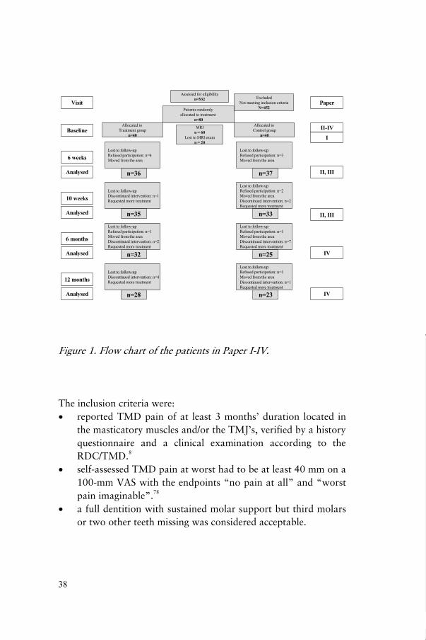
Figure 1. Flow chart of the patients in Paper I-IV. The inclusion criteria were:
reported TMD pain of at least 3 months’ duration located in the masticatory muscles and/or the TMJ’s, verified by a history questionnaire and a clinical examination according to the RDC/TMD.8 self-assessed TMD pain at worst had to be at least 40 mm on a 100-mm VAS with the endpoints “no pain at all” and “worst pain imaginable”.78 a full dentition with sustained molar support but third molars or two other teeth missing was considered acceptable.

39
The exclusion criteria were: previous treatment with occlusal appliances symptoms related to disease in other parts of the stomatog-nathic system (e.g., toothache, neuralgia) pain due to systemic disease (e.g., rheumatoid arthritis) fibromyalgia pain due to a whiplash-associated disorder history of psychiatric disorders inability to answer a questionnaire due to difficulties with the Swedish language
Procedures (I-IV)
Diagnosing and imaging (II)
After having filled in the history questionnaire, the studied patients were examined clinically by two dentists, according to the RDC/TMD system at the Department of Stomatognathic Physiol-ogy at Malmö University, Malmö, Sweden. The patients received their clinical diagnosis/es accordingly and all 80 participating pa-tients were then referred to the Department of Oral and Maxillofa-cial Radiology at Malmö University, Malmö, Sweden, for MRI of the TMJ’s and for panoramic radiography, to exclude dental rea-sons for their pain. Twenty patients declined or were unable to participate in the MRI examination (Fig. 1). The reasons for pa-tients not participating in the MRI examination were, among oth-ers, obesity, pregnancy, and claustrophobia. Sixty patients were examined with MRI of the TMJs.
Bilateral MR images were taken of the TMJs in a Siemens Magne-tom Vision, 1.5 Tesla machine (Siemens, Erlangen, Germany) with a TMJ surface coil at the Department of Radiology, Skåne Univer-sity Hospital, Malmö, Sweden.
MRIs were taken in the closed mouth position and the patients were instructed to close their mouth with the teeth in maximal con-tact. Images were also taken in the open mouth position with the
40
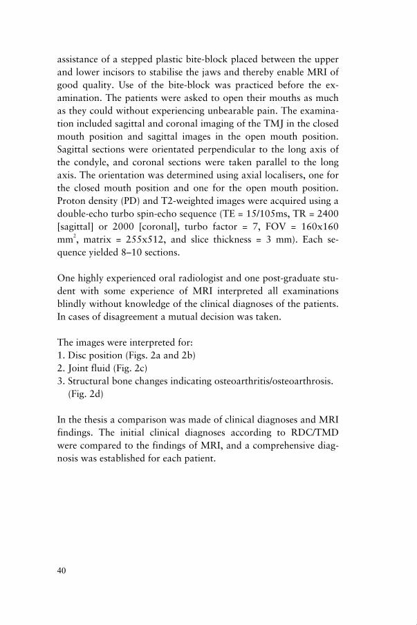
assistance of a stepped plastic bite-block placed between the upper and lower incisors to stabilise the jaws and thereby enable MRI of good quality. Use of the bite-block was practiced before the ex-amination. The patients were asked to open their mouths as much as they could without experiencing unbearable pain. The examina-tion included sagittal and coronal imaging of the TMJ in the closed mouth position and sagittal images in the open mouth position. Sagittal sections were orientated perpendicular to the long axis of the condyle, and coronal sections were taken parallel to the long axis. The orientation was determined using axial localisers, one for the closed mouth position and one for the open mouth position. Proton density (PD) and T2-weighted images were acquired using a double-echo turbo spin-echo sequence (TE = 15/105ms, TR = 2400 [sagittal] or 2000 [coronal], turbo factor = 7, FOV = 160x160 mm2, matrix = 255x512, and slice thickness = 3 mm). Each se-quence yielded 8–10 sections.
One highly experienced oral radiologist and one post-graduate stu-dent with some experience of MRI interpreted all examinations blindly without knowledge of the clinical diagnoses of the patients. In cases of disagreement a mutual decision was taken.
The images were interpreted for: 1. Disc position (Figs. 2a and 2b) 2. Joint fluid (Fig. 2c) 3. Structural bone changes indicating osteoarthritis/osteoarthrosis. (Fig. 2d)
In the thesis a comparison was made of clinical diagnoses and MRI findings. The initial clinical diagnoses according to RDC/TMD were compared to the findings of MRI, and a comprehensive diag-nosis was established for each patient.

Figure 2a. Figure 2b.
Figure 2c. Figure 2d. Figure 2a. MRI of normal temporomandibular joint. PD weighted. Figure 2b. MRI of temporomandibular joint with disc displace-ment. PD weighted. Figure 2c. MRI on temporomandibular joint with effusion. T2 weighted. Figure 2d. MRI of temporomandibular joint with structural bone changes, osteophyte formation and subcondral cyst. PD weighted.
Figure 2a Figure 2b
Figure 2c Figure 2d

42
Experimental Methods (III-IV)
At baseline, two examiners performed initial screening, history-taking, and clinical examination. Patient characteristics such as age, gender, ethnicity, marital status, level of education, occupa-tion, place of residence, and duration of TMD pain were studied. All patients were informed about their clinical diagnoses according to RDC/TMD and the lack of a clear-cut cause of their TMD pain and contributing factors. They were reassured and informed about the nature of TMD; about the relationship between muscle fatigue, TMD pain, and psychophysiologic aspects of stress; and about how to self-monitor TMD symptoms.
At baseline and at all follow-ups, patients filled in an earlier Swed-ish version of the RDC/TMD history questionnaire, where VAS was used instead of NRS when current, average and worst pain was measured. Treatment outcome variables were calculated at baseline and at follow-ups. RDC/TMD examination techniques were used in the clinical examination at baseline and at follow-ups.
The studies were performed as RCT’s, and patients were random-ised to the treatment group, treated with a resilient appliance or to the control group treated with a palatal hard non-occluding appli-ance after the initial screening. Patients were allocated by two in-dependent dental assistants to exclude any involvement of the den-tists who were kept “blind” during examination and follow-up procedures throughout the studies. The dental assistants random-ised the patients in blocks of 10, with 5 concealed sheets with the text “resilient appliance” and 5 with the text “control appliance”, to one of the two groups. The randomisation procedure was re-peated until 80 patients were included (Fig.1). The same examiners evaluated the patients after treatment. Both examiners were blinded to group assignment. A TMD specialist, not involved in examination and evaluation of treatment outcome, prepared for, delivered and adjusted the appliances.
The 4 domains according to IMMPACT recommendations, pain intensity, physical functioning, emotional functioning, and partici-

43
pant ratings of overall, global improvement, were considered when evaluating treatment outcome.
Pain intensity Primary treatment outcome measures were considered a 30% re-duction of TMD pain at worst registered on VAS and a 30% re-duction of CPI at 10-week follow-up and the 12-month follow-up. A reduction of at least 30% on VAS was considered clinically sig-nificant.73
Physical functioningJaw function was measured by the Jaw Disability Checklist in Axis II, at baseline, at the 10-week follow-up and at the 12-month fol-low-up.
Emotional functioning The modified SCL-90-R instrument in Axis II was used when measuring grade of depression, non-specific physical symptoms, and the 29-item SoC questionnaire was used measuring the SoC. Measurements with the modified SCL-90-R were performed at baseline and the 12-month follow-up, and with the SoC question-naire at baseline.
Global improvementGlobal improvement was measured according to a 6-point verbal rating scale (0=symptom-free, 1= much better, 2= better, 3= un-changed, 4= worse, 5= much worse) at the 12-month follow-up.
Additional outcome measures included frequency of TMD pain, and occlusal changes after using the appliances. Wear and use of the appliances were registered, and the patients were asked to re-port any kind of discomfort associated with the appliance therapy. The number of patients needed to be treated (NNT), which is the number of patients who must receive a particular therapy for one to benefit,79 was calculated on the basis of primary outcome at the 10-week follow-up and at the 12-month follow-up. Questions about frequency and influence of headache were added to the Axis II questionnaire and thereby measured.

44
Treatment Method (III-IV)Two different intraoral appliances were used as treatment alterna-tives in the study, a resilient appliance (treatment group) and a control appliance (control group). The resilient appliance had a rather smooth, flat surface and made contact with supporting teeth (Fig. 3a). The appliance covered all occlusal surfaces in the maxilla and occluded with contacts in the molar, premolar, and canine re-gions after adjustment. The resilient appliance was produced of a 4-mm thick BIOPLAST® clear-transparent film (Scheu Dental GmbH, Iserlohn, Germany) in a BIOSTAR® heat and vacuum press (Scheu Dental GmbH, Iserlohn, Germany).
The hard non-occlusal control appliance, had palatal coverage and clasps to attach to one molar on each side of the maxilla. All the control appliances were produced by the same dental technician. The appliance did not cover occlusal surfaces and did not alter the intermaxillary relationship (Fig. 3b).
Figure 3a. The resilient appliance. Figure 3b. The control app- liance.
The TMD specialist who delivered all the appliances after adjust-ments, informed the patients about how to use them. They were instructed to use the appliances at night for 10 weeks and after that period, when needed. All patients were informed in the same way and they were scheduled to attend the same number of visits. Some patients demanded minor adjustments in between follow-ups due to comfort reasons.

45
SoC, grade of depression, non-specific physical symptoms and general health (IIII)
To measure the patients coping ability, the SoC scale was used.41
The scale is available in a 29 question format, and in a 13 ques-tion, short format. In study (IIII) patients filled in the original 29-item questionnaire which results in an overall index where a high score indicates a strong SoC (graded in weak 136, moderate 137-148, and strong 149), depending on SoC score according to Lan-gius which represents a good coping ability.42, 80
Grade of depression and non-specific physical symptoms was measured by the modified SCL-90-R in Axis II (RDC/TMD) in three grades (normal, moderate, severe).39 It consists of 32 ques-tions including 20 measuring tendency of depression and 12 meas-uring the tendency of non-specific physical symptoms. The figures of all items are added and the sum is divided by the numbers of items answered. Grade of depression and grade of non-specific physical symptoms was assessed as normal, moderate or severe.
Patients self-reported general health was assessed at follow-ups, by a five-grade scale (1=poor, 2=fair, 3=good, 4=very good, 5=excellent).
A regression analysis was performed in order to evaluate age, gender, SoC grade, grade of depression, grade of non-specific phys-ical symptoms, and general health as possible influencing factors for the treatment outcome.
To test any association, SoC was compared to grade of depression, grade of non-specific physical symptoms, and general health.

46
Statistical methods (I-IV)
Descriptive statistics with means and standard deviations were used when presenting values such as for age and intensity of TMD pain. (II-IV)
Chi-square test and Fisher’s exact test were used to compare the distribution of categorical variables. (II, II, IV)
Mann-Whitney U test were used for differences between groups for ordinal variables such as pain measurements with VAS. (III, IV)
McNemar's test was used for categorical variables for within-group comparisons. (III, IV)
Wilcoxon’s signed-rank test was used for analysing ordinal vari-ables within groups. (III, IV)
Paired t-test was used for interval variables within groups. (II, II, IV)
Kappa statistics were used to evaluate observer agreement when analysing MRI. The kappa values were interpreted according to the guidelines of Landis and Koch81 adapted by Altman82. (II)
Logistic regression models with likelihood ratio tests were used when analysing data. The effect of the treatment was tested in two regression models. In the first model the treatment outcome was tested, whereas in the second model, the treatment outcome was tested after correcting for six possible background variables. One-way ANOVA test was used when testing the association between SoC and grades of depression, non-specific physical symptoms and general health. A significance level of = 5% was used in all the tests. (IIII)
Variance analysis was performed to investigate differences between SoC, and grade of depression, non-specific physical symptoms, and general health. A confidence interval of 95% was used. (IIII)

47
Intent to treat (ITT) was used when analysing primary treatment outcome and when assessing number-needed-to-treat (NNT). (IIV)
Results for differences were considered statistically significant at p<0.05. The statistical analyses were made using the Statistical Package for the Social Sciences (SPSS) 13.0 for Windows (SPSS Inc., Chicago, Ill, USA). (II-IV)
Ethical considerations
The patients were thoroughly informed about the study by the ex-aminers, and all participants gave their written consent. After ap-plication to the Ethics Committee of Lund University, Lund, Swe-den the study was approved (ref. no. LU 327-00 and ref. no. LU 505-00).

48

49
RESULTS
Seventy-three patients attended the 6-week follow-up, 68 patients the 10-week follow-up, 57 patients the 6-month follow-up, and 51 patients the 12-month follow-up (Fig 1). None of the patients at-tending the follow-ups received additional treatment for their TMD pain during the 12 months of appliance therapy. Patient characteristics, and intensity of TMD pain, and duration of TMD pain, and CPI, at baseline are presented in Table 1. The duration of TMD pain prior to baseline was 3 to 360 months in the treatment group (median 24 months) and 4 to 72 months in the control group (median 24 months). No statistically significant differences (P > 0.05) in the variables before treatment were found between the treatment and control groups.
Drop-out patients did not differ regarding age and gender from the participating patients attending follow-ups (Fig 1). Thus the group of responders can be considered representative of the initial sample of patients with TMD pain, who had been randomly selected for participation.
Paper I and thesis
Of the 60 patients examined with MRI, 19 were clinically diag-nosed as having myofascial pain (myofascial pain group) and 41 as having arthralgia/osteoarthritis in combination with myofascial pain (arthralgia/osteoarthritis group). The number of days between clinical examination and MRI examination varied from 17 to 157 days, with a mean of 67 and a median of 63.

50
Two patients in each TMD pain group had no MRI findings. A statistical difference in MRI findings between the two TMD pain groups was only found regarding disc displacement in combination with joint fluid, which was found significantly (P = 0.047) more often in the arthralgia/osteoarthritis group compared to the myo-fascial pain group. A high percentage of disc displacement was found in the TMJs in both groups, significantly higher in the ar-thralgia/osteoarthritis group (P = 0.002). Structural bone changes and joint fluid were found in both groups. Flattening and osteo-phytes were the most common structural bone changes.
The agreement between the clinical diagnosis of disc displacement and the corresponding MRI findings was studied.. In 101 joints di-agnosed clinically as having no disc displacement, only 37 were confirmed at MRI. In 14 TMJs with the clinical diagnosis disc dis-placement with reduction, the diagnosis was confirmed in 11 TMJs (79%) on MRI. In 17 TMJs with the MRI diagnosis disc displace-ment without reduction the diagnosis was set in two joints clini-cally.
Taking the MRI findings into consideration, 7 of the 19 patients in the myofascial pain group received a diagnosis of osteoarthrosis. Of the originally 41 patients in the arthralgia/osteoarthritis group, 21 had their initial clinical diagnosis changed from arthralgia to osteoarthritis. Twelve patients in the myofascial pain group and 35 in the arthralgia/osteoarthrosis group had MRI findings of disc displacement. Of the 12 patients in the myofascial pain group only 1 had a clinical diagnosis of disc displacement. In the arthral-gia/osteoarthritis group 11 of the 35 patients with MRI findings of disc displacement, had a clinical diagnosis of disc displacement. In total 28 of the 60 (47%) patients had the initial clinical diagnosis changed.
Papers II and IV
Twenty-four patients in the treatment group and 23 in the control group reported that they were aware of grinding or clenching their

51
teeth at night. At baseline all patients had a mean value of 57 forCPI and 72 for TMD pain at worst, with no differences betweengroups (Table 1).
Treatmentgroup
(n= 36)
Controlgroup
(n= 37)
GenderMaleFemale
927
433
Age (years)MeanMin-Max< 2020-40> 40
3514-67
62010
3313-68
91711
TMD pain
Intensityat worst VAS mean(mm, ±SD)
75 (±18) 69 (±20)
CPI (mean, ±SD) 60 (±17) 54 (±21)
Frequencypersistentrecurrentone-time experience
15201
12214
Duration3–6 months > 6 months
234
235
Headache
Frequencypersistentrecurrentone-time experience
81612
62011
Table 1. Patient characteristics, intensity, frequency, and durationof TMD pain, and frequency of headache at baseline in the treat-ment group and the control group.

52
Clinical subdiagnoses according to RDC/TMD are presented inTable 2. All patients had a diagnosis of myofascial pain and 50 pa-tients also had arthralgia/osteoarthritis. No significant between-group differences (P > 0.05) were observed in the subdiagnoses.
Table 2. Clinical diagnoses according to RDC/TMD, of the par-ticipating patients in the treatment group and the control group.
Treatment outcome IMMPACT
Pain intensityThe primary treatment outcome was calculated in a per-protocolanalysis at the 10-week follow-up. A 30% reduction in TMD painat worst, at 10-week follow-up was 22 patients (63%) in thetreatment group and 17 patients (52%) in the control group, withno significant difference (P > 0.05) between the groups. Mean VAS
TMDTreatment
group(n= 36)
Controlgroup
(n= 37)Pain diagnoses:
Myofascial painwithout limited openingwith limited opening
315
343
ArthralgiaOsteoarthritis
231
260
Other diagnoses:
Disc displacementwith reductionwithout reduction
with limited openingwithout limited opening
Osteoarthrosis
12
10
3
6
01
2

53
TMD pain at worst
0
10
20
30
40
50
60
70
80
90
100
Baseline 10 w eeks 12 months
mm
T group
C group
for patients in the treatment group not reaching a 30% reduction in TMD pain at the 10-week follow-up was 76, and 64 in the con-trol group. A 30% reduction in CPI was found in 24 patients in the T group and in 20 patients in the C group at 10 weeks without any statistically significant differences (P > 0.05) between the groups.
Due to the fact that 30% of the treated patients were drop-outs at the 12-month follow-up, an intent-to-treat analysis was performed to show a more realistic way to report the primary treatment out-come. A 30% reduction of TMD pain at worst, registered on VAS, was found in 19 patients (48%) in the treatment group and 14 pa-tients (35%) in the control group, with no difference between groups (P = 0.256). The analysis at the 12-month follow-up showed that 20 patients (50%) in the treatment group and 17 pa-tients (43%) in the control group had 30% reduction in CPI, with no difference between groups (P = 0.501) (Figs. 4 and 5).
Figure 4. Mean VAS of worst experienced TMD pain at baseline, at the 10-week follow-up and at the 12-month follow-up in the treatment group (T group) and in the control group (C group).

54
Figure 5. Mean CPI at baseline, at the 10-week follow-up and at the 12-month follow-up in the treatment group (T group) and in the control group (C group).
Physical functioning Limited or prevented activities were reported by 28 patients (78%) in the treatment group and 29 patients (78%) in the control group at baseline, without significant differences between groups (Fig). The number of patients reporting no limited jaw activities due to TMD pain (jaw disability checklist) increased significantly in the treatment group compared to the control group (P=0.013) at the 10-week follow-up. Jaw functioning improved within both groups at 12 months (T group=9 and P=0.000, C group=13 and P=0.062). There were no statistically significant differences at the follow-ups between the groups (P > 0.05) (Fig. 6).
CPI
0
10
20
30
40
50
60
70
80
90
100
Baseline 10 w eeks 12 months
mm
T group
C group

55
Figure 6. Patients’ self-reported jaw disability presented as percent in the treatment group (T group) and in the control group (C), at baseline, at the 10-week follow-up, and at the 12-month follow-up.
Emotional functioning The scores for depression and non-specific physical symptoms at baseline and at follow-ups in the treatment and control groups are presented in Figures 7 and 8. In the treatment group there was a significant decrease in grade of depression at the 6-month follow-up (P = 0.021), but not at the 12-month follow-up (P = 0.227). Re-garding grade of non-specific physical symptoms there was a statis-tically significant decrease at 6-month but not at the 12-month fol-low-up (P = 0.057) in the treatment group. In the control group there were statistically significant decreases regarding both grade of depression (P = 0.002) and grade of non-specific physical symp-toms (P = 0.008) at the 6-month follow-up and at the 12-monthsfollow-up (P = 0.008 and P = 0.008). No significant differ-ences were found between groups regarding grade of depression and non-specific physical symptoms (P > 0.05).
Jaw disability check-list
0
10
20
30
40
50
60
70
80
90
100
Baseline 10 w eeks 12 months
%
T group
C group

56
Gra
de o
f dep
ress
ion
0102030405060708090100
T no
rmal
T m
oder
ate
T se
vere
C n
orm
alC
mod
erat
eC
sev
ere
%
Bas
elin
e12
mon
ths
Figu
re 7
. Gra
de o
f dep
ress
ion
pres
ente
d as
per
cent
, in
the
trea
tmen
t gro
up (T
) and
in th
e co
ntro
l gro
up(C
), at
bas
elin
e an
d at
the
12-m
onth
follo
w-u
p.

57
Gra
de o
f non
-spe
cific
phy
sica
l sym
ptom
s
0102030405060708090100
T no
rmal
T m
oder
ate
T se
vere
C n
orm
alC
mod
erat
eC
sev
ere
%
Bas
elin
e12
mon
ths
Figu
re 8
.Gra
de o
f non
-spe
cific
phy
sica
l sym
ptom
s pr
esen
ted
as p
erce
nt, i
n th
e tr
eatm
ent
grou
p (T
) and
in th
e co
ntro
l gro
up (C
), at
bas
elin
e an
d at
the
12-m
onth
follo
w-u
p.

58
Global improvement At the 12-month follow-up 25 patients out of 28 (89%) in the treatment group reported that they were “better, much better or symptom-free”, and in the control group 16 patients out of 23 (70%) reported an improvement. There were no statistically sig-nificant differences (P > 0.05) between the groups at this follow-up.
Additional outcomes The frequency of persistent TMD pain decreased significantly within both groups over time. Fifteen patients in the treatment group and 12 patients in the control group at baseline, two pa-tients in the treatment group and 5 patients in the control group at the 10-week follow-up, and 1 patient in the treatment group and 3 patients in the control group at the 12-month follow-up, reported persistent TMD pain. No significant differences were found be-tween groups.
NNT was 10 for the resilient appliance at 10 weeks, when calcu-lated on a 30% reduction of TMD pain at worst, in a per-protocol analysis. NNT calculated on a 30 % reduction on CPI was 14 for the resilient appliance at 12 months, when analysed as ITT.
At baseline 26 patients (72%) in the treatment group and 19 pa-tients (51%) in the control group had experienced headache within the last six months prior to start. Self-reported distress of headache decreased significantly within both groups at the follow-ups, with-out any differences between groups. Recurrent and persistent headache decreased over time from 24 (67%) to 10 (36%) in the treatment group and 26 (70%) to 9 (39%) in the control group, without statistically significant differences between groups.
Occlusal appliances During the study self-reported use of appliance decreased similarly in the two groups. Thirty-two of the patients, at the 10-week fol-low-up, in the treatment group and 31 of the patients in the con-trol group reported that they used their appliances most nights per week, corresponding figures at the 6-month follow-up were 19 in the treatment group and 14 in the control group, and at the 12-

59
month follow-up 13 in the treatment group and 10 in the control group.
One patient in each group reported discomfort during appliance use, at the 10-week follow-up and at the 12-month follow-up. One patient reported bad smell from the resilient appliance, and one re-ported unpleasantness during control appliance use. Mild to mod-erate wear of the resilient appliance was found in 21 patients (75%) and none of the appliances had a severe wear at the 12-month follow-up. Eleven resilient appliances were discoloured, compared to 7 control appliances at the 12-month follow-up.
Adverse effects A reduction of 4 occlusal contacts, or more, were considered rele-vant and not as a result of intra-observer variety. No occlusal changes were observed in patients treated with a resilient appli-ance. Occlusal changes in the frontal region were registered at the 12-month follow-up in 3 patients (patients were unaware of the change/s) in the control group.
Paper III
The mean value for SoC was 142 (±25) for women and 150 (±24)for men, where 22 (37%) of the women and 3 (23%) of the men had a low grade of SoC. Patients between 20 and 40 years of age had a mean SoC of 145 (±24), while the corresponding figure for older patients was 141 (±27). Mean SoC was 147 (±23) in the treatment group (n=36) and 139 (±27) in the control group (n=37).
In the treatment group 20 patients had a moderate or severe self-reported grade of depression according to SCL-90-R, and in the control group, the corresponding figure was 25. Self-reported non-specific physical symptoms were moderate or severe in 24 patients in the treatment group and in 27 patients in the control group. Good to excellent perceived general health was reported by 32 pa-tients in the treatment group and by 30 patients in the control group.

60
A logistic regression analysis was carried out to test whether there were differences among the background variables in relation to treatment outcome. No statistically significant difference in efficacy between the resilient appliance and the non-occluding control ap-pliance was found (p=0.344). When testing for the six possible background variables one by one, in relation to treatment outcome no statistically significant differences were found (Table 3).
A severe grade of depression and a low grade of SoC (mean 136) were found in 26 patients (37%). Thirty-one (44%) of the studied patients had registered a severe grade of non-specific physical symptoms with a moderate grade of SoC (mean 140). A fairly good health and a low grade of mean SoC (mean 129) were found in 9 patients (12%). Two patients with bad general health had high grades of SoC (mean 154). In a variance analysis there were no sta-tistically significant associations found between mean SoC and grades of depression, non-specific physical symptoms or general health.

61
.448
.13-
2.55
.57
.77
.57
Gen
eral
hea
lth.2
10.8
1-2.
591.
45.3
0.3
7D
epre
ssio
n (S
CL-
90R
)
.363
.72-
2.41
1.32
.31
.28
Non
-spe
cific
phy
sica
l
sym
ptom
s (SC
L-90
R)
.258
.99-
1.03
1.01
.01
.01
SOC
.801
.96-
1.03
1.00
.02
-.00
Age
.172
.67-
8.99
2.45
.66
.90
Gen
der
.344
.61-
4.19
1.59
.49
.47
Trea
tmen
t
P-va
lue
95%
CI
Odd
s rat
ioSt
anda
rd e
rror
Reg
ress
ion
coef
ficie
nt
Tab
le 3
.Log
istic
reg
ress
ion
anal
ysis
mad
e on
var
iabl
es th
at m
ay in
fluen
ce tr
eatm
ent o
utco
me,
a 3
0% r
educ
tion
of w
orst
TM
D p
ain,
in t
he 6
8 T
MD
pa
in p
atie
nts.

62
63
DISCUSSION
The overall objective of this thesis was to investigate treatment outcome in a RCT of resilient appliance therapy in patients suffer-ing from TMD pain, diagnosed according to the RDC/TMD. A further objective was to compare subdiagnoses of TMD with find-ings on MRI. The pain coping ability of the patients was measured by SoC in order to study influence of psychosocial factors on treatment outcome.
Treatment outcome efficacy of the resilient appliance therapy was studied in a RCT in the short- and long term. There were no statis-tically significant differences regarding primary treatment outcome measures between the treatment group and the control group at the 10-week follow-up. In the long-term follow-up similar results were found between the investigated groups, with regard to the four treatment outcome domains described by IMMPACT: pain inten-sity, global improvement, physical functioning, and emotional functioning.69
Diagnosing
Through the years a variety of clinical diagnostic approaches have been used when diagnosing TMD and orofacial pain conditions. Unspecific criteria and different systems have been used. The RDC/TMD was constructed and presented in 1992 since standard-ised diagnostic criteria for defining clinical subtypes of TMD were lacking.8 The instrument was initially meant to be used for research

64
purposes as it allows standardisation and replication of research into the most common forms of muscle- and joint-related TMD. The system for calibrating users is one of the reasons for the good inter- and intraobserver reproducibility. Due to clear-cut diagnostic criteria it has been found to be a useful tool in everyday clinical practice when managing patients with TMD. Ongoing refinement of the instrument is continuously performed and the scientific work with the upgraded version has recently been published in 2010 by the international RDC/TMD consortium.26, 31-36 In contrast to other RCT studies on the resilient splint63, 65 our study used the validated and reliable RCT/TMD instrument in the diagnostic process, which strengthens the results, since comparisons may be made in the fu-ture if other research groups is about to investigate the resilient ap-pliance as a treatment modality in TMD patients. A shortcoming, however, of the study is that the RDC/TMD instrument was slightly modified by using VAS instead of NRS when measuring self-reported intensity of worst, average and present TMD pain, in Axis II.
Abnormal TMJ findings on MR images were registered in both the myofascial pain group and in the arthralgia/osteoarthritis pain group. This was in agreement with the hypothesis that clinical di-agnoses according to RDC/TMD could not be confirmed with MRI findings. Similarly, in a systematic review no clear evidence was found for a relationship between clinical and MRI diagnoses and findings.28
A study by Wiese et al.83, using RDC/TMD and sagittal TMJ to-mography as imaging technique, found that the clinical diagnosis was changed for about 30% of the patients, mostly from arthralgia to osteoarthritis when the radiographic findings were taken into consideration. This is in line with the results from this thesis when it comes to the diagnosis, which should have been changed in 47% of cases, when combining clinical and MRI findings. Structural bone changes in the TMJ indicating osteoarthritis/osteoarthrosis were found in both pain groups. Minimal flattenings of the condyle and/or articular eminence is commonly found, and probably of no clinical significance.84 Therefore, in patients with structural bone

65
changes visible on a MR image, a single flattening was considered normal. MRI is not considered an optimal modality when analys-ing structural bone changes.85 The use of MRI may have lead to underdiagnosing findings of structural bone changes as a sign of osteoarthritis and osteoarthrosis. In a project validating image analysis criteria for the RDC/TMD computed tomography was recommended for assessing osteoarthritis and MRI for disc posi-tion and effusion.86 However a recent systematic review concluded that conventional tomography is currently the imaging modality of choice for diagnosing erosions and osteophytes of the TMJ, when compared to the more expensive CT modality.87
According to Wiese et al.83, who also studied the influence of a changed diagnosis on management, no change of management was considered necessary in 73% of the patients due to altered diagno-ses. According to these results, even though MRI findings may con-tribute to a changed clinical TMD diagnosis, it may leave the treatment initially planned unchanged, this due to current treat-ment regimen for TMD pain. Since the investigated patients in this thesis were predetermined for a treatment study, a change of treatment was not possible to investigate. Studies are needed to in-vestigate the diagnostic thinking efficacy and therapeutic efficacy for MRI.29 The MRI findings in this thesis were not taken into con-sideration for the final comprehensive diagnoses according to RDC/TMD, which is stated in the criteria for the diagnostic sys-tem. However, the RCT treatment part of the study was possible to perform since one of the inclusion criteria incorporated patients with TMD pain, and the pain diagnoses of RDC/TMD are not de-pendant of MRI findings.
There was a time interval between the clinical and the MRI exami-nation in this study. In an ideal situation is that clinical examina-tion and MRI examination should be performed as soon as possi-ble after each other. However, the everyday clinical situation in Sweden, means that it is difficult to perform the MRI examination during the same visit as the clinical examination. This is due to the long waiting time to gain access to a MRI machine for a TMJ ex-amination. Long time interval could, of course, influence findings

66
on MRI due to natural fluctuation of particularly TMJ disc dis-placement with reduction. Some patients may have developed a disc displacement during the waiting time and others may have re-covered when compared to the clinical examination. Findings of disc displacement without reduction, structural bone changes and joint fluid, however, were probably not influenced by the time in-terval.
The recommendations by IMMPACT were followed for evaluating treatment outcome in the long-term follow-up, which strengthens the structure of the study. John et al.23 reported that the reliability of diagnostic classification improved when diagnoses were grouped into pain versus non-pain diagnoses, which further strengthens the results of this thesis, since only patients with TMD pain was stud-ied. The authors concluded that in clinical decision-making and re-search, an adequate and reliable diagnosis is critical in establishing a clinical condition and when considering a rational approach to treatment. They further concluded that the RDC/TMD demon-strates sufficiently high reliability for the most common TMD di-agnoses, supporting its use in clinical research and decision mak-ing.23 In a recently published study32 on the work with an updated RDC/TMD Axis I version the authors concluded that the revised RDC/TMD Axis I TMD diagnostic algorithms were recommended for myofascial pain and joint pain as reliable and valid. However, revised clinical criteria alone, without recourse to imaging, were considered inadequate for valid diagnosis of two of the three disc displacements as well as osteoarthritis and osteoarthrosis.
According to Hofmann and Eriksen3, referring to the triad of dis-ease-illness-sickness, TMD belongs to the spheres of illness and sickness but not the sphere of disease. A common cause for seeking dental or medical care among TMD patients is the pain and dis-comfort they may experience, therefore TMD is classified as an ill-ness. In cases of dysfunction and/or chronic TMD pain, the sick-ness sphere can also be relevant. However, today when using the systematic RDC/TMD system, with clear-cut diagnostic criteria, subdiagnoses such as myofascial pain and osteoarthritis may be classified a disease. The TMJ disc displacement, which is often a

67
harmless condition, is no more to be classified as a disease, but rather a prevalent illness and/or sickness. When the triad was de-scribed in the 1960’s classification systems such as RDC/TMD did not exist. Regardless of the categorisation of conditions and ail-ments used, the interpretation changes as research continues and further knowledge develops. Therefore the spheres for the TMD subdiagnoses, according to the triad, may change with time.
Resilient appliance therapy There were no statistically significant differences between the treatment group and the control group regarding treatment out-come efficacy, however 50% of the investigated TMD pain pa-tients benefited from treatment with both the resilient appliance and the control appliance, when calculating ITT of a 30% reduc-tion of CPI. The control appliance has been used in other RCTs studying the stabilisation appliance showing a treatment effect of about 50% in the short-term perspective and around 30% in a longer perspective, which is slightly above the expected placebo ef-fect.88, 89
The results of our study are in line with a study by Truelove et al.62
All three studied groups in their investigation showed a significant decrease in CPI at follow-ups, at 3, 6 and 12months, however no significant differences in TMD-related pain was found between the groups at baseline or at any follow-up. All participating patients in that study had a RDC/TMD group I diagnosis according to their inclusion criteria. The changes from baseline were comparable for all three groups. The authors did not note any significant differ-ences at any follow-up for compliance with study protocols or for occurrences of adverse effects from either splint type. Muscular pain, studied by Truelove et al., decreased significantly in all groups including the soft splint group.
In a study by Okeson90 the author found an increased nocturnal muscle activity in healthy subjects when wearing soft appliance. This may indicate that the resilient appliance can increase clench-

68
ing, however only 10 patients in each studied group were partici-pating in that study, where a soft occlusal splint was compared to a hard occlusal splint. Increased clenching caused by the use of a soft splint could be an explanation to why no difference was found when comparing the soft appliance to control treatment.62, 91 Espe-cially, as more than 60% of the patients before the start of our study were aware of grinding or clenching their teeth at night.91
When taking the established results of this thesis into considera-tion, it would have been more appropriate to perform an effective-ness study at this stage, since most of the resilient appliances made are made by general practitioners.56 There is a lack of effectiveness studies, and the different clinical indications for general practitio-ners to use resilient appliances are mostly unknown. Therefore it would be interesting to investigate current indications for using re-silient appliances as treatment for TMD patients in the general practice, and when this is elucidated efficacy studies could be per-formed from a clearer clinical point of view. Then cost benefit analysis would be more interesting to perform, since cost-effectiveness is highly dependant on the indication for treatment and treatment outcome.
Jaw functioning was improved in both studied groups. According to the jaw disability checklist in Axis II, patients in both groups re-ported that limited jaw activities decreased significantly at the 10-week follow-up. However, no difference was found at the 12-month follow-up, which is in line with no differences being found between the groups regarding intensity and frequency of TMD pain. One important contributor to an individual´s oral-health-related quality of life is limitations in orofacial functioning 92.
The studied patients showed moderate to high baseline scores for non-specific physical symptoms and depression, which corresponds well with other studies 93, 94. It seemed that TMD-pain patients were more distressed than healthy individuals. All participating patients had a diagnosis of myofascial pain, and non-specific physical symptoms has been found to be a significant risk factor for devel-oping myofascial pain 95. High scores for depression were found in
69
36% of the patients in this study and depression has been found to be related to pain perception 96, 97.
Headache within the last 6 months prior to treatment start was re-ported by 72% of the patients in the treatment group and in 51% of the patients in the control group at baseline. In both groups a significant decrease of self-reported distress caused by headache was found at follow-ups, however with no difference between groups. The results are in line with a study by Ekberg and Nilner88,
98 who found that a reduction of muscles tender to palpation corre-lated significantly to improvement of headache at follow-ups in pa-tients with TMD with myogenous pain. They concluded that the studied stabilisation appliance seemed to have a statistically signifi-cant better positive short- and long-term effect compared to the control appliance on tension-type headache, in the studied category of patients. The control appliance was the same design as used in our study.
In the short-term NNT value was calculated to be 10 and in the long-term the NNT value increased to 14, which could be ex-plained by the smaller number of studied patients in the long-term follow-up, due to several drop-outs. When calculating the different treatment outcome variables intent to treat figures were used to present the result in a more realistic way, compensating for the drop-outs. However, the high drop-out figure has to be considered a shortcoming of the study. When comparing the resilient appli-ance with the stabilisation appliance, which have shown better NNT value (3) when treating TMD patients, the resilient one has to be considered to be a treatment modality with poor efficacy when treating adult patients with TMD pain.88
In a retrospective and prospective study by Lindfors et al.99 the soft appliance was investigated as a treatment combined with a stabili-sation appliance in the opposing jaw, in patients, refractory to pre-vious TMD treatment. The authors concluded that the investiga-tion showed that a combined treatment with a hard acrylic stabili-sation appliance and a soft appliance in the opposing jaw seemed to give an improvement of TMD signs and symptoms in apparently

70
therapy resistant TMD patients. A further conclusion drawn was that their results should be taken with caution due to the fact that the study did not include any control group, and that there was an obvious need for RCTs concerning the efficacy and effectiveness of the combined treatment presented. A combination treatment like this may booster pain reduction of the treatment, however prospec-tive RCTs should be conducted before this can be recommended.
In a study by Harkins et al.100 it was found that 67% of patients with TMD pain received transient occlusal changes after soft ap-pliance use. The changes consisted of minor intrusion of molars and premolars. These kinds of changes were not found in our study, in which the resilient appliance was adjusted to bilateral stable occlusal contacts in the canine, premolar and the molar re-gions (even though not easy to perform), and the control appliance was adjusted so that obvious occlusal contacts were eliminated. However, three patients in the control group were found to have minor occlusal changes in the frontal region, which the affected pa-tients were unaware of. These findings in our study may be due to intraobserver bias, even though occluding foil was used to mini-mise such bias.
Mechanisms of action of occlusal splints have been discussed for many years. The exact mechanism is still unclear, and the treating ability of intraoral appliances is probably not dependant on one specific factor, but several both intrinsic (the appliance it self) and extrinsic (patient related) factors may interact and contribute. One mechanism that has been discussed is the influence of the appliance on occlusion.57 The resilient appliance may reduce some of the heavy loading that occurs during parafunctional activity, however the control appliance in our study showed no significant difference in treatment outcome, which may be interpreted as other mecha-nisms of action being more important. Another factor possible in-fluencing the effect of an occlusal appliance is change in the condyle-fossa relationship.101, 102 In a study by Limchaichana et al. the effect on condyle position on MRI was studied with and with-out a resilient appliance.64 The resilient appliance changed the condyle in 76% of the TMJs and mostly in an anterior direction.

71
However, treatment outcome was not related to changes in condyle position.
For a TMD treatment to be effective one should consider the tim-ing of introducing the treatment. This may be one factor influenc-ing treatment outcome. Due to the current psychosocial status of the individual patient, motivation for treatment compliance may be high or low. Regardless, it has to be kept in mind that placebo ef-fect, spontaneous remission and regression to the mean are impor-tant factors for positive treatment outcome, especially in the long-term.6 However, the impact of these factors on the result ought to be the same in both groups, due to the randomisation process.
In a study by Van Selms et al.75 time course of myofascial TMD complaints was investigated over a 12 month period. The authors showed that baseline values of CPI had a positive correlation with CPI during follow-up. Patients with a low score on non-specific physical symptoms showed a further decline in CPI during follow-up, whereas patients with a high score showed a gradual increase. Patients with high scores on non-specific physical symptoms showed a significant increase in values of mandibular function im-pairment. They concluded that baseline self-reports of pain and impairment, oral parafunctional activities, pain elsewhere in the body, and non-specific physical symptoms are associated with the severity and time course of myofascial TMD complaints following treatment. These factors should be taken into consideration in clinical practice, when predicting treatment outcome, which is im-portant in order to be able to give patients a proper and cost-effective treatment. In our study moderate to severe grade of self-reported non-specific physical symptoms were found in 24 patients in the treatment group and in 27 patients in the control group. No significant differences were found between the treatment groups and no correlation was found with treatment outcome in the logis-tic regression analysis, which is not in line with the study by Van Selms et al.

72
SoC as an influencing factor for the treatment outcome
The psychosocial factor was measured by the SoC 29-item instru-ment. A statistically significant higher number of patients with low grade SoC were found in the control group at baseline. Despite this, the regression analysis showed no statistical difference in treatment outcome between the two groups. Initial low SoC, there-fore, did not appear to predict a negative treatment outcome when treating TMD pain patients with a resilient appliance or a palatal, non-occluding (control) appliance. A shortcoming of the study is the low number of participating patients. This makes it difficult to draw a conclusion on SoC in TMD patients in general. However, SoC has recently been investigated in a Finnish epidemiologic study43 which revealed that low SoC is associated with myogenous TMD findings. The authors also concluded that SoC as a psycho-social aspect has a role in the background of TMD. SoC is an in-strument increasingly used in studies on different types of pain throughout the human body. Future studies of SoC with the 29-item questionnaire in epidemiological surveys, may therefore be of interest in order to compare TMD pain with other types of pain.
Implications for general practitioners and future research
MRI should be seen as a complementary examination in the TMD pain diagnostic process. The MRI findings may influence the clini-cal diagnoses. However, the effect on planned treatment is un-known and referral criteria for MRI are unclear. In patients with suspected osteoarthritis, which include structural bone changes, other radiographic modalities should be considered, as MRI may underdiagnose bone changes.
In future research on the next version of RDC/TMD, it would be interesting to study when the complement of radiographic imaging may contribute to the clinical diagnosis, which in the future may result in more clear-cut indications for when imaging may be useful in the diagnostic process. This would enable improved TMD diag-

73
nosing and more effective treatment strategies, in favour of the suf-fering TMD pain patient.
To evaluate the evidence for efficacy of MRI a prospective study of consecutive TMD pain patients should be performed without any delay of the MRI examination. The influence of MRI findings on the clinical diagnoses and treatment outcome could be tested in a RCT with groups with and without MRI.
Lindfors et al.56 have shown that stabilisation appliances and resil-ient appliances are widely used by the general practitioners in Swe-den. The resilient type of appliance is used due to its relatively low cost, and has therefore been considered a cost-effective treatment modality in patients with TMD. The results presented in this thesis, however, questions the benefit of the treatment modality as a suc-cessful choice when treating adult patients suffering from chronic TMD pain. Possible treatment indications for the resilient appli-ance may be treatment of TMD in children and adolescents chang-ing teeth due to a demand for flexibility for erupting teeth, and as a short-term treatment of acute TMD in adults. However, this was not the scope for this thesis. To make a statement, evidence-based, future high quality RCTs are necessary.

74

75
CONCLUSIONS
Conclusions which can be drawn from the results of this thesis:
Clinical diagnoses according to RDC/TMD are not always confirmed by findings on MRI. (II)
The clinical diagnoses were changed depending on the MRI findings. (TThesis)
The capabilities of the resilient appliance and the non-occluding control appliance to reduce symptoms in patients with TMD pain were similar in the short term. (III)
None of the studied background variables (age, gender, SOC, depression, non-specific physical symptoms, general health) seem to be of importance for the short-term treatment out-come in patients treated with resilient or non-occluding intra-oral appliances. (IIII)
No association was found for SOC compared to depression, non-specific physical symptoms and general health in TMD-pain patients. (IIII)
There was no statistically significant difference between the re-silient appliance and the non-occluding control appliance in reducing TMD pain, physical functioning, emotional function-ing and headache in a 12-month perspective. (IIV)

76
The resilient appliance seems to have good durability, since most of the appliances showed no or mild wear at the 12-month follow-up. (IIV)

77
ACKNOWLEDGEMENTS
I wish to express my sincere gratitude to all who have supported and helped me through the years, making this thesis possible. In particular:
My primary supervisor, co-author and friend Associate Professor EwaCarin Ekberg, for excellent professional guidance through out my doctoral education, for unlimited patience in tough times, and for sharing your great knowledge in the scientific field of Stoma-tognathic physiology.
My co-supervisor and co-author Professor AArne Petersson,for valuable scientific guidance and excellent teaching in the field of Oral and Maxillofacial Radiology, and for inspiring discussions through out the work with this thesis.
My co-author and supervisor Professor MMaria Nilner, for intro-ducing me to the scientific field, for never-failing scientific support and guidance, and for helping me develop my professional self-esteem.
My co-author, colleague, and friend Associate Professor DDanilaVallon, for scientific and personal advise, and for treating all the participating patients in this thesis.
My co-author, Odont. Dr NNapat Limchaichana, for coopera-tion, and interesting meetings and discussions in English.
My boss and colleague, Professor and Head of the Department of Stomatognathic physiology, Faculty of Odontology in Malmö Thomas List, for inspiring scientific discussions, for practical ad-vise, and for giving me the time to complete this thesis.

78
My research patient coordinator, dental assistant, and friend Gunilla Millman, for perfect clinical administration, for never fail-ing loyalty, and for the hearty laughs and at the same time being so serious in your work.
My research patient coordinator and dental assistant, BBirgittaZahlée, for clinical administrative support, and for skilful clinical assistance throughout the years.
Head of the PDS in Kalmar county, dr AAnders Nylander and the organization, for generously supporting me and creating the possibility to complete this thesis.
My boss and colleague, dr HHåkan Westerling at the Specialist Dental Centre in Kalmar, for supporting discussions and gener-ously giving me the time to conclude this work.
All my friends and colleagues at the Department of Stomatog-nathic Physiology, Faculty of Odontology, Malmö University, for encouragement, discussions and support through out the years, and especially dr AAndreas Dawson, for sharp discussions, and for all practical help with tables and figures and, of course, for my enlarged biceps muscles!
All my friends and colleagues at the Specialist Dental Centre in Kalmar, for encouragement and loyal support. Especially the staff at the Department of Stomatognathic Physiology YYvonne Halling,Jeanette Wernersson and LLeena Norell for all support and pa-tience throughout these years. Without your understanding attitude and help this work had been impossible to complete.
All my friends and colleagues at the Centre of Oral Rehabilita-tion, County of Östergötland, and the Department of Stomatog-nathic Physiology in Linköping for your kind support. My deepest gratitude to Odont. Dr KKerstin Wahlund, for generous support and help through turbulent periods, and for being such a good friend and colleague.
All my friends and colleagues at the Department of Stomatog-nathic Physiology, Institution for Postgraduate Dental Education, Jönköping, especially Professor MMartti Helkimo and Professor Tomas Magnusson, for supporting my research work during my period of specialist training.
Elisabeth Edwinson and DDaniel Bengmark for invaluable ad-ministrative help and for interesting discussions.

79
Dental technician Jan-Erik Persson for making all the appli-ances in this study.
Per Egevad and JJenny Widmark at Malmö University, for ex-cellent literature search and computer support.
Per-Erik Isberg for excellent help with the statistical analyses. Cecilia Hallström for perfect linguistic revision of this thesis.
My dear friend PPär-Magnus Möller, for our special friendship throughout life.
My parents LLeif and UUlla, my brother AAnders and my sister Ulrika and their families, for their love, support and understanding during these years.
My life companion RRoger, for understanding, patience, over-whelming discussions, and endless love.
This research was financially supported by grants from: The Faculty of Odontology, Malmö University, Malmö The Public Dental Service of Kalmar, Kalmar County Council The Public Dental Service of Östergötland, Östergötland County Council The Swedish Dental Society Praktikertjänst AB, Sweden

80

81
REFERENCES
1. Preamble to the Constitution of the World Health Organization as adopted by the International Health Conference, New York. 1946.
2. Twaddle A. Influence and illness: definitions and definers of illness behaviour among older males in Providence, Rhode Island. : Brown University; 1968.
3. Hofmann BM, Eriksen HM. The concept of disease: ethical challenges and relevance to dentistry and dental education. Eur J Dent Educ. 2001;5:2-8; discussion 9-11.
4. www.who.int/topics/oral_health/en/5. Laskin DM. Temporomandibular disorders: an evidence-based ap-
proach to diagnosis and treatment. Chicago: Quintessence; 2006. 6. Von Korff M, Dworkin SF, Le Resche L, Kruger A. An epidemiologic
comparison of pain complaints. Pain. 1988;32:173-83. 7. Olesen. Headache Classification Committee. The international classifi-
kation of headache disorders, 2nd ed. Cephalalgia. 2004;24:1-160. 8. Dworkin SF, LeResche L. Research diagnostic criteria for temporo-
mandibular disorders: review, criteria, examinations and specifica-tions, critique. J Craniomandib Disord. 1992;6:301-55.
9. Carlson CR, Okeson JP, Falace DA, Nitz AJ, Curran SL, Anderson D. Comparison of psychologic and physiologic functioning between pa-tients with masticatory muscle pain and matched controls. J Orofac Pain. 1993;7:15-22.
10. LeResche L. Epidemiology of Temporomandibular Disorders: Implica-tions for the Investigation of Etiologic Factors. Critical Review. Oral Biology & Medicine. 1997;8:291-305.
11. Dworkin SF, Huggins KH, LeResche L, Von Korff M, Howard J, Truelove E, et al. Epidemiology of signs and symptoms in temporo-mandibular disorders: clinical signs in cases and controls. J Am Dent Assoc. 1990;120:273-81.
12. Fricton JR. Epidemiology of temporomandibular disorders. In. Fricton JR, Dubner R (eds). Orofacial pain and temporomandibular disorders. New York: Raven Press Ltd.; 1995.

82
13. De Kanter RJ, Truin GJ, Burgersdijk RC, Van 't Hof MA, Battistuzzi PG, Kalsbeek H, et al. Prevalence in the Dutch adult population and a meta-analysis of signs and symptoms of temporomandibular disorder. J Dent Res. 1993;72:1509-18.
14. Locker D, Slade G. Prevalence of symptoms associated with temporo-mandibular disorders in a Canadian population. Community Dent Oral Epidemiol. 1988;16:310-3.
15. Magnusson T, Carlsson GE. Recurrent headaches in relation to temporomandibular joint pain-dysfunction. Acta Odontol Scand. 1978;36:333-8.
16. Pettengill C. A comparison of headache symptoms between two groups: a TMD group and a general dental practice group. Cranio. 1999;17:64-9.
17. Ballegaard V, Thede-Schmidt-Hansen P, Svensson P, Jensen R. Are headache and temporomandibular disorders related? A blinded study. Cephalalgia. 2008;28:832-41.
18. Al-Jundi MA, John MT, Setz JM, Szentpetery A, Kuss O. Meta-analysis of treatment need for temporomandibular disorders in adult nonpatients. J Orofac Pain. 2008;22:97-107.
19. Okeson. Orofacial pain. Guidelines for assessment, diagnosis, and management. 1:st ed. Carol Stream: Quintessence; 1996.
20. Merskey H BN. Classification of Chronic Pain. Descriptions of Chron-ic Pain Syndromes and Definitions of Pain Terms. Seattle: IASP Press; 1994.
21. Dworkin SF, LeResche L, DeRouen T, Von Korff M. Assessing clinical signs of temporomandibular disorders: reliability of clinical examiners. J Prosthet Dent. 1990;63:574-9.
22. Schmitter M, Ohlmann B, John MT, Hirsch C, Rammelsberg P. Research diagnostic criteria for temporomandibular disorders: a cali-bration and reliability study. Cranio. 2005;23:212-8.
23. John MT, Dworkin SF, Mancl LA. Reliability of clinical temporoman-dibular disorder diagnoses. Pain. 2005;118:61-9.
24. List T, John MT, Dworkin SF, Svensson P. Recalibration improves inter-examiner reliability of TMD examination. Acta Odontol Scand. 2006;64:146-52.
25. Turk DC, Rudy TE. A dual-diagnostic approach assesses TMD pa-tients. J Mass Dent Soc. 1995;44:16-9.
26. www.rdc-tmdinternational.org/27. Fryback DG, Thornbury JR. The efficacy of diagnostic imaging. Med
Decis Making. 1991;11:88-94. 28. Koh KJ, List T, Petersson A, Rohlin M. Relationship between clinical
and magnetic resonance imaging diagnoses and findings in degenera-tive and inflammatory temporomandibular joint diseases: a systematic literature review. J Orofac Pain. 2009;23:123-39.
83
29. Limchaichana N, Petersson A, Rohlin M. The efficacy of magnetic resonance imaging in the diagnosis of degenerative and inflammatory temporomandibular joint disorders: a systematic literature review. Oral Surg Oral Med Oral Pathol Oral Radiol Endod. 2006;102:521-36.
30. Nilner M, Petersson A. Clinical and radiological findings related to treatment outcome in patients with temporomandibular disorders. Dentomaxillofac Radiol. 1995;24:128-31.
31. Anderson GC, Gonzalez YM, Ohrbach R, Truelove EL, Sommers E, Look JO, et al. The Research Diagnostic Criteria for Temporomandi-bular Disorders. VI: future directions. J Orofac Pain.24:79-88.
32. Look JO, John MT, Tai F, Huggins KH, Lenton PA, Truelove EL, et al. The Research Diagnostic Criteria For Temporomandibular Disord-ers. II: reliability of Axis I diagnoses and selected clinical measures. J Orofac Pain.24:25-34.
33. Ohrbach R, Turner JA, Sherman JJ, Mancl LA, Truelove EL, Schiff-man EL, et al. The Research Diagnostic Criteria for Temporomandi-bular Disorders. IV: evaluation of psychometric properties of the Axis II measures. J Orofac Pain.24:48-62.
34. Schiffman EL, Ohrbach R, Truelove EL, Tai F, Anderson GC, Pan W, et al. The Research Diagnostic Criteria for Temporomandibular Dis-orders. V: methods used to establish and validate revised Axis I diag-nostic algorithms. J Orofac Pain.24:63-78.
35. Schiffman EL, Truelove EL, Ohrbach R, Anderson GC, John MT, List T, et al. The Research Diagnostic Criteria for Temporomandibular Disorders. I: overview and methodology for assessment of validity. J Orofac Pain.24:7-24.
36. Truelove E, Pan W, Look JO, Mancl LA, Ohrbach RK, Velly AM, et al. The Research Diagnostic Criteria for Temporomandibular Disord-ers. III: validity of Axis I diagnoses. J Orofac Pain.24:35-47.
37. Dworkin SF, Von Korff M, LeResche L. Multiple pains and psychiatric disturbance. An epidemiologic investigation. Arch Gen Psychiatry. 1990;47:239-44.
38. Gatchel RJ, Garofalo JP, Ellis E, Holt C. Major psychological disord-ers in acute and chronic TMD: an initial examination. J Am Dent As-soc. 1996;127:1365-70, 72, 74.
39. Derogatis L. SCL-90-R; administration, scoring and procedures manual II. Towson (MD): Clinical Psychometric Research; 1983.
40. Dworkin SF, Sherman J, Mancl L, Ohrbach R, LeResche L, Truelove E. Reliability, validity, and clinical utility of the research diagnostic criteria for Temporomandibular Disorders Axis II Scales: depression, non-specific physical symptoms, and graded chronic pain. J Orofac Pain. 2002;16:207-20.
84
41. Antonovsky A. Unraveling the Mystery of Health. How People Man-age Stress and Stay Well. San Fransisco: Jossey-Bass Publishers; 1987.
42. Antonovsky A. The structure and properties of the sense of coherence scale. Soc Sci Med. 1993;36:725-33.
43. Sipila K, Ylostalo P, Kononen M, Uutela A, Knuuttila M. Association of sense of coherence and clinical signs of temporomandibular disord-ers. J Orofac Pain. 2009;23:147-52.
44. Schumacher J, Wilz G, Gunzelmann T, Brahler E. [The Antonovsky Sense of Coherence Scale. Test statistical evaluation of a representative population sample and construction of a brief scale]. Psychother Psy-chosom Med Psychol. 2000;50:472-82.
45. Eriksson M, Lindstrom B. Antonovsky's sense of coherence scale and the relation with health: a systematic review. J Epidemiol Community Health. 2006;60:376-81.
46. Soderberg S, Lundman B, Norberg A. Living with fibromyalgia: sense of coherence, perception of well-being, and stress in daily life. Res Nurs Health. 1997;20:495-503.
47. Johansson A, Unell L, Carlsson G, Soderfeldt B, Halling A, Widar F. Associations between social and general health factors and symptoms related to temporomandibular disorders and bruxism in a population of 50-year-old subjects. Acta Odontol Scand. 2004;62:231-7.
48. Johansson A, Unell L, Carlsson GE, Soderfeldt B, Halling A. Risk factors associated with symptoms of temporomandibular disorders in a population of 50- and 60-year-old subjects. J Oral Rehabil. 2006;33:473-81.
49. Osterberg T, Carlsson GE. Relationship between symptoms of tempo-romandibular disorders and dental status, general health and psycho-somatic factors in two cohorts of 70-year-old subjects. Gerodontolo-gy. 2007;24:129-35.
50. Reissmann DR, John MT, Schierz O, Wassell RW. Functional and psychosocial impact related to specific temporomandibular disorder diagnoses. J Dent. 2007;35:643-50.
51. John MT, Reissmann DR, Schierz O, Wassell RW. Oral health-related quality of life in patients with temporomandibular disorders. J Orofac Pain. 2007;21:46-54.
52. Carlsson GE, Magnusson T. Management of temporomandibular disorders in the general practice. Carol Stream: Quintessence 1999.

85
53. Fricton J. Current evidence providing clarity in management of tempo-romandibular disorders: summary of a systematic review of rando-mized clinical trials for intra-oral appliances and occlusal therapies. J Evid Based Dent Pract. 2006;6:48-52.
54. DuPont JS, Jr., Brown CE. Occlusal splints from the beginning to the present. Cranio. 2006;24:141-5.
55. Goodwillie D. Arthritis of the temporo-maxillary articulation. Arch Med. 1881;5:259-63.
56. Lindfors E, Magnusson T, Tegelberg A. Interocclusal appliances--indications and clinical routines in general dental practice in Sweden. Swed Dent J. 2006;30:123-34.
57. Clark GT. A critical evaluation of orthopedic interocclusal appliance therapy: design, theory, and overall effectiveness. J Am Dent Assoc. 1984;108:359-64.
58. Forssell H, Kalso E, Koskela P, Vehmanen R, Puukka P, Alanen P. Occlusal treatments in temporomandibular disorders: a qualitative systematic review of randomized controlled trials. Pain. 1999;83:549-60.
59. Turp JC, Komine F, Hugger A. Efficacy of stabilization splints for the management of patients with masticatory muscle pain: a qualitative systematic review. Clin Oral Investig. 2004;8:179-95.
60. Al-Ani Z, Gray RJ, Davies SJ, Sloan P, Glenny AM. Stabilization splint therapy for the treatment of temporomandibular myofascial pain: a systematic review. J Dent Educ. 2005;69:1242-50.
61. Forssell H, Kalso E. Application of principles of evidence-based medicine to occlusal treatment for temporomandibular disorders: are there lessons to be learned? J Orofac Pain. 2004;18:9-22; discussion 3-32.
62. Truelove E, Huggins KH, Mancl L, Dworkin SF. The efficacy of traditional, low-cost and nonsplint therapies for temporomandibular disorder: a randomized controlled trial. J Am Dent Assoc. 2006;137:1099-107; quiz 169.
63. Wright E, Anderson G, Schulte J. A randomized clinical trial of intraoral soft splints and palliative treatment for masticatory muscle pain. J Orofac Pain. 1995;9:192-9.
64. Limchaichana N, Nilsson H, Petersson A, Ekberg E. Resilient ap-pliance-therapy treatment outcome in patients with TMD pain corre-lated to MRI-determined changes in condyle position. Cranio. 2009;27:185-93.
65. Naikmasur V, Bhargava P, Guttal K, Burde K. Soft occlusal splint therapy in the management of myofascial pain dysfunction syndrome: a follow-up study. Indian J Dent Res. 2008;19:196-203.

86
66. Pettengill CA, Growney MR, Jr., Schoff R, Kenworthy CR. A pilot study comparing the efficacy of hard and soft stabilizing appliances in treating patients with temporomandibular disorders. J Prosthet Dent. 1998;79:165-8.
67. Nilner M. Application of principles of evidence-based medicine to occlusal treatments for temporomandibular disorders: Are there les-sons to be learned? J Orofac Pain. 2004;18:27-30.
68. Dworkin RH, Turk DC, Farrar JT, Haythornthwaite JA, Jensen MP, Katz NP, et al. Core outcome measures for chronic pain clinical trials: IMMPACT recommendations. Pain. 2005;113:9-19.
69. Dworkin RH, Turk DC, Wyrwich KW, Beaton D, Cleeland CS, Farrar JT, et al. Interpreting the clinical importance of treatment outcomes in chronic pain clinical trials: IMMPACT recommendations. J Pain. 2008;9:105-21.
70. Turk DC, Dworkin RH, Allen RR, Bellamy N, Brandenburg N, Carr DB, et al. Core outcome domains for chronic pain clinical trials: IM-MPACT recommendations. Pain. 2003;106:337-45.
71. Ohrbach R, Dworkin SF. Five-year outcomes in TMD: relationship of changes in pain to changes in physical and psychological variables. Pain. 1998;74:315-26.
72. Magnusson T, List T, Helkimo M. Self-assessment of pain and discom-fort in patients with temporomandibular disorders: a comparison of five different scales with respect to their precision and sensitivity as well as their capacity to register memory of pain and discomfort. J Oral Rehabil. 1995;22:549-56.
73. Farrar JT, Young JP, Jr., LaMoreaux L, Werth JL, Poole RM. Clinical importance of changes in chronic pain intensity measured on an 11-point numerical pain rating scale. Pain. 2001;94:149-58.
74. Moher D, Schulz KF, Altman D. The CONSORT Statement: revised recommendations for improving the quality of reports of parallel-group randomized trials 2001. Explore (NY). 2005;1:40-5.
75. van Selms MK, Lobbezoo F, Naeije M. Time courses of myofascial temporomandibular disorder complaints during a 12-month follow-up period. J Orofac Pain. 2009;23:345-52.
76. Greene CS, Goddard G, Macaluso GM, Mauro G. Topical review: placebo responses and therapeutic responses. How are they related? J Orofac Pain. 2009;23:93-107.
77. Ekberg EC, Vallon D, Nilner M. Occlusal appliance therapy in pa-tients with temporomandibular disorders. A double-blind controlled study in a short-term perspective. Acta Odontol Scand. 1998;56:122-8.
78. Huskisson EC. Visual analogue scales. In: R M, editor. Pain Measure-ment and Assessment. New York: Raven Press; 1983. p. 33-7.
79. www.ebem.org

87
80. Langius A, Bjorvell H, Antonovsky A. The sense of coherence concept and its relation to personality traits in Swedish samples. Scand J Car-ing Sci. 1992;6:165-71.
81. Landis JR, Koch GG. The measurement of observer agreement for categorical data. Biometrics. 1977;33:159-74.
82. Altman D. Practical statistics for medical research. 1st ed. London: Chapman & Hall; 1991.
83. Wiese M. Cross-sectional tomography and temporomandibular joint disorders. Aarhus: University of Aarhus; 2008.
84. Brooks SL, Westesson PL, Eriksson L, Hansson LG, Barsotti JB. Prevalence of osseous changes in the temporomandibular joint of asymptomatic persons without internal derangement. Oral Surg Oral Med Oral Pathol. 1992;73:118-22.
85. Westesson PL, Katzberg RW, Tallents RH, Sanchez-Woodworth RE, Svensson SA. CT and MR of the temporomandibular joint: compari-son with autopsy specimens. AJR Am J Roentgenol. 1987;148:1165-71.
86. Ahmad M, Hollender L, Anderson Q, Kartha K, Ohrbach R, Truelove EL, et al. Research diagnostic criteria for temporomandibular disord-ers (RDC/TMD): development of image analysis criteria and examiner reliability for image analysis. Oral Surg Oral Med Oral Pathol Oral Radiol Endod. 2009;107:844-60.
87. Hussain AM, Packota G, Major PW, Flores-Mir C. Role of different imaging modalities in assessment of temporomandibular joint erosions and osteophytes: a systematic review. Dentomaxillofac Radiol. 2008;37:63-71.
88. Ekberg EC, Nilner M. Treatment outcome of short- and long-term appliance therapy in patients with TMD of myogenous origin and ten-sion-type headache. J Oral Rehabil. 2006;33:713-21.
89. Greene CS, Laskin DM. Splint therapy for the myofascial pain--dysfunction (MPD) syndrome: a comparative study. J Am Dent Assoc. 1972;84:624-8.
90. Okeson JP. The effects of hard and soft occlusal splints on nocturnal bruxism. J Am Dent Assoc. 1987;114:788-91.
91. Nilsson H, Limchaichana N, Nilner M, Ekberg EC. Short-term treat-ment of a resilient appliance in TMD pain patients: a randomized con-trolled trial. J Oral Rehabil. 2009;36:547-55.
92. Reisine ST, Weber J. The effects of temporomandibular joint disorders on patients' quality of life. Community Dent Health. 1989;6:257-70.
93. Manfredini D, Marini M, Pavan C, Pavan L, Guarda-Nardini L. Psychosocial profiles of painful TMD patients. J Oral Rehabil. 2009;36:193-8.

88
94. Yap AU, Tan KB, Chua EK, Tan HH. Depression and somatization in patients with temporomandibular disorders. J Prosthet Dent. 2002;88:479-84.
95. Rammelsberg P, LeResche L, Dworkin S, Mancl L. Longitudinal outcome of temporomandibular disorders: a 5-year epidemiologic study of muscle disorders defined by research diagnostic criteria for temporomandibular disorders. J Orofac Pain. 2003;17:9-20.
96. Baad-Hansen L, Leijon G, Svensson P, List T. Comparison of clinical findings and psychosocial factors in patients with atypical odontalgia and temporomandibular disorders. J Orofac Pain. 2008;22:7-14.
97. Slade GD, Diatchenko L, Bhalang K, Sigurdsson A, Fillingim RB, Belfer I, et al. Influence of psychological factors on risk of temporo-mandibular disorders. J Dent Res. 2007;86:1120-5.
98. Ekberg E, Vallon D, Nilner M. Treatment outcome of headache after occlusal appliance therapy in a randomised controlled trial among pa-tients with temporomandibular disorders of mainly arthrogenous ori-gin. Swed Dent J. 2002;26:115-24.
99. Lindfors E, Nilsson H, Helkimo M, Magnusson T. Treatment of temporomandibular disorders with a combination of hard acrylic sta-bilisation appliance and a soft appliance in the opposing jaw. A retro- and prospective study. Swed Dent J. 2008;32:9-16.
100. Harkins S, Marteney JL, Cueva O, Cueva L. Application of soft occlusal splints in patients suffering from clicking temporomandibular joints. Cranio. 1988;6:71-6.
101. Ekberg EC, Sabet ME, Petersson A, Nilner M. Occlusal appliance therapy in a short-term perspective in patients with temporomandibu-lar disorders correlated to condyle position. Int J Prosthodont. 1998;11:263-8.
102. Okeson J. Orofacial pain. Guidelines for assessment, diagnosis, and management. 3rd ed. Chicago: Quintessence; 1996.

MALMÖ UNIVERSITY
205 06 MALMÖ, SWEDEN
WWW.MAH.SE
isbn/issn 91-7104-311-x / 0348-6672