HIV/AIDS 2 - USMF
79
HIV/AIDS 2
Transcript of HIV/AIDS 2 - USMF
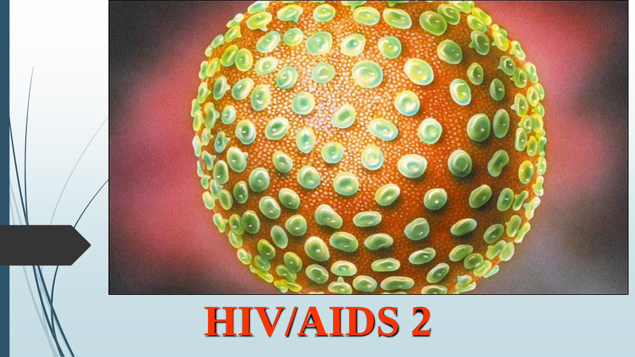
HIV/AIDS 2

Stages of HIV infection
Acute
Clinical latency - asymptomatic
Chronic – symptomatic (including AIDS)
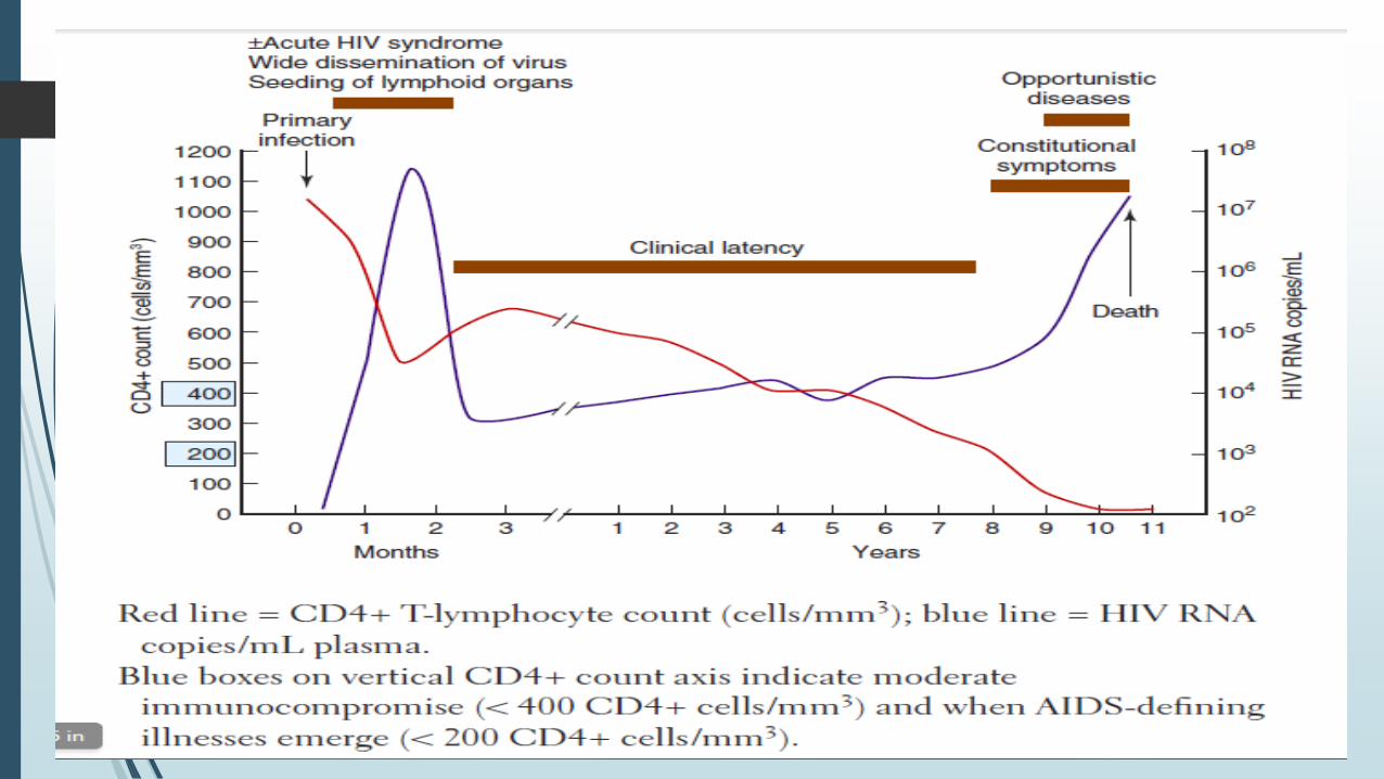
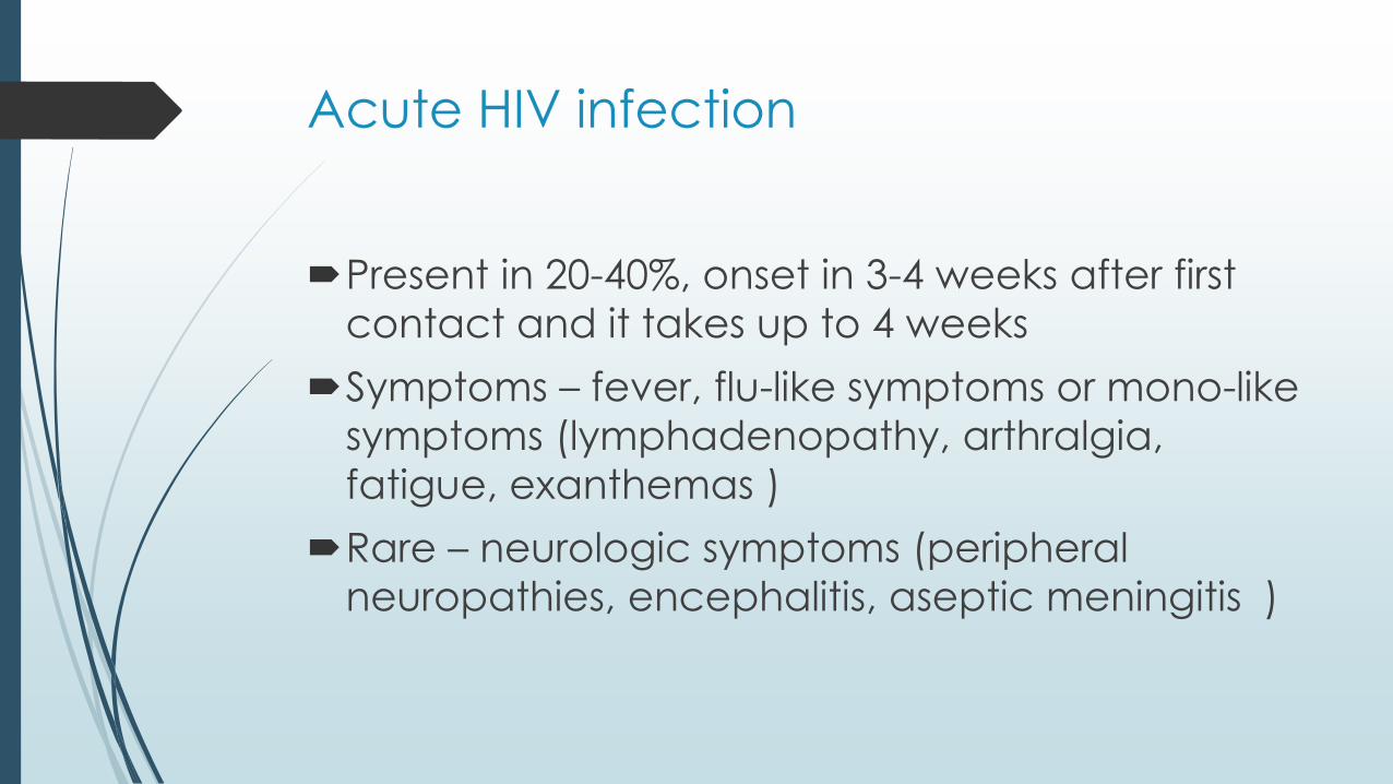
Acute HIV infection
Present in 20-40%, onset in 3-4 weeks after first
contact and it takes up to 4 weeks
Symptoms – fever, flu-like symptoms or mono-like
symptoms (lymphadenopathy, arthralgia,
fatigue, exanthemas )
Rare – neurologic symptoms (peripheral
neuropathies, encephalitis, aseptic meningitis )
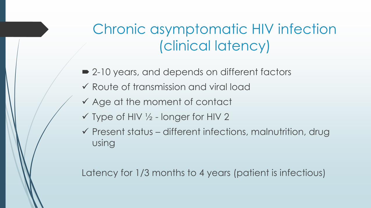
Chronic asymptomatic HIV infection
(clinical latency)
2-10 years, and depends on different factors
Route of transmission and viral load
Age at the moment of contact
Type of HIV ½ - longer for HIV 2
Present status – different infections, malnutrition, drug
using
Latency for 1/3 months to 4 years (patient is infectious)
Chronic symptomatic HIV infection
Painless generalized persistent lymphadenopathy (2 peripheral groups –
most common cervical, axillar, occipital)
It contain :
1. Non – specific symptoms – unknown fever 3-4 weeks, weight loss >10%,
persistent diarrhea >1 month; for children - chronic bilateral parotidites,
physical retard
2. Opportunistic infections related to decreasing immunity – respiratory
recurrent infections (otitis, sinusitis, pneumonia), oral symptoms
(candidiasis, hairy leukoplakia, gingivitis), skin manifestations (herpes
simplex, recidivate shingles, molluscum contagiosum, seboreic dermatitis,
prurigo etc.)
3. Suggestive clinical symptoms, specific for HIV (AIDS Related Complex –
ARC).
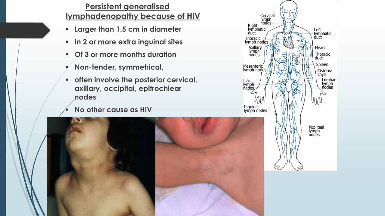
Persistent generalised
lymphadenopathy because of HIV
Larger than 1.5 cm in diameter
In 2 or more extra inguinal sites
Of 3 or more months duration
Non-tender, symmetrical,
often involve the posterior cervical,
axillary, occipital, epitrochlear
nodes
No other cause as HIV
CAUSES OF LYMPHADENOPATHY in HIV
Bacterial infection:
• pyogenic bacteria
• syphilis
Mycobacterial:
• tuberculosis
• MAC
Fungal:
• histoplasmosis
• coccidioidomycosis
Chlamydial:
• lymphogranuloma venereum
Parasitic:
• toxoplasmosis
Viral:
• EBV
• CMV
Malignant disorders of the immune system:
• Hodgkin's disease
• non-Hodgkin's disease
• other malignancies
Lymphomas that develop in HIV+ patients (LHIV)
• 4-10% of AIDS pts
• 10% of non-Hodgkin lymphomas in US/Europe are AIDS related
• Most common LHIV:
• diffuse large B cell lymphoma (DLBCL),
• Burkitt lymphoma,
• primary effusion lymphoma (PEL),
• plasmablastic lymphoma
• HIV is NOT directly involved in the malignant transformation of B
cells
• several pathogenetic mechanisms:
• chronic antigen stimulation,
• genetic abnormalities,
• cytokine dysregulation,
• EBV and HHV8
• Lymph node involv.: 1/3 of patients;
• extranodal: GI, CNS, liver, bone marrow
• Aggressive, poor outcome
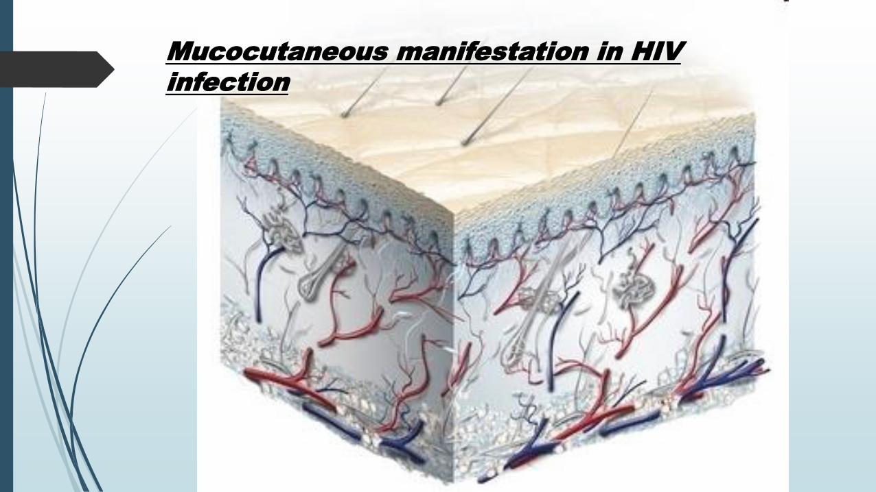
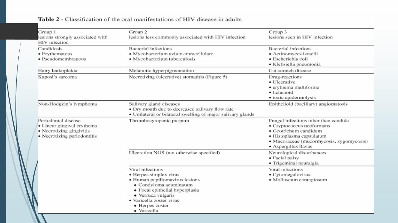
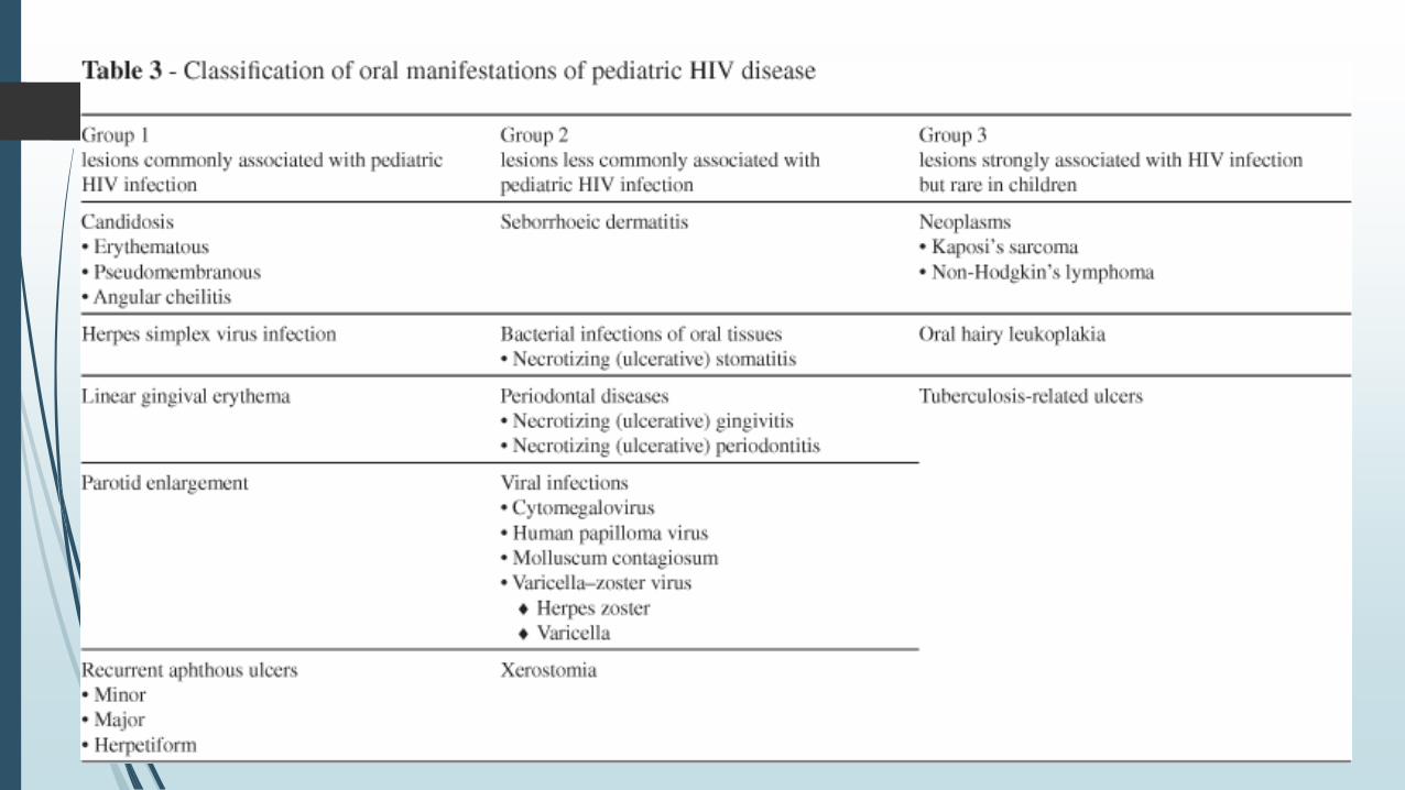
Mucocutaneous manifestation in HIV
infection

3 groups of oral manifestations of AIDS
Group 1 (strongly associated with HIV infection):
1. oral candidosis,
2. hairy leukoplakia,
3. Kaposi sarcoma,
4. linear gingival erythema,
5. necrotizing ulcerative gingivitis,
6. necrotizing ulcerative periodontitis, 7. non-Hodgkin lymphoma.
Group 2
1. atypical ulcers,
2. salivary glands diseases,
3. viral infection such (CMV, HSV, HPV, HZV).
Group 3 (rarer)
1. diffuse osteomyelitis
2. squamous cell carcinoma.
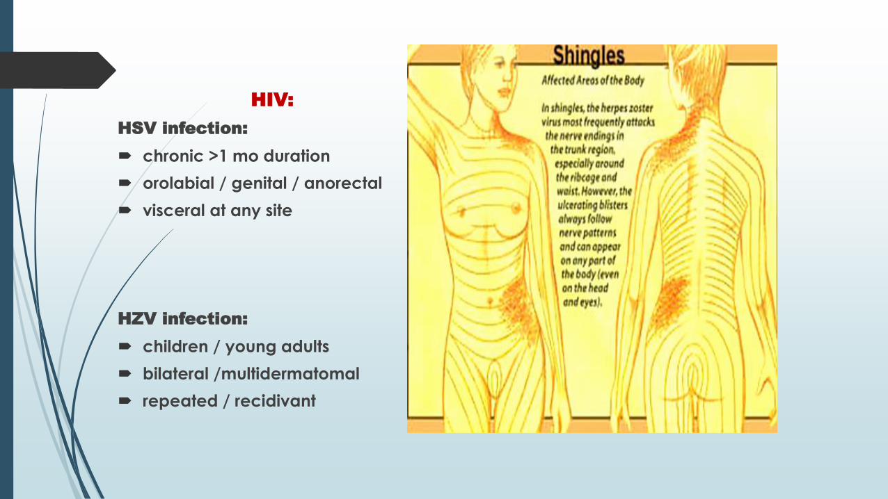
HIV:
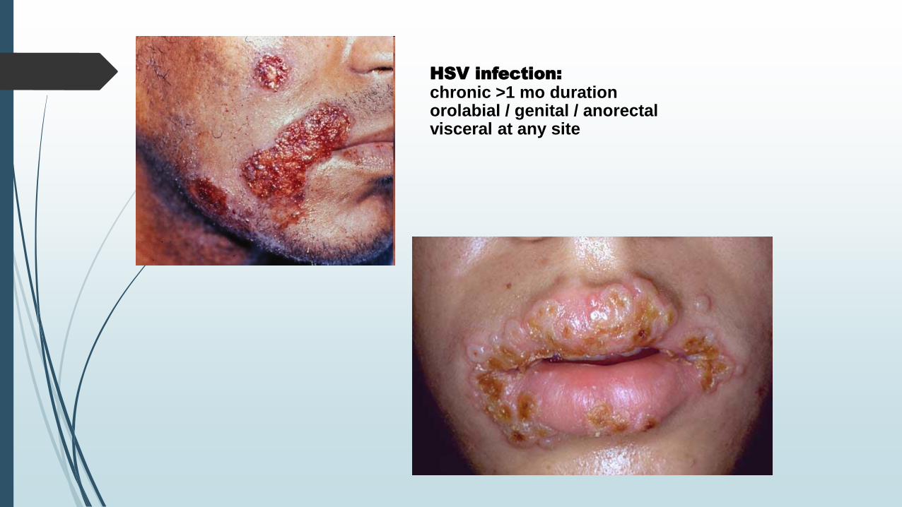
HSV infection:
chronic >1 mo duration
orolabial / genital / anorectal
visceral at any site
HZV infection:
children / young adults
bilateral /multidermatomal
repeated / recidivant
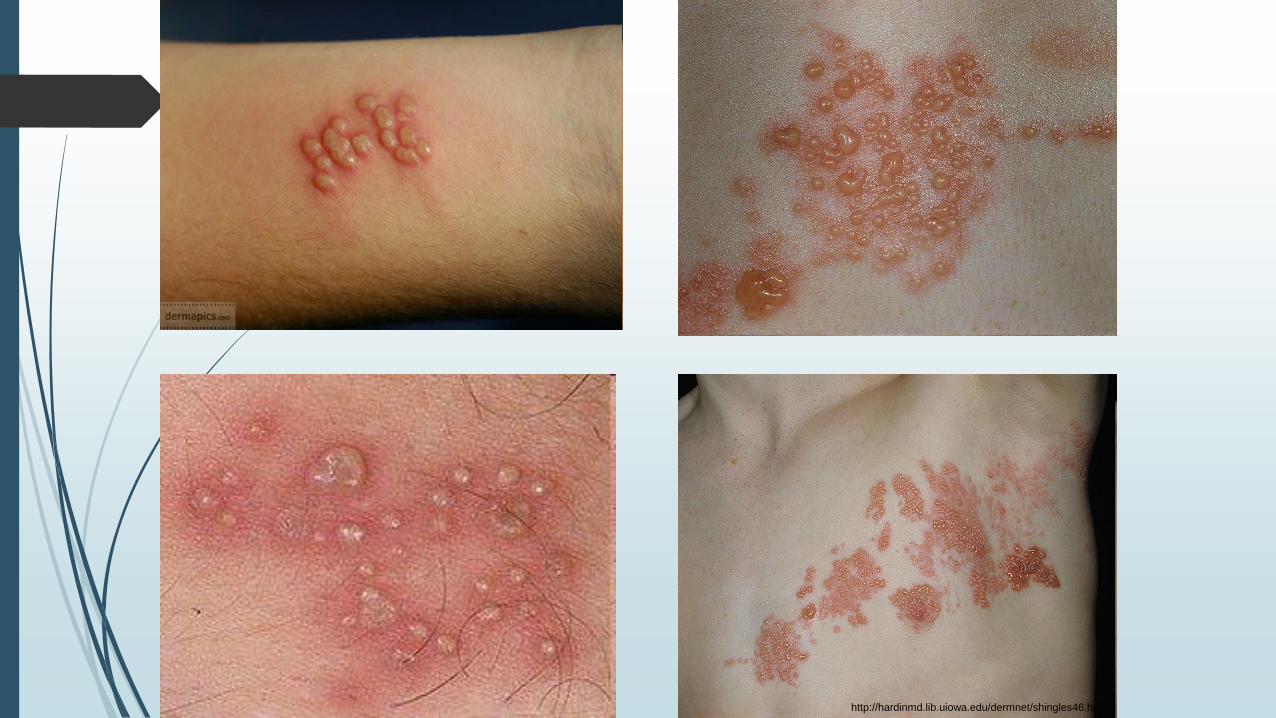
http://hardinmd.lib.uiowa.edu/dermnet/shingles46.html
HSV infection:
chronic >1 mo duration orolabial / genital / anorectalvisceral at any site
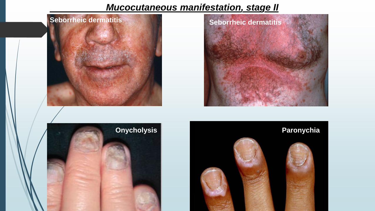
Seborrheic dermatitis Seborrheic dermatitis
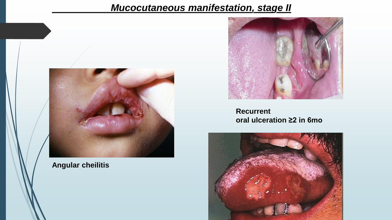
Mucocutaneous manifestation, stage II
Onycholysis Paronychia
Mucocutaneous manifestation, stage II
Angular cheilitis
Recurrent
oral ulceration ≥2 in 6mo

Pseudomembranous
Erythematous
Persistent or recurring oral candidiasis (stage III)• associated with more frequent progression to AIDS
• has been also used as a clinical marker to define the severity of HIV infection
• pseudomembranous candidosis usually followed by erythematous candidosis
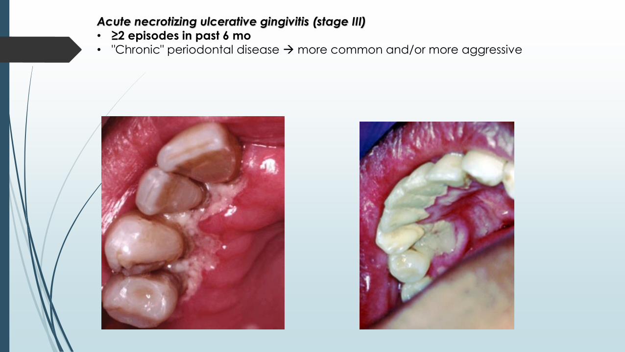
Acute necrotizing ulcerative gingivitis (stage III)• ≥2 episodes in past 6 mo• "Chronic" periodontal disease more common and/or more aggressive
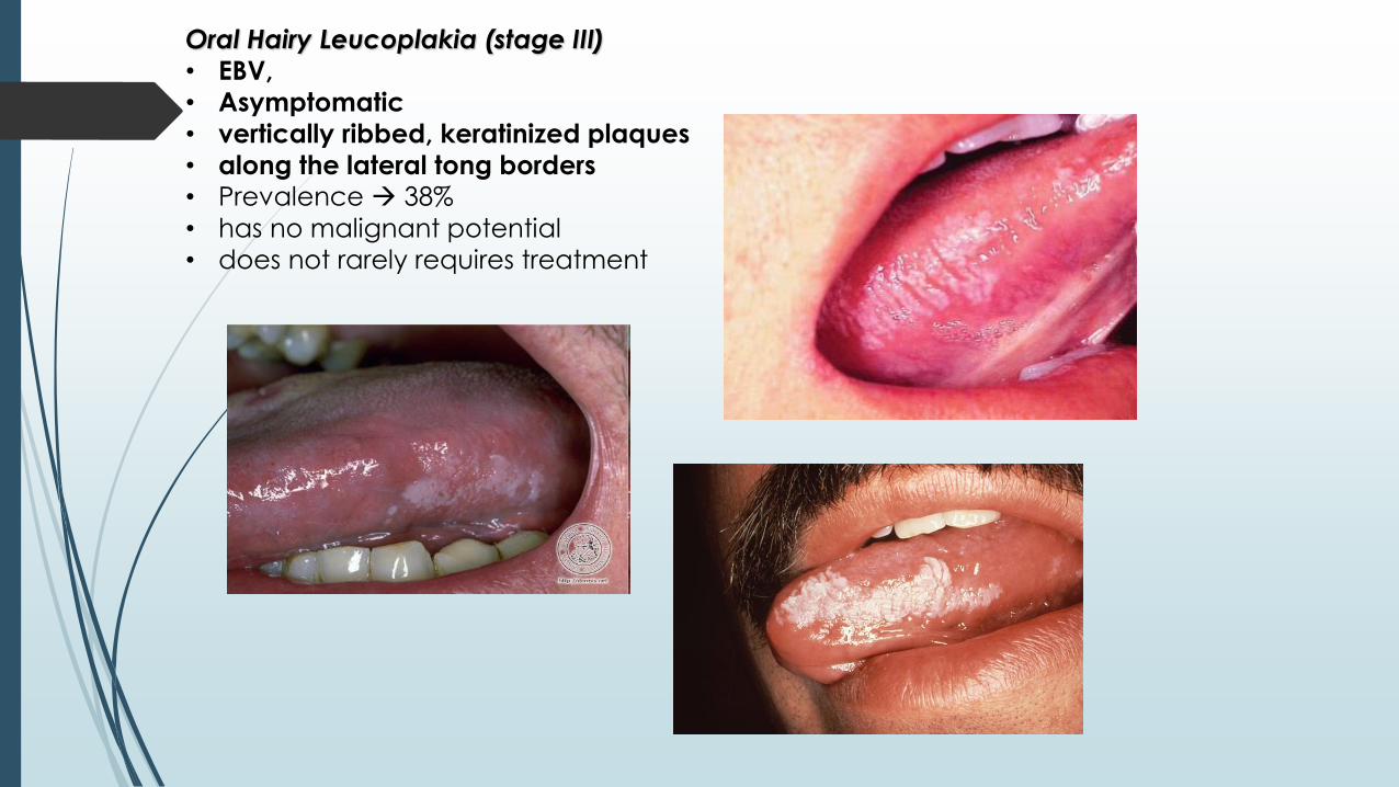
Oral Hairy Leucoplakia (stage III)• EBV,
• Asymptomatic
• vertically ribbed, keratinized plaques
• along the lateral tong borders• Prevalence 38%
• has no malignant potential
• does not rarely requires treatment
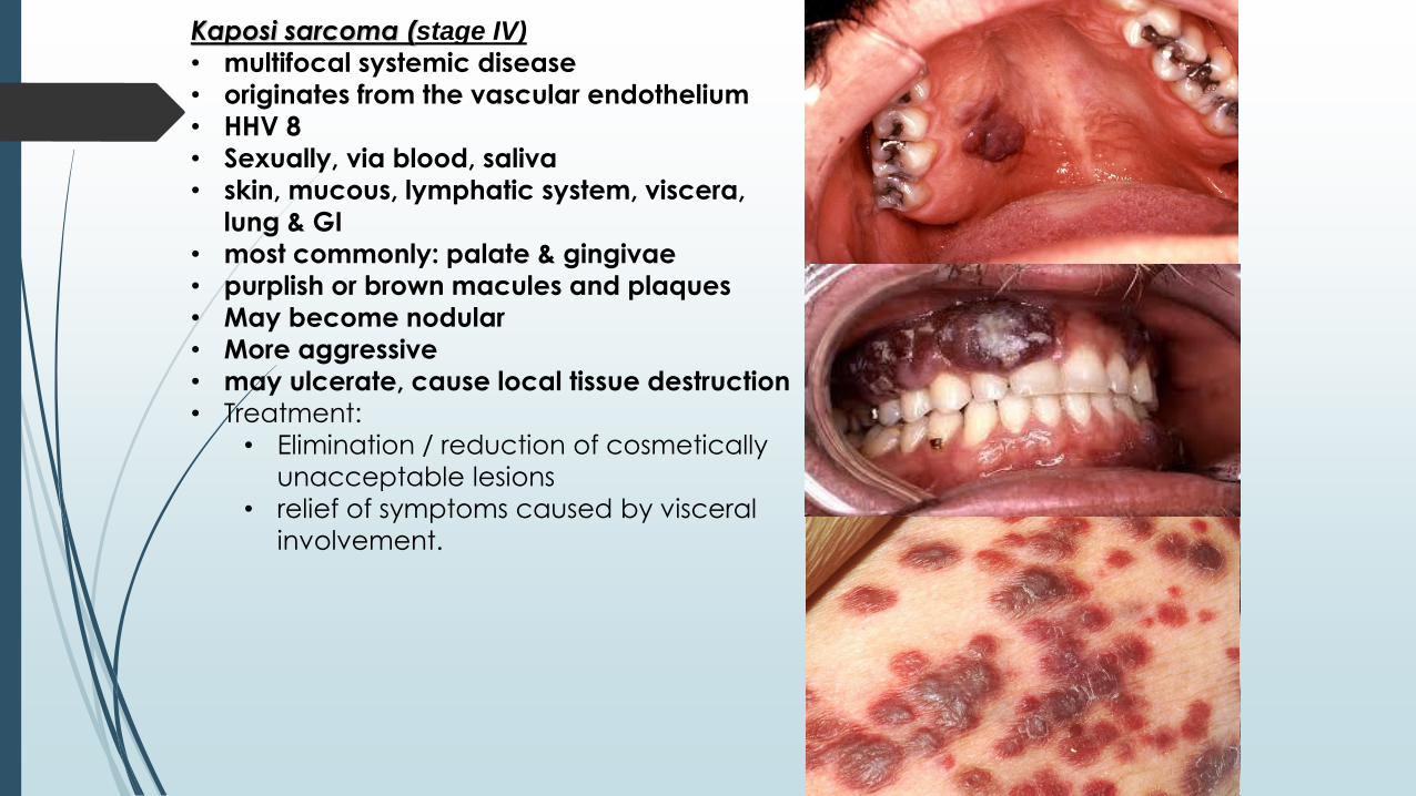
Kaposi sarcoma (stage IV)
• multifocal systemic disease
• originates from the vascular endothelium
• HHV 8
• Sexually, via blood, saliva
• skin, mucous, lymphatic system, viscera,
lung & GI
• most commonly: palate & gingivae
• purplish or brown macules and plaques
• May become nodular
• More aggressive
• may ulcerate, cause local tissue destruction
• Treatment:
• Elimination / reduction of cosmetically
unacceptable lesions
• relief of symptoms caused by visceral
involvement.

Molluscum contagiosum
MC virus, DNA, Poxviridae family
replicates in cytoplasm epidermal cells
small papules with central umbilication
Crusted or “Norwegian” scabies
widespread eczematous eruption,
no characteristic papules and burrows
Pulmonary manifestation in HIV
infection
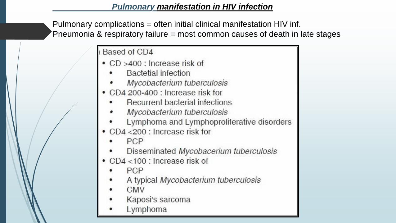
Pulmonary manifestation in HIV infection
Pulmonary complications = often initial clinical manifestation HIV inf.
Pneumonia & respiratory failure = most common causes of death in late stages
suggestive of HIV infection is bac. pneum ≥2 episodes in 6 mo
most common causes:
Streptococcus pneumoniae,
Hemophilus influenzae,
Staph. aureus (parenchymal necrosis + cavitation commonly seen)
The signs and symptoms:
fever and cough (90%),
tachypnea,
purulent sputum production,
pleuritic chest pain
Chest x-ray: focal infiltrates
Response to antimicrobial therapy is generally prompt;
If no prompt improvement, suggests complications such as:
empyema (infected parapneumonic pleural effusion),
lung abscess
or another opportunistic infection
Bacterial Pneumonias in HIV
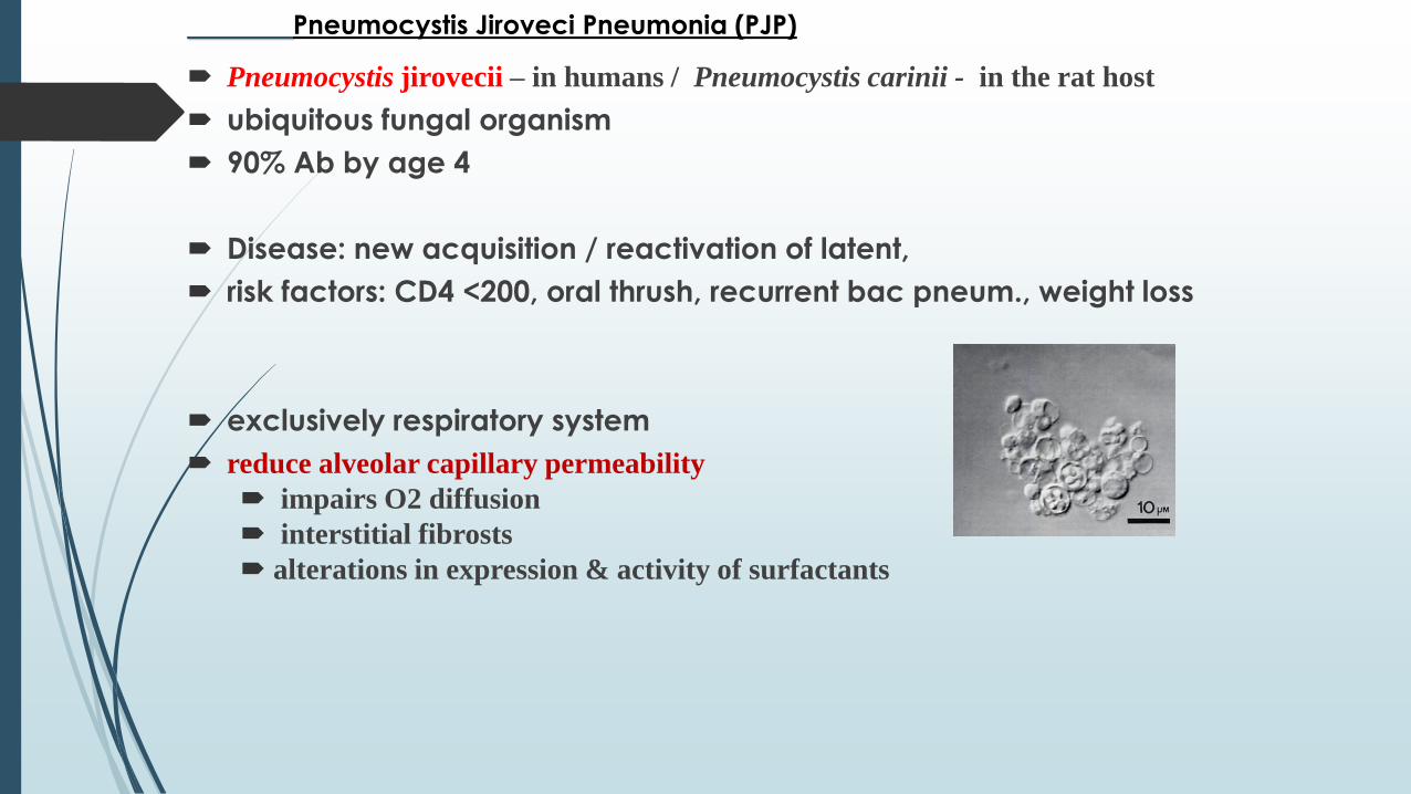
Pneumocystis jirovecii – in humans / Pneumocystis carinii - in the rat host
ubiquitous fungal organism
90% Ab by age 4
Disease: new acquisition / reactivation of latent,
risk factors: CD4 <200, oral thrush, recurrent bac pneum., weight loss
exclusively respiratory system
reduce alveolar capillary permeability
impairs O2 diffusion
interstitial fibrosts
alterations in expression & activity of surfactants
Pneumocystis Jiroveci Pneumonia (PJP)
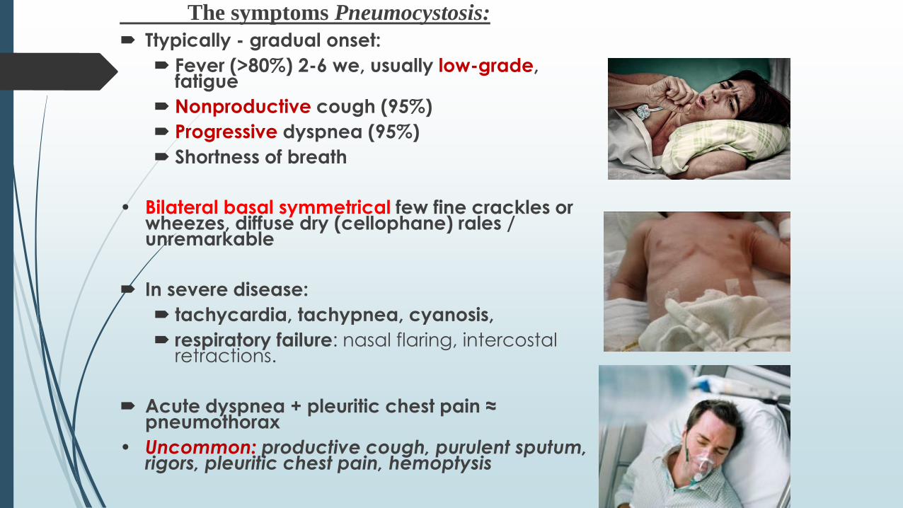
The symptoms Pneumocystosis:
Ttypically - gradual onset:
Fever (>80%) 2-6 we, usually low-grade, fatigue
Nonproductive cough (95%)
Progressive dyspnea (95%)
Shortness of breath
• Bilateral basal symmetrical few fine crackles or wheezes, diffuse dry (cellophane) rales / unremarkable
In severe disease:
tachycardia, tachypnea, cyanosis,
respiratory failure: nasal flaring, intercostalretractions.
Acute dyspnea + pleuritic chest pain ≈ pneumothorax
• Uncommon: productive cough, purulent sputum, rigors, pleuritic chest pain, hemoptysis
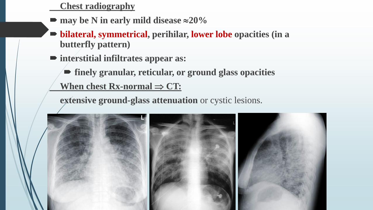
Chest radiography
may be N in early mild disease 20%
bilateral, symmetrical, perihilar, lower lobe opacities (in a butterfly pattern)
interstitial infiltrates appear as:
finely granular, reticular, or ground glass opacities
When chest Rx-normal CT:
extensive ground-glass attenuation or cystic lesions.
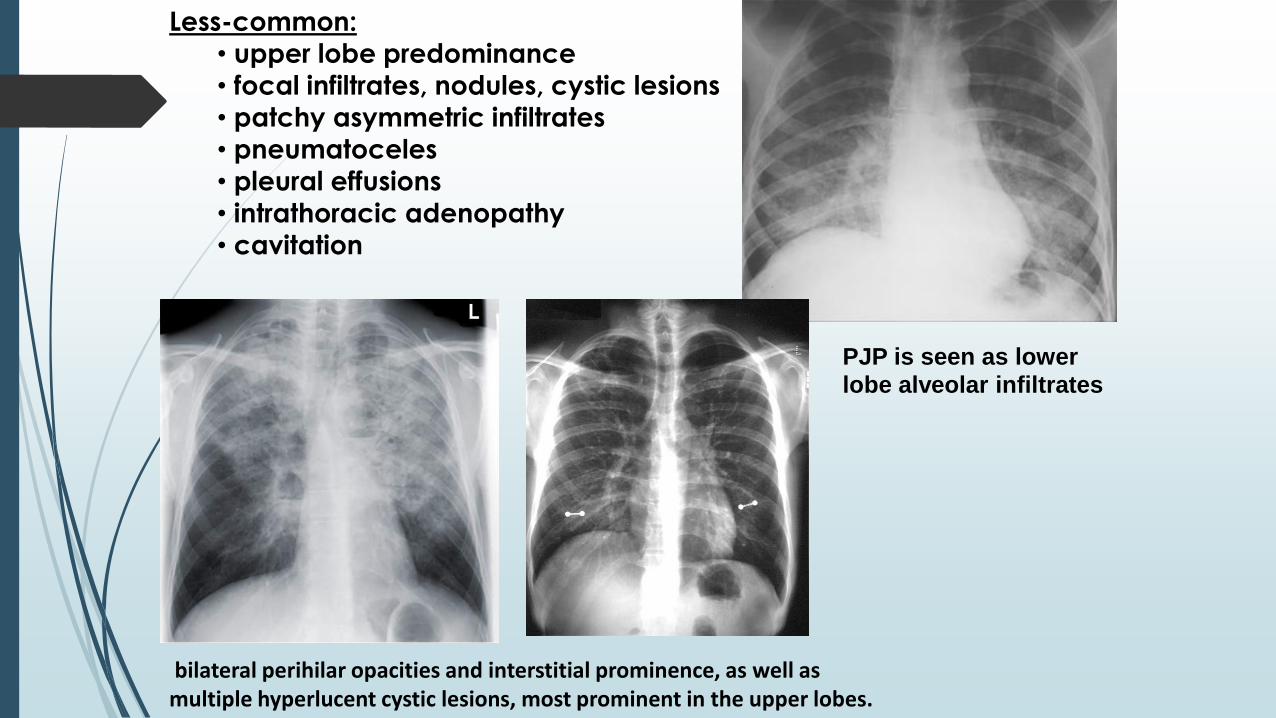
Less-common:
• upper lobe predominance
• focal infiltrates, nodules, cystic lesions
• patchy asymmetric infiltrates
• pneumatoceles
• pleural effusions
• intrathoracic adenopathy
• cavitation
bilateral perihilar opacities and interstitial prominence, as well as multiple hyperlucent cystic lesions, most prominent in the upper lobes.
PJP is seen as lower
lobe alveolar infiltrates
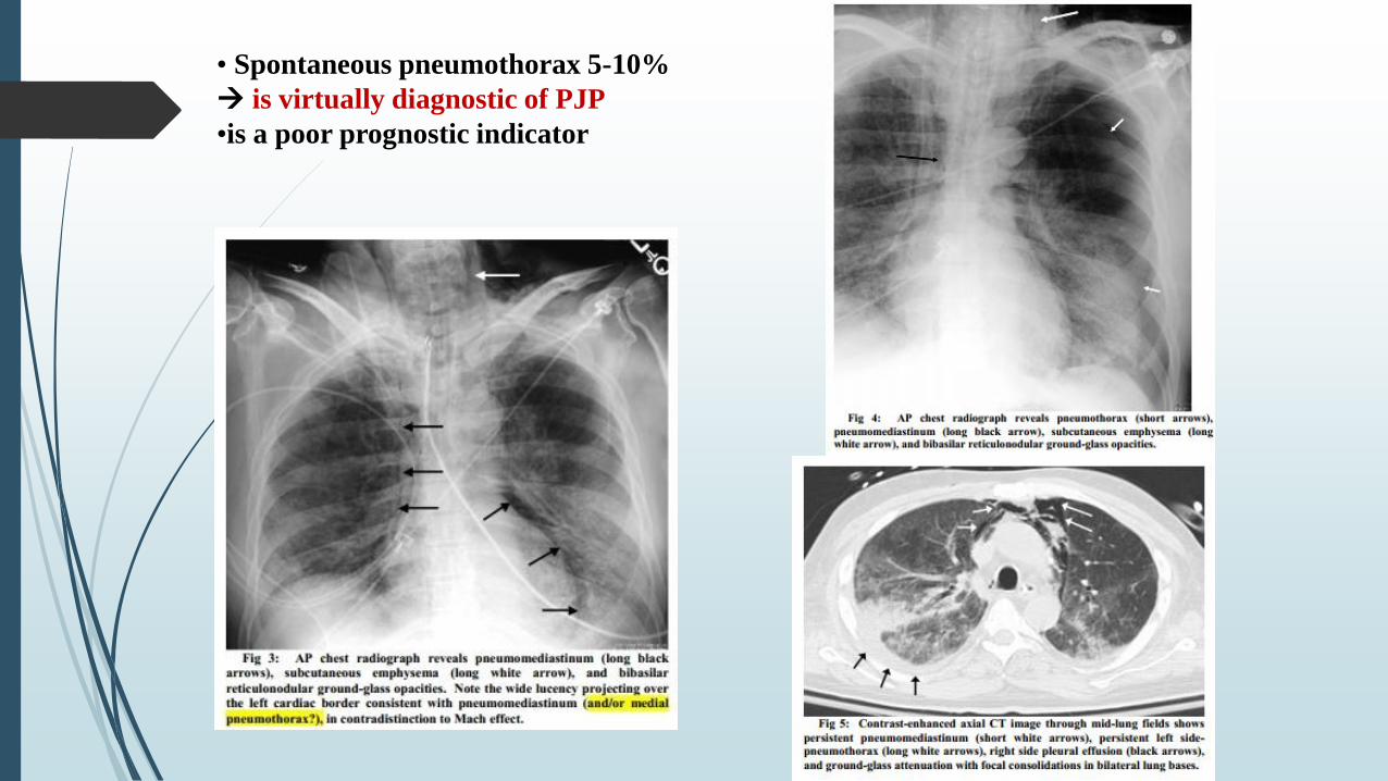
• Spontaneous pneumothorax 5-10%
is virtually diagnostic of PJP
•is a poor prognostic indicator
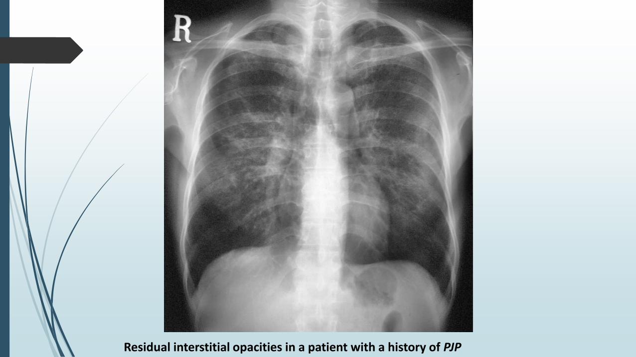
Residual interstitial opacities in a patient with a history of PJP
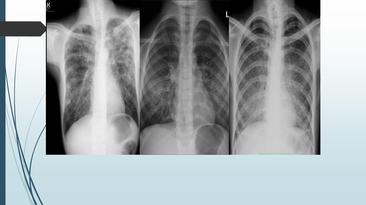
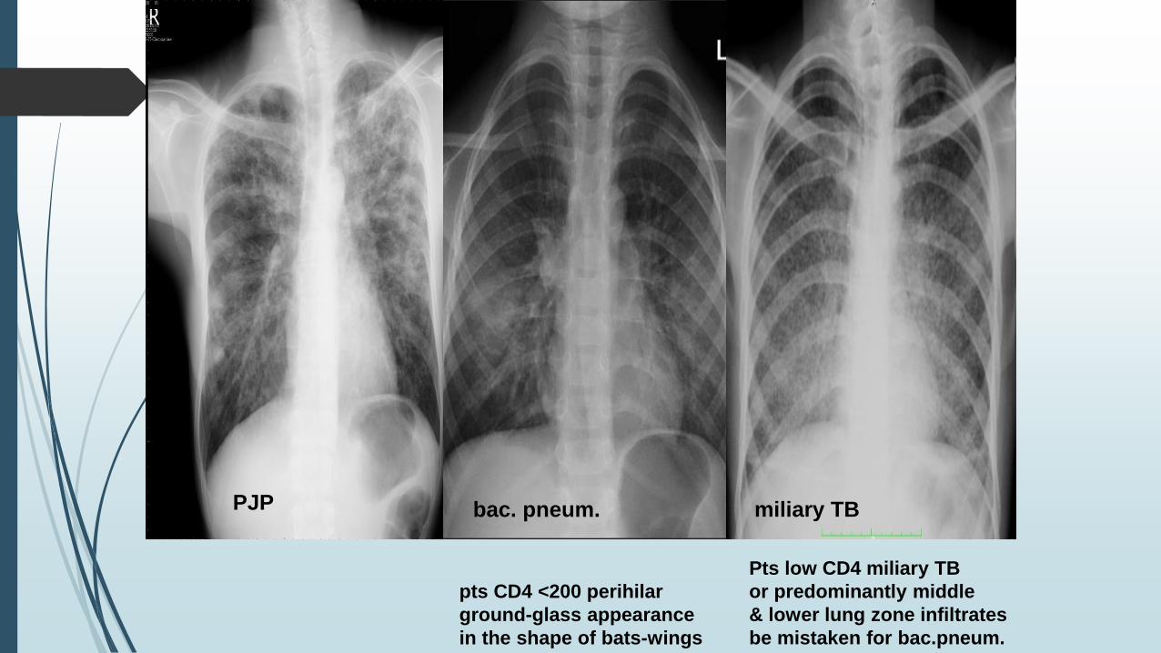
PJP
pts CD4 <200 perihilar
ground-glass appearance
in the shape of bats-wings
bac. pneum.
Pts low CD4 miliary TB
or predominantly middle
& lower lung zone infiltrates
be mistaken for bac.pneum.
miliary TB
Diagnosis
Specimens:
induced sputum with hypertonic saline
bronchoscopy with BAL (bronchoalveolar lavage)
transbronchial biopsy
open lung biopsy 95% to 100%
Stains preferred:
Giemsa
Toluidine blue
Methenamine silver
Culturing yet not possible
Rapid method - direct fluorescent method with monoclonal`s
Serology – to establish prevalence for epidemiology purpose
PCR – (ability to distinguish colonization from disease is less clear)
LDH are usually elevated (>220 U/L) in 90% of pts:
reflect the degree of lung injury
elevated LDH may indicate therapy failure
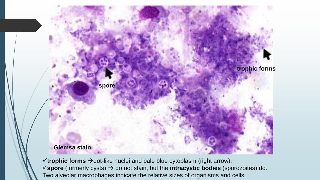
trophic forms dot-like nuclei and pale blue cytoplasm (right arrow).
spore (formerly cysts) do not stain, but the intracystic bodies (sporozoites) do.
Two alveolar macrophages indicate the relative sizes of organisms and cells.
Giemsa stain
trophic forms
spore
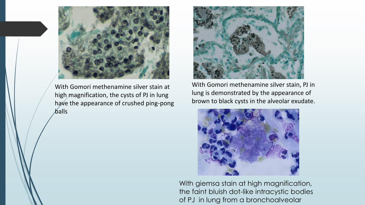
With Gomori methenamine silver stain, PJ in lung is demonstrated by the appearance of brown to black cysts in the alveolar exudate.
With Gomori methenamine silver stain at high magnification, the cysts of PJ in lung have the appearance of crushed ping-pong balls
With giemsa stain at high magnification,
the faint bluish dot-like intracystic bodies
of PJ in lung from a bronchoalveolar
lavage.
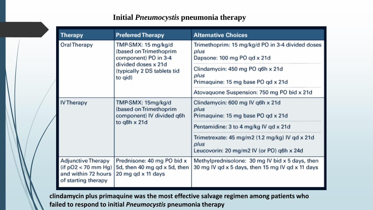
Initial Pneumocystis pneumonia therapy
clindamycin plus primaquine was the most effective salvage regimen among patients who failed to respond to initial Pneumocystis pneumonia therapy
Pneumocystiosis
Primary prophylaxis
when CD4 <200
a history of oropharyngeal candidiasis
Pts receiving pyrimethamine sulfadiazine for suppression of toxo do not require additional prophylaxis for PCP
TMP-SMX 5 mg/kg/day (1single-strength tab/day / 1double-strength tab/3xwe )
Dapsone 100mg/day
Dapsone 50mg/day + pyrimethamine 50mg/we + leucovorin 25 mg/we
atovaquone
Cotrimoxazole preventive therapy (WHO)
Age Criteria for initiation Criteria for
discontinuationa
Dose of
cotrimoxazole
HIV exposed
infants
In all, starting at 4–6
weeks after birth
Until the risk of HIV
transmission ends or
HIV inf. is excluded
<1 year In all Until 5 years of age
regardless of CD4% or
clinical symptoms (if
for PJP / toxo)
1–5 years WHO stages 2 - 4 or
CD4 <25% or
in all (limited health
infrastructure)
Never
≥5 years,
including
adults
Any WHO stage and
CD4 <350 WHO (<200
USA) or WHO 3-4 any
CD4
Never or CD4 ≥350
after 6 mo of ART
(high bac inf/malaria)
or CD4 ≥200 after
6 mo (3 mo USA) of
ART (some countries)
>30kg =
960mg/day
Contraindications to CTX preventive therapy:
severe allergy to sulfa; severe liver disease, severe renal disease and G6PD deficiency.
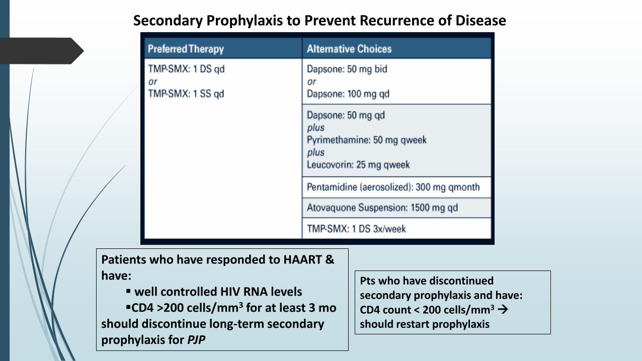
Secondary Prophylaxis to Prevent Recurrence of Disease
Patients who have responded to HAART & have:
well controlled HIV RNA levels CD4 >200 cells/mm3 for at least 3 mo
should discontinue long-term secondary prophylaxis for PJP
Pts who have discontinued secondary prophylaxis and have:CD4 count < 200 cells/mm3
should restart prophylaxis
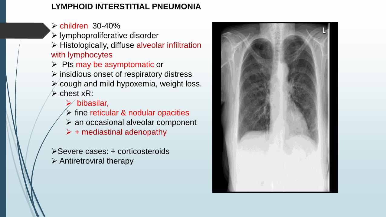
LYMPHOID INTERSTITIAL PNEUMONIA
children 30-40%
lymphoproliferative disorder
Histologically, diffuse alveolar infiltration
with lymphocytes
Pts may be asymptomatic or
insidious onset of respiratory distress
cough and mild hypoxemia, weight loss.
chest xR:
bibasilar,
fine reticular & nodular opacities
an occasional alveolar component
+ mediastinal adenopathy
Severe cases: + corticosteroids
Antiretroviral therapy
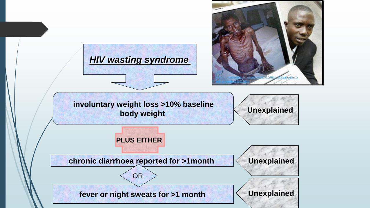
HIV wasting syndrome
involuntary weight loss >10% baseline
body weight
PLUS EITHER
Unexplained
chronic diarrhoea reported for >1month
fever or night sweats for >1 month
Unexplained
Unexplained
OR
mbahdukunbagong.blogspot.com/2010/09/definition-patient-
ask-to-mbah-dukun.html
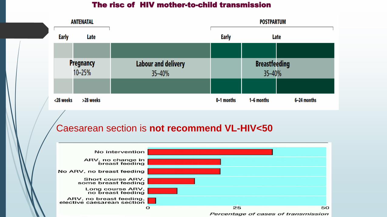
Caesarean section is not recommend VL-HIV<50
The risc of HIV mother-to-child transmission
http://sti.bmj.com/content/86/Suppl_2/ii16.full
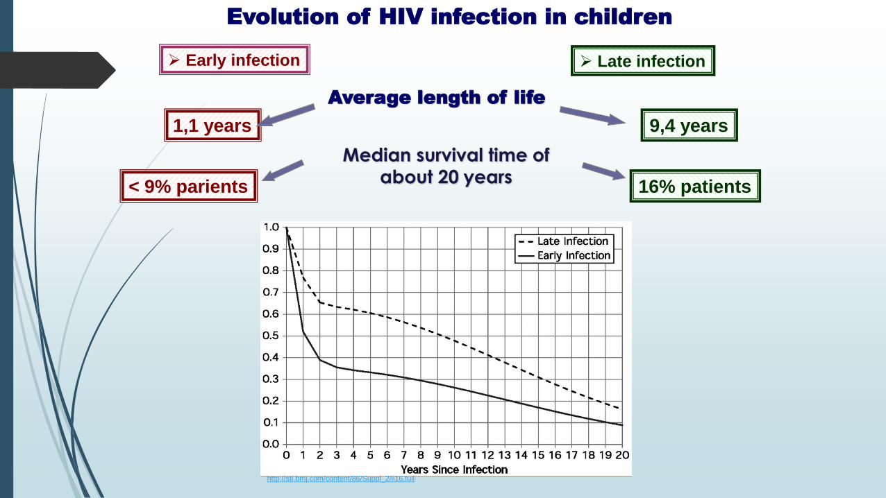
Evolution of HIV infection in children
Early infection
Median survival time of about 20 years
Late infection
Average length of life
1,1 years 9,4 years
< 9% parients 16% patients
Symptoms of pediatric HIV infection:
vary by age and individual child, the more common symptoms:
• Unusually frequent / severe / recurrent / not responding to
standard treatment
• bac inf. (otitis media, sinusitis, pneumonia)
• fungal inf. (candid.)
• viral infections (HSV, HZV, CMV)
• Growth failure
• Failure to gain weight
• Failure to reach developmental milestones during the expected
time frame
• Behavioral abnormalities (in older children), such as loss of
concentration and memory

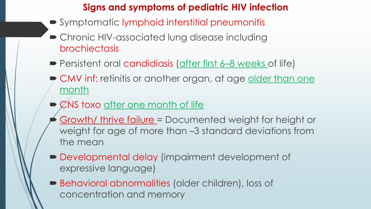
Signs and symptoms of pediatric HIV infection
Unexplained persistent hepatosplenomegaly
Lineal gingival erythema
Extensive wart virus infection,
Extensive molluscum contagiosum
Unexplained persistent parotid enlargement
• Unusually frequent / severe / recurrent / not responding to
standard treatment
• bac inf. (otitis media, sinusitis, bronchitis, pneumonia)
• fungal inf. (candid.)
• viral infections (HSV, HZV, CMV)
• Unexplained persistent diarrhoea (14 days or more) not
responding atment
Lymph node tuberculosis
Signs and symptoms of pediatric HIV infection
Symptomatic lymphoid interstitial pneumonitis
Chronic HIV-associated lung disease including
brochiectasis
Persistent oral candidiasis (after first 6–8 weeks of life)
CMV inf: retinitis or another organ, at age older than one
month
CNS toxo after one month of life
Growth/ thrive failure = Documented weight for height or
weight for age of more than –3 standard deviations from
the mean
Developmental delay (impairment development of
expressive language)
Behavioral abnormalities (older children), loss of
concentration and memory
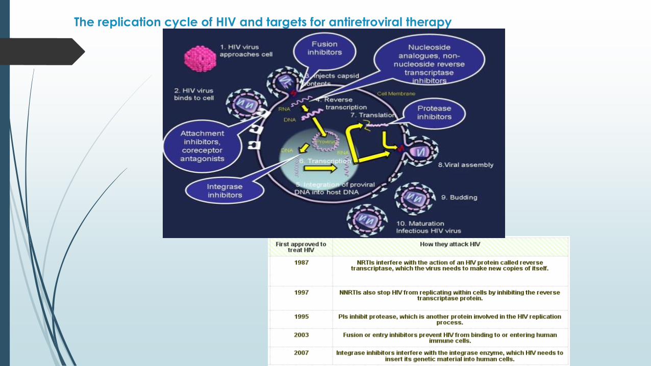
The replication cycle of HIV and targets for antiretroviral therapy
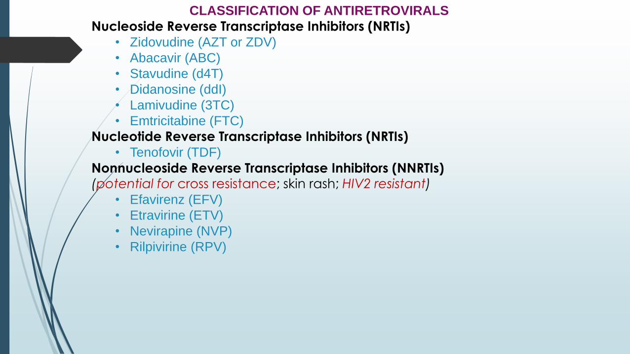
CLASSIFICATION OF ANTIRETROVIRALS
Nucleoside Reverse Transcriptase Inhibitors (NRTIs)
• Zidovudine (AZT or ZDV)
• Abacavir (ABC)
• Stavudine (d4T)
• Didanosine (ddI)
• Lamivudine (3TC)
• Emtricitabine (FTC)
Nucleotide Reverse Transcriptase Inhibitors (NRTIs)
• Tenofovir (TDF)
Nonnucleoside Reverse Transcriptase Inhibitors (NNRTIs)(potential for cross resistance; skin rash; HIV2 resistant)
• Efavirenz (EFV)
• Etravirine (ETV)
• Nevirapine (NVP)
• Rilpivirine (RPV)
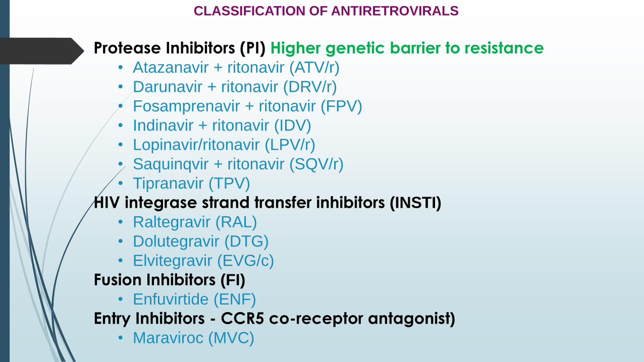
CLASSIFICATION OF ANTIRETROVIRALS
Protease Inhibitors (PI) Higher genetic barrier to resistance
• Atazanavir + ritonavir (ATV/r)
• Darunavir + ritonavir (DRV/r)
• Fosamprenavir + ritonavir (FPV)
• Indinavir + ritonavir (IDV)
• Lopinavir/ritonavir (LPV/r)
• Saquinqvir + ritonavir (SQV/r)
• Tipranavir (TPV)
HIV integrase strand transfer inhibitors (INSTI)
• Raltegravir (RAL)
• Dolutegravir (DTG)
• Elvitegravir (EVG/c)
Fusion Inhibitors (FI)
• Enfuvirtide (ENF)
Entry Inhibitors - CCR5 co-receptor antagonist)
• Maraviroc (MVC)
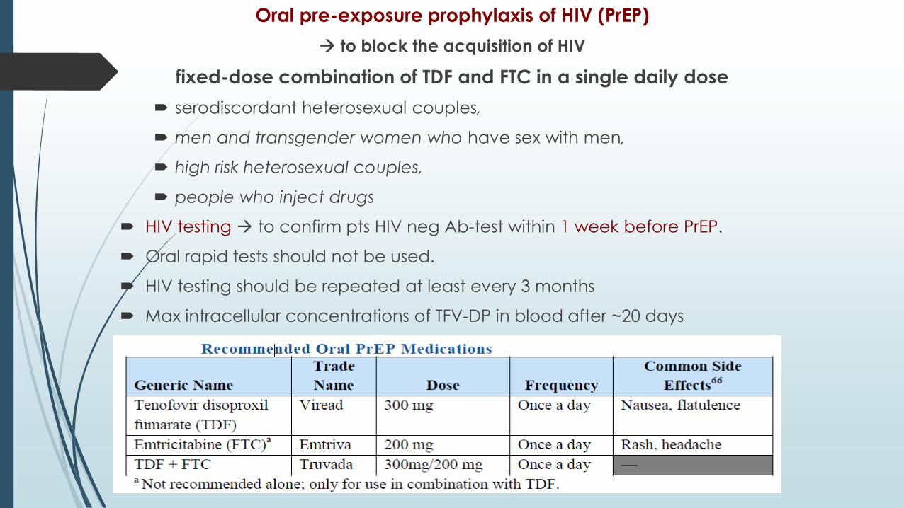
Oral pre-exposure prophylaxis of HIV (PrEP)
to block the acquisition of HIV
fixed-dose combination of TDF and FTC in a single daily dose
serodiscordant heterosexual couples,
men and transgender women who have sex with men,
high risk heterosexual couples,
people who inject drugs
HIV testing to confirm pts HIV neg Ab-test within 1 week before PrEP.
Oral rapid tests should not be used.
HIV testing should be repeated at least every 3 months
Max intracellular concentrations of TFV-DP in blood after ~20 days
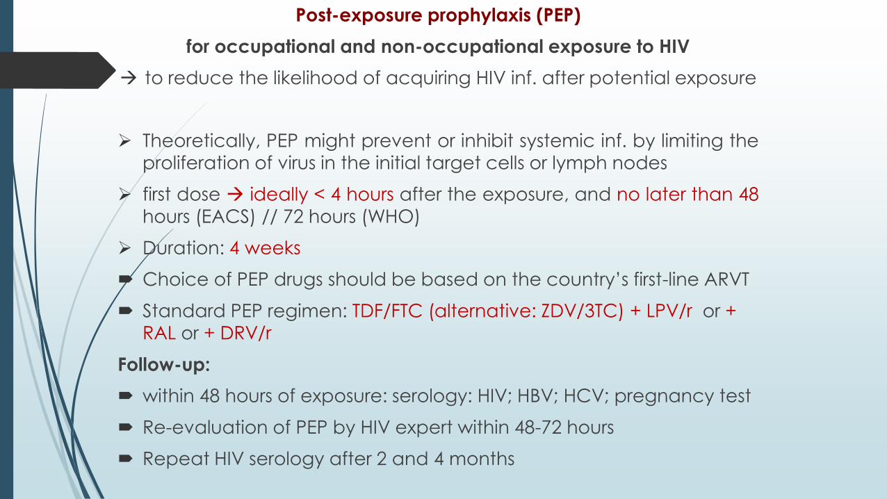
Post-exposure prophylaxis (PEP)
for occupational and non-occupational exposure to HIV
to reduce the likelihood of acquiring HIV inf. after potential exposure
Theoretically, PEP might prevent or inhibit systemic inf. by limiting the
proliferation of virus in the initial target cells or lymph nodes
first dose ideally < 4 hours after the exposure, and no later than 48
hours (EACS) // 72 hours (WHO)
Duration: 4 weeks
Choice of PEP drugs should be based on the country’s first-line ARVT
Standard PEP regimen: TDF/FTC (alternative: ZDV/3TC) + LPV/r or +
RAL or + DRV/r
Follow-up:
within 48 hours of exposure: serology: HIV; HBV; HCV; pregnancy test
Re-evaluation of PEP by HIV expert within 48-72 hours
Repeat HIV serology after 2 and 4 months
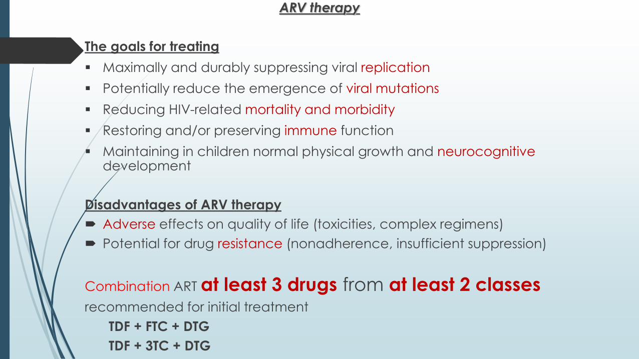
ARV therapy
The goals for treating
Maximally and durably suppressing viral replication
Potentially reduce the emergence of viral mutations
Reducing HIV-related mortality and morbidity
Restoring and/or preserving immune function
Maintaining in children normal physical growth and neurocognitivedevelopment
Disadvantages of ARV therapy
Adverse effects on quality of life (toxicities, complex regimens)
Potential for drug resistance (nonadherence, insufficient suppression)
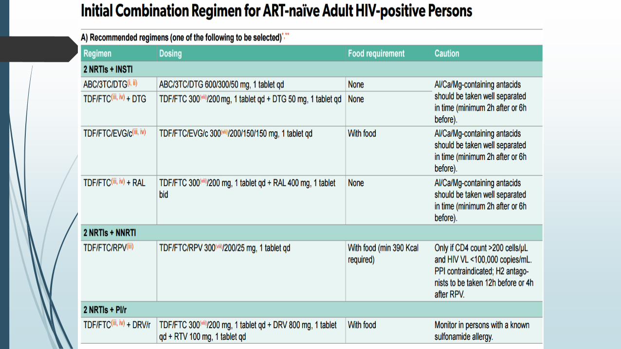
Combination ART at least 3 drugs from at least 2 classes recommended for initial treatment
TDF + FTC + DTG
TDF + 3TC + DTG
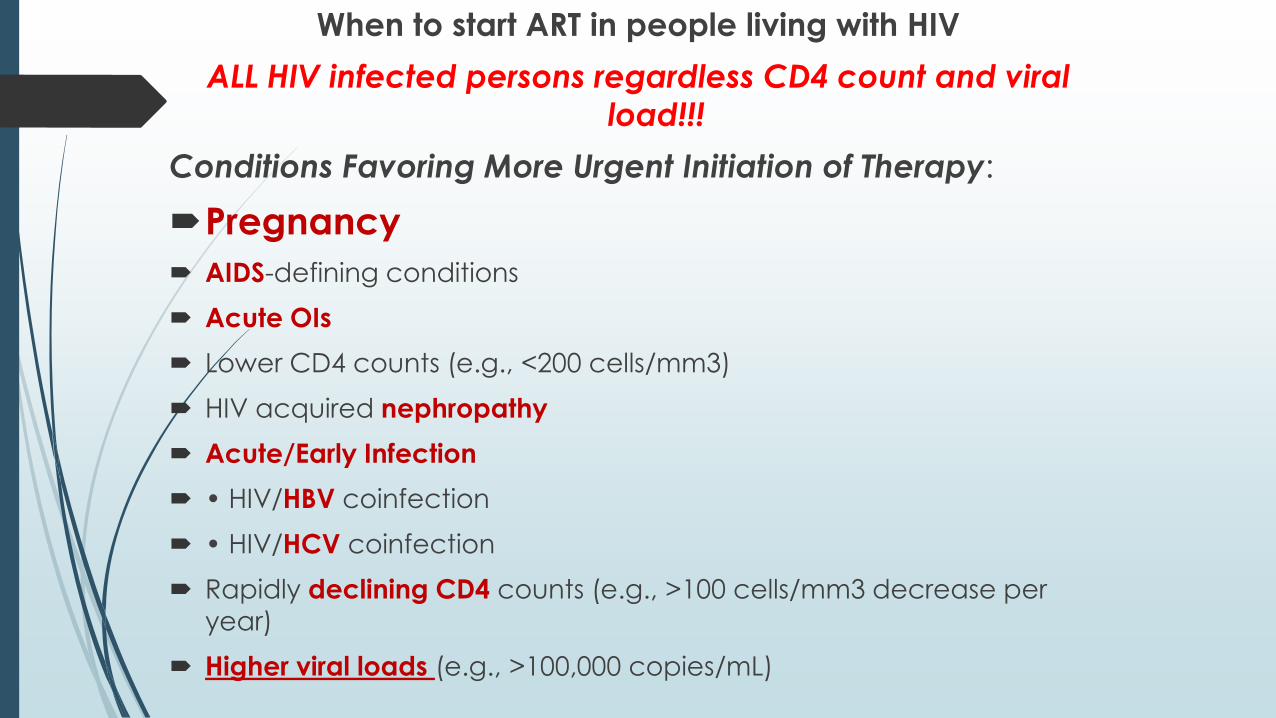
When to start ART in people living with HIV
ALL HIV infected persons regardless CD4 count and viral
load!!!
Conditions Favoring More Urgent Initiation of Therapy:
Pregnancy
AIDS-defining conditions
Acute OIs
Lower CD4 counts (e.g., <200 cells/mm3)
HIV acquired nephropathy
Acute/Early Infection
• HIV/HBV coinfection
• HIV/HCV coinfection
Rapidly declining CD4 counts (e.g., >100 cells/mm3 decrease per
year)
Higher viral loads (e.g., >100,000 copies/mL)
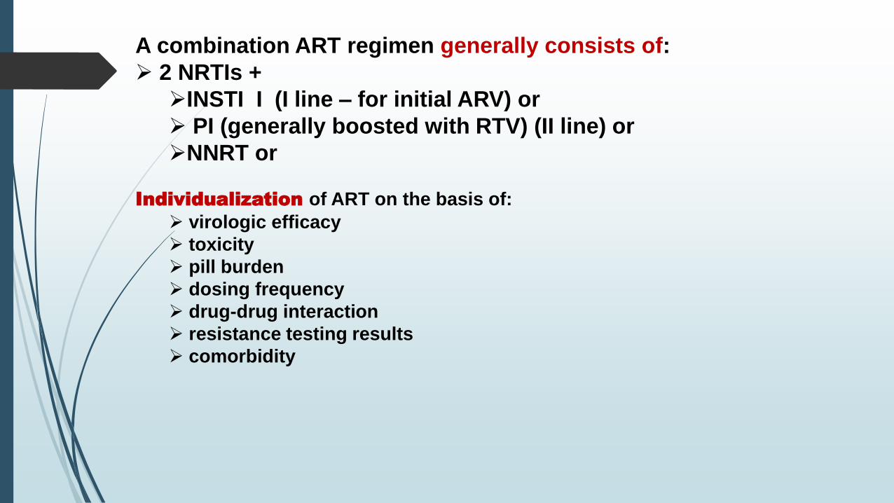
A combination ART regimen generally consists of:
2 NRTIs +
INSTI I (I line – for initial ARV) or
PI (generally boosted with RTV) (II line) or
NNRT or
Individualization of ART on the basis of:
virologic efficacy
toxicity
pill burden
dosing frequency
drug-drug interaction
resistance testing results
comorbidity

Antiretroviral Regimens Not Recommended
Monotherapy with NRTI
• Rapid resistance
• Inferior ARV activity
Dual-NRTI regimens
• Rapid resistance
• Inferior ARV activity
Triple-NRTI regimens except for ABC/ZDV/3TC or possibly
TDF + ZDV/3TC
• High rate of early virologic nonresponse (ABC/TDF/3TC;
TDF/ddI/3TC).
Adherence once-daily fixed-dose
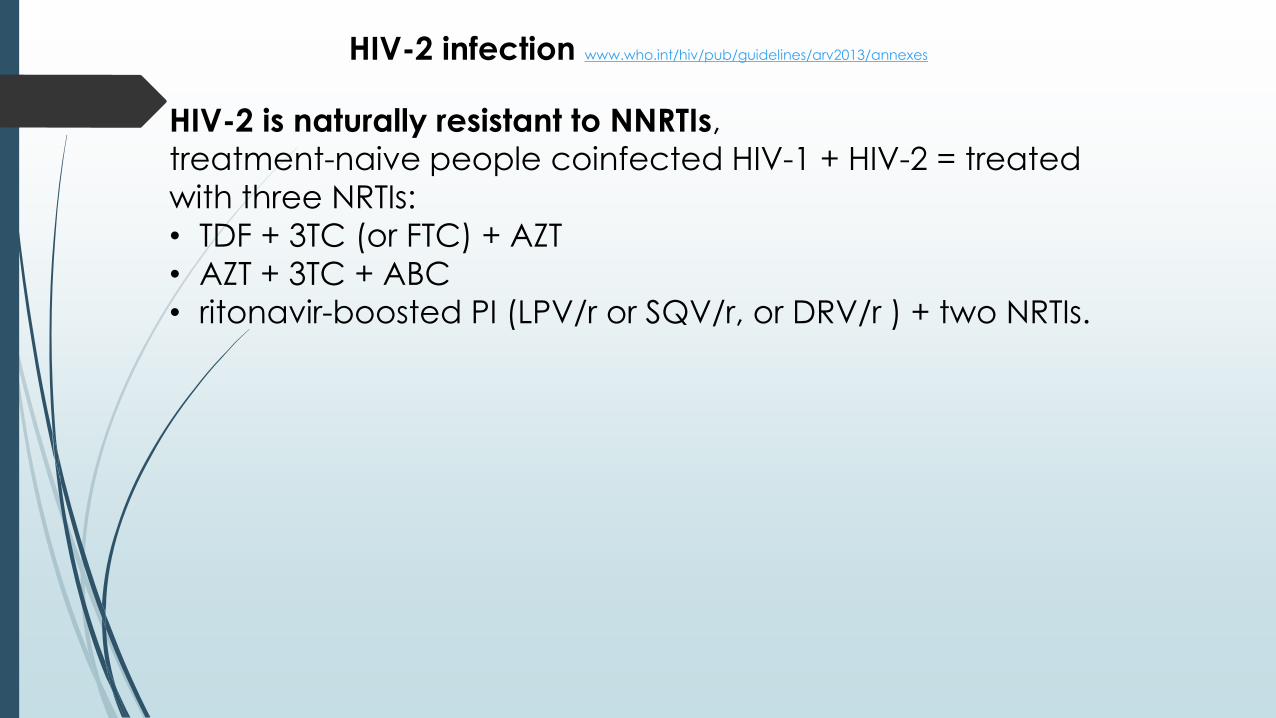
HIV-2 infection www.who.int/hiv/pub/guidelines/arv2013/annexes
HIV-2 is naturally resistant to NNRTIs,
treatment-naive people coinfected HIV-1 + HIV-2 = treated
with three NRTIs:
• TDF + 3TC (or FTC) + AZT
• AZT + 3TC + ABC
• ritonavir-boosted PI (LPV/r or SQV/r, or DRV/r ) + two NRTIs.
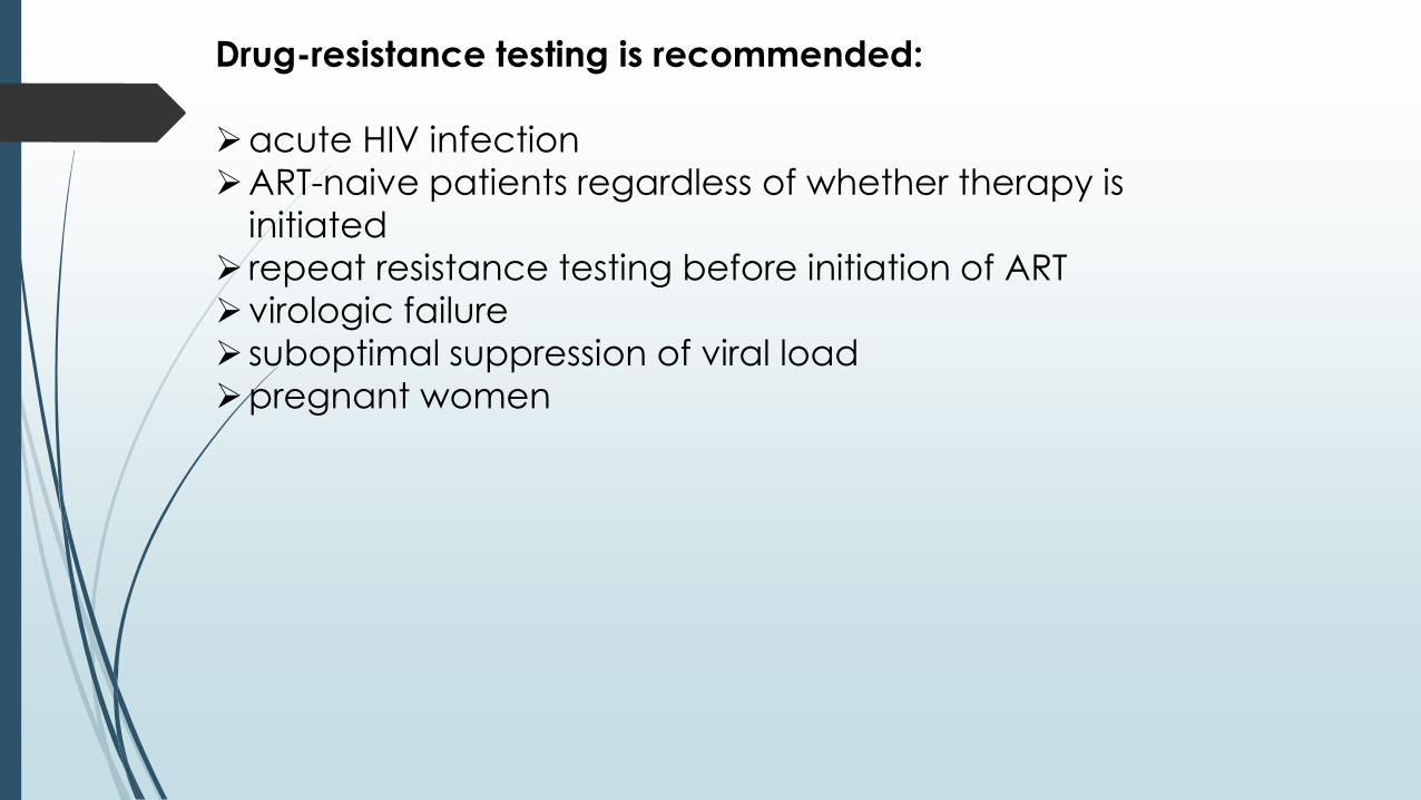
Drug-resistance testing is recommended:
acute HIV infection
ART-naive patients regardless of whether therapy is
initiated
repeat resistance testing before initiation of ART
virologic failure
suboptimal suppression of viral load
pregnant women
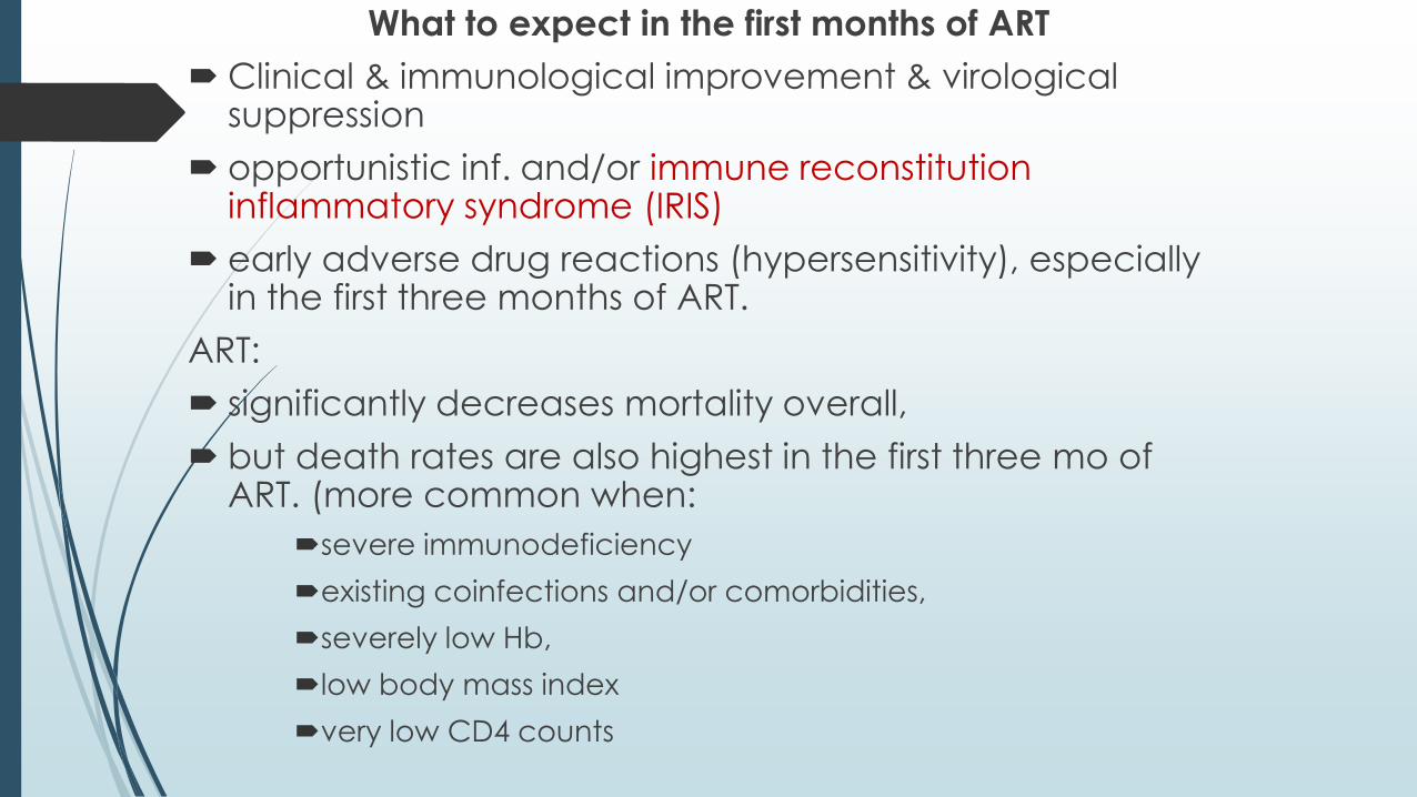
What to expect in the first months of ART
Clinical & immunological improvement & virologicalsuppression
opportunistic inf. and/or immune reconstitution inflammatory syndrome (IRIS)
early adverse drug reactions (hypersensitivity), especially in the first three months of ART.
ART:
significantly decreases mortality overall,
but death rates are also highest in the first three mo of ART. (more common when:
severe immunodeficiency
existing coinfections and/or comorbidities,
severely low Hb,
low body mass index
very low CD4 counts
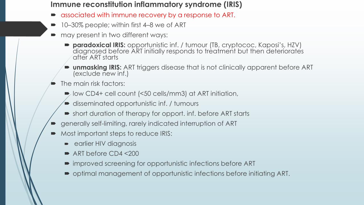
Immune reconstitution inflammatory syndrome (IRIS)
associated with immune recovery by a response to ART.
10–30% people; within first 4–8 we of ART
may present in two different ways:
paradoxical IRIS: opportunistic inf. / tumour (TB, cryptococ, Kaposi’s, HZV) diagnosed before ART initially responds to treatment but then deteriorates after ART starts
unmasking IRIS: ART triggers disease that is not clinically apparent before ART (exclude new inf.)
The main risk factors:
low CD4+ cell count (<50 cells/mm3) at ART initiation,
disseminated opportunistic inf. / tumours
short duration of therapy for opport. inf. before ART starts
generally self-limiting, rarely indicated interruption of ART
Most important steps to reduce IRIS:
earlier HIV diagnosis
ART before CD4 <200
improved screening for opportunistic infections before ART
optimal management of opportunistic infections before initiating ART.
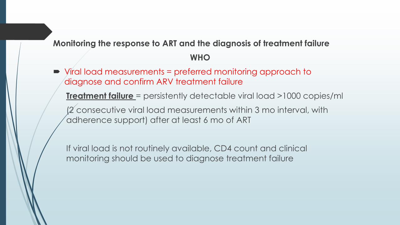
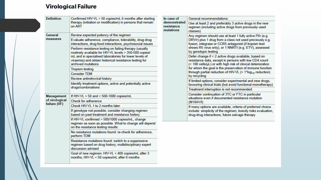
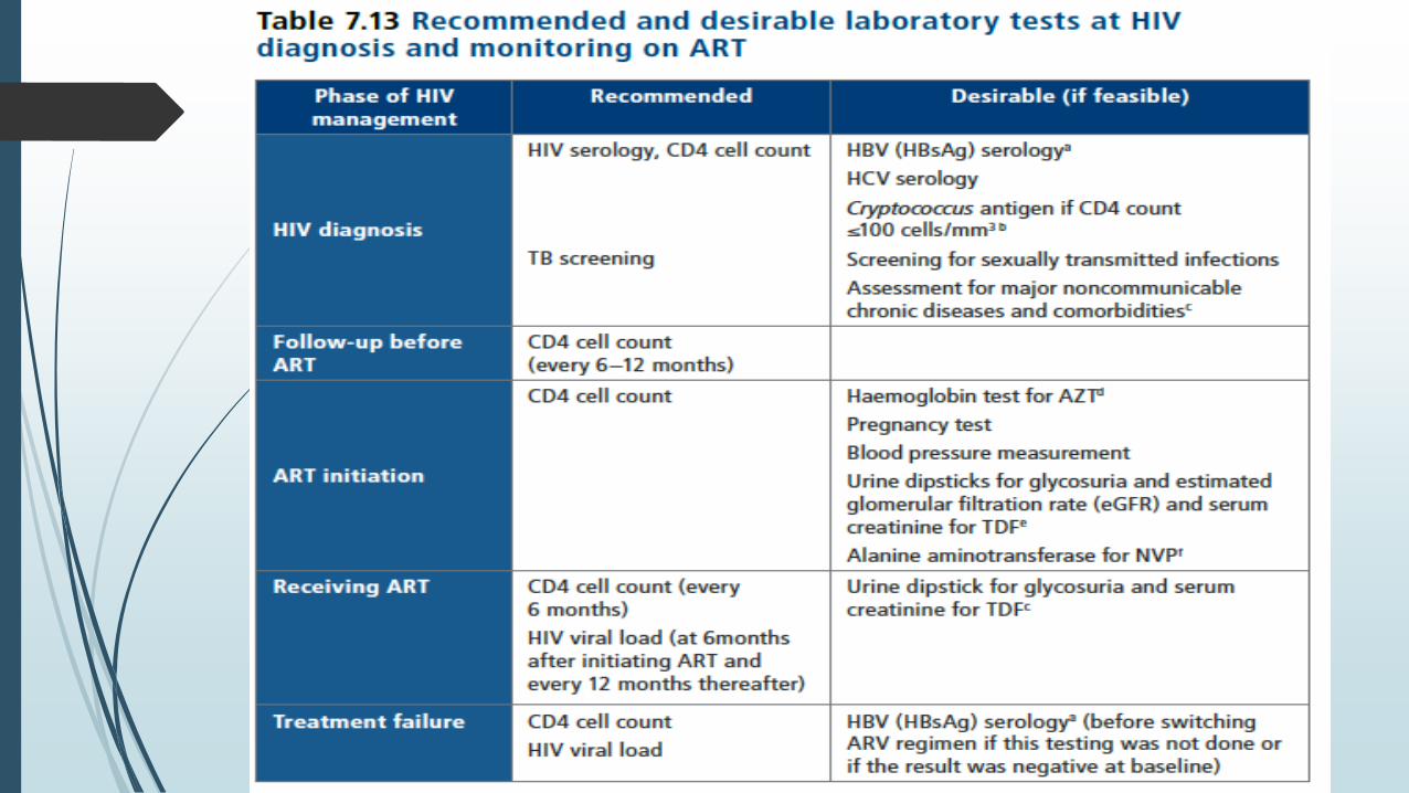
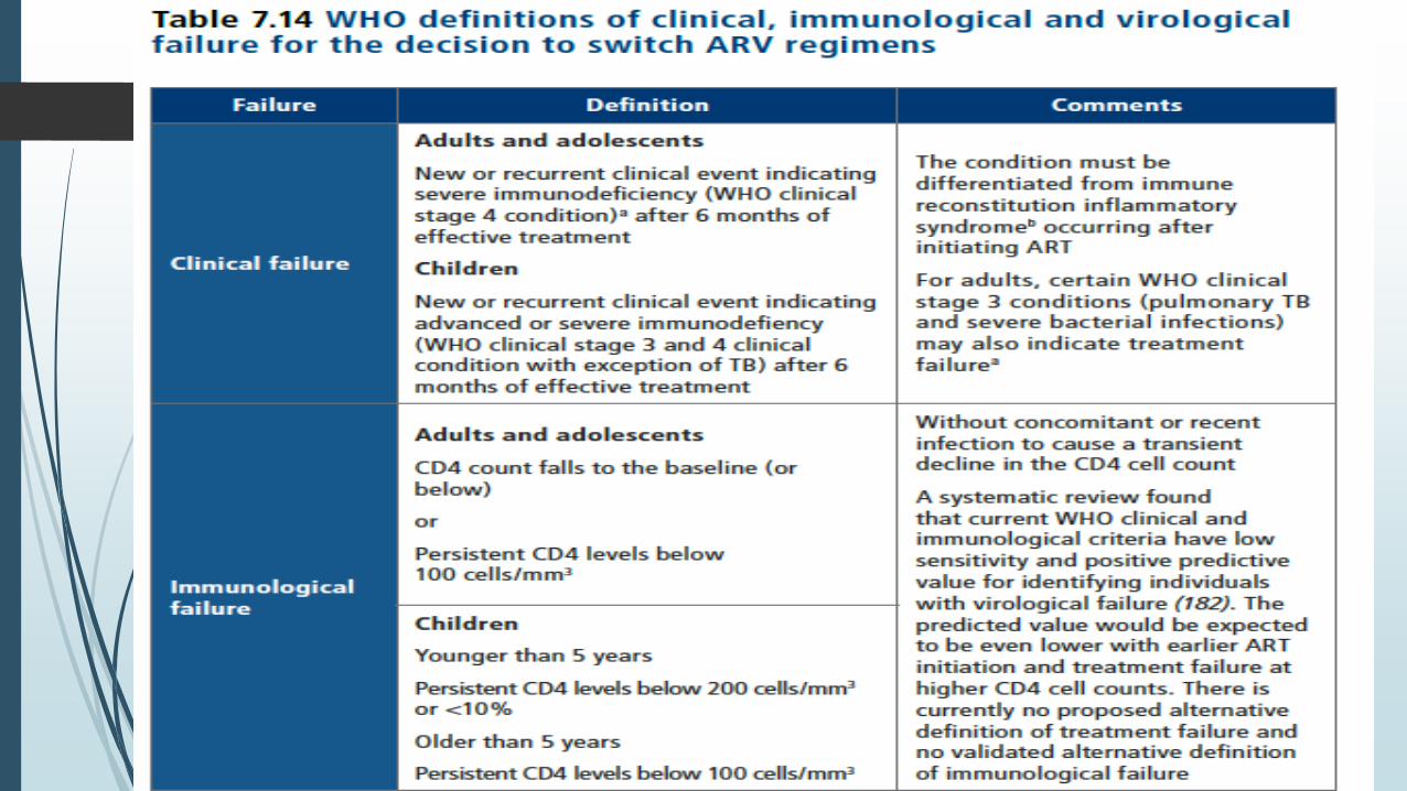
Monitoring the response to ART and the diagnosis of treatment failure
WHO
Viral load measurements = preferred monitoring approach to
diagnose and confirm ARV treatment failure
Treatment failure = persistently detectable viral load >1000 copies/ml
(2 consecutive viral load measurements within 3 mo interval, with
adherence support) after at least 6 mo of ART
If viral load is not routinely available, CD4 count and clinical
monitoring should be used to diagnose treatment failure
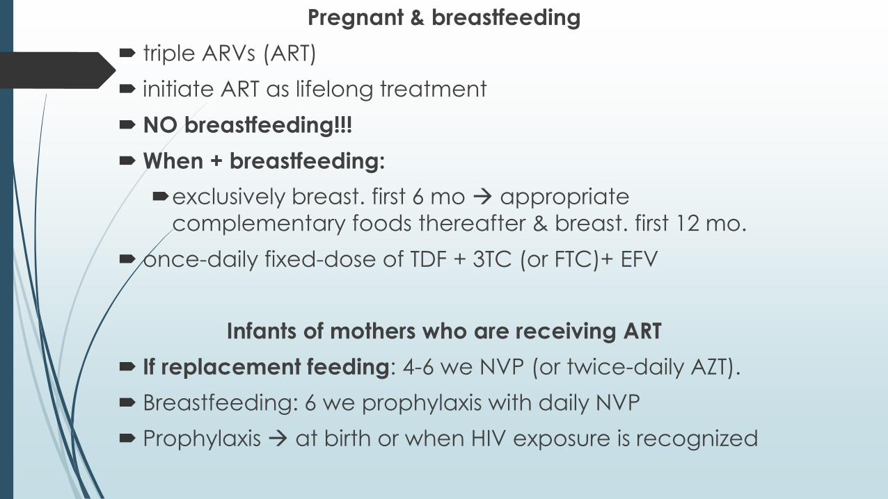
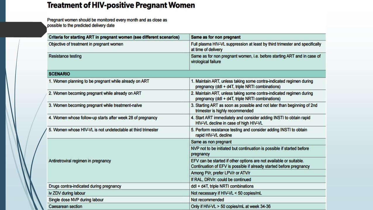
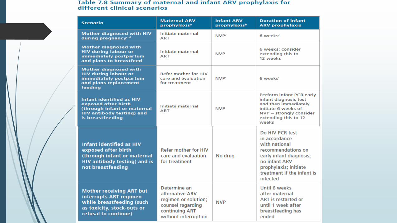
Pregnant & breastfeeding
triple ARVs (ART)
initiate ART as lifelong treatment
NO breastfeeding!!!
When + breastfeeding:
exclusively breast. first 6 mo appropriate
complementary foods thereafter & breast. first 12 mo.
once-daily fixed-dose of TDF + 3TC (or FTC)+ EFV
Infants of mothers who are receiving ART
If replacement feeding: 4-6 we NVP (or twice-daily AZT).
Breastfeeding: 6 we prophylaxis with daily NVP
Prophylaxis at birth or when HIV exposure is recognized
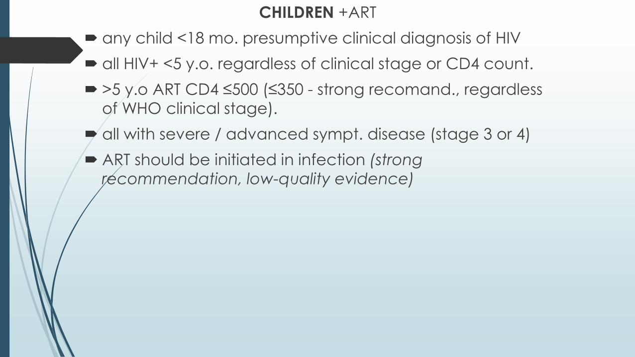
CHILDREN +ART
any child <18 mo. presumptive clinical diagnosis of HIV
all HIV+ <5 y.o. regardless of clinical stage or CD4 count.
>5 y.o ART CD4 ≤500 (≤350 - strong recomand., regardless
of WHO clinical stage).
all with severe / advanced sympt. disease (stage 3 or 4)
ART should be initiated in infection (strong
recommendation, low-quality evidence)
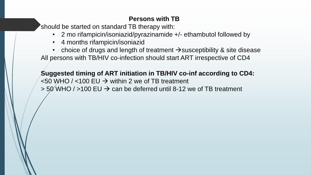
Persons with TB
should be started on standard TB therapy with:
• 2 mo rifampicin/isoniazid/pyrazinamide +/- ethambutol followed by
• 4 months rifampicin/isoniazid
• choice of drugs and length of treatment susceptibility & site disease
All persons with TB/HIV co-infection should start ART irrespective of CD4
Suggested timing of ART initiation in TB/HIV co-inf according to CD4:
<50 WHO / <100 EU within 2 we of TB treatment
> 50 WHO / >100 EU can be deferred until 8-12 we of TB treatment
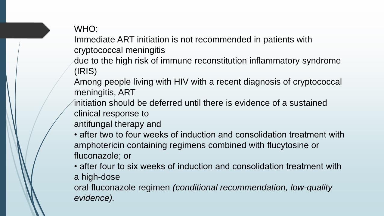
WHO:
Immediate ART initiation is not recommended in patients with
cryptococcal meningitis
due to the high risk of immune reconstitution inflammatory syndrome
(IRIS)
Among people living with HIV with a recent diagnosis of cryptococcal
meningitis, ART
initiation should be deferred until there is evidence of a sustained
clinical response to
antifungal therapy and
• after two to four weeks of induction and consolidation treatment with
amphotericin containing regimens combined with flucytosine or
fluconazole; or
• after four to six weeks of induction and consolidation treatment with
a high-dose
oral fluconazole regimen (conditional recommendation, low-quality
evidence).
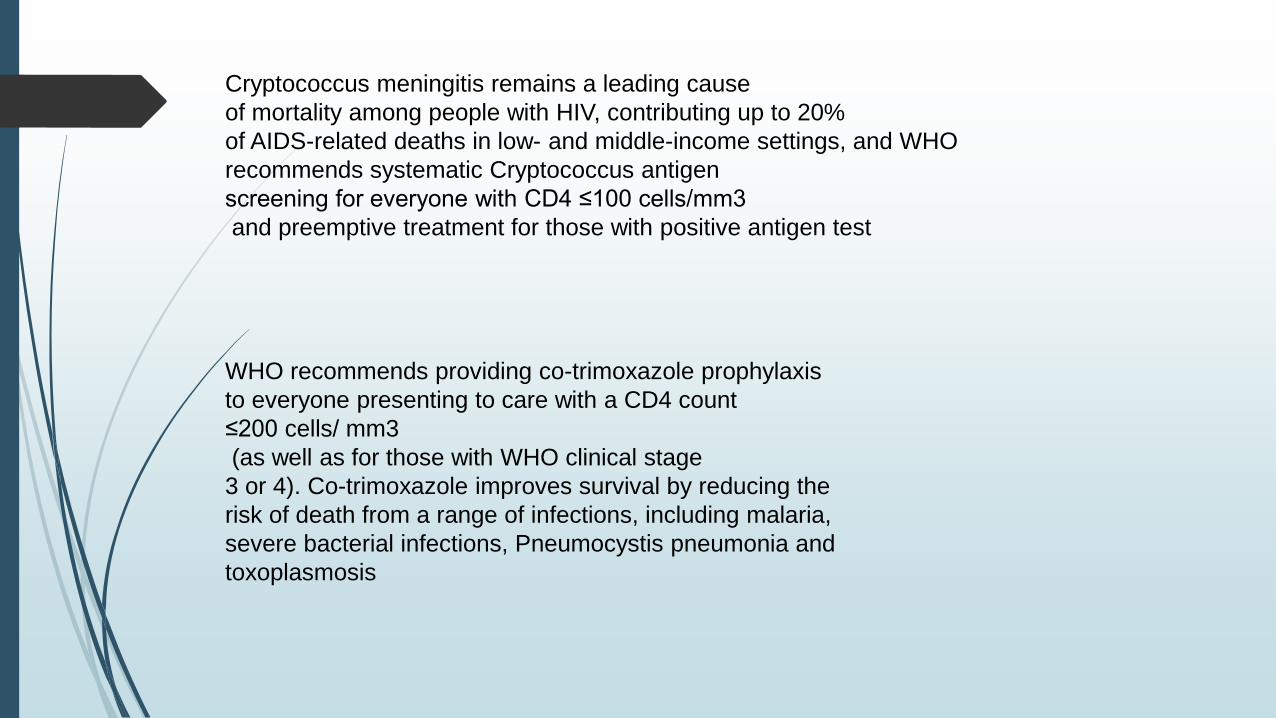
Cryptococcus meningitis remains a leading cause
of mortality among people with HIV, contributing up to 20%
of AIDS-related deaths in low- and middle-income settings, and WHO
recommends systematic Cryptococcus antigen
screening for everyone with CD4 ≤100 cells/mm3
and preemptive treatment for those with positive antigen test
WHO recommends providing co-trimoxazole prophylaxis
to everyone presenting to care with a CD4 count
≤200 cells/ mm3
(as well as for those with WHO clinical stage
3 or 4). Co-trimoxazole improves survival by reducing the
risk of death from a range of infections, including malaria,
severe bacterial infections, Pneumocystis pneumonia and
toxoplasmosis
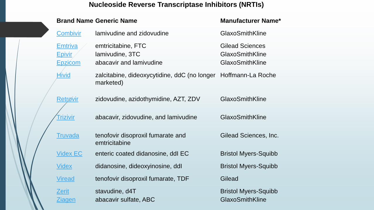
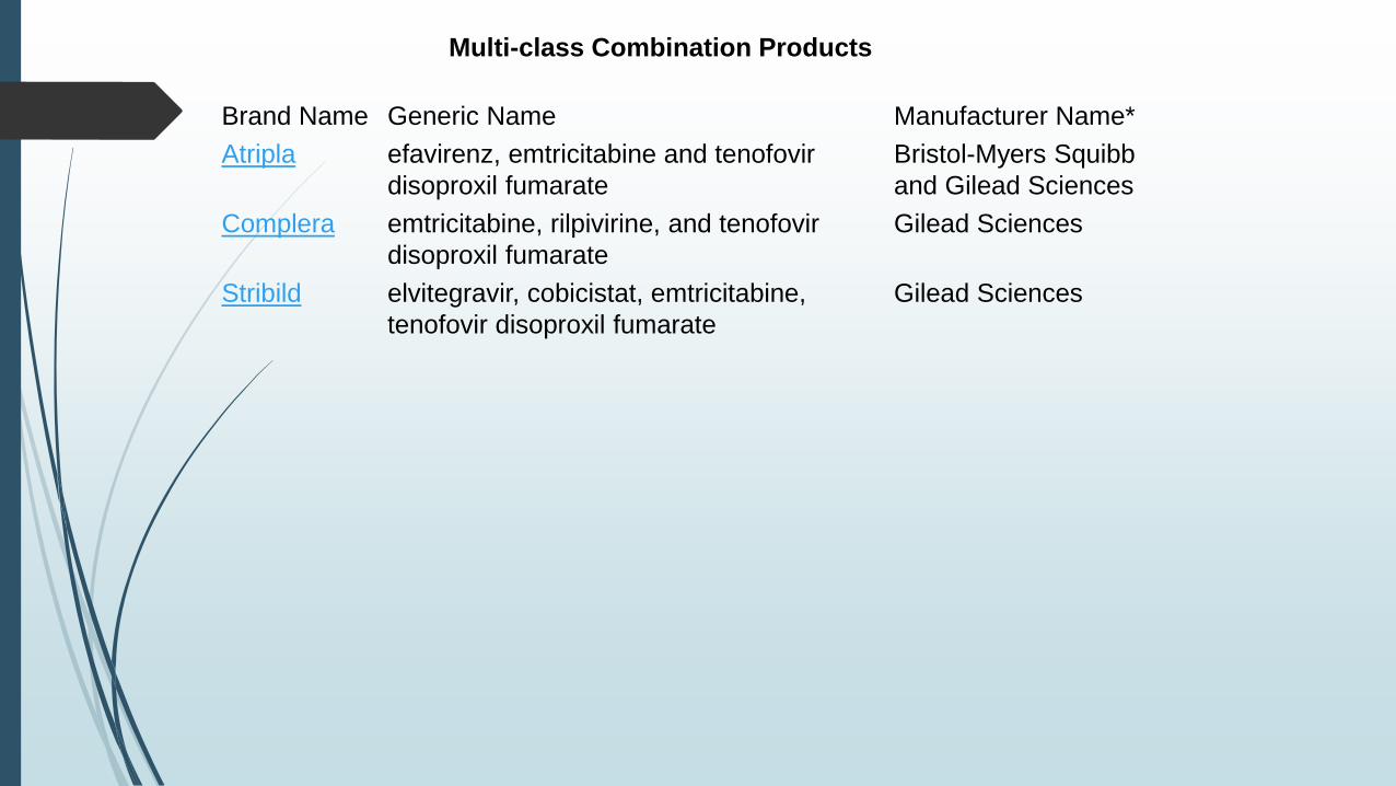
Brand Name Generic Name Manufacturer Name*
Combivir lamivudine and zidovudine GlaxoSmithKline
Emtriva emtricitabine, FTC Gilead Sciences
Epivir lamivudine, 3TC GlaxoSmithKline
Epzicom abacavir and lamivudine GlaxoSmithKline
Hivid zalcitabine, dideoxycytidine, ddC (no longer
marketed)
Hoffmann-La Roche
Retrovir zidovudine, azidothymidine, AZT, ZDV GlaxoSmithKline
Trizivir abacavir, zidovudine, and lamivudine GlaxoSmithKline
Truvada tenofovir disoproxil fumarate and
emtricitabine
Gilead Sciences, Inc.
Videx EC enteric coated didanosine, ddI EC Bristol Myers-Squibb
Videx didanosine, dideoxyinosine, ddI Bristol Myers-Squibb
Viread tenofovir disoproxil fumarate, TDF Gilead
Zerit stavudine, d4T Bristol Myers-Squibb
Ziagen abacavir sulfate, ABC GlaxoSmithKline
Nucleoside Reverse Transcriptase Inhibitors (NRTIs)
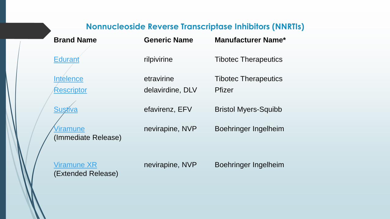
Nonnucleoside Reverse Transcriptase Inhibitors (NNRTIs)
Brand Name Generic Name Manufacturer Name*
Edurant rilpivirine Tibotec Therapeutics
Intelence etravirine Tibotec Therapeutics
Rescriptor delavirdine, DLV Pfizer
Sustiva efavirenz, EFV Bristol Myers-Squibb
Viramune
(Immediate Release)
nevirapine, NVP Boehringer Ingelheim
Viramune XR
(Extended Release)
nevirapine, NVP Boehringer Ingelheim
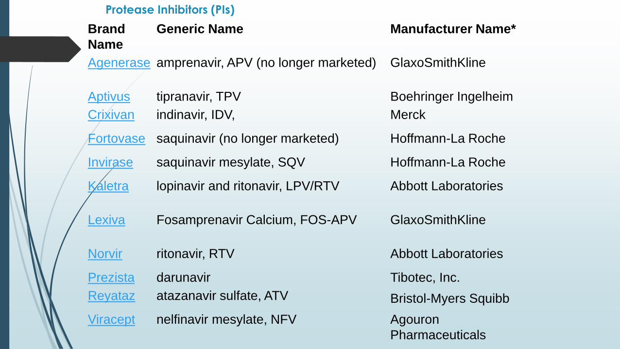
Protease Inhibitors (PIs)
Brand
Name
Generic Name Manufacturer Name*
Agenerase amprenavir, APV (no longer marketed) GlaxoSmithKline
Aptivus tipranavir, TPV Boehringer Ingelheim
Crixivan indinavir, IDV, Merck
Fortovase saquinavir (no longer marketed) Hoffmann-La Roche
Invirase saquinavir mesylate, SQV Hoffmann-La Roche
Kaletra lopinavir and ritonavir, LPV/RTV Abbott Laboratories
Lexiva Fosamprenavir Calcium, FOS-APV GlaxoSmithKline
Norvir ritonavir, RTV Abbott Laboratories
Prezista darunavir Tibotec, Inc.
Reyataz atazanavir sulfate, ATV Bristol-Myers Squibb
Viracept nelfinavir mesylate, NFV Agouron
Pharmaceuticals
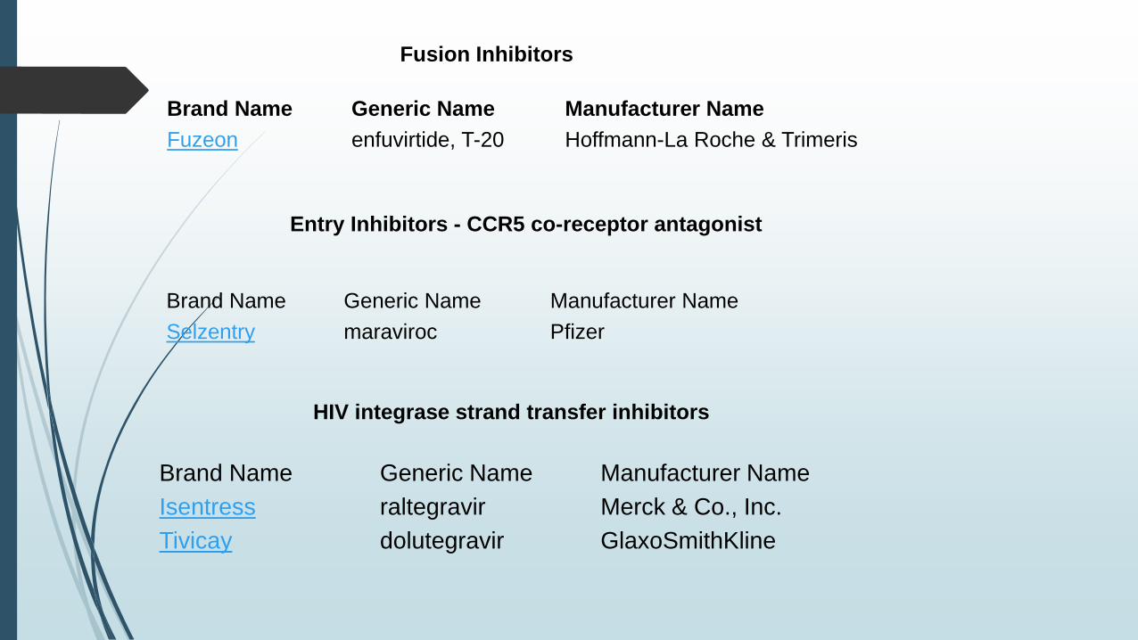
Brand Name Generic Name Manufacturer Name
Fuzeon enfuvirtide, T-20 Hoffmann-La Roche & Trimeris
Brand Name Generic Name Manufacturer Name
Selzentry maraviroc Pfizer
Brand Name Generic Name Manufacturer Name
Isentress raltegravir Merck & Co., Inc.
Tivicay dolutegravir GlaxoSmithKline
HIV integrase strand transfer inhibitors
Fusion Inhibitors
Entry Inhibitors - CCR5 co-receptor antagonist
Brand Name Generic Name Manufacturer Name*
Atripla efavirenz, emtricitabine and tenofovir
disoproxil fumarate
Bristol-Myers Squibb
and Gilead Sciences
Complera emtricitabine, rilpivirine, and tenofovir
disoproxil fumarate
Gilead Sciences
Stribild elvitegravir, cobicistat, emtricitabine,
tenofovir disoproxil fumarate
Gilead Sciences
Multi-class Combination Products