HISTOPATOLOGY OF DENTAL CARIES
27
HISTOPATOLOGY OF DENTAL CARIES GÜLBİ KE DEMİ REL, DDS,PhD
Transcript of HISTOPATOLOGY OF DENTAL CARIES

HISTOPATOLOGY OF DENTAL CARIES
GÜLBİKE DEMİREL, DDS,PhD

CLASSIFICATION OF DENTAL CARIES
Pit and Fissure Caries: This caries is usually seen in pit and fissures on occlusal surface of posterior teeth and buccal and lingual surfaces of molars and on lingual surface of maxillary anteriors
According to their Anatomical Site

CLASSIFICATION OF DENTAL CARIES
Smooth Surface Caries:This is usually seen on all smooth surface of teeth; gingival third of buccal and lingual surfaces and proximal surfaces.
According to their Anatomical Site

CLASSIFICATION OF DENTAL CARIES
Root Caries: Root caries occurs on exposed root surface
According to their Anatomical Site

CLASSIFICATION OF DENTAL CARIES
Primary Caries: A caries lesion not adjacent to an existing restoration or crown.
According to New Lesion or Recurrent Lesion
Clinical picture showing primary caries

CLASSIFICATION OF DENTAL CARIES
Recurrent Caries: A caries lesion adjacent to an existing restoration, crown, or sealant. Other term used is caries adjacent to restorations and sealants (CARS). Also referred to as recurrent caries, implying that a primary caries lesion was restored but that the lesion reoccurred.
According to New Lesion or Recurrent Lesion
Radiograph showing recurrent caries adjacent to proximal restoration

CLASSIFICATION OF DENTAL CARIES
Residual Caries: It is caries that remains in the prepared tooth surface even after placing the restoration Sometimes caries close to pulp are left intentionally so as to prevent pulp exposure.
According to New Lesion or Recurrent Lesion
Figure showing accident and intentional residual caries

CLASSIFICATION OF DENTAL CARIES
Active Carious Lesion: A caries lesion that is considered to be biologically active—that is, a lesion in which tooth demineralization is in frank activity at the time of examination.
According to Speed of Caries Progression
Inactive/Arrested Carious Lesion: A caries lesion that is considered to be biologically inactive at the time of examination—that is, in which tooth demineralization caused by caries may have happened in the past but has stopped and is currently stalled. Also referred to as arrested caries, meaning that the caries process has been arrested but that the clinical signs of the lesion itself are still present.

CLASSIFICATION OF DENTAL CARIES
Rampant Caries: Term used to describe the presence of extensive and multiple cavitated and active caries lesions in the same person. Typically used in association with “baby bottle caries,” “radiation therapy caries,” or “meth-mouth caries.” These terms refer to the etiology of the condition.
According to Speed of Caries Progression
Rampant caries in a school-age child

Caries Lesion Progression
The time for progression from a noncavitated caries lesion to a cavitated caries lesion on smooth surfaces is estimated to be 18 months (± 6 months)
Peak rates for the incidence of new lesions occur 3 years after the eruption of the tooth.
Occlusal pit-and-fissure lesions develop in less time than smooth-surface caries lesions.

Caries Lesion Progression
Poor oral hygiene and frequent exposures to sucrose-containing or acidic food can produce noncavitated initial (“white spot”) lesions (first clinical evidence of demineralization) in 3 weeks.
Radiation-induced hyposalivation (dry mouth) can lead to development of caries lesions in 3 months from the onset of the radiation.
Caries lesion progression in healthy individuals is usually slow compared with the progression in compromised persons.

Enamel Caries Lesions
On clean, dry teeth the earliest evidence of caries lesion on the smooth enamel surface of a crown is the initial lesion or a “white spot”
Facial and lingual smooth-surface caries. This patient has high caries activity with rapidly advancing caries lesions. Cariogenic biofilm extends entirely around the cervical areas of the posterior teeth. Several levels of caries involvement can be seen, including cavitation (c); initial (noncavitated) white spot lesions (i); and stained, roughened, partially remineralized initial (noncavitated) lesions (s).

Enamel Caries Lesions
These lesions usually are observed on the facial and lingual surfaces of teeth. Initial lesions are chalky white, opaque areas that are revealed only when the tooth surface is desiccated and are termed noncavitated enamel caries lesions or initial lesions.
These areas of enamel lose their translucency because of the extensive subsurface porosity caused by demineralization.
Care must be exercised in distinguishing white spots of initial caries lesions from developmental white spot hypocalcifications or other developmental defects of enamel.

Enamel Caries Lesions

Enamel Caries Lesions
The surface texture of an initial lesion is unaltered and is undetectable by tactile examination with an explorer.
A more advanced lesion develops an irregular surface that is rougher than the unaffected, normal enamel. Softened chalky enamel that can be chipped away with an explorer is a sign of active caries. However, injudicious use of an explorer tip can cause actual cavitation in a previously noncavitated area, requiring, in most cases, restorative intervention.

Enamel Caries Lesions
Enamel Caries Lesions

Enamel Caries Lesions
It has been shown experimentally and clinically that initial enamel caries lesions can remineralize whether the dentin is involved or not.
Initial, noncavitated enamel lesions retain most of the original crystalline framework of the enamel rods, and the etched crystallites serve as nucleating agents for remineralization

Enamel Caries Lesions
Calcium and phosphate ions from saliva can penetrate the enamel surface and precipitate on the highly reactive crystalline surfaces in the enamel lesion.
The presence of trace amounts of fluoride ions during this remineralization process greatly enhances the precipitation of calcium and phosphate, resulting in the remineralized enamel becoming more resistant to subsequent caries activity because of the incorporation of more acid-resistant fluorapatite

Enamel Caries Lesions

Enamel Caries Lesions

Enamel Caries Lesions
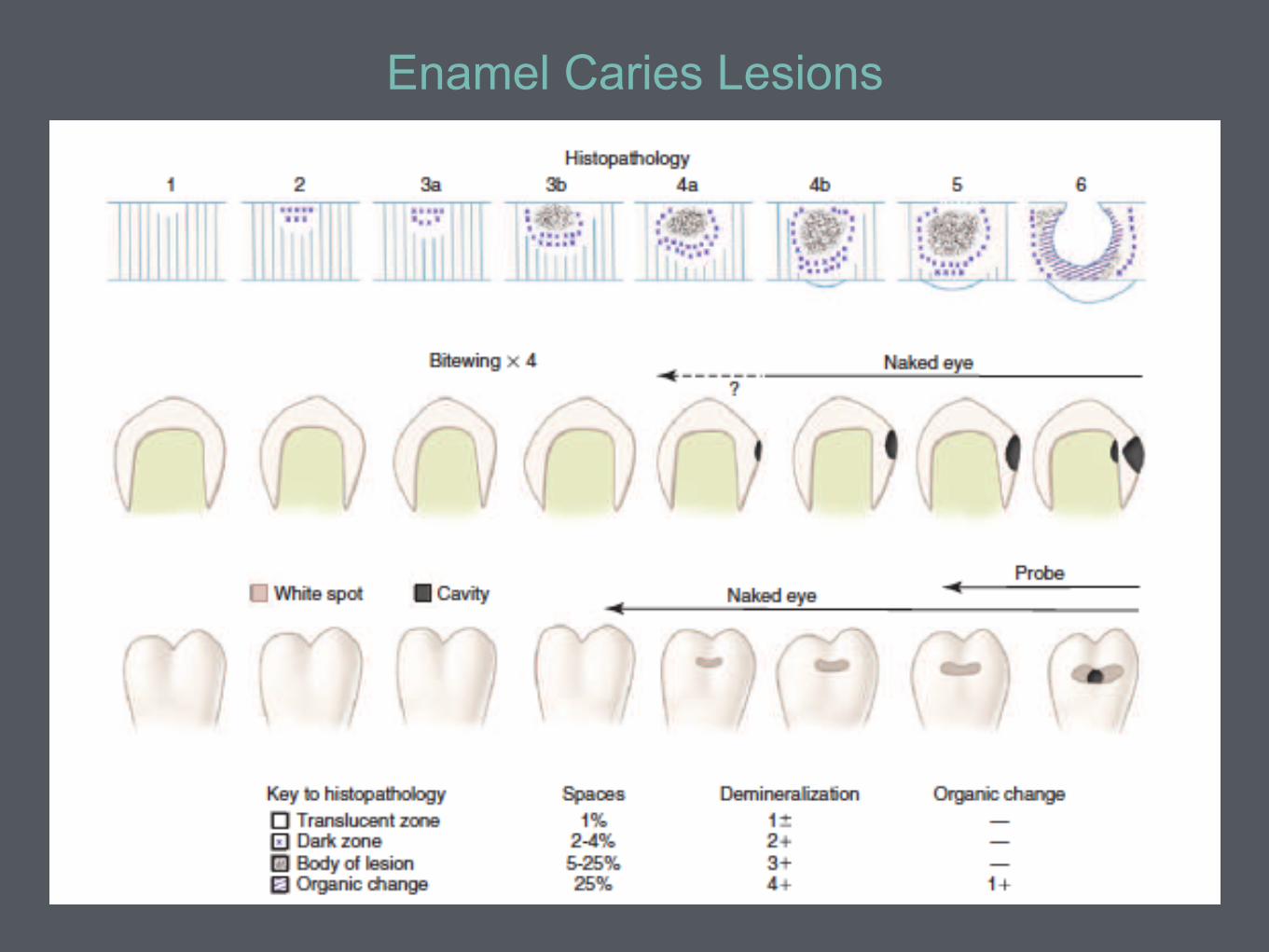
Different zones are seen before complete disintegration of enamel. Early enamel lesion seen under polarized light reveals four distinct zones of mineralization.These zones begin from the dentinal side of the lesion.
Zones in Enamel Caries

Enamel Caries LesionsZones in Enamel Caries
Zone 1: Translucent zone
– Represent the advancing front of the lesion
– Ten times more porous than sound enamel
– Not always present.

Enamel Caries LesionsZones in Enamel Caries
• Zone 2: Dark zone
– It lies adjacent and superficial to the translucent zone
– Called dark zone because it does not transmit polarized light
– Formed due to demineralization.

Enamel Caries LesionsZones in Enamel Caries
Zone 3: Body of the lesion
– Largest portion of the incipient caries
– Found between the surface and the dark zone
– It is the area of greatest demineralization making it
more porous.

Enamel Caries LesionsZones in Enamel Caries
• Zone 4: Surface zone
– This zone is affected area from caries
– Greater resistance probably due to greater degree of mineralization and greater fluoride concentration
– It radiopacity is comparable to adjacent enamel.

Enamel Caries LesionsZones in Enamel Caries
Translucent zone
Dark zone
Body of the lesion
Surface zone
Sound enamel