Histopathology and the diagnosis of lysosomal storage ...
43
Histopathology and the diagnosis of lysosomal storage disorders – part 1 Glenn Anderson Clinical Electron Microscopist Great Ormond Street Hospital for Children London, UK Paediatric EM training day, Southampton University, 4 th October 2013
Transcript of Histopathology and the diagnosis of lysosomal storage ...

Histopathology and thediagnosis of lysosomalstorage disorders – part 1
Glenn AndersonClinical Electron MicroscopistGreat Ormond Street Hospital for ChildrenLondon, UK
Paediatric EM training day, Southampton University, 4th October 2013
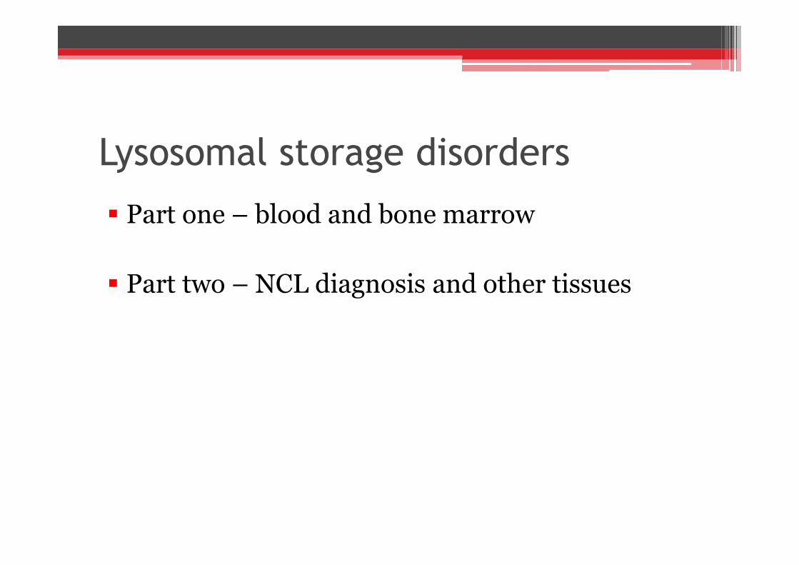
Lysosomal storage disorders
Part one – blood and bone marrow
Part two – NCL diagnosis and other tissues
Part one – blood and bone marrow
Part two – NCL diagnosis and other tissues

Lysosomal storage disorders
Group of 50 diseases
Rare with a frequency of about 1:8000 live births
Inherited in an autosomal-recessive fashion,exceptions X-linked
Accumulation of waste products in lysosomes dueto missing or reduced enzyme
Group of 50 diseases
Rare with a frequency of about 1:8000 live births
Inherited in an autosomal-recessive fashion,exceptions X-linked
Accumulation of waste products in lysosomes dueto missing or reduced enzyme

Clinical features - variable
Appear normal at birth – progressive
Failure to thrive
Dysmorphic features
Neurological symptoms – seizures, behaviour
Organomegaly
Appear normal at birth – progressive
Failure to thrive
Dysmorphic features
Neurological symptoms – seizures, behaviour
Organomegaly

Clinical features
Visual problems – cherry red spot
Skeletal dysplasia
Muscle weakness including cardiomyopathy
Loss of skills – speech and learning
Many disorders can present in different forms –infantile, juvenile and adult
Hydrops fetalis
Visual problems – cherry red spot
Skeletal dysplasia
Muscle weakness including cardiomyopathy
Loss of skills – speech and learning
Many disorders can present in different forms –infantile, juvenile and adult
Hydrops fetalis

Clinical PresentationClinical Presentation
Sample to specialised laboratorySample to specialised laboratory
Preliminary screenurine or blood
Preliminary screenurine or blood
Algorithm for Diagnosis
Preliminary screenurine or blood
Preliminary screenurine or blood
Defect in lysosomal enzyme ornon-enzymatic protein
Defect in lysosomal enzyme ornon-enzymatic protein
DiagnosisDiagnosis Prenatal diagnosisPrenatal diagnosisGenetic counsellingGenetic counselling
Treatment/managementTreatment/management

LSD - classification Sphingolipidoses – GM1 & GM2 gangliosidosis, Fabrys, MLD,
Gaucher, Krabbe, Niemann-Pick A & B
Mucopolysaccharidoses – Hurler, Hunter, Sanfilippo, Morquio
Glycoproteinoses (Oligos) – Mannosidosis, Fucosidosis, Sialidosis
Other enzyme defects – Wolman, CESD, GSD II
Neuronal ceroid lipofuscinoses – CLN 1-14
Disorders of lysosome-related organelle – Chediak-Higashi,Griscelli, Hermansky-Pudlak
Lysosomal membrane defects – Cystinosis, Sialic acid storagedisease, Niemann-Pick C
Sphingolipidoses – GM1 & GM2 gangliosidosis, Fabrys, MLD,Gaucher, Krabbe, Niemann-Pick A & B
Mucopolysaccharidoses – Hurler, Hunter, Sanfilippo, Morquio
Glycoproteinoses (Oligos) – Mannosidosis, Fucosidosis, Sialidosis
Other enzyme defects – Wolman, CESD, GSD II
Neuronal ceroid lipofuscinoses – CLN 1-14
Disorders of lysosome-related organelle – Chediak-Higashi,Griscelli, Hermansky-Pudlak
Lysosomal membrane defects – Cystinosis, Sialic acid storagedisease, Niemann-Pick C
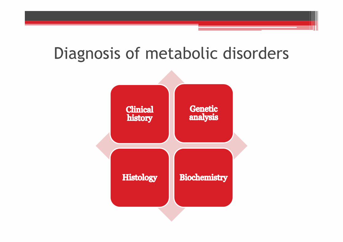
Diagnosis of metabolic disorders

Morphological examination
Light microscopyroutine stains, lipid & enzyme histochemistryprocedures
Transmission electron microscopy
Light microscopyroutine stains, lipid & enzyme histochemistryprocedures
Transmission electron microscopy
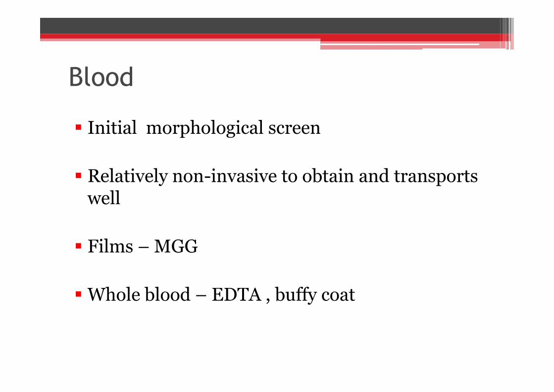
Blood
Initial morphological screen
Relatively non-invasive to obtain and transportswell
Films – MGG
Whole blood – EDTA , buffy coat
Initial morphological screen
Relatively non-invasive to obtain and transportswell
Films – MGG
Whole blood – EDTA , buffy coat

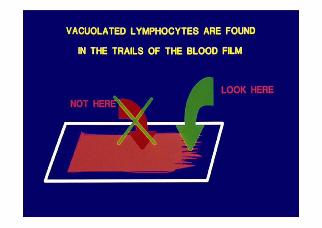
Blood – film assessment
Vacuolated lymphocytes(enlarged lyosomes)
- small vacuoles- large vacuoles
Other white cells and platelets
Vacuolated lymphocytes(enlarged lyosomes)
- small vacuoles- large vacuoles
Other white cells and platelets
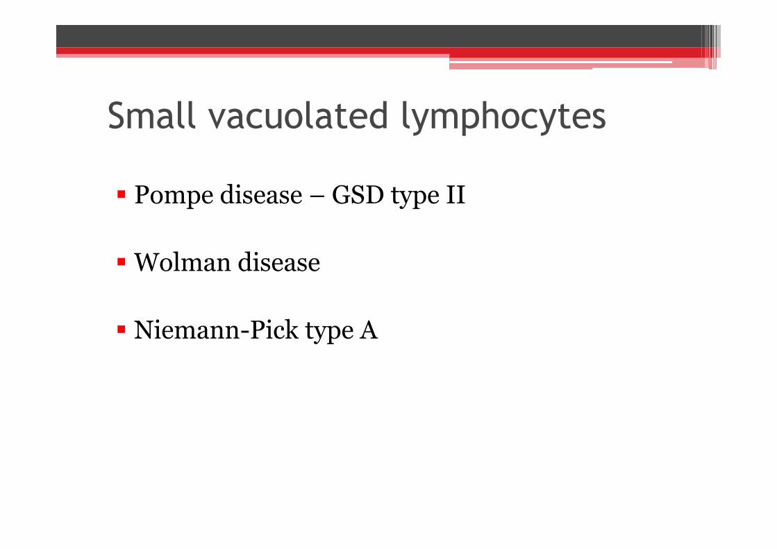
Small vacuolated lymphocytes
Pompe disease – GSD type II
Wolman disease
Niemann-Pick type A
Pompe disease – GSD type II
Wolman disease
Niemann-Pick type A
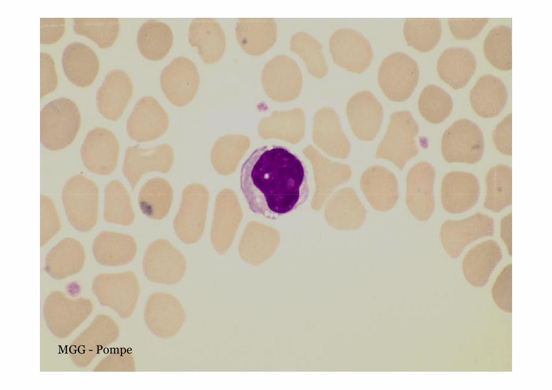
MGG - Pompe

Celloidinised PAS - Pompe

H&E - Pompe

Pompe

OROWolman
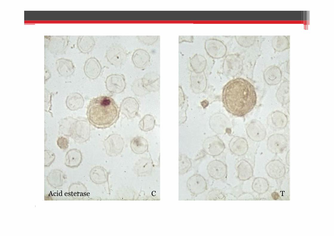
Acid esterase C T

Small bowel - ORO

Liver

BMA - ORO
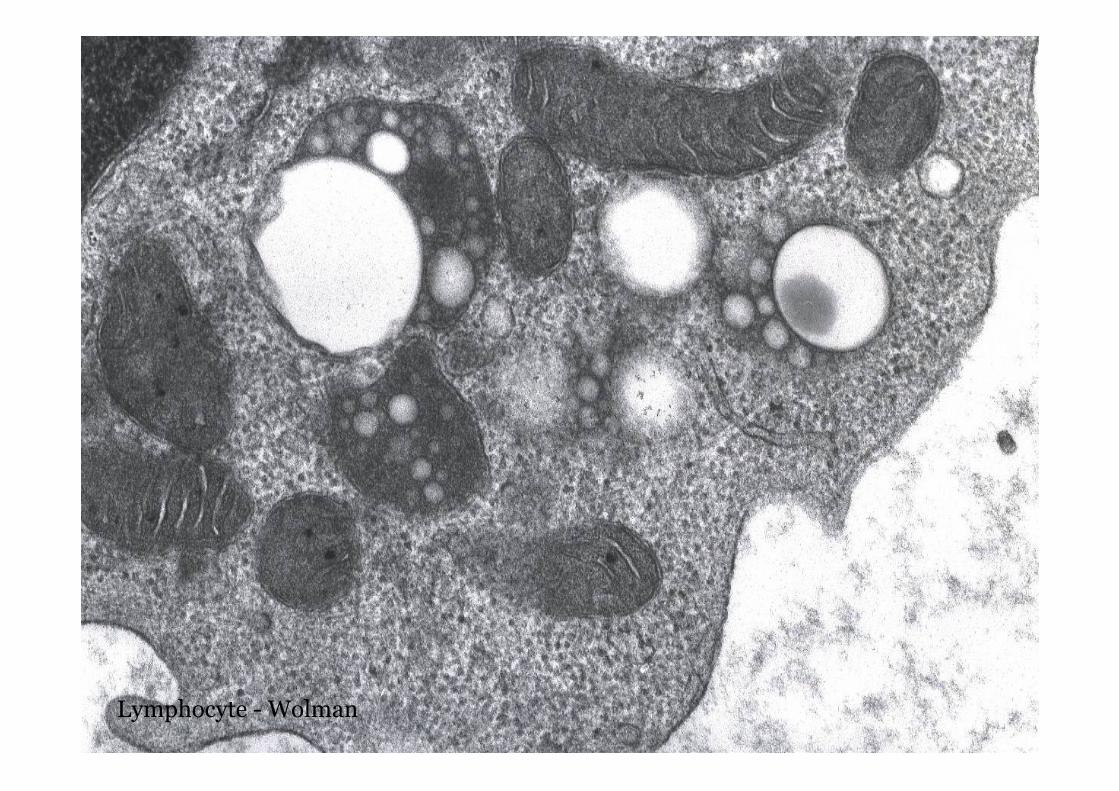
Lymphocyte - Wolman

Large vacuolated lymphocytes
GM1 gangliosidosis type I
Juvenile Batten disease
Mannosidosis
Sialidosis
I cell disease
Sialic acid and Salla disease
GM1 gangliosidosis type I
Juvenile Batten disease
Mannosidosis
Sialidosis
I cell disease
Sialic acid and Salla disease

MGG - GM1 gangliosidosis
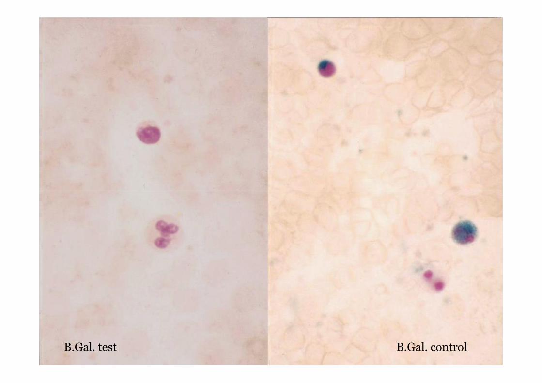
B.Gal. controlB.Gal. test

GM1 EM

Lymphocytes - other inclusions
Cytoplasmic inclusions – Chediak-Higashi, Gassercells MPS
Metachromatic inclusions – Sanfillipo MPS III
Cytoplasmic inclusions – Chediak-Higashi, Gassercells MPS
Metachromatic inclusions – Sanfillipo MPS III

White cell changes - Neutrophils
Vacuolation – nonspecific, neutral lipid storage
Toxic granulation – inflammatory states ‘sepsis’
Alder granulation – MPS
Atypical granules – Chediak-Higashi
Vacuolation – nonspecific, neutral lipid storage
Toxic granulation – inflammatory states ‘sepsis’
Alder granulation – MPS
Atypical granules – Chediak-Higashi

Neutrophil images
Neutral lipid SD

White cell changes - Eosinophils
Atypical granulation – GM1, Sialic acid storage

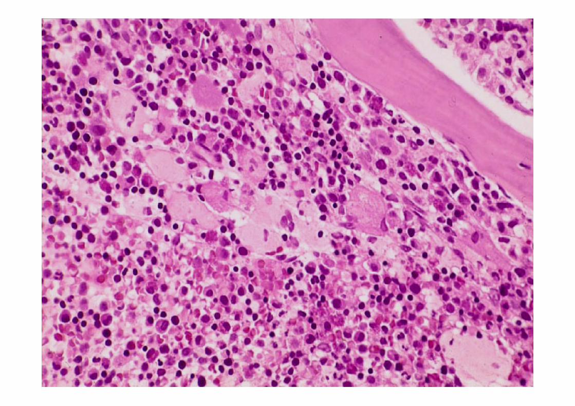
Bone marrow
Bone marrow aspirates- preferred- enzymes, lipids preserved- histochemical techniques, EM
Bone marrow trephines- more material- routine stains, immunocytochemistry,EM
Bone marrow aspirates- preferred- enzymes, lipids preserved- histochemical techniques, EM
Bone marrow trephines- more material- routine stains, immunocytochemistry,EM

Storage cells
Foamy cells- Niemann-Pick, Mannosidosis, Wolman
Fibrillary cells- Gaucher, GM1 gangliosidosis type II
Foamy cells- Niemann-Pick, Mannosidosis, Wolman
Fibrillary cells- Gaucher, GM1 gangliosidosis type II

Niemann-Pick C

Sea blue histiocytes
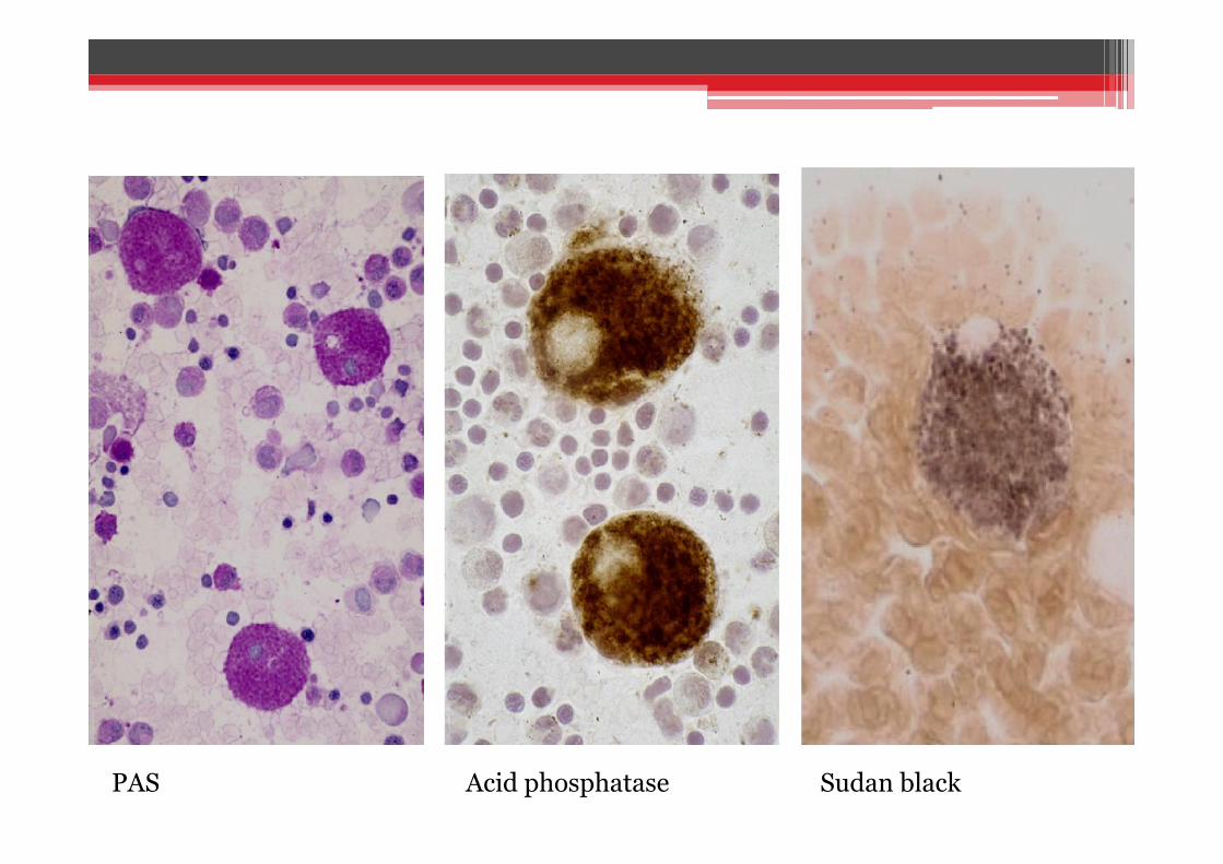
PAS Acid phosphatase Sudan black

Niemann-Pick

MGG - Gaucher

MGG – Gaucher, spleen

Gaucher

Gaucher
Part 2 to follow.