Histopathologic investigation of the dimensions of the cochlear nerve canal in normal temporal bones
4
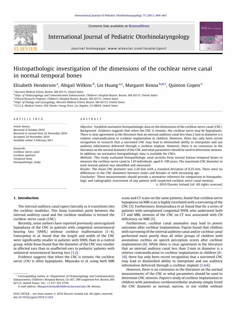
Histopathologic investigation of the dimensions of the cochlear nerve canal in normal temporal bones Elisabeth Henderson a , Abigail Wilkins b , Lin Huang a,c , Margaret Kenna b,d, *, Quinton Gopen e a Harvard Medical School, Boston, MA 02115, United States b Dept. of Otolaryngology and Communication Enhancement, Children’s Hospital Boston, Boston, MA 02115, United States c Clinical Research Program, Children’s Hospital Boston, Boston, MA 02115, United States d Dept. of Otology and Laryngology, Harvard Medical School, Boston, MA 02115, United States e U.C.L.A. Medical Center, 650 Charles Young Drive, Los Angeles, CA 90024, United States 1. Introduction The internal auditory canal tapers laterally as it transitions into the cochlear modiolus. This bony transition point between the internal auditory canal and the cochlear modiolus is termed the cochlear nerve canal (CNC). Recently, some authors have reported previously unrecognized hypoplasia of the CNC in patients with congenital sensorineural hearing loss (SNHL) without cochlear malformation [1–4]. Fatterpekar et al. found that the length and width of the CNC were significantly smaller in patients with SNHL than in a control group, while Kono found that the diameter of the CNC was smaller in affected ears than in unaffected ears in pediatric patients with unilateral sensorineural hearing loss [1,2]. Evidence suggests that when the CNC is stenotic the cochlear nerve (CN) is often hypoplastic. Miyasaka et al. using both MRI scans and CT scans on the same patients, found that cochlear nerve hypoplasia on MRI scan is highly correlated with a narrowing of the CNC [3]. Furthermore, Komatsubara et al. found that for a series of patients with unexplained congenital SNHL who underwent both CT and MRI, stenosis of the CNC on CT was associated with CN deficiency on MRI [5]. Furthermore, cochlear canal anomalies may lead to poorer outcomes after cochlear implantation. Papsin found that children with narrowing of the internal auditory canal and/or cochlear canal performed more poorly than all other groups of children with anomalous cochlea on speech perception scores after cochlear implantation [6]. While there is clear agreement in the literature that an internal auditory canal less than 2 mm in diameter is a relative contraindication to cochlear implantation in children [6– 10], there has only been recent recognition that a narrowed CNC may lead to diminished ability to interpolate and use auditory information delivered through a cochlear implant [1,4,6]. However, there is no consensus in the literature on the normal measurements of the CNC or what parameters should be used to determine CNC stenosis. Papsin’s study of cochlear implantation in children with anomalous cochleovestibular anatomy simply listed the CNC diameter as normal, narrow, or not visible without International Journal of Pediatric Otorhinolaryngology 75 (2011) 464–467 ARTICLE INFO Article history: Received 4 October 2010 Received in revised form 22 November 2010 Accepted 24 November 2010 Available online 5 February 2011 Keywords: Cochlear nerve canal Cochlear aperture Temporal bone Histopathologic study ABSTRACT Objective: Establish normative histopathologic data on the dimensions of the cochlear nerve canal (CNC). Background: Evidence suggests that when the CNC is stenotic, the cochlear nerve may be hypoplastic. There is clear agreement in the literature that an internal auditory canal less than 2 mm in diameter is a relative contraindication to cochlear implantation in children. However, there has only been recent recognition in research that a narrowed CNC may lead to diminished ability to interpolate and use auditory information delivered through a cochlear implant. However, there is no consensus in the literature on the normal diameter of the CNC and what parameters should be used to determine stenosis. In addition, no normative histopathologic data is available for CNCs. Methods: This study evaluated histopathologic axial sections from normal human temporal bones to measure the cochlear nerve canal in 110 individuals, aged 0–100 years. The maximum CNC diameter in each normal patient was identified and measured. Results: The mean CNC diameter was 2.26 mm with a standard deviation of 0.25 mm. There were no differences in the CNC diameters between males and females or with increasing age. Conclusion: These measurements should provide a normative reference for comparison in histopatho- logic and radiographic assessment of any patient with suspected cochlear nerve canal stenosis. ß 2010 Elsevier Ireland Ltd. All rights reserved. * Corresponding author at: Department of Otolaryngology and Communication Enhancement, Children’s Hospital Boston, LO-367, 300 Longwood Ave, Boston, MA 02115, United States. Tel.: +1 617 355 5759. E-mail address: [email protected] (M. Kenna). Contents lists available at ScienceDirect International Journal of Pediatric Otorhinolaryngology journal homepage: www.elsevier.com/locate/ijporl 0165-5876/$ – see front matter ß 2010 Elsevier Ireland Ltd. All rights reserved. doi:10.1016/j.ijporl.2010.11.024
-
Upload
elisabeth-henderson -
Category
Documents
-
view
212 -
download
0
Transcript of Histopathologic investigation of the dimensions of the cochlear nerve canal in normal temporal bones
International Journal of Pediatric Otorhinolaryngology 75 (2011) 464–467
Histopathologic investigation of the dimensions of the cochlear nerve canalin normal temporal bones
Elisabeth Henderson a, Abigail Wilkins b, Lin Huang a,c, Margaret Kenna b,d,*, Quinton Gopen e
a Harvard Medical School, Boston, MA 02115, United Statesb Dept. of Otolaryngology and Communication Enhancement, Children’s Hospital Boston, Boston, MA 02115, United Statesc Clinical Research Program, Children’s Hospital Boston, Boston, MA 02115, United Statesd Dept. of Otology and Laryngology, Harvard Medical School, Boston, MA 02115, United Statese U.C.L.A. Medical Center, 650 Charles Young Drive, Los Angeles, CA 90024, United States
A R T I C L E I N F O
Article history:
Received 4 October 2010
Received in revised form 22 November 2010
Accepted 24 November 2010
Available online 5 February 2011
Keywords:
Cochlear nerve canal
Cochlear aperture
Temporal bone
Histopathologic study
A B S T R A C T
Objective: Establish normative histopathologic data on the dimensions of the cochlear nerve canal (CNC).
Background: Evidence suggests that when the CNC is stenotic, the cochlear nerve may be hypoplastic.
There is clear agreement in the literature that an internal auditory canal less than 2 mm in diameter is a
relative contraindication to cochlear implantation in children. However, there has only been recent
recognition in research that a narrowed CNC may lead to diminished ability to interpolate and use
auditory information delivered through a cochlear implant. However, there is no consensus in the
literature on the normal diameter of the CNC and what parameters should be used to determine stenosis.
In addition, no normative histopathologic data is available for CNCs.
Methods: This study evaluated histopathologic axial sections from normal human temporal bones to
measure the cochlear nerve canal in 110 individuals, aged 0–100 years. The maximum CNC diameter in
each normal patient was identified and measured.
Results: The mean CNC diameter was 2.26 mm with a standard deviation of 0.25 mm. There were no
differences in the CNC diameters between males and females or with increasing age.
Conclusion: These measurements should provide a normative reference for comparison in histopatho-
logic and radiographic assessment of any patient with suspected cochlear nerve canal stenosis.
� 2010 Elsevier Ireland Ltd. All rights reserved.
Contents lists available at ScienceDirect
International Journal of Pediatric Otorhinolaryngology
journa l homepage: www.e lsev ier .com/ locate / i jpor l
1. Introduction
The internal auditory canal tapers laterally as it transitions intothe cochlear modiolus. This bony transition point between theinternal auditory canal and the cochlear modiolus is termed thecochlear nerve canal (CNC).
Recently, some authors have reported previously unrecognizedhypoplasia of the CNC in patients with congenital sensorineuralhearing loss (SNHL) without cochlear malformation [1–4].Fatterpekar et al. found that the length and width of the CNCwere significantly smaller in patients with SNHL than in a controlgroup, while Kono found that the diameter of the CNC was smallerin affected ears than in unaffected ears in pediatric patients withunilateral sensorineural hearing loss [1,2].
Evidence suggests that when the CNC is stenotic the cochlearnerve (CN) is often hypoplastic. Miyasaka et al. using both MRI
* Corresponding author at: Department of Otolaryngology and Communication
Enhancement, Children’s Hospital Boston, LO-367, 300 Longwood Ave, Boston, MA
02115, United States. Tel.: +1 617 355 5759.
E-mail address: [email protected] (M. Kenna).
0165-5876/$ – see front matter � 2010 Elsevier Ireland Ltd. All rights reserved.
doi:10.1016/j.ijporl.2010.11.024
scans and CT scans on the same patients, found that cochlear nervehypoplasia on MRI scan is highly correlated with a narrowing of theCNC [3]. Furthermore, Komatsubara et al. found that for a series ofpatients with unexplained congenital SNHL who underwent bothCT and MRI, stenosis of the CNC on CT was associated with CNdeficiency on MRI [5].
Furthermore, cochlear canal anomalies may lead to pooreroutcomes after cochlear implantation. Papsin found that childrenwith narrowing of the internal auditory canal and/or cochlear canalperformed more poorly than all other groups of children withanomalous cochlea on speech perception scores after cochlearimplantation [6]. While there is clear agreement in the literaturethat an internal auditory canal less than 2 mm in diameter is arelative contraindication to cochlear implantation in children [6–10], there has only been recent recognition that a narrowed CNCmay lead to diminished ability to interpolate and use auditoryinformation delivered through a cochlear implant [1,4,6].
However, there is no consensus in the literature on the normalmeasurements of the CNC or what parameters should be used todetermine CNC stenosis. Papsin’s study of cochlear implantation inchildren with anomalous cochleovestibular anatomy simply listedthe CNC diameter as normal, narrow, or not visible without

[()TD$FIG]
Fig. 1. Slides for Patient A at low power (top image) and high power (bottom image)
with cochlear nerve canal aperture measurement in millimeters.
[()TD$FIG]
Fig. 2. Histogram of cochlear nerve canal diameter with a normal density curve.
E. Henderson et al. / International Journal of Pediatric Otorhinolaryngology 75 (2011) 464–467 465
defining the difference between normal and narrow [6]. Fatterpe-kar et al. used CTs from patients evaluated with inflammatory andneoplastic disease to measure the dimensions of the bony canalslocated at the fundus of the internal auditory canal [1,11].Stjernholm used casts from temporal bone specimens and CTsfrom patients evaluated for cholesteatoma or chronic otitis mediato measure the mean dimensions of the CNC but arrived at a meancanal width that was smaller than that reported by Fatterpekaret al. [1,11,12]. To date, there are no histopathologic studiesmeasuring the diameter of the cochlear nerve canal in normativepatients.
Establishing the normal diameter of the cochlear nerve canal isan important step that must be accomplished before the effect ofCNC stenosis on hearing can be assessed. The purpose of this studywas to measure the CNC in normal human temporal bones acrossboth genders and a wide age range using standard histopathologictechniques. Normal temporal bones from the newborn periodthrough 100 years of age were measured to determine the averagesize of the cochlear nerve canal and to determine whether thecochlear nerve canal changes with age. Although the anatomy inthis area is complex, horizontal sections were selected for allmeasurements as horizontal sections are most similar to standardaxial sections obtained on CT scans.
2. Methods
This study was reviewed and approved by the MassachusettsEye and Ear Infirmary Institutional Review Board.
Selected human temporal bones from the Massachusetts Eyeand Ear Infirmary Otopathology Laboratory, part of the HumanTemporal Bone Consortium for Research Resource Enhancement,were used for this study. They were harvested at autopsy by use ofthe temporal bone plug technique. The specimens were fixed informalin, decalcified in ethylenediamine tetra-acetic acid, embed-ded in celloidin or polyester wax and serially sectioned in the axialplane at 20 mm for celloidin and 10 mm for polyester wax.Approximately every 10th section was stained with hematoxylinand eosin and mounted on slides. Slides were examined byprojection light microscopy and measurements in millimeterswere scaled using a 2 mm slide.
To determine the normal size of the cochlear nerve canals,normal temporal bones from 10 patients aged 0–1 year and 10patients in each 10 year increment from ages 1 to 100 wereselected for a total of 110 temporal bones from 110 patients, 62(56.4%) men and 48 (43.6%) women. Temporal bones wereincluded if the bony labyrinth had been coded by an expertpathologist as being normal at the time of inclusion in the temporalbone bank. Exclusion criteria for temporal bones included cochlearartifact caused by removal damage, cases of prenatal birth,diseases of the bone (such as Paget’s disease), clinical history oftemporal bone trauma or surgery on the same side as thespecimen, or clinical diagnosis of syndromes associated withabnormal temporal bone formation or hearing loss. Specimenswith a history of membranous abnormalities, acquired otologicdisease, acute meningitis, or ototoxic drug use were includedunder the assumption that these conditions would not change thebony anatomy.
Measurements were made by selecting the mid-cochlear slidein the series and then a minimum of 2 sections before and after themid-cochlear section in order to find the maximum diameter. TheCNC is continuous with the axis of the modiolus and it is oriented inthe anterolateral direction with varying angles between it and theinternal auditory canal from which it opens [12]. In order to ensureconstant measurements of the CNC between individuals, the CNCdiameter was measured for each slide at the area of bony contourchange where the CNC opened off of the internal auditory canal
(see Fig. 1). Slides before and after the mid-cochlear slide in theseries were examined and measured until the maximummeasurement of the cochlear nerve canal was found. The numberof sections evaluated for each patient varied due to variations inthe angle of the extraction and sectioning during the preparation ofthe temporal bones.
3. Results
The mean cochlear nerve canal diameter was 2.26 mm with astandard deviation of 0.25 mm (maximum was 2.81 mm, mini-mum was 1.75 mm). The histogram (Fig. 2) showed that the

Table 1Mean and standard deviation of cochlear nerve canal diameter by age group.
Age range Mean CNC diameter (mm) Standard deviation
0–1 2.29 0.21
1–10 2.36 0.23
11–20 2.41 0.37
21–30 2.16 0.28
31–40 2.23 0.19
41–50 2.34 0.27
51–60 2.22 0.22
61–70 2.17 0.14
71–80 2.24 0.23
81–90 2.20 0.26
91–100 2.25 0.28
E. Henderson et al. / International Journal of Pediatric Otorhinolaryngology 75 (2011) 464–467466
cochlear nerve canal diameter roughly followed a normaldistribution. The Shapiro–Wilk test had a p-value 0.25, whichsupported the normality hypothesis. The maximum CNC diameterdid not show any significant difference between male and femalewith means 2.29 mm (std. 0.24 mm) vs. 2.22 mm (std. 0.25 mm)(p-value from two sample t-test was 0.12). There was no significantchange in maximum CNC diameter with increasing age (slope of�0.0013, p-value = 0.098; Pearson’s correlation coefficient= �0.16, p-value = 0.098). Table 1 provides the means and standarddeviations for the different age groups.
4. Discussion
CNC stenosis is a recently described anatomical finding thatmay be associated with sensorineural hearing loss. A similarcondition, internal auditory canal stenosis, has been wellcorrelated with sensorineural hearing loss in both unilateral andbilateral internal auditory canal stenosis cases [7–9,13–16]. Duringembryologic development, a layer of mesoderm surrounds thevestibulocochlear complex. This mesoderm layer becomes carti-laginous and ultimately transforms into bone as the internalauditory canal. Some investigators feel that the internal auditorycanal develops with a smaller caliber when there is a problem withembryogenesis of the vestibulocochlear complex. Other investi-gators have theorized that the small caliber internal auditory canalmay consequently cause dysfunction of the auditory or vestibularnerves from compressive damage [16]. In either case, there is aclear association between the radiographic findings of a smallinternal auditory canal and documented sensorineural hearingloss, both unilaterally and bilaterally.
Both theories can be extrapolated to the etiopathogenesis ofCNC stenosis. If absence of the cochlear nerve results in asmaller caliber of the internal auditory canal during develop-ment, then certainly a narrowing of the end point of the internalauditory canal may also be associated with some auditory nerveanomalies. If a small internal auditory canal can cause damageor dysfunction to the vestibulocochlear complex, a narrowaperture where the cochlear nerve leaves the internal auditorycanal and enters the cochlea could easily cause analogous orunique auditory problems.
In the current study, the mean diameter of the cochlear nervecanal was 2.26 mm with a standard deviation of 0.25 mm. Theminimum diameter of any cochlear nerve canal seen in this seriesof 110 patients was 1.75 mm, and the maximum diameter was2.81 mm. Our histopathologic results are similar to the results ofStjernholm and Muren, who found a mean diameter of 2.58 mm(SD 0.31, range 2.1–3.6) on inspection of 117 silicone rubber castsof temporal bones [12]. Stjernholm and Muren also found a meandiameter of 1.91 mm (SD 0.24, range 1.3–2.4) on inspection of CTsof 50 patients referred for middle ear disease [12]. They attributedthe smaller diameter on CT to partial volume averaging affecting
the outer contours of the canal [12]. Fatterpekar et al. found alarger mean diameter of 2.13 (SD 0.44) on review of 50 CTs ofpatients with inflammatory or neoplastic disease and Teissier et al.found a similar diameter of 2.16 mm (SD 0.24) in CTs of 174 earsfrom patients with conductive hearing loss or unilateral SNHL(only the hearing ear was used in the control group) [4,11].
Radiographic studies have not yet established a definitivediameter of the CNC which might indicate SNHL and multipleinvestigators have offered varying cut-offs. Stjernholm andMuren stated that if a measurement of the CNC was <1.4 mmcochlear nerve hypoplasia or aplasia should be considered [12].In comparison, Miyasaka et al. and Komatsubara et al. bothreported that a diameter of <1.5 mm suggests hypoplasia, whileTeissier et al. and Kono found that the larger diameter of<1.7 mm on transverse images suggests hypoplasia [1,2,4,5].These proposed cut-offs are all smaller than the cut-off forinternal auditory canal stenosis, which is generally agreed to beless than 2 mm [6–8,10].
Our results suggest that when a CNC has a diameter of <1.76, itis two standard deviations below the average width on histopath-ologic exam. It would therefore seem likely that any patient with acochlear nerve canal measured radiographically under 1.76 mmrepresents an abnormally small cochlear aperture. Histopathologicand radiographic measurements of temporal bone anatomy havebeen found to correlate well [12,17,18]. Therefore, althoughpreoperative evaluations of cochlear implant candidates are maderadiographically, measuring the CNC diameter histopathologicallycan accurately help establish a normal CNC diameter and assessproposed definitions of CNC stenosis.
5. Conclusion
On average, the cochlear nerve canal was 2.26 mm in width atits widest point on axial histopathologic sections. There were nodifferences with increasing age or between males and females interms of the CNC measurements. This paper serves as a referencepoint for comparison with imaging studies to better determinewhether a finding of CNC stenosis may or may not be pathologicbased on size criteria.
Financial disclosure
None.
Acknowledgements
We thank Dr. Saumil Merchant for access to the temporal bonecollection at the Massachusetts Eye and Ear Infirmary and for hisassistance on this paper.
References
[1] G.M. Fatterpekar, S.K. Mukherji, J. Alley, Y. Lin, M. Castillo, Hypoplasia of the bonycanal for the cochlear nerve in patients with congenital sensorineural hearingloss: initial observations, Radiology 215 (1) (2000) 243–246.
[2] T. Kono, Computed tomographic features of the bony canal of the cochlear nervein pediatric patients with unilateral sensorineural hearing loss, Radiat. Med. 26 (3)(2008) 115–119.
[3] M. Miyasaka, S. Nosaka, N. Morimoto, H. Taiji, H. Masaki, CT and MR imaging forpediatric cochlear implantation: emphasis on the relationship between thecochlear nerve canal and the cochlear nerve, Pediatric Radiol. 40 (9) (2010)1509–1516.
[4] N. Teissier, T. Van Den Abbeele, G. Sebag, M. Elmaleh-Berges, Computed tomog-raphy measurements of the normal and the pathologic cochlea in children,Pediatric Radiol. 40 (3) (2010) 275–283.
[5] S. Komatsubara, A. Haruta, Y. Nagano, T. Kodama, Evaluation of cochlear nerveimaging in severe congenital sensorineural hearing loss, ORL 69 (3) (2007)198–202.
[6] B.C. Papsin, Cochlear implantation in children with anomalous cochleovestibularanatomy, Laryngoscope 115 (S106) (2005) 1–26.

E. Henderson et al. / International Journal of Pediatric Otorhinolaryngology 75 (2011) 464–467 467
[7] P.D. Phelps, Cochlear implants for congenital deformaties, J. Laryngol. Otol. 106(1992) 967–970.
[8] C.L.W. Shelton, L.L. Tonokawa, W.W. Lo, W.F. House, The narrow internal auditorycanal in children: a contraindication to cochlear implants, Otolaryngol. Head NeckSurg. 100 (3) (1989) 227–231.
[9] W. Lo, Imaging of cochlear and auditory brain stem implantation, Am. J. Neuror-adiol. 19 (6) (1998) 1147–1154.
[10] D. Bamiou, S. Worth, P. Phelps, T. Sirimanna, K. Rajput, Eighth nerve aplasia andhypoplasia in cochlear implant candidates: the clinical perspective, Otol. Neu-rotol. 22 (July(4)) (2001) 492–496.
[11] G.M. Fatterpekar, S.K. Mukherji, Y. Lin, J.G. Alley, J.A. Stone, M. Castillo, Normalcanals at the fundus of the internal auditory canal: CT evaluation. [Miscella-neous article], J. Comput. Assisted Tomogr. 23 (September/October(5)) (1999)776–780.
[12] C. Stjernholm, C. Muren, Dimensions of the cochlear nerve canal: a radioanatomicinvestigation, Acta Oto-Laryngol. 122 (1) (2002) 43–48.
[13] K. Ito, S. Suzuki, T. Murofushi, Ishimoto S-i, S. Iwasaki, S. Karino, Neuro-otologicfindings in unilateral isolated narrow internal auditory meatus. [Miscellaneousarticle], Otol. Neurotol. 26 (July(4)) (2005) 767–772.
[14] M.F. Mafee, J.E. Selis, D.A.Yannias, G.E.Valvassori, S. Pruzansky, E.L. Applebaum, et al.,Congenital sensorineural hearing loss, Radiology 150 (February(2)) (1984) 427–434.
[15] K. Nakamura, J. Koda, Y. Koike, Stenosis of the internal auditory canal with VIIthand VIIIth cranial nerve dysfunctions, ORL 61 (1) (1999) 16–18.
[16] M.A. Rothschild, P.A. Wackym, A.R. Silvers, P.M. Som, Isolated primary unilateralstenosis of the internal auditory canal, Int. J. Pediatric Otorhinolaryngol. 50 (3)(1999) 219–224.
[17] L.L. Chan, S. Manolidis, K.H. Taber, L. Anne Hayman, In vivo measurements oftemporal bone on reconstructed clinical high-resolution computed tomographyscans, Laryngoscope 110 (8) (2000) 1375–1378.
[18] S. Hirai, S. Cureoglu, P.A. Schachern, H. Hayashi, M.M. Paparella, T. Harada, Largevestibular aqueduct syndrome: a human temporal bone study, Laryngoscope 116(11) (2006) 2007–2011.