
Histology of the Cardiovascular System
35
Histology of the Cardiovascular System • Curriculum : Phase 1/Semeser 3/Cardiovascular/Session 1 2020/20201 • Lecturer : Dr. Rajaa Ali Al- Taee • Msc. PhD. Histology • Hammurabi Medical Collage / Babylon University
Transcript of Histology of the Cardiovascular System

Histology of the Cardiovascular System
• Curriculum : Phase 1/Semeser 3/Cardiovascular/Session 1
2020/20201
• Lecturer : Dr. Rajaa Ali Al- Taee
• Msc. PhD. Histology
• Hammurabi Medical Collage / Babylon University

Histology of the Cardiovascular System
• Objectives :
By the end of this lecture and following completion of the self study you should be able to:
describe how blood vessels (arteries, arterioles, capillaries, venules and veins)
are named.
describe the structure of different types of blood vessels in relation to their
function in supplying blood to and from the tissues of the body.
You should also revise the structure of cardiac muscle covered in the Tissues
of the Body module.

References:
Histology Textbooks ‘Basic Histology’, Junqueira,13 th
Edition chapter 1,2,3.pp:1-72
‘Colour Atlas of Histology’ Gartner and Hiatt 5 th Edition.

Blood Vessels
Arteries
Distribute blood to tissues
Capillaries
Permit exchange
between blood and tissue
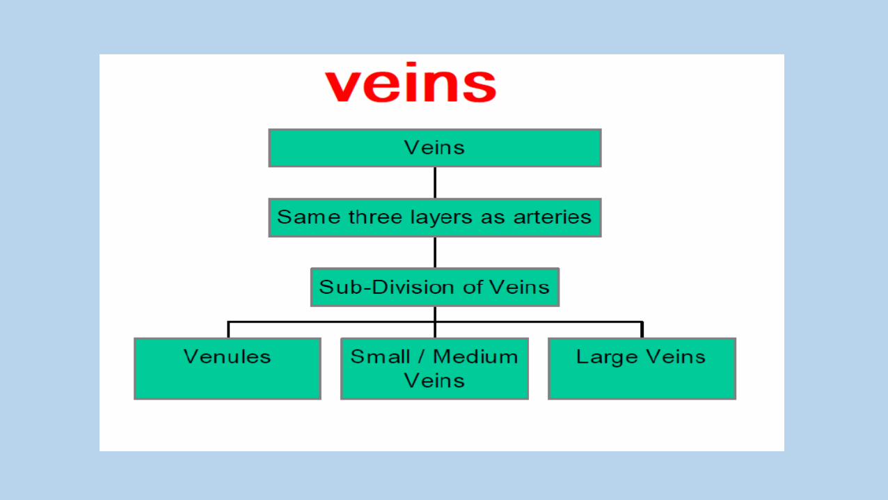
Veins
Return the blood to the Heart
Types of Blood Vessels

Structural Plan of Blood Vessels
1.The tunica intima :
(layer of endothelial cells ,a thin
subendothelial layer of loose con.
T. with occasional smooth muscle
cells). In arteries, the intima is separated
from the media by an internal elastic
lamina

2.The tunica media:
(consists of concentric layers of smooth
muscle cells and amounts of elastic
fibers and reticular fibers of collagen
type III, proteoglycans, and
glycoproteins)
In arteries, the media has a thinner
external elastic lamina

3.The tunica adventitia
:
consists of :
• connective tissue.
• Lymphatic
capillaries, vasa
vasorum, and
nerves.

Figre : summarized the comparison between veins & arteries

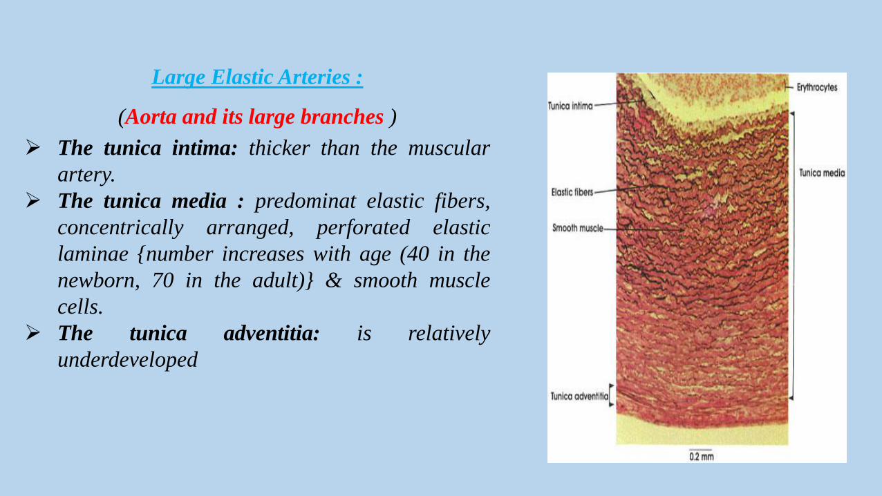
Large Elastic Arteries :
(Aorta and its large branches )
The tunica intima: thicker than the muscular
artery.
The tunica media : predominat elastic fibers,
concentrically arranged, perforated elastic
laminae {number increases with age (40 in the
newborn, 70 in the adult)} & smooth muscle
cells.
The tunica adventitia: is relatively
underdeveloped

The muscular arteries:1.The intima :
• a very thin subendothelial layer
• internal elastic lamina,
2.The tunica media :
• up to 40 layers of more prominent
smooth muscle
• An external elastic lamina
(is present only in the larger
muscular arteries.)
3.The adventitia: consists of :
• connective tissue.
• Lymphatic capillaries, vasa
vasorum, and nerves.

Arterioles
• Less than 0.5 mm in diameter.
• The subendothelial layer is very thin.
• Elastic laminae are absent .
• Tunica media is composed of
circularly arranged smooth muscle
cells.
• In both small arteries and arterioles,
the tunica adventitia is very thin.


? Identify the characteristic tunica of arteries?

Capillaries
• permit metabolic exchange between
blood and tissues.
• single layer of endothelial cells
• diameter varies (5 to 10 micro m ).
• their individual length is not more
than 50 micro m.
• comprise over 90% of all blood
vessels in the body.

Type of capillaries
1. The continuous
(somatic) capillaries.
2. The fenestrated
(visceral) capillaries.3. The discontinuous
sinusoidal capillaries.
●Absence of fenestrae in their wall.
●Found in all type of muscular
tissue, C.T., exocrine gland and
nervous tissue.
●Transport of macromolecules
across the endothelial cytoplasm by
pinocytotic vesicle.
●Presence of fenestrae (channels)
obliterated by a diaphragm .
●basal lamina is continuous.
●In kidney.
➢The endothelium (discontinuous
layer).
➢Multiple fenestrations without
diaphragms.
➢The basal lamina is
discontinuous.
➢In liver and hematopoietic
organs (Bone Marrow and spleen).
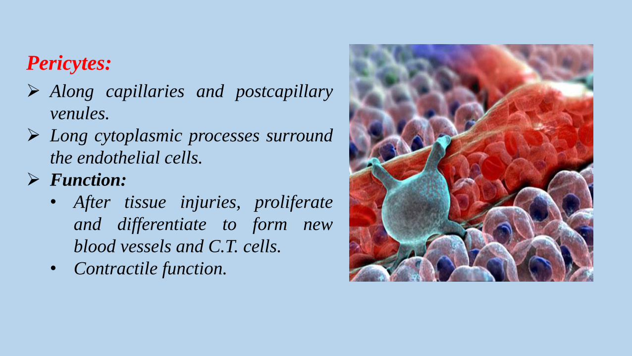
Pericytes:
Along capillaries and postcapillary
venules.
Long cytoplasmic processes surround
the endothelial cells.
Function:
• After tissue injuries, proliferate
and differentiate to form new
blood vessels and C.T. cells.
• Contractile function.

? Memorize types of capillaries.

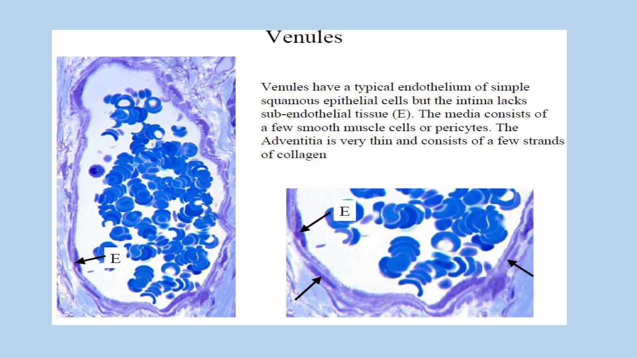
Postcapillary Venules
• T. intima:
– endothelium
– a very thin subendothelial layer.
• T. media : may contain:
only contractile pericytes.
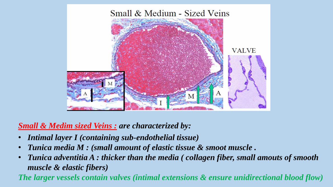
Small & Medim sized Veins : are characterized by:
• Intimal layer I (containing sub-endothelial tissue)
• Tunica media M : (small amount of elastic tissue & smoot muscle .
• Tunica adventitia A : thicker than the media ( collagen fiber, small amouts of smooth
muscle & elastic fibers)
The larger vessels contain valves (intimal extensions & ensure unidirectional blood flow)

Large Veins: (ex: vena cava)
• Well developed intimal ,medial
and adventitial tunics.
• T. media: (smooth muscule
,reticular & collagen fiber).
• T. Adventitia well-developed
layer (longtitudinally
arranged collagen & smooth
muscle fibers, adipose A and
vessels V =vasa vasorum)
• Contain valves.

A Comparison of a Typical Artery and a Typical Vein

Two features of microcirculation are important in the amount of
blood perfusing the capillary beds:
The pre-capillary sphincter (PS), can restrict the flow into the
capillaries.
Thoroughfare channels (TC) can provide a route for blood to
enter the venous system without traversing the capillary bed.

vasa vasorum (vessels of vessels).
Amicrovasculature lie in the tunica adventitia
of large blood vessels ( ex. Aorta ).
function: to bring O2 and nutrients to local
cells too far from the lumen to be nourished by
blood there.
arterioles (A),capillaries and venules (V)
constitute the vasa vasorum (vessels of
vessels).
? Can you diffrentiate between veins & arteries?

Heart:
• Muscular organ , when
contracts, pumping the
blood through the
circulatory system.
• Producing a hormone called
atrial natriuretic factor.

The walls of all four heart chambers consist
of three tunics:
• Endocardium
• Myocardium
• Epicardium
Structure of heart1. superior vena cava
2. pulmonary semilunar
3. right atrium
4. tricuspid valve
5. right ventricle
6. inferior vena cava
7. septum
8. left ventricle
9. bicuspid) or mitral valve
10. aortic semilunar valve
11. left atrium
12. aorta

Endocardium
• Consists of:
➢ squamous endothelial cells.
➢ subendothelial layer of loose C. T.
(contains elastic, collagen fibers, smooth m.
, veins, nerves, and branches of the
impulse – conducting system of the heart
(Purkinje cells).
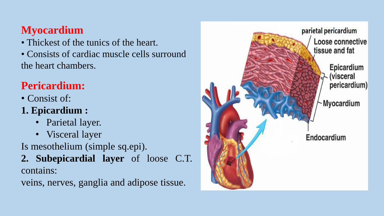
Myocardium• Thickest of the tunics of the heart.
• Consists of cardiac muscle cells surround
the heart chambers.
Pericardium:• Consist of:
1. Epicardium :
• Parietal layer.
• Visceral layer
Is mesothelium (simple sq.epi).
2. Subepicardial layer of loose C.T.
contains:
veins, nerves, ganglia and adipose tissue.

The heart has a specialized system to generate a
rhythmic stimulus (spread to the entire myocardium)
consist of:
➢ sinoatrial node (pacemaker). modified cardiac
muscle cells (fusiform),
with fewer myofibrils
➢ atrioventricular node: similar to those of the
sinoatrial node, but their cytoplasmic projections
branch ,forming a network.
➢ atrioventricular bundle
(bundle of His): formed by cells similar to those
of the atrioventricular node.
➢ Purkinje fibers:
• End of atrioventricular bundle
• 1-2 central nuclei.
• few myofibrils


The Lymphatic Vascular System:
Lymphatic capillaries:
In the various tissues as thin, closed-ended
vessels , consist of:
• A single layer of endothelium
• An incomplete basal lamina.
The larger lymphatics :similar to veins
except:
• They have thinner walls and lack
separation between tunics.
• More numerous internal valves


![[HISTOLOGY] Endocrine System](https://static.fdocuments.in/doc/165x107/56d6bfef1a28ab30169849e6/histology-endocrine-system.jpg)