
Histological study on the role of ginger against cisplatin ...
9
1110-0559 © 2013 The Egyptian Journal of Histology DOI: 10.1097/01.EHX.0000428403.28567.0f 312 Introduction Cisplatin is an important chemotherapeutic agent because of its therapeutic effect against testicular germ cell cancer. As a heavy metal compound, cisplatin produces DNA crosslinks that are presumably responsible for its antineoplastic effect [1]. In many previous animal studies, it was noted that within days of cisplatin injection animals showed severe testicular damage characterized by spermatogenic damage, germ cell apoptosis, Leydig cell dysfunction, and testicular steroidogenic disorder [2,3]. However, the severity of cisplatin-induced testicular damage was dose dependent [4]. Many natural products of plant origin might have a partially protective role against drug-induced toxicity of chemotherapeutic agents [5,6]. However, to date, virtually no medicinal plant has been described that relieves cisplatin-induced reproductive side effects accompanying chemotherapy in cancer patients. Meanwhile, ginger rhizome (Zingiber officinale), which is used worldwide as a spice, was reported to have both antioxidant [7] and androgenic activity [8], but at the ultrastructural level there is no reliable study on the effect of ginger on cisplatin-induced testicular injury. Thus, the present study aimed at investigating the ultrastructural changes in the testes of male albino rats after cisplatin administration in doses comparable to the therapeutic doses used in cancer chemotherapy and at postulating the possible protective effects of ginger against it. Materials and methods Materials Cisplatin Cisplatin vials were obtained from Ebewe Company, (Ebewe, Unterach, AUSTRIA). Each 20 ml vial contained 10 mg cisplatin. a Histology Department, Faculty of Medicine, Cairo University, Cairo and b Histology Department, Faculty of Medicine, Fayoum University, Fayoum, Egypt Correspondence to Mohamed Salah Elgendy, Histology Department, Faculty of Medicine, Fayoum University, 63514, Fayoum, Egypt Tel: +20 100 539 6843; fax: +20 846 302 350; e-mail: [email protected] Received 11 September 2012 Accepted 28 October 2012 The Egyptian Journal of Histology 2013, 36:312-320 29 (1368-2013) Aim of the work Chemotherapy with cisplatin has adverse effects on spermatogenesis. Therefore, this work aimed at investigating the protective role of ginger against cisplatin-induced testicular toxicity in male albino rats. Materials and methods Twenty-four adult albino rats were used in this study. They were divided into three groups. The first group served as the control group; the second group was injected with cisplatin (12 mg/kg once); and the third group was injected with cisplatin (12 mg/kg once) and then given ginger (310 mg/kg orally) for 26 days. Testicular specimens were processed for light microscopic examination using H&E. Other specimens were processed for electron microscopic examination. Results Cisplatin had damaging effects on the seminiferous tubules. Some areas of the tubules showed complete depletion of germ cells. Other areas showed some spermatogonia or primary spermatocytes. Sertoli cells showed a variable degree of degenerative changes in the form of destruction of cellular processes and cell junction. Interruption of the nuclear envelope of spermatids and loss of intercellular bridges were noticed. Treating with ginger resulted in normal Sertoli cells and cell junctions. The germ cells lining the tubules were more or less normal except for some intercellular vacuolations. Conclusions The use of ginger has some protective effects on the testicular structure; hence, a larger number of experiments with higher doses of ginger or longer administration period could be beneficial for patients taking chemotherapeutic drugs. Keywords: cisplatin, ginger, seminiferous tubule, spermatogenic cells, testis Egypt J Histol 36:312-320 © 2013 The Egyptian Journal of Histology 1110-0559 Histological study on the role of ginger against cisplatin- induced testicular toxicity in albino rats Abeer F. Abdel-Mohsen a , Mohamed S. Elgendy b , Dalia M. Elmarakby a and Doaa I. Eldsouky b Original article Copyright © The Egyptian Journal of Histology. Unauthorized reproduction of this article is prohibited.
Transcript of Histological study on the role of ginger against cisplatin ...

1110-0559 © 2013 The Egyptian Journal of Histology DOI: 10.1097/01.EHX.0000428403.28567.0f
312
IntroductionCisplatin is an important chemotherapeutic agent because of its therapeutic effect against testicular germ cell cancer. As a heavy metal compound, cisplatin produces DNA crosslinks that are presumably responsible for its antineoplastic effect [1].
In many previous animal studies, it was noted that within days of cisplatin injection animals showed severe testicular damage characterized by spermatogenic damage, germ cell apoptosis, Leydig cell dysfunction, and testicular steroidogenic disorder [2,3]. However, the severity of cisplatin-induced testicular damage was dose dependent [4].
Many natural products of plant origin might have a partially protective role against drug-induced toxicity of chemotherapeutic agents [5,6]. However, to date, virtually no medicinal plant has been described that relieves cisplatin-induced reproductive side effects
accompanying chemotherapy in cancer patients. Meanwhile, ginger rhizome (Zingiber officinale), which is used worldwide as a spice, was reported to have both antioxidant [7] and androgenic activity [8], but at the ultrastructural level there is no reliable study on the effect of ginger on cisplatin-induced testicular injury.
Thus, the present study aimed at investigating the ultrastructural changes in the testes of male albino rats after cisplatin administration in doses comparable to the therapeutic doses used in cancer chemotherapy and at postulating the possible protective effects of ginger against it.
Materials and methodsMaterialsCisplatinCisplatin vials were obtained from Ebewe Company, (Ebewe, Unterach, AUSTRIA). Each 20 ml vial contained 10 mg cisplatin.
aHistology Department, Faculty of Medicine, Cairo University, Cairo and bHistology Department, Faculty of Medicine, Fayoum University, Fayoum, Egypt
Correspondence to Mohamed Salah Elgendy, Histology Department, Faculty of Medicine, Fayoum University, 63514, Fayoum, EgyptTel: +20 100 539 6843; fax: +20 846 302 350;e-mail: [email protected]
Received 11 September 2012Accepted 28 October 2012
The Egyptian Journal of Histology2013, 36:312-32029 (1368-2013)
Aim of the workChemotherapy with cisplatin has adverse effects on spermatogenesis. Therefore, this work aimed at investigating the protective role of ginger against cisplatin-induced testicular toxicity in male albino rats.Materials and methodsTwenty-four adult albino rats were used in this study. They were divided into three groups. The first group served as the control group; the second group was injected with cisplatin (12 mg/kg once); and the third group was injected with cisplatin (12 mg/kg once) and then given ginger (310 mg/kg orally) for 26 days. Testicular specimens were processed for light microscopic examination using H&E. Other specimens were processed for electron microscopic examination.ResultsCisplatin had damaging effects on the seminiferous tubules. Some areas of the tubules showed complete depletion of germ cells. Other areas showed some spermatogonia or primary spermatocytes. Sertoli cells showed a variable degree of degenerative changes in the form of destruction of cellular processes and cell junction. Interruption of the nuclear envelope of spermatids and loss of intercellular bridges were noticed. Treating with ginger resulted in normal Sertoli cells and cell junctions. The germ cells lining the tubules were more or less normal except for some intercellular vacuolations.ConclusionsThe use of ginger has some protective effects on the testicular structure; hence, a larger number of experiments with higher doses of ginger or longer administration period could be beneficial for patients taking chemotherapeutic drugs.
Keywords: cisplatin, ginger, seminiferous tubule, spermatogenic cells, testis
Egypt J Histol 36:312-320© 2013 The Egyptian Journal of Histology1110-0559
Histological study on the role of ginger against cisplatin-induced testicular toxicity in albino ratsAbeer F. Abdel-Mohsena, Mohamed S. Elgendyb, Dalia M. Elmarakbya and Doaa I. Eldsoukyb
Original article
Copyright © The Egyptian Journal of Histology. Unauthorized reproduction of this article is prohibited.

313Ginger role in Cisplatin testicular toxicity Abdel-Mohsen et al.
Histological studyAt the end of the experimental period, on day 26, rats from all groups were anesthetized using an injection of 50 mg/kg thiopental sodium subcutaneously and then sacrificed.
Samples from the left testes were cut into small cubes (∼1 mm) and immediately fixed in 3% phosphate-buffered glutaraldehyde (pH 7.3) for 3 h. Ultrathin sections were prepared and stained with uranyl acetate and lead citrate and examined through an electron microscope [13].
Samples from the right testes were fixed in Bouin’s solution for 48 h and processed using the paraffin technique, whereas sections of 7-µm thickness were prepared and stained with H&E [14] to be examined through a light microscope.
This study was approved by the ethics committee on animal research in the animal house of Kasr-El-Aini Faculty of Medicine, Cairo University, Cairo, Egypt, following international ethics and regulations for animal research in laboratory applications [15].
Morphometric studyThe following parameters were measured and analyzed:(1) Mean body weight at the time of sacrifice.(2) Mean testicular weight at the time of sacrifice.(3) Diameter of the seminiferous tubules.(4) The number of normal seminiferous tubules (rounded
in transverse section, 150–250 µm in diameter, lined by stratified spermatogenic epithelium resting on clear basement membrane and narrow lumen) and abnormal tubules (those that did not have the above-mentioned features).
Parameters 3 and 4 were measured and analyzed using the image analyzer computer system (Leica Qwin 500, Hessen, Germany).
Statistical analyses of the obtained results were performed using analysis of variance and the t-test.
ResultsLight microscopic resultsThe control groupThe testes of the rats in the control group consisted of transversely cut closely packed seminiferous tubules, separated from each other by narrow interstitial spaces containing interstitial Leydig cells around blood capillaries. These seminiferous tubules were lined by spermatogenic cells and Sertoli cells. The spermatogenic cells were formed of spermatogonia, primary spermatocytes, and spermatids. The spermatogonia appeared as small cells with round dark nuclei forming the basal layer. The primary spermatocytes were the largest cells in the section with central rounded nuclei. Elongated spermatids also appeared (Fig. 1).
The cisplatin groupIn the cisplatin group, some of the seminiferous tubules appeared distorted with small diameter and wide lumina
The applied dose in many chemotherapeutic protocols is calculated according to body surface area. To be effective with the least side effects its dose should range from 20 to 100 mg/m2 if applied once or repeated either as a monotherapy or in combination therapy [9]. In the present study, we used a dose of 12 mg/kg, obtained by converting an equivalent dose of 75 mg/m2 for humans to a rat equivalent dose in mg/kg [10].
This conversion was done using the following formula:Human equivalent dose (mg/kg) / human k
m =
animal
equivalent dose (mg/kg) / animal km
, km constant
(coefficient factor for species); km (coefficient factor for
humans)=37 and km (coefficient factor for rats)=6.
GingerGinger extracts were obtained from Arab Company for Pharmaceuticals and Medicinal Plants (MEPACO, Cairo, Egypt) in tablet form. Each tablet contained 400 mg of ginger extract.
The recommended doses for humans range from 1 to 3 g daily in many different indications [11,12]. In the present study, we used a dose equivalent to 3 g/day for an average human body weight of 60 kg and converted it to a rat equivalent dose in mg/kg [10], which was found to be 310 mg/kg/day. The tablet was crushed and dissolved in 4 ml saline; hence, each ml contained 100 mg ginger.
AnimalsThe present study included 24 adult albino rats of 200–220 g body weight; they were obtained from and housed in the animal house of Kasr-El-Aini Faculty of Medicine, Cairo University, Cairo, Egypt. The animals received a standard diet for rodents and were allowed free access to water. They were distributed in three cages, each containing eight rats.
Treatment protocolThe rats were divided into three groups of eight animals each.
The first group served as the control group. The rats in this group received 1 ml saline 0.9% NaCl daily for 26 successive days through a gastric tube and were injected intraperitoneally with 6 ml saline on day 21 of the experiment. They were sacrificed at the same time as the rats of the experimental groups.
The second group was the cisplatin group. The rats in this group received 1 ml saline 0.9% NaCl daily for 26 successive days through a gastric tube. On day 21 of the experiment, each rat was injected intraperitoneally with a single dose of 12 mg/kg cisplatin.
The third group was the cisplatin and ginger group. The rats in this group received ginger extract dissolved in saline orally at a dose of 310 mg/kg daily for 26 successive days through a gastric tube on day 21 of ginger extract administration. Each rat was injected intraperitoneally with a single dose of 12 mg/kg cisplatin.
Copyright © The Egyptian Journal of Histology. Unauthorized reproduction of this article is prohibited.

314 Ginger role in Cisplatin testicular toxicity Abdel-Mohsen et al.
In cisplatin and cisplatin and ginger groups, there was a significant decrease (P≤0.001) in mean body weight when compared with the control group. The decrease was more obvious in the cisplatin group.
In cisplatin and cisplatin and ginger groups, there was a significant decrease (P≤0.001) in mean testicular weight when compared with the control group. The decrease was more obvious in the cisplatin group.
In cisplatin and cisplatin and ginger groups, there was a significant decrease (P≤0.001) in seminiferous tubule diameter when compared with the control group. The decrease was more obvious in the cisplatin group.
In cisplatin and cisplatin and ginger groups, there was significant decrease (P≤0.001) in the percentage of normal seminiferous tubules when compared with the control group. The decrease was more obvious in the cisplatin group.
lined by few spermatogonia with many intercellular vacuolations. Other tubules were severely affected with near-total depletion of their germ cells. The interstitial spaces appeared wide with apparently normal interstitial Leydig cells (Fig. 2).
The ginger and cisplatin groupExamination of testicular sections from rats given ginger and cisplatin showed seminiferous tubules with a relatively normal morphology. They were lined by many layers of spermatogenic cells. Some tubules still showed intercellular vacuolations (Fig. 3).
Electron microscopic resultsThe control groupExamination of testicular sections of the control group showed that seminiferous tubules were lined by spermatogonia, primary spermatocytes, and Sertoli cells. Sertoli cells had euchromatic nuclei and nucleoli resting on the irregular basement membrane. Spermatogonia with euchromatic nuclei and primary spermatocytes (present within infolding of Sertoli cells in the adluminal compartment) were separated from the basal compartment by an ectoplasmic specialized junction between Sertoli cells (Figs 4 and 5).
Early spermatids appeared with rounded nuclei with finely granular cytoplasm rich in mitochondria. Spermatids at ‘Golgi phase’ contained prominent acrosomal vesicles in association with the nucleus. Elongated heads of spermatids with flattened condensed nuclei were present within infoldings of Sertoli cells (Fig. 6).
The cisplatin groupExamination of testicular sections from rats taking cisplatin showed many primary spermatocytes. The cells were separated from the basement membrane by basal vacuolation and destroyed processes of Sertoli cells (Fig. 7). Spermatogonia type B appeared with central round nuclei containing peripheral clumps of heterochromatin condensation at the periphery in close association with the nuclear membrane; in addition, some degenerated cells could be observed (Fig. 8).
Primary spermatocytes appeared with loss of intercellular bridges. Interruption of nuclear envelope of spermatids was noticed (Fig. 9).
The ginger and cisplatin groupExamination of testicular sections from rats given ginger and cisplatin showed that most of the seminiferous tubules were lined by apparently normal primary spermatocytes and early spermatids. Sertoli cells with euchromatic indented nuclei and ectoplasmic specialization junction were seen. Intercellular spaces were filled with cytoplasmic processes of Sertoli cells (Figs 10 and 11).
Morphometric resultsStatistical analysis of the obtained results comparing the different experimental groups is summarized in Table 1 and reveals the following:
Figure 1. A photomicrograph of a testicular section from a control rat showing seminiferous tubules (T) lined by normal spermatogenic cells (brace). The spermatogenic cells are spermatogonia (thin arrows), primary spermatocytes (thick arrows), and elongated spermatids (curved arrows). The interstitial spaces (stars) are clearly visualized and contain Leydig cells (crossed arrows) and blood vessels (bv). H&E stain (a), × 200; (b), × 400.
Copyright © The Egyptian Journal of Histology. Unauthorized reproduction of this article is prohibited.

315Ginger role in Cisplatin testicular toxicity Abdel-Mohsen et al.
Figure 5. An electron photomicrograph of a testicular section from the control group showing primary spermatocytes (thick arrows) present within infoldings of Sertoli cells (S) in the adluminal compartment. Note ectoplasmic specialized junctions (thin arrows) between Sertoli cell processes (S) and germ cells. TEM, × 10 000.
Figure 2. A photomicrograph of a testicular section from a rat given cisplatin showing degenerative changes in some tubules with a few germ cells (thin arrows) with marked intercellular vacuolation (thick arrows). Other tubules (T) are markedly disorganized with near-total depletion of their germ cells. Only a single layer of spermatogonia and Sertoli cell processes are present. Note the wide interstitium (stars). H&E stain (a), × 200; (b), × 400.
Figure 3. A photomicrograph of a testicular section from a rat given ginger and cisplatin showing some seminiferous tubules with more or less normal cellularity (brace). Other tubules still show intercellular vacuolation (thick arrows) with relatively normal spermatogenic cells. Note the wide interstitium (stars). H&E stain (a), × 200; (b), × 400.
Figure 4. An electron photomicrograph of a testicular section from the control group showing one Sertoli cell with a euchromatic nucleus and nucleolus and other Sertoli cells with indented nuclei (double arrow), spermatogonia (thin arrows), and primary spermatocytes (thick arrows). Note irregular boundary tissue, basement membrane (dotted arrows), and myoid cells beneath it (arrow heads). TEM, × 4000.
Copyright © The Egyptian Journal of Histology. Unauthorized reproduction of this article is prohibited.
316 Ginger role in Cisplatin testicular toxicity Abdel-Mohsen et al.
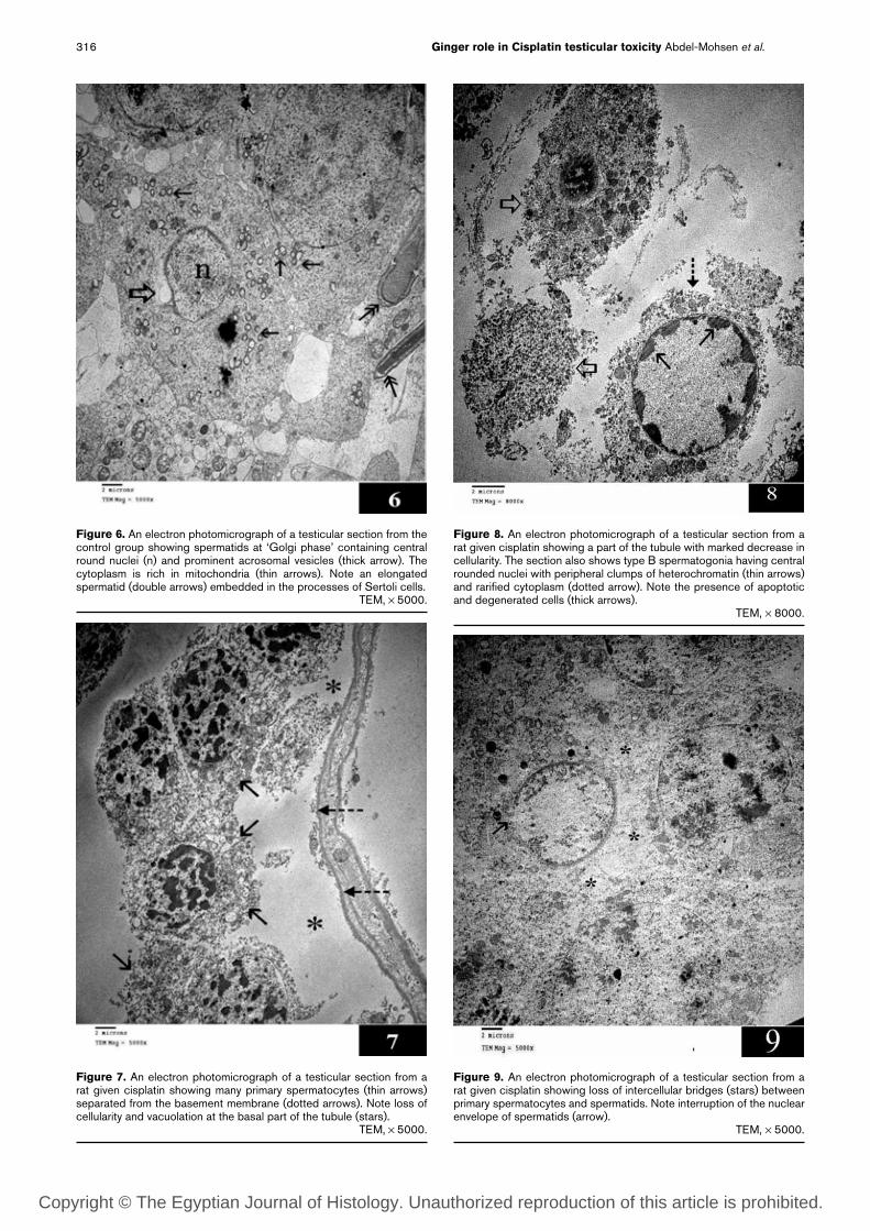
Figure 6. An electron photomicrograph of a testicular section from the control group showing spermatids at ‘Golgi phase’ containing central round nuclei (n) and prominent acrosomal vesicles (thick arrow). The cytoplasm is rich in mitochondria (thin arrows). Note an elongated spermatid (double arrows) embedded in the processes of Sertoli cells. TEM, × 5000.
Figure 7. An electron photomicrograph of a testicular section from a rat given cisplatin showing many primary spermatocytes (thin arrows) separated from the basement membrane (dotted arrows). Note loss of cellularity and vacuolation at the basal part of the tubule (stars). TEM, × 5000.
Figure 8. An electron photomicrograph of a testicular section from a rat given cisplatin showing a part of the tubule with marked decrease in cellularity. The section also shows type B spermatogonia having central rounded nuclei with peripheral clumps of heterochromatin (thin arrows) and rarified cytoplasm (dotted arrow). Note the presence of apoptotic and degenerated cells (thick arrows). TEM, × 8000.
Figure 9. An electron photomicrograph of a testicular section from a rat given cisplatin showing loss of intercellular bridges (stars) between primary spermatocytes and spermatids. Note interruption of the nuclear envelope of spermatids (arrow). TEM, × 5000.
Copyright © The Egyptian Journal of Histology. Unauthorized reproduction of this article is prohibited.

317Ginger role in Cisplatin testicular toxicity Abdel-Mohsen et al.
Figure 10. An electron photomicrograph of a testicular section from a rat given ginger and cisplatin showing normal Sertoli cells with euchromatic indented nuclei (n2) and cytoplasm rich in mitochondria (thin arrows) and cellular processes connected by ectoplasmic specialization (dotted arrows). Note normal primary spermatocytes (thick arrow) containing central round nuclei (n1). TEM, × 5000.
Figure 11. An electron photomicrograph of a testicular section from a rat given ginger and cisplatin showing normal primary spermatocytes (thick arrows) and spermatids (thin arrows). Note the processes of Sertoli cells filling the intercellular spaces (curved arrows). TEM, × 3000.
Table 1. Results of statistical analysis of morphometric results
Mean ± SD G1 (control) G2 (cisplatin) G3 (cisplatin and ginger)
Mean body weight (g) 245.6 ± 7.9 214.8 ± 4.6∗ 230.2 ± 6.7∗,#
Mean testicular weight (mg) 1316.8 ± 3.6 934.4 ± 8.1∗ 1186.3 ± 4.7∗,#
Mean diameter of seminiferous tubules (µm) 243.9 ± 36.4 191.8 ± 44.8∗ 211.3 ± 24.2∗,#
% of normal seminiferous tubules 87.3 ± 5.7 14.2 ± 3.2∗ 74.4 ± 6.7∗,#
∗Marked differences are significant at P<0.001 compared with group 1 (control).#Marked differences are significant at P<0.001 compared with group 2 (cisplatin).
DiscussionThis study aimed at investigating the protective effects of ginger against cisplatin-induced testicular toxicity in male albino rats and studying the possible mechanisms underlying these effects.
In the cisplatin group, there was a significant decrease (P≤0.001) in body and testicular weights when compared with the control group. This result was confirmed by other studies, which found a significant reduction in the weight of testes expressed in relation to body weight [16].
Testicular sections from rats that received cisplatin showed some distorted seminiferous tubules with a statistically significant increase in the number of tubules with small diameter as well as a number of abnormal and
degenerated tubules. Similar results were achieved in other studies [17–19].
In the present study, some seminiferous tubules showed loss of cellularity with intercellular vacuolation. Other tubules were severely affected with total depletion of germ cells. These results were similar to the results of other investigators [17].
Loss of intercellular bridges was seen in the present study. Some investigators recorded loss of intercellular bridges and even formation of multinucleated giant cells in the testes exposed to toxic agents. This may lead to the formation of abnormal sperms [20].
In addition, there was interruption of the nuclear envelope in some spermatids. Other researchers declared that cisplatin was capable of binding to several cellular
Copyright © The Egyptian Journal of Histology. Unauthorized reproduction of this article is prohibited.

318 Ginger role in Cisplatin testicular toxicity Abdel-Mohsen et al.
with the cisplatin group. This result might be attributed to the androgenic effect of ginger on the testes [28].
Light microscopic and electron microscopic examination of testicular sections from the ginger and cisplatin group showed seminiferous tubules with relatively normal morphology. Most of the seminiferous tubules are lined with many layers of relatively normal spermatogenic cells, including early and late spermatids. This was confirmed by a statistically significant increase in seminiferous tubule diameter, number of normal seminiferous tubules, and the statistically significant decrease in the number of abnormal tubules compared with the cisplatin group. The diminished degenerative changes in the ginger-treated group could prove the protective effect of ginger against cisplatin-induced testicular damage. However, this protective effect is not total, as some seminiferous tubules were separated by wide interstitial spaces, were surrounded by partially separated basement membrane, and were lined by some dispersed and degenerated spermatogenic cells. The protective effect of ginger could be dose dependent, as many investigators found that administration of ginger of 50 and 100 mg/kg in rats for 20 consecutive days significantly increased sperm motility and viability in rat testes [20]. In contrast, other investigators found that ginger aids in the production of healthy sperm but should be taken with care as it can distort membranes of the seminiferous tubules, affecting the epithelial lining of the tubules [29].
With regard to the possible mechanisms of ginger’s protective effects, many studies revealed that ginger significantly lowered lipid peroxidation by maintaining the activities of antioxidant enzymes such as superoxide dismutase, catalase, and glutathione peroxidase in rats [30]. Other studies revealed that all major active ingredients of ginger, such as zingerone, gingerdiol, zingibrene, gingerols, and shogaols, had antioxidant activity [31,32].
In addition, normalization of antioxidant activities, and the concurrent decrease of malondialdehyde in the testes, was protected with ginger before cisplatin treatment [16].
AcknowledgementsConflicts of interestThere is no conflict of interest to declare.
References1 Kartalou M, Essigmann JM. Recognition of cisplatin adducts by cellular
proteins. Mutat Res 2001; 478:1–21.2 Malarvizhi D, Manimaran RR, Aruldhas MM, Mathur PP. Quantitative maintenance
of spermatogenesis in cisplatin-treated rats by exogenous administration of testosterone propionate. J Endocrinol Reprod 1998; 1:67–72.
3 Cherry SM, Hunt PA, Hassold TJ. Cisplatin disrupts mammalian spermatogenesis, but does not affect recombination or chromosome segregation. Mutat Res 2004; 564:115–128.
4 Colpi GM, Contalbi GF, Nerva F, Sagone P, Piediferro G. Testicular function following chemo-radiotherapy. Eur J Obstet Gynecol Reprod Biol 2004; 113 (Suppl):S2–S6.
5 Greggi Antunes LM, Darin JD’AC, Bianchi MDLP. Effects of the antioxidants curcumin or selenium on cisplatin-induced nephrotoxicity and lipid peroxidation in rats. Pharmacol Res 2001; 43:145–150.
6 Shirwaikar A, Issac D, Malini S. Effect of Aerva lanata on cisplatin and gentamicin models of acute renal failure. J Ethnopharmacol 2004; 90:81–86.
components, including membrane phospholipids and cytoskeletal microfilaments [21].
Sertoli cell affection was recorded in the present study represented by the absence of cytoplasmic processes and ectoplasmic specialization in electron microscopic studies. However, in severely affected tubules in the present study, mainly Sertoli cells were present. This was similar to the study by the investigators who recorded that Sertoli cells were more resistant than spermatogenic cells to toxic agents [22].
The present study showed wide interstitial spaces in the cisplatin-treated group, which can be explained by a significant reduction in the diameter of tubules in this group [21].
The mechanism by which cisplatin alters Sertoli cell function has been discussed by other authors. They suggested that the destructive effect of cisplatin on Sertoli cells occurs at high doses of the drug, leading to release of hydrolytic enzymes, destruction of blood–testis barrier, and consequent destruction of adjacent cells. Overproduction of reactive oxygen species by cisplatin causes damage to the DNA, breakdown of polyunsaturated fatty acids of cell membrane lipoproteins, and damage of protein contents of the cell and subsequent testicular cell degeneration [23]. Moreover, cisplatin treatment decreases the testicular activities of antioxidants such as reduced glutathione and catalase, which prevent generation of reactive oxygen species. Thus, the concurrent decrease in antioxidants in cisplatin-treated tissues might potentially explain the cisplatin-induced complications [24].
Cisplatin can inhibit DNA replication and RNA transcription by the formation of interstrand and intrastrand DNA crosslinks, leading to impairment of DNA replication [25].
It was recorded that most stem spermatogonia survive chemotherapy if the cumulative dose of cytotoxic drugs does not exceed 600 mg/m2 [26]. These results were in accordance with our results on electron microscopic examination. Moreover, type B spermatogonia can survive after an almost complete depletion of differentiating germ cells [17].
Testicular sections examined by the electron microscope confirmed the light microscopic findings and showed shrinkage of some cells, degenerated (vacuolated and rarefied) cytoplasm, condensed small nuclei, nonnucleated degenerated cytoplasmic remnants, wide separation between spermatogenic cells, and separation of the basement membrane from overlying cells. Also, electron microscopic examination of the cisplatin-treated group showed degenerated mitochondria in some spermatogenic cells. These findings could be features of early apoptosis as seen in the present study. The apoptotic changes associated with cisplatin treatment were recorded in many other studies [17,19,27].
In the ginger and cisplatin group, there was a significant increase in body and testicular weights when compared
Copyright © The Egyptian Journal of Histology. Unauthorized reproduction of this article is prohibited.

319Ginger role in Cisplatin testicular toxicity Abdel-Mohsen et al.
21 Goel R, Andrews PA, Pfeifle CE, Abramson IS, Kirmani S, Howell SB. Comparison of the pharmacokinetics of ultrafilterable cisplatin species detectable by derivatization with diethyldithiocarbamate or atomic absorption spectroscopy. Eur J Cancer 1990; 26:21–27.
22 Ishii T, Matsuki S, Iuchi Y, Okada F, Toyosaki S, Tomita Y, et al. Accelerated impairment of spermatogenic cells in sod1-knockout mice under heat stress. Free Radic Res 2005; 39:697–705.
23 D’Cruz SC, Mathur PP. Effect of piperine on the epididymis of adult male rats. Asian J Androl 2005; 7:363–368.
24 Kaur P, Bansal MP. Influence of selenium induced oxidative stress on spermatogenesis and lactate dehydrogenase-X in mice testis. Asian J Androl 2004; 6:227–232.
25 Boekelheide K, Fleming SL, Allio T, Embree-Ku ME, Hall SJ, Johnson KJ, et al. 2,5-Hexanedione-induced testicular injury. Ann Rev Pharmacol Toxicol 2003; 43:125–147.
26 Schrader M, Muller M, Straub B, Miller K. Testicular sperm extraction in azoospermic patients with gonadal germ cell tumors prior to chemotherapy – a new therapy option. Asian J Androl 2002; 4:9–15.
27 Mohammadnejad D, Abedelahi A, Soleimani-Rad J, Mohammadi-Roshandeh A, Rashtbar M, Azami A. Degenerative effect of cisplatin on testicular germinal epithelium. Adv Pharmaceut Bull 2012; 2:173–177.
28 Khaki A, Fathiazad F, Nouri M, Khaki AA, Ozanci CC, Ghafari-Novin M, Hamadeh M. The effects of ginger on spermatogenesis and sperm parameters of rat. Iran J Reprod Med 2009; 7:7–12.
29 Oyewo OO, Onyije FM, Ashamu EA, Akintude OW, Akinola AE. Evaluation of ethanolic extract of ginger on the histology of the testes and sperm of adult wistar rats. Int J Sci Technol Res 2012; 1:50–53.
30 Jeyakumar SM, Nalini N, Menon VP. Antioxidant activity of ginger (Zingiber officinale rosc) in rats fed a high fat diet. Med Sci Res 1999; 27:341–344.
31 Zancan KC, Marques MOM, Petenate AJ, Meireles MAA. Extraction of ginger (Zingiber officinale roscoe) oleoresin with CO2 and co-solvents: a study of the antioxidant action of the extracts. J Supercrit Fluids 2001; 24:57–76.
32 Masuda Y, Kikuzaki H, Hisamoto M, Nakatani N. Antioxidant properties of gingerol related compounds from ginger. BioFactors 2004; 21:293–296.
7 Sekiwa Y, Kubota K, Kobayashi A. Isolation of novel glucosides related to gingerdiol from ginger and their antioxidative activities. J Agric Food Chem 2000; 48:373–377.
8 Kamtchouing P, Fandio GYM, Dimo T, Jatsa HB. Evaluation of androgenic activity of Zingiber officinale and Pentadiplandra brazzeana in male rats. Asian J Androl 2002; 4:299–301.
9 Kindler HL. Systemic treatments for mesothelioma: standard and novel. Curr Treat Options Oncol 2008; 9:171–179.
10 Reagan-Shaw S, Nihal M, Ahmad N. Dose translation from animal to human studies revisited. FASEB J 2008; 22:659–661.
11 Apariman S, Ratchanon S, Wiriyasirivej B. Effectiveness of ginger for prevention of nausea and vomiting after gynecological laparoscopy. J Med Assoc Thai 2006; 89:2003–2009.
12 Ozgoli G, Goli M, Simbar M. Effects of ginger capsules on pregnancy, nausea, and vomiting. J Altern Complement Med 2009; 15:243–246.
13 Hayat M. Basic technique for transmission electron microscopy. 9th ed. Florida, USA: Academic Press; 1986. pp. 31–38.
14 Drury RAB, Wallington EB. Carlton’s histological techniques. 5th ed. England: Oxford University Press; 1980. pp. 139–239.
15 Gluck JP, Di Pasquale T, Orlans B. Applied ethics in animal research (philosophy, regulation and laboratory applications). West Lafayette, Indiana. USA: Purdue University Press; 2002.
16 Amin A, Hamza AEA. Effects of roselle and ginger on cisplatin-induced reproductive toxicity in rats. Asian J Androl 2006; 8:607–612.
17 Sawhney P, Giammona CJ, Meistrich ML, Richburg JH. Cisplatin-induced long-term failure of spermatogenesis in adult C57/Bl/6J mice. J Androl 2005; 26:136–145.
18 Silici S, Ekmekcioglu O, Eraslan G, Demirtas A. Antioxidative effect of royal jelly in cisplatin-induced testes damage. Urology 2009; 74:545–551.
19 Sajjad H. Toxicity effect of cisplatin treatment on rat testis tissue. Ann Biol Res 2012; 3:2297–2303.
20 Miething A. The formation and dissolution of the bridge-partitioning complex in intercellular bridges of dividing germ cells in the testis of the golden hamster. J Submicros Cytol Pathol 2003; 35:271–276.
Copyright © The Egyptian Journal of Histology. Unauthorized reproduction of this article is prohibited.

320 Ginger role in Cisplatin testicular toxicity Abdel-Mohsen et al.
الملخص العربى
دراسة هستولوجية على دور الزنجبيل ضد السمية المستحثة للسيسبلاتن على خصية الفأر الأبيض البالغ
عبير فؤاد عبد المحسن، محمد صلاح الجندي1، داليا محمد المراكبى، دعاء إبراهيم الدسوقى محمد الحسينى1
قسم الهستولوجيا - كلية الطب - جامعة القاهرة و1جامعة الفيوميعانى مرضى المنوية وغالبا الحيوانات إنتاج السيسبلاتن على باستخدام عقار الكيميائي العلاج يؤثر المقدمة:
السرطان تحت العلاج الكيميائي من عقم مؤقت أو دائم حسب جرعة السيسبلاتن المستخدمة.هدف الدراسة: ويهدف هذا العمل إلى بحث دور الزنجبيل فى حماية الخصية من التأثير الضار للسيسبلاتن في الفئران.الأدوات و الطرق المستخدمة: وقد تم استخدام 24 فأرا مقسمة إلى ثلاث مجموعات كل مجموعة عددها 8 فئران نفس فى )% 0.9 ملح محلول ( الأدوية مع المستخدمة المذيبات نفس أعطيت )الضابطة(: الاولى المجموعة
التوقيت مع حيوانات التجربة وتم التضحية بها فى نفس التوقيت مع نظائرها من المجموعات التجريبية المجموعة الثانية )مجموعة السيسبلاتن(: أعطيت عن طريق الفم 0.9 مليلتر محلول ملح %0.9 يوميا لمدة 26 يوم وفى اليوم 21 تم حقنها في الغشاء البروتونى بجرعة واحدة من عقار السيسبلاتن مقدارها 12 ملى جرام لكل
كيلو جرام من الوزن الفم طريق عن ملح محلول في مذاب الزنجبيل أعطيت والزنجبيل(: السيسبلاتن )مجموعة الثالثة المجموعة بجرعة 350 ملى جرام لكل كيلو جرام وزن لمدة 26 يوم وفى اليوم 21 تم حقنها في الغشاء البرتونى بجرعة واحدة من عقار السيسبلاتن مقدارها 12 ملى جرام لكل كيلو جرام من الوزن و فى نهاية التجربة )اليوم 26( تم استئصال الخصية اليمنى من فئران المجموعات الثلاثة حيث عولجت لأعداد قطاعات شمعية والتي تم صباغتها بهيماتوكسلين وايوسين، و فحصها بالميكروسكوب الضوئي وتم تقطيع الخصية اليسرى من فئران المجموعات
الثلاثة إلى قطع صغيرة وتجهيزها للفحص تحت الميكروسكوب الالكتروني .النتائج: وقد أظهرت الدراسة التأثير الضار لعقار السيسبلاتن على الخصية في صورة ضمور في بعض الأنابيب المنوية، اتساع المناطق البينية، اختفاء وضمور الخلايا المنوية مع وجود فجوات بها ماعدا خلايا سيرتولى التي الميكروسكوب اثبته ما الالكتروني بالميكروسكوب العينات فحص وأكد السيسبلاتن لعقار الضار التأثير تقاوم الضوئي من ضمور الخلايا المنوية هذا بالإضافة الى ضمور السيتوبلازم في الخلايا المنوية الأولية و كذلك فى
النطيفات المتأخرة بالإضافة إلى بعض التغرات فى خلايا سيرتولى من تدمير لزوائدها و نقاط الإتصال بينها.وعند استخدام الزنجبيل مع السيسبلاتن لوحظ إن الأنابيب المنوية كانت اقرب إلى الطبيعي فأصبحت مبطنة ببضعة طبقات من الخلايا المنوية الطبيعية مع وجود بعض اتساع فى المناطق البينية وانفصال الخلايا المنوية عن بعضها الزنجبيل باستخدام المنوية الخلايا أن الالكتروني الميكروسكوب وأكد السيتوبلازم فى فجوات ووجود البعض الشكل المبطنة قريبة من الخلايا الطبيعي مع شكل الخلوي الترتيب مازالت طبيعية ملتصقة بعضها ببعض مع الطبيعى و كذلك ظهرت خلايا سيرتولى ممتدة من الغشاء القاعدى حتى تجويف الأنبوبة المنوية وتحتوى على نواة
غير منتظمة الشكل وسيتوبلازم غنى بالميتوكوندريا.المستخلصات: ان الدراسة الحالية اظهرت دور الزنجبيل فى حماية الخصية من التأثير الضار لعقار السيسبلاتن.
التوصيات:ينصح باستخدام عقار السيسبلاتن بجرعات صغيرة لتقليل إثارة الجانبية. . 1ينصح باستخدام الزنجبيل مع السيسبلاتن للحماية من اثار السيسبلاتن الجانبية الضارة و من الممكن استخدام . 2
الزنجبيل لمدة أطول لتحقيق الحماية المرجوة من التأثير الضار للسيسبلاتن.
Copyright © The Egyptian Journal of Histology. Unauthorized reproduction of this article is prohibited.


![Effects of Roselle and Ginger on cisplatin-induced ... · PDF fileEffects of Roselle and Ginger on cisplatin-induced reproductive toxicity in rats Amr Amin, ... fects [4]. Many natural](https://static.fdocuments.in/doc/165x107/5ab2b84f7f8b9a6b468dbc19/effects-of-roselle-and-ginger-on-cisplatin-induced-of-roselle-and-ginger-on.jpg)
