Histological study of the intestine and liver tissues in...
7
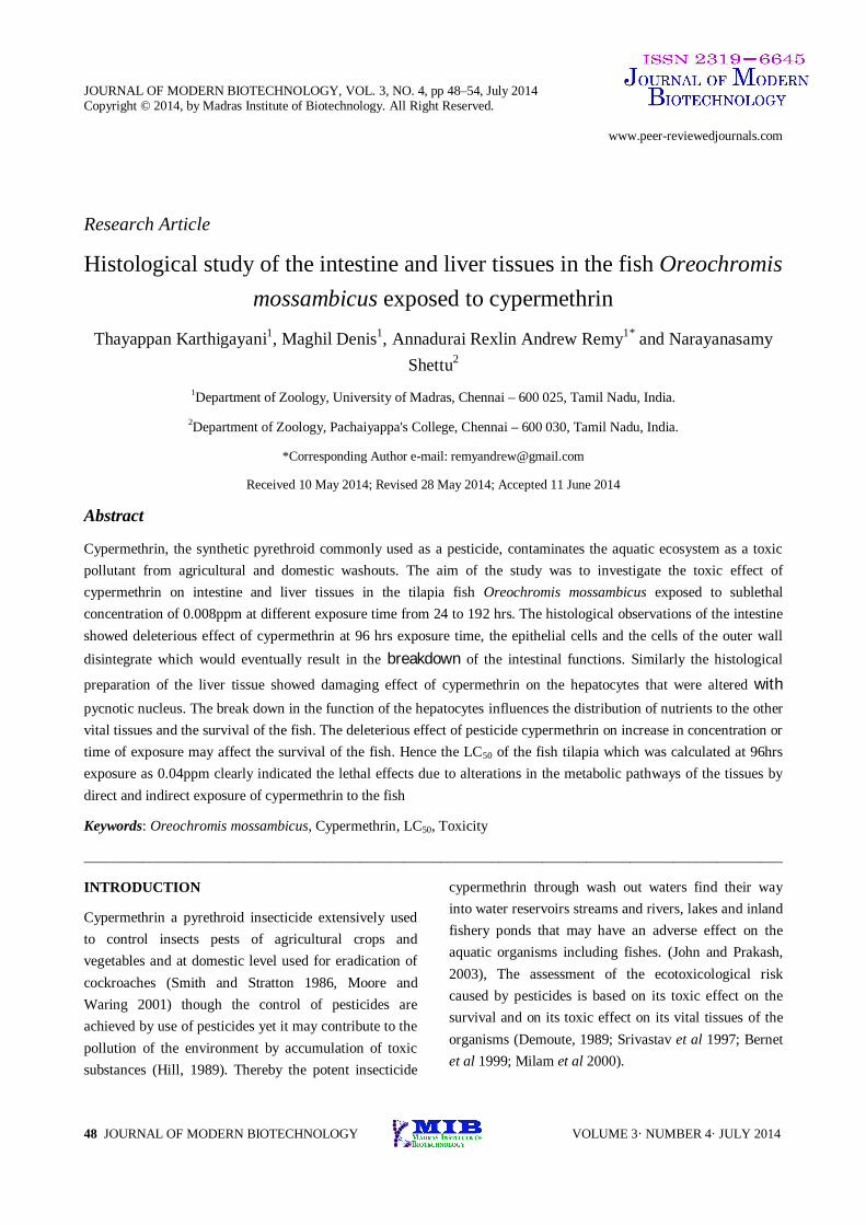
JOURNAL OF MODERN BIOTECHNOLOGY, VOL. 3, NO. 4, pp 48–54, July 2014 Copyright © 2014, by Madras Institute of Biotechnology. All Right Reserved. www.peer-reviewedjournals.com 48 JOURNAL OF MODERN BIOTECHNOLOGY VOLUME 3· NUMBER 4· JULY 2014 Research Article Histological study of the intestine and liver tissues in the fish Oreochromis mossambicus exposed to cypermethrin Thayappan Karthigayani 1 , Maghil Denis 1 , Annadurai Rexlin Andrew Remy 1* and Narayanasamy Shettu 2 1 Department of Zoology, University of Madras, Chennai – 600 025, Tamil Nadu, India. 2 Department of Zoology, Pachaiyappa's College, Chennai – 600 030, Tamil Nadu, India. *Corresponding Author e-mail: [email protected] Received 10 May 2014; Revised 28 May 2014; Accepted 11 June 2014 Abstract Cypermethrin, the synthetic pyrethroid commonly used as a pesticide, contaminates the aquatic ecosystem as a toxic pollutant from agricultural and domestic washouts. The aim of the study was to investigate the toxic effect of cypermethrin on intestine and liver tissues in the tilapia fish Oreochromis mossambicus exposed to sublethal concentration of 0.008ppm at different exposure time from 24 to 192 hrs. The histological observations of the intestine showed deleterious effect of cypermethrin at 96 hrs exposure time, the epithelial cells and the cells of the outer wall disintegrate which would eventually result in the breakdown of the intestinal functions. Similarly the histological preparation of the liver tissue showed damaging effect of cypermethrin on the hepatocytes that were altered with pycnotic nucleus. The break down in the function of the hepatocytes influences the distribution of nutrients to the other vital tissues and the survival of the fish. The deleterious effect of pesticide cypermethrin on increase in concentration or time of exposure may affect the survival of the fish. Hence the LC 50 of the fish tilapia which was calculated at 96hrs exposure as 0.04ppm clearly indicated the lethal effects due to alterations in the metabolic pathways of the tissues by direct and indirect exposure of cypermethrin to the fish Keywords: Oreochromis mossambicus, Cypermethrin, LC 50 , Toxicity ________________________________________________________________________________________________ INTRODUCTION Cypermethrin a pyrethroid insecticide extensively used to control insects pests of agricultural crops and vegetables and at domestic level used for eradication of cockroaches (Smith and Stratton 1986, Moore and Waring 2001) though the control of pesticides are achieved by use of pesticides yet it may contribute to the pollution of the environment by accumulation of toxic substances (Hill, 1989). Thereby the potent insecticide cypermethrin through wash out waters find their way into water reservoirs streams and rivers, lakes and inland fishery ponds that may have an adverse effect on the aquatic organisms including fishes. (John and Prakash, 2003), The assessment of the ecotoxicological risk caused by pesticides is based on its toxic effect on the survival and on its toxic effect on its vital tissues of the organisms (Demoute, 1989; Srivastav et al 1997; Bernet et al 1999; Milam et al 2000).
Transcript of Histological study of the intestine and liver tissues in...
JOURNAL OF MODERN BIOTECHNOLOGY, VOL. 3, NO. 4, pp 48–54, July 2014 Copyright © 2014, by Madras Institute of Biotechnology. All Right Reserved. www.peer-reviewedjournals.com
48 JOURNAL OF MODERN BIOTECHNOLOGY VOLUME 3· NUMBER 4· JULY 2014
Research Article
Histological study of the intestine and liver tissues in the fish Oreochromis mossambicus exposed to cypermethrin
Thayappan Karthigayani1, Maghil Denis1, Annadurai Rexlin Andrew Remy1* and Narayanasamy Shettu2
1Department of Zoology, University of Madras, Chennai – 600 025, Tamil Nadu, India.
2Department of Zoology, Pachaiyappa's College, Chennai – 600 030, Tamil Nadu, India.
*Corresponding Author e-mail: [email protected]
Received 10 May 2014; Revised 28 May 2014; Accepted 11 June 2014
Abstract
Cypermethrin, the synthetic pyrethroid commonly used as a pesticide, contaminates the aquatic ecosystem as a toxic pollutant from agricultural and domestic washouts. The aim of the study was to investigate the toxic effect of cypermethrin on intestine and liver tissues in the tilapia fish Oreochromis mossambicus exposed to sublethal concentration of 0.008ppm at different exposure time from 24 to 192 hrs. The histological observations of the intestine showed deleterious effect of cypermethrin at 96 hrs exposure time, the epithelial cells and the cells of the outer wall disintegrate which would eventually result in the breakdown of the intestinal functions. Similarly the histological
preparation of the liver tissue showed damaging effect of cypermethrin on the hepatocytes that were altered with pycnotic nucleus. The break down in the function of the hepatocytes influences the distribution of nutrients to the other vital tissues and the survival of the fish. The deleterious effect of pesticide cypermethrin on increase in concentration or time of exposure may affect the survival of the fish. Hence the LC50 of the fish tilapia which was calculated at 96hrs exposure as 0.04ppm clearly indicated the lethal effects due to alterations in the metabolic pathways of the tissues by direct and indirect exposure of cypermethrin to the fish
Keywords: Oreochromis mossambicus, Cypermethrin, LC50, Toxicity
________________________________________________________________________________________________
INTRODUCTION
Cypermethrin a pyrethroid insecticide extensively used to control insects pests of agricultural crops and vegetables and at domestic level used for eradication of cockroaches (Smith and Stratton 1986, Moore and Waring 2001) though the control of pesticides are achieved by use of pesticides yet it may contribute to the pollution of the environment by accumulation of toxic substances (Hill, 1989). Thereby the potent insecticide
cypermethrin through wash out waters find their way into water reservoirs streams and rivers, lakes and inland fishery ponds that may have an adverse effect on the aquatic organisms including fishes. (John and Prakash, 2003), The assessment of the ecotoxicological risk caused by pesticides is based on its toxic effect on the survival and on its toxic effect on its vital tissues of the organisms (Demoute, 1989; Srivastav et al 1997; Bernet et al 1999; Milam et al 2000).
Karthigayani et al Effect of Cypermethrin Toxicity in the intestine and liver of the fish
49 VOLUME 3· NUMBER 4· JULY 2014 JOURNAL OF MODERN BIOTECHNOLOGY
The present study was aimed to study the effect of pyrethroid insecticide cypermethrin on the intestine and liver of the fish Mozambique tilapia, Oreochromis mossambicus. Histological changes in the organs that are directly exposed to the pesticide such as the intestine was taken as a parameter to assess the impact or toxic effect of cypermethrin. Also histological studies of the liver were undertaken to understand the effect of absorbed cypermethrin on the internal soft tissue.
MATERIALS AND METHODS
Chemical
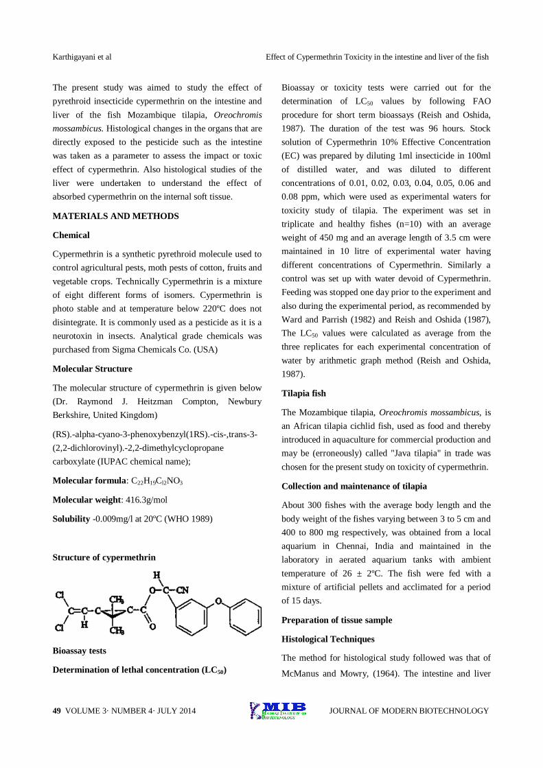
Cypermethrin is a synthetic pyrethroid molecule used to control agricultural pests, moth pests of cotton, fruits and vegetable crops. Technically Cypermethrin is a mixture of eight different forms of isomers. Cypermethrin is photo stable and at temperature below 220ºC does not disintegrate. It is commonly used as a pesticide as it is a neurotoxin in insects. Analytical grade chemicals was purchased from Sigma Chemicals Co. (USA)
Molecular Structure
The molecular structure of cypermethrin is given below (Dr. Raymond J. Heitzman Compton, Newbury Berkshire, United Kingdom)
(RS).-alpha-cyano-3-phenoxybenzyl(1RS).-cis-,trans-3-(2,2-dichlorovinyl).-2,2-dimethylcyclopropane carboxylate (IUPAC chemical name);
Molecular formula: C22H19Cl2NO3
Molecular weight: 416.3g/mol
Solubility -0.009mg/l at 20ºC (WHO 1989)
Structure of cypermethrin
Bioassay tests
Determination of lethal concentration (LC50)
Bioassay or toxicity tests were carried out for the determination of LC50 values by following FAO procedure for short term bioassays (Reish and Oshida, 1987). The duration of the test was 96 hours. Stock solution of Cypermethrin 10% Effective Concentration (EC) was prepared by diluting 1ml insecticide in 100ml of distilled water, and was diluted to different concentrations of 0.01, 0.02, 0.03, 0.04, 0.05, 0.06 and 0.08 ppm, which were used as experimental waters for toxicity study of tilapia. The experiment was set in triplicate and healthy fishes (n=10) with an average weight of 450 mg and an average length of 3.5 cm were maintained in 10 litre of experimental water having different concentrations of Cypermethrin. Similarly a control was set up with water devoid of Cypermethrin. Feeding was stopped one day prior to the experiment and also during the experimental period, as recommended by Ward and Parrish (1982) and Reish and Oshida (1987), The LC50 values were calculated as average from the three replicates for each experimental concentration of water by arithmetic graph method (Reish and Oshida, 1987).
Tilapia fish
The Mozambique tilapia, Oreochromis mossambicus, is an African tilapia cichlid fish, used as food and thereby introduced in aquaculture for commercial production and may be (erroneously) called "Java tilapia" in trade was chosen for the present study on toxicity of cypermethrin.
Collection and maintenance of tilapia
About 300 fishes with the average body length and the body weight of the fishes varying between 3 to 5 cm and 400 to 800 mg respectively, was obtained from a local aquarium in Chennai, India and maintained in the laboratory in aerated aquarium tanks with ambient temperature of 26 ± 2ºC. The fish were fed with a mixture of artificial pellets and acclimated for a period of 15 days.
Preparation of tissue sample
Histological Techniques
The method for histological study followed was that of
McManus and Mowry, (1964). The intestine and liver
Effect of Cypermethrin Toxicity in the intestine and liver of the Fish Karthigayani et al
50 JOURNAL OF MODERN BIOTECHNOLOGY VOLUME 3· NUMBER 4· JULY 2014
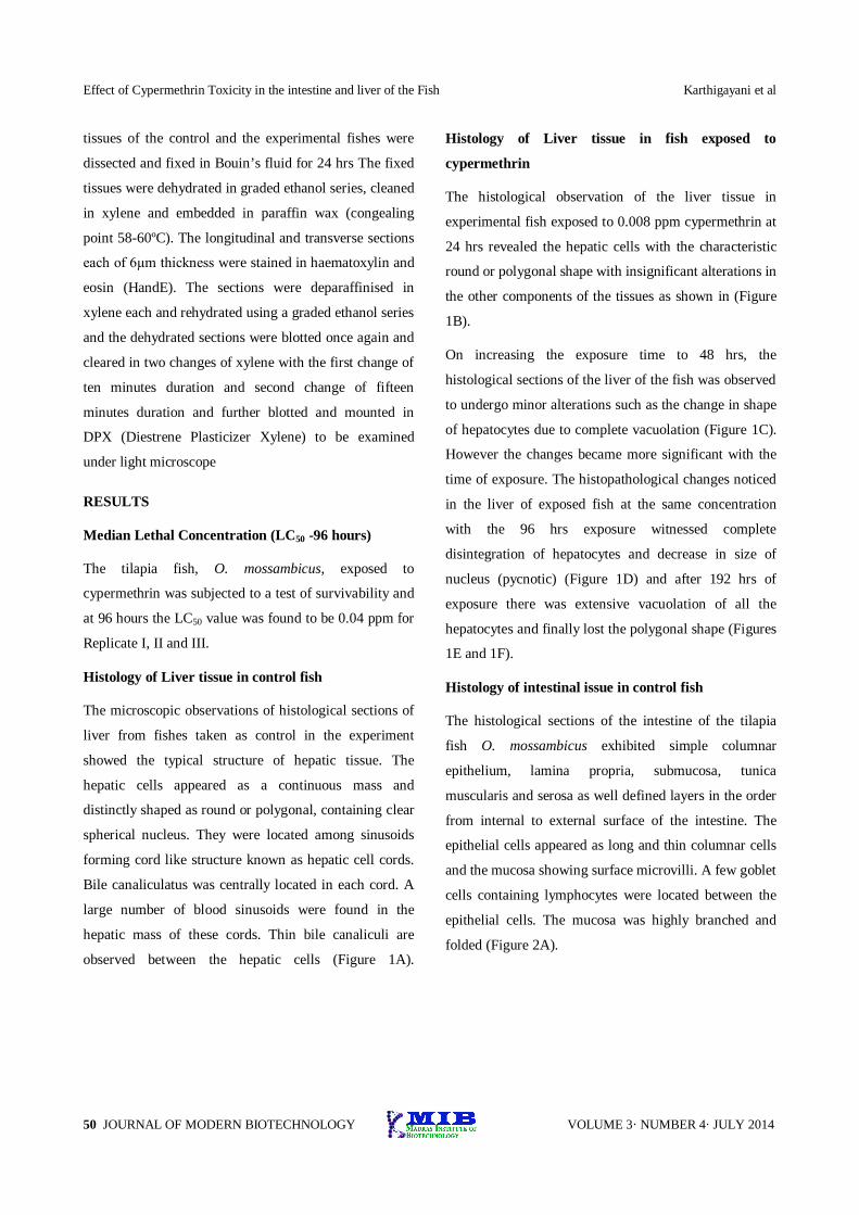
tissues of the control and the experimental fishes were
dissected and fixed in Bouin’s fluid for 24 hrs The fixed
tissues were dehydrated in graded ethanol series, cleaned
in xylene and embedded in paraffin wax (congealing
point 58-60ºC). The longitudinal and transverse sections
each of 6µm thickness were stained in haematoxylin and
eosin (HandE). The sections were deparaffinised in
xylene each and rehydrated using a graded ethanol series
and the dehydrated sections were blotted once again and
cleared in two changes of xylene with the first change of
ten minutes duration and second change of fifteen
minutes duration and further blotted and mounted in
DPX (Diestrene Plasticizer Xylene) to be examined
under light microscope
RESULTS
Median Lethal Concentration (LC50 -96 hours)
The tilapia fish, O. mossambicus, exposed to
cypermethrin was subjected to a test of survivability and
at 96 hours the LC50 value was found to be 0.04 ppm for
Replicate I, II and III.
Histology of Liver tissue in control fish
The microscopic observations of histological sections of
liver from fishes taken as control in the experiment
showed the typical structure of hepatic tissue. The
hepatic cells appeared as a continuous mass and
distinctly shaped as round or polygonal, containing clear
spherical nucleus. They were located among sinusoids
forming cord like structure known as hepatic cell cords.
Bile canaliculatus was centrally located in each cord. A
large number of blood sinusoids were found in the
hepatic mass of these cords. Thin bile canaliculi are
observed between the hepatic cells (Figure 1A).
Histology of Liver tissue in fish exposed to
cypermethrin
The histological observation of the liver tissue in
experimental fish exposed to 0.008 ppm cypermethrin at
24 hrs revealed the hepatic cells with the characteristic
round or polygonal shape with insignificant alterations in
the other components of the tissues as shown in (Figure
1B).
On increasing the exposure time to 48 hrs, the
histological sections of the liver of the fish was observed
to undergo minor alterations such as the change in shape
of hepatocytes due to complete vacuolation (Figure 1C).
However the changes became more significant with the
time of exposure. The histopathological changes noticed
in the liver of exposed fish at the same concentration
with the 96 hrs exposure witnessed complete
disintegration of hepatocytes and decrease in size of
nucleus (pycnotic) (Figure 1D) and after 192 hrs of
exposure there was extensive vacuolation of all the
hepatocytes and finally lost the polygonal shape (Figures
1E and 1F).
Histology of intestinal issue in control fish
The histological sections of the intestine of the tilapia
fish O. mossambicus exhibited simple columnar
epithelium, lamina propria, submucosa, tunica
muscularis and serosa as well defined layers in the order
from internal to external surface of the intestine. The
epithelial cells appeared as long and thin columnar cells
and the mucosa showing surface microvilli. A few goblet
cells containing lymphocytes were located between the
epithelial cells. The mucosa was highly branched and
folded (Figure 2A).

Karthigayani et al Effect of Cypermethrin Toxicity in the intestine and liver of the fish
51 VOLUME 3· NUMBER 4· JULY 2014 JOURNAL OF MODERN BIOTECHNOLOGY
Figure 1A: Transverse section of the intestine of the control fish Oreochromis mossambicus showing the normal organization of intestine. Columnar Epithelium (CE), sub mucosa (SM) and serosa (S). Bouin, 6 µm, HE. X 110.
Figure 1B: Transverse section of the intestine of the fish Oreochromis mossambicus exposed to 24 hrs, sublethal exposure of cypermethrin at 0.008ppm concentrations showing less damage cell wall of the intestine columnar epithelium (EC) sub mucosa (SM) and Serosa (S) in higher magnification Bouin, 6 µm, HE. X 950.
Figure 1C: Transverse section of the intestine of the fish Oreochromis mossambicus, exposed to 48 hrs, sublethal exposure of cypermethrin at 0.008 ppm concentration showing slight damage in the periphery of the cell wall (indicated in arrows). Bouin, 6µm, HE.X 950.
Figure 1D: Transverse section of the intestine of the fish Oreochromis mossambicus exposed to 72 hrs, sublethal exposure of cypermethrin at 0.008 ppm concentration showing disintegration of the cell wall. (indicated in arrows) Bouin, 6 µm, HE. X 950.
Figure 1E: Transverse section of the intestine of the fish Oreochromis mossambicus, exposed to 96 hrs, sublethal exposure of cypermethrin at 0.008ppm concentration showing shrinkage of columnar epithelial cells. Bouin, 6 µm, HE. X 950.
Figure 1F: Transverse section of the intestine of the fish Oreochromis mossambicus exposed to 192 hrs, sublethal exposure of cypermethrin at 0.008 ppm concentration showing severe damage of columnar epithelial cells. Bouin, 6 µm, HE. X 950.
Effect of Cypermethrin Toxicity in the intestine and liver of the Fish Karthigayani et al
52 JOURNAL OF MODERN BIOTECHNOLOGY VOLUME 3· NUMBER 4· JULY 2014
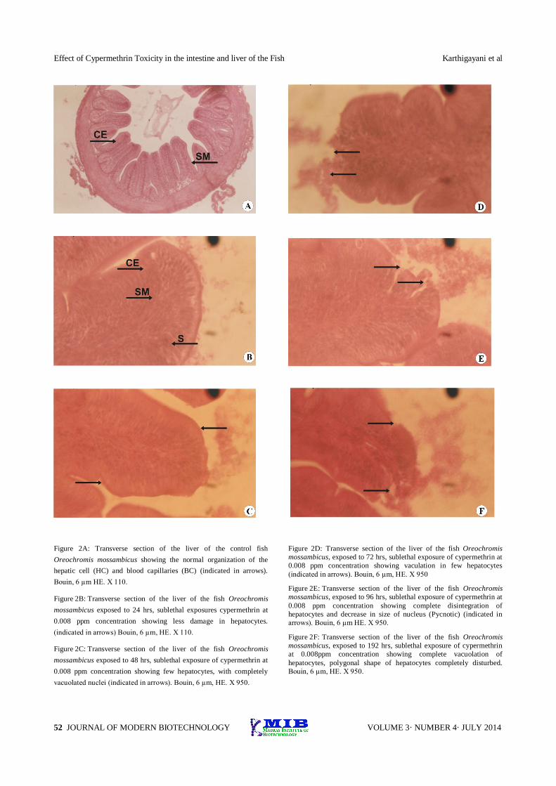
Figure 2A: Transverse section of the liver of the control fish Oreochromis mossambicus showing the normal organization of the hepatic cell (HC) and blood capillaries (BC) (indicated in arrows). Bouin, 6 µm HE. X 110.
Figure 2B: Transverse section of the liver of the fish Oreochromis mossambicus exposed to 24 hrs, sublethal exposures cypermethrin at 0.008 ppm concentration showing less damage in hepatocytes. (indicated in arrows) Bouin, 6 µm, HE. X 110.
Figure 2C: Transverse section of the liver of the fish Oreochromis mossambicus exposed to 48 hrs, sublethal exposure of cypermethrin at 0.008 ppm concentration showing few hepatocytes, with completely vacuolated nuclei (indicated in arrows). Bouin, 6 µm, HE. X 950.
Figure 2D: Transverse section of the liver of the fish Oreochromis mossambicus, exposed to 72 hrs, sublethal exposure of cypermethrin at 0.008 ppm concentration showing vaculation in few hepatocytes (indicated in arrows). Bouin, 6 µm, HE. X 950
Figure 2E: Transverse section of the liver of the fish Oreochromis mossambicus, exposed to 96 hrs, sublethal exposure of cypermethrin at 0.008 ppm concentration showing complete disintegration of hepatocytes and decrease in size of nucleus (Pycnotic) (indicated in arrows). Bouin, 6 µm HE. X 950.
Figure 2F: Transverse section of the liver of the fish Oreochromis mossambicus, exposed to 192 hrs, sublethal exposure of cypermethrin at 0.008ppm concentration showing complete vacuolation of hepatocytes, polygonal shape of hepatocytes completely disturbed. Bouin, 6 µm, HE. X 950.

Karthigayani et al Effect of Cypermethrin Toxicity in the intestine and liver of the fish
53 VOLUME 3· NUMBER 4· JULY 2014 JOURNAL OF MODERN BIOTECHNOLOGY
Histology of intestinal tissues in fish exposed to cypermethrin
The observation of the histological preparations of intestine of fish O. mossambicus exposed to 0.008 ppm of cypermethrin for 24 hrs showed no definite alterations except for an increase in number of goblet cells (Figure 2B) and on increasing the exposure to 48 hrs the intestinal wall, the serosa was eroded (Figure 2C). However, at 72 hrs exposure period the goblet cells increased in number and the disintegration of intestinal serosa continued up to 96 hrs of exposure (Figure 2D). The epithelial columnar cells were observed to change in structural form from well-defined columnar cell to shrunken cells from 96 hrs of exposure to finally disintegrate at 192 hrs of exposure (Figures 2E and 2F).
DISCUSSION
The LC50 value of 0.04 ppm at 96 hrs exposure of cypermethrin a synthetic pyrethroid pesticide has been reported in the present study for tilapia fish O. mossambicus. The reported LC50 value of 14, 50µg/l of deltamethrin (synthetic pyrethroid) in Oreochromis niloticus fingerlings suggested acute pyrethroid sensitivity and toxicity to fish (Glow and Godzi 1994; Karthigayani et al (2014). In Poecilia reticulata the pyrethroid dieldrin was considered more two times more toxic to the fish species than other pesticides (Mittal et al 1994). Many reports have demonstrated the acute toxicity of pyrethroid insecticides on fish species and its developmental stages (Mestres and Mestres 1992 ; Koprucii and Aydin 2004). Evidently the pyrethroid pesticide exposure to fish had deleterious effects on tissues that eventually had lethal effects (Yildririm et al 2006, Singh and Singh 2007).
The present study confirms and demonstrates the toxic effect of cypermethrin on intestine of tilapia fish O. mossambicus that was directly exposed to cypermethrin by ingested water and liver which was exposed to the absorbed cypermethrin. Since, cypermethrin is a chemical of different iso forms and is stable in extreme conditions can withstand disintegration which enhances
its capacity to react with the living tissues without undergoing degradation. The present study was designed to study the toxic effect of cypermethrin with an increase of exposure time to analyse the histological alterations of intestine and liver of tilapia fish O. mossambicus. Recently Cengiz and Unlu, (2006) reported histopathological effects of deltamethrin on the gills, liver, and gut tissues in mosquito fish (Gambusia affinis) after exposure to sublethal concentrations of 0.25 and 0.50 g/L. These results are in agreement with our findings on histological alterations observed in intestine and liver tissues.
The exposure of sublethal concentration of 0.008 ppm of cypermethrin on histological preparations of intestine demonstrated alterations in the intestinal wall. On increasing the exposure of cypermethrin, had a deleterious effect on the internal epithelial lining cells and such changes in intestine, probably induce changes in biological functions of the fish that may contribute to lethargic behavioural responses.
The histological alterations of the liver tissues increase with the exposure time of cypermethrin also indicate that absorption of cypermethrin was normal up to 48hrs and at 96hrs of exposure and the cells were unable to differentially diffuse cypermethrin and thereby higher concentration of cypermethrin possibly accumulated in the liver. The hepatocytes were able to withstand the toxic effect of cypermethrin at sub lethal concentration (0.008ppm) up to 48hrs as the concentration of cypermethrin increased in the liver cells, the hepatocytes apparently showed alterations in shape with more cells changing from polygonal to round shape and the cells becoming more vacuolated due to probable change in an osmatic stress induced by cypermethrin.
Also the pycnosis of nucleus proved the disintegration of regulative functioning of the nucleus and resulting changes in the metabolism of the nutrients reaching the liver cells. This change in metabolic pathway of the essential nutrients such as glucose and amino acid may have direct or indirect influence on the other vital tissue of the fish such as brain and kidney.
Effect of Cypermethrin Toxicity in the intestine and liver of the Fish Karthigayani et al
54 JOURNAL OF MODERN BIOTECHNOLOGY VOLUME 3· NUMBER 4· JULY 2014
The results obtained by the study on toxic effect of cypermethrin clearly demonstrate that, at sublethal concentrations itself the pesticide has deleterious effect on the tissues exposed to it directly or even indirectly. Hence, cypermethrin can be considered as a potent toxic pollutant capable of destroying the balance of aquatic ecosystem.
REFERENCES
Bernet, D, Schmidt H, Meier W, Burkhardt-Holm P and Wahli T. 1999. Histopathology in fish: roposal for a protocol to assess aquatic pollution. Journal of Fish Disease 22: 25–34.
Cengiz, E. I.; Unlu, E. 2006. Sublethal effects of commercial deltamethrin on the structure of the gill, liver and gut tissues of mosquitofish, Gambusia affinis: A microscopic study. Environmental Toxicology and Pharmacology 21: 246–253.
Demoute JP 1989. A brief review of the environmental fate and metabolism of pyrethroids. Pesticide Science 27:375–385
Glow and Godzi 1994. Acute toxicity of deltamethrin and dieldrin to Oreochromis niloticus (LIN). Bull Environ ContamToxicol 52:351–354. Andrew Moore a,*, Colin P. Waring bthe effects of a synthetic pyrethroid pesticide on someaspects of reproduction in Atlantic salmon (Salmosalar L.). Aquatic Toxicology 52 (2001) 1–12
Hill IR. 1989. Aquatic organisms and pyrethroids. PesticSci 27: 429–465.
John, P.J and Prakash, A., 2003. Bioaccumulation of pesticides on some organs of freshwater catfish Mystusvitatus. Bull. Environ. Contam.Toxicol., 70: 1013 – 1016.
Karthigayani, T, Denis M, Andrew Remy AR and Shettu, N. 2014. Histological study of the intestine and liver tissues in the fish Oreochromis mossambicus exposed to cypermethrin. Journal of Modern Biotechnology 3:35–41.
Ko¨pru¨cu¨ K, Aydın R. 2004. The toxic effects of pyrethroid deltamethrin on the common carp (Cyprinuscarpio L.) embryos and larvae. Pestic Biochem Phsiol 80:47–53.
McManus and Mowry, 1964 Staining methods: histological and histochemical. Harper and Row. New York.
Mestres R, Mestres G. 1992. Deltamethrin: Uses and environmental safety. Rev Environ ContamToxicol 124:1–18.
Milam CD, Farris JL, Wilhide JD. 2000. Evaluating mosquito control pesticides for effect on target and non-target organisms. Arch Environ ContamToxicol 39:324–328.10 Demoute 1989
Mittal PK, Adak T, Sharma VP. 1994. Comparative toxicity of certain mosquitocidal compounds to larvivorous fish, Poeciliareticulata. Indian J Malariol 31:43–47.
Pratap B. Singh and, Vandana Singh 2007, Cypermethrin induced histological changes in gonadotrophic cells, liver ,gonads, plasma levels of estradiol-17β And 11-ketotestosterone, and sperm motility in Heterpneustes fossilis (Bloch) Chemosphere 72 (2008) 422-431.
Raymond J. Heitzman Compton, Newbury Berkshire, United Kingdom. Cypermethrin
Reish, D.L., and Oshida, P.S. 1987 Manual of method’s in Aquatic environmental Research, part -10 short –term bioassays. FAO-Tech. 247 - 262
Singh,A., Srivastava, V.K., 1999. Toxic effect of synthetic pyrethroid permethrin on the enzyme system of the freshwater fish Channa striatus. Chemosphere 39, 1951-1956
Smith TM, Stratton GW. 1986. Effects of synthetic pyrethroids insecticides on non-target organisms. Res Rev 97:93–119.
Srivastav AK, Srivastava SK, Srivastav SK. 1997. Impact of deltamethrin on serum calcium and inorganic phosphate of freshwater catfish, Heteropneustesfossilis. Bull Environ ContamToxicol 59:841–846.
Ward GS, Parish PR. 1982.Toxicity tests. In: Manuals of Methods in Aquatic Environment Research, Part 6. FAO Fish. Tech. Pap. 185: 1-23.
ZıynetYildirim, A. CaglanKarasuBenlı, MahmutSelvı, AyhanOzkul,FıgenErkoc¸ OnerKocakAcute Toxicity, Behavioural Changes, and Histopathological Effects of Deltamethrin on Tissues (Gills, Liver, Brain, Spleen, Kidney, Muscle, Skin) of Nile Tilapia (Oreochromis niloticus L.) Fingerlings 2006 Wiley Periodicals, Inc. Environ Toxicology 21: 614–620.