Histological and histochemical observations on the testis of Gobius paganellus
13
Zeitsehrift ffir Zellforschung 65, 350--362 (1965) From the Institute of Zoology and Comparative Anatomy of the University of Camerino and the Zoological Station of Naples, Italy HISTOLOGICAL AND HISTOCHEMICAL OBSERVATIONS ON THE TESTIS OF GOBIUS PAGANELLUS* By HUGH STANLEY * *, GIOVAI~I~I CHIEFFI and VIRGILIO BOTTE With I0 Figures in the Text (Received July 8, 1964) Introduction Although the testes of the teleost fishes have been the subject of numerous histological investigations, some major problems have still not been fully clarified. One of these problems is the presence or absence of steroid-producing interstitial tissue homologous with the Leydig cells of the mammalian testis. COURRIER (1921), KOLMEa and SCHEMINZKY (1922), WEISEL (1949), and POLDEIr (1961) could find no such tissue in some teleost species. Other investigators (C~Am- BEbI~ET, 1930; FOLLENIUS, 1953) report rather typical interstitial tissue which undergoes seasonal variation in relation to the reproductive cycle. MARSHALL and LOFTS (1956) suggested that the apparent absence of interstitial tissue in some teleosts (Esox, Salvelinus, Labeo) is due to a different arrangement of these cells as "lobule boundary cells" instead of interstitial clumps lying among the seminiferous tubules. In Gobius paganellus glandular tissue does occur in small clumps between seminiferous tubules and also as a large mass lying along the mesorchial side of the testis (CouRRIER 1921, EGGERT 1931, ~u 1936, and VIVlEN 1939, 1941). A similarly placed gland in the testis of Blennius was first described as endocrine in nature by C~AM1)Y (1921) and then as exocrine by CHAMeY and GLEu (1922). COU~RIEn (1922) confirmed the latter interpretation, but he further described clusters of small cells as Leydig cell homologous among the exocrine cells. The question of the presence or absence of Sertoli cell homologues is also un- clear from the teleost literature. Most authors agree that the germ cells occur in "cysts" or "follicles" within each of which the germ cells develop synchronously. Some investigators have reported that "connective tissue" cells (TuR~]~R 1919, BOWERS and HOLLIDAu 1961) or "follicle" cells (HANN 1927, WEISEL 1943) make up the "cyst" wall. These cells were interpreted as Sertoli cells by VAUPEL This investigation was supported by research grant RG-6455 from the Division of General Medical Sciences, U.S. Public Health Service. * Postdoctoral Fellow from the Division of General Medical Sciences, U.S. Public Health Service. Supported in part by U.S. Public Health Service Training Grant 5 T1 GM-136, Department of Biological Structure, University of Washington, School of Medicine, Seattle, Washington, U.S.A. Acknowledgements. The authors wish to express their appreciation to Drs. NEWTON lB. EVERETT, JOH1% H. LU]~T and DA:NIEL SZOLLOSI for reading the manuscript ~nd suggesting improvements in it. Special thanks are extended to Professor EDWARD C. ROOSEN-RvNoE for his very thoughtful criticisms during the preparation of this paper.
-
Upload
hugh-stanley -
Category
Documents
-
view
212 -
download
0
Transcript of Histological and histochemical observations on the testis of Gobius paganellus

Zeitsehrift ffir Zellforschung 65, 350--362 (1965)
From the Institute of Zoology and Comparative Anatomy of the University of Camerino and the Zoological Station of Naples, Italy
HISTOLOGICAL AND HISTOCHEMICAL OBSERVATIONS ON THE TESTIS OF GOBIUS PAGANELLUS*
By
HUGH STANLEY * *, GIOVAI~I~I CHIEFFI and VIRGILIO BOTTE
With I0 Figures in the Text
(Received July 8, 1964)
Introduction
Although the testes of the teleost fishes have been the subject of numerous histological investigations, some major problems have still not been fully clarified. One of these problems is the presence or absence of steroid-producing interst i t ial tissue homologous with the Leydig cells of the mammalian testis. COURRIER (1921), KOLMEa and SCHEMINZKY (1922), WEISEL (1949), and POLDEIr (1961) could find no such tissue in some teleost species. Other investigators (C~Am- BEbI~ET, 1930; FOLLENIUS, 1953) report rather typical intersti t ial tissue which undergoes seasonal variation in relation to the reproductive cycle. MARSHALL and LOFTS (1956) suggested tha t the apparent absence of intersti t ial tissue in some teleosts (Esox, Salvelinus, Labeo) is due to a different arrangement of these cells as "lobule boundary cells" instead of interst i t ial clumps lying among the seminiferous tubules.
In Gobius paganellus glandular tissue does occur in small clumps between seminiferous tubules and also as a large mass lying along the mesorchial side of the testis (CouRRIER 1921, EGGERT 1931, ~u 1936, and VIVlEN 1939, 1941). A similarly placed gland in the testis of Blennius was first described as endocrine in nature by C~AM1)Y (1921) and then as exocrine by CHAMeY and GLEu (1922). COU~RIEn (1922) confirmed the la t ter interpretation, but he further described clusters of small cells as Leydig cell homologous among the exocrine cells.
The question of the presence or absence of Sertoli cell homologues is also un- clear from the teleost l i terature. Most authors agree tha t the germ cells occur in "cysts" or "follicles" within each of which the germ cells develop synchronously. Some investigators have reported tha t "connective tissue" cells (TuR~]~R 1919, BOWERS and HOLLIDAu 1961) or "follicle" cells (HANN 1927, WEISEL 1943) make up the "cyst" wall. These cells were interpreted as Sertoli cells by VAUPEL
�9 This investigation was supported by research grant RG-6455 from the Division of General Medical Sciences, U.S. Public Health Service.
�9 * Postdoctoral Fellow from the Division of General Medical Sciences, U.S. Public Health Service. Supported in part by U.S. Public Health Service Training Grant 5 T1 GM-136, Department of Biological Structure, University of Washington, School of Medicine, Seattle, Washington, U.S.A.
Acknowledgements. The authors wish to express their appreciation to Drs. NEWTON lB. EVERETT, JOH1% H. LU]~T and DA:NIEL SZOLLOSI for reading the manuscript ~nd suggesting improvements in it. Special thanks are extended to Professor EDWARD C. ROOSEN-RvNoE for his very thoughtful criticisms during the preparation of this paper.

Histophysiology of Gobius testis 351
(1929), C R A T G - B ~ T (1930), CHAWN and GO~DO~ (1951) and POLDER (1961). Other invest igators have seen no cells they would designate as Sertoli cells (MELAMPY and CAVAZOS 1954, RO]3~RTSON 1958, H~NDERSON 1962). Manyo the r s made no specific character izat ion of the "cys t" wall.
The current s tudy presents a general histological description of the testis of Gobius paganellus at various seasons of the year, along with histochemical evidence of steroidogenic ac t iv i ty in the g landular elements. Special a t t en t ion is given to the quest ion of Sertoli cell homologues.
Materials and Methods
Specimens of Gobius paganellus were collected in shallow waters in the Gulf of Naples during March, April, June, September, October, November and December of 1962 and Febru- ary and April of 1963. For histological observations testis tissue was fixed in Bouin's or Stieve's fixatives. 5 # to 7 # paraffin sections were stained with Weigert's iron haematoxylin or Masson's trichrome stain.
Tissues from the same animals were fixed in 10 % neutral formalin. Frozen sections were then stained with Sudan black B for general lipids. Special tests for the demonstration of cholesterol and its esters, acidic and neutral lipids, phospholipids and lipofuseins were perform- ed as described by PEARSE (1961).
Frozen fresh tissues sectioned on a cryostat were used for enzyme localization. A 5-3fl- hydroxysteroid dehydrogenase was demonstrated according to the technique of WATTE1KB]~RG (1958) as modified by LEvr, DEA~E and RvBI~ (1959). For lactic dehydrogenase the tech- nique as used by the latter authors was used.
Observations
Histology
The testes of immatu re Gobius paganellus are f lat tened, blade-like s t ructures a t tached by a mesorchium to the dorso-lateral aspect of the body wall in the posterior por t ion of the coelom. Anter ior ly the testes are suspended from the ventro- la tera l aspect of the swim bladder.
D T TA
I
GA Fig. 1. Immature testis of GoMus paganellus in transverse section. M. mesorehium ; V. vein; .A. artery ;
GA. gland, anlage; D. deferent duet; T. seminiferous tubule; TA. tuniea albu~nea. 100 •
The immatu re testis is composed of two general par ts approximate ly equal in mass: the seminiferous and the g landular port ions (Fig. 1). The g landular por t ion lies nearest the mesorchium. Along its mesorchial side a large ar te ry and vein course longi tudinal ly , par t ia l ly surrounded by the g landular cells. F r o m these vessels, small branches extend la teral ly into the g landular and seminfferous port ions of the testis.

3 5 2 H . STANLEY, G . C t I I E F F [ a n d V . B O T T E :
F i g . 2. T r a n s v e r s e s e c t i o n t h r o u g h m a t u r e , a c t i v e , J u n e t e s t i s of Gobius paganellus. 1. i n t e r s t i t i a l g l a n d ; S. f r e e s p e r m a t o z o a in t u b u l e l u m i n a . O t h e r a b b r e v i a t i o n s a s i n F i g . 1. T h e s e m i n i f c r o u s t u b u l e s a re
l i n e d b y fol l ic lcs c o n t a i n i n g g e r m cells i n v a r i o u s s t a g e s of d e v c l o p m e n t . A b o u t 50 •
F i g . 3. S e c t i o n t h r o u g h a n a c t i v e ( J u n e ) t e s t i s of G. paganeUus. I . i n t e r s t i t i a l g l a n d t i s s u e ; S. f r ee s p e r m a t o z o a i n l u m e n of s e m i n i f e r o u s t u b u l e . Fo l l ic les c o n t a i n i n g s p e r m a t o c y t e s a n d s p e r m a t i d s l ine
t h e t u b u l e wal l s . A s p e r m a t i d fol l icle (Sp) is o p e n to t h e t u b u l e l u m e n . A b o u t 250 •
F i g . 4. S e c t i o n t h r o u g h t h e w a l l of a s e m i n i f e r o u s t u b u l e s h o w i n g a p r i m a r y s p e r m a t o g o n i u m (G.) a n d fol l ic le c o n t a i n i n g p r i m a r y s p e r m a t o e y t e s (P.S . ) . A n o t h e r p r i m a r y s p e r m a t o c y t e fol l icle is a t t h e
u p p e r lef t . F.IV . fol l ic le wa l l ; F . N . fol l ic le cell n u c l e u s ; T . W . t u b u l e wa] l . A b o u t 1000 •
Histophysiology of Gobius testis 353
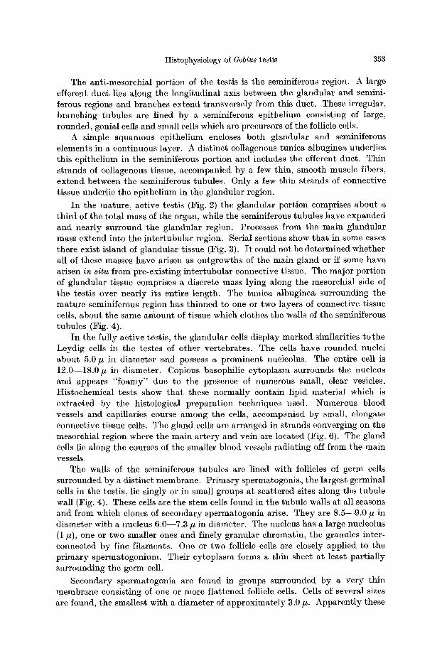
The anti-mesorchial portion of the testis is the seminiferous region. A large efferent duct lies along the longitudinal axis between the glandular and semini- ferous regions and branches extend transversely from this duct. These irregular, branching tubules are lined by a seminiferous epithelium consisting of large, rounded, gonial cells and small cells which are precursors of the follicle cells.
A simple squamous epithelium encloses both glandular and seminiferous elements in a continuous layer. A distinct collagenous tunica albuginea underlies this epithelimn in the seminiferous portion and includes the efferent duct. Thin strands of eollagenous tissue, accompanied by a few thin, smooth muscle fibers, extend between the seminiferous tubules. Only a few thin strands of connective tissue underlie the epithelium in the glandular region.
In the mature, active testis (Fig. 2) the glandular portion comprises about a third of the total mass of the organ, while the scminiferous tubules have expanded and nearly surround the glandular region. Processes from the main glandular mass extend into the intertubular region. Serial sections show tha t in some cases there exist island of glandular tissue (Fig. 3). I t could not be determined whether all of these masses have arisen as outgrowths of the main gland or if some have arisen in situ from pre-existing intertubular connective tissue. The major portion of glandular tissue comprises a discrete mass lying along the mcsorchial side of the testis over nearly its entire length. The tunica albuginea surrounding the mature seminiferous region has thinned to one or two layers of connective tissue cells, about the same amount of tissue which clothes the walls of the seminiferous tubules (Fig. 4).
In the fully active testis, the glandular cells display marked similarities to the Leydig cells in the testes of other vertebrates. The cells have rounded nuclei about 5.0 # in diameter and possess a prominent nucleolus. The entire cell is 12.0--18.0 # in diameter. Copious basophilic cytoplasm surrounds the nucleus and appears " foamy" due to the presence of numerons small, clear vesicles. Histochemical tests show tha t these normally contain lipid material which is extracted by the histological preparation techniques used. Numerous blood vessels and capillaries course among the cells, accompanied by small, elongate connective tissue cells. The gland cells are arranged in strands converging on the mesorchial region where the main ar tery and vein are located (Fig. 6). The gland cells lie along the courses of the smaller blood vessels radiating off from the main vessels.
The walls of the seminiferous tubules are lined with follicles of germ cells surrounded by a distinct membrane. Pr imary spermatogonia, the largest germinal cells in the testis, lie singly or in sma]l groups at scattered sites along the tubule wall (Fig. 4). These ceils are the stem cells found in the tubule walls at all seasons and from which clones of secondary spermatogonia arise. They are 8.5--9.0 # in diameter with a nucleus 6.0--7.3 # in diameter. The nucleus has a large nucleolus (1 lu), one or two smaller ones and finely granular chromatin, the granules inter- connected by fine filaments. One or two follicle cells are closely applied to the pr imary spermatogonium. Their cytoplasm forms a thin sheet at least partially surrounding the germ cell.
Secondary spermatogonia are found in groups surrounded by a very thin membrane consisting of one or more flattened follicle cells. Cells of several sizes are found, the smallest with a diameter of approximately 3.0 #. Apparent ly these

354 H. STANLEY, G. C~IEFFI and V. BOTTE:
secondary spermatogonial nests result from repeated divisions of a single primary spermatogonium and thus constitute a clone. The more numerous the cells are within a follicle, the smaller is their size. This circumstance indicates that there are several spermatogonial divisions and that the germ cells gradually decrease in size with successive divisions. All cells within a single follicle are of similar size and are in the same stage of development. With successive divisions of the spermatogonia, their chromatin gradually takes the form of coarse flakes; this phenomenon resembles the chromatin transformation of comparable stages in other vertebrates (for review see ROOSE~-RtrNGE 1962).
Primary spermatocytes have a diameter of 5.0--6.0/~ and a dense, somewhat contracted nucleus. There is then some enlargement of the cells during the tran- sition from the final spermatogonial stage to the primary spermatocyte.
Secondary spermatocytes are about 3.6 # in diameter while early spermatids have a diameter of about 3.0/~. In a spermatid follicle the cells are often oriented with their heads aligned in one direction, usually against that part of the wall where the follicle cell nuclei are located. In some spermatid follicles the wall of the follicle is open to the lumen of the tubule (Fig. 3). The spermatids are sub- sequently released from the follicle and lie free in the tubule. These luminal spermatozoa have a nucleus about 1.0 # in width and 1.5 # in length.
The follicle cells have an elongate nucleus which may be up to 9.0 # in longest diameter. The nucleus is very pale with one or two prominent nucleo]i and very faint-staining chromatin (Fig. 4). There is often a crease in the nucleus fairly characteristic of Sertoli cell nuclei (ROOSEN-RuNGE 1962). These nuclei usually lie near the tubule wall in the area between two adjacent germinal follicles. They are larger, lighter staining and quite distinct from the connective tissue cells at the periphery of the tubules. Their cytoplasm is drawn out into a thin sheet which forms the wall of the follicle and encloses a nest of synchronously developing germ cells. In some cases only a single cell appears to form the entire follicle wall; in other cases there may be two or more cells. Cytoplasmic projections from the follicle cells extend for at least a short distance between germ cells toward the interior of the follicle.
Spawning by the females of this species occurs in the summer and by August the ovaries of most females are spent. Coincident with the spawning period of the female, the summer testis (June--July) shows dilated seminiferous tubules packed with free spermatozoa (Figs. 2, 3). Most intact follicles contain sperma- tocyte or spermatid stages although a few primary and secondary spermatogonial follicles may be seen. The glandular cells are enlarged and contain many vacuoles. By October the testicular tubules are somewhat collapsed and contain only a few free spermatozoa. Spermatogonial follicles are present in large number, while spermatocyte follicles are less frequent and spermatid follicles are absent. Connec- tive tissue appears relatively more abundant than in the June testis. In February the testis contains a large number of spermatogonial follicles and a few contain spermatocytes or early spermatids. Some free spermatozoa are found in the tubules. In March many follicles contain spermatocytes and some have spermatids. A few free spermatozoa are found in the tubule lumen. Fewer spermatogonial follicles are seen than in February. By April or May the number of spermatid follicles has increased significantly and the tubules are again packed with free spermatozoa as in the June testis in preparation for the summer spawning season.

Histophysiology of Gobius testis 355
Fig. 5. G. paganellus ( June) . T r a n s v e r s e sect ion of the tes t is . F rozen sec t ion s t a ined w i t h Sudan b lack B. Lip ids are concen t r a t ed in the cells of t he m a i n g land a n d in the is lets of in te r s t i t i a l g land
t issue. 50 •
Fig . 6. De ta i l of the in te r s t i t i a l g l and of the preceding f igure . L ip ids are d ispersed as smal l droplets . Sudan b lack B. 224 •
Histochemical tests
An intense sudanophilia could be demonstrated in the glandular cells in all periods of the sexual cycle, even when the spermatogenetic activity appeared to be very low (October--November). All sudanophilia was intracellular in this period. While in Apri l - -June the sudanophilic material occurred in the form of small, uniform droplets throughout the cytoplasm (Figs. 5, 6), toward the end of the spawning season (October--November) the droplets seemed to have coalesced into larger masses (Figs. 7,8). In this latter period, slight sudanophilia was also present in the lumina of the seminiferous tubules.
Throughout the year small droplets of cholesterol are present in the glandular cells. The greatest amount was found in December, that is at the beginning phase of the sexual cycle, while weak but consistently positive reactions were observed

356 H. STANLEY, G. CtIIEFFI and V. BOTTE:
F i g . 7. G. paganelhts (November testis). Transverse sect ion of the testis. Frozen sect ion s ta ined wi th S u d a n b l a c k B . Lipids in g land tissue, some sudanophi l ia in the shmmkcn seminiferous tissue. 55 •
F i g . 8. Detai l of the interst i t ia l g land of the proceeding fignre. Note the distribution of large sndano- philic drops. 350 •
at other t imes of the year. The presence of csterified cholesterol and absence of free cholesterol was indicated by the lack of birefringence in the cells after treat- ment with digitonin and staining with Sudan black B. Fixed unstained sections examined with the polarization microscope show no birefringence in the glandular or seminiferous tissue.
Acidic fats, as indicated by staining with Nile blue sulphate, were more abun- dant than neutral fats, but the latter increased somewhat in the April to June period. Baker's reaction for phospholipids gave consistent ly negative results.
Table. Results o/ various histoehemical reactions per/ormed o~ the interstitial tissue o/ the testis
D e c e m b e r February I Septem-ber Octobcr Novem-ber
Color of non- treated sections
Sudan black B
Schultz Digitonin Birefringence Nile blue
Baker Schmorl js.3fl.hydroxy.
steroid dehydrogenase
Lactate de- hydrogenase
+ d - § small
droplets + - 5
blue + d - + , red d-
d - d - + d - small
droplets d-
blue d-d-d-, red d-
d - §
d-d-
April June
d-
blue ' blue d - § ~ d - + ,
red Jrd- red-~d-
§ + d-d-d-d-
-++d- d - d - +
§ small
droplets
blue § + d-, red d-
§ §
§ moderate droplets
d-
blue § red -4-
d- d--4 + large
droplets §
blue d - - c + , red ~-
d-d-d-d-
§
d- very weakly positive ; d- + weakly positive ; d- d- d- moderately positive; d- ~ =[- d- in- tensely positive; - - negative.

Histophysiology of Gobius testis 357
Fig. 9. G. I)aganellus (April testis). The reac t ion for A5-3 f l -hydroxysteroid dehydrogenase is posi t ive in the cells of the inters t i t ia l tissue. Carmalum, used as a counters ta in , has colored intensely the free
spe rma tozoa ir~ the tubule lumina (S.). I . in ters t i t ia l tissue. 22 •
Fig. 10. G. Paganellus (November testis). Reac t ion for zis-3 f l -hydroxysteroid dehydrogenase is posi t ive only in the inters t i t ia l tissue. 70 •
Histological preparations of Oe tobe r~November testes showed an orange- staining substance in the cytoplasm of the glandular cells. The test for lipofuscins with the reaction of Schmorl gave negative results.
zjs-3fi-hydroxysteroid dehydrogenasc is present exclusively in the glandular cells and absent from the seminiferons tissues (Figs. 9, 10). I t s act ivi ty is easily demonstrable at all seasons, even in November at the end of the sexual cycle. Only in December, at the beginning of the new sexual cycle, is the reaction less intense, and this only in the more central cells of the glandular islets.
Lactic dehydrogenase is present in both seminiferous and glandular tissue, though it appears to be more concentrated in the latter. The reaction is very weak in the period September to December (just after spawning) but becomes increasingly more intense in the period of major spermatogenetic activity.
Discussion
In previous studies on Gobius the glandular tissue was considered homologous to the interstitial tissue of other vertebrates by COURRIE~ (1921), EG(~ERT (1931) and VIvIE~ (1939, 1941). KoLraE~ and SCHEMINZK~ (1922), on the other hand, interpreted these tissues as elements of the suprarenal bodies, while WIART (1936) did not state an opinion as to homology. None of the above studies offered histo- chemical data.

358 H. STA1WLEY, G. CItIEFFI and V. BOTTE:
The presence of cholesterol-positive lipids and especially of zJs-3fl-hydro - xysteroid dehydrogenase activity in the glandular tissue of Gobius constitutes strong if not conclusive evidence of steroidogenic function. I t appears probable tha t this tissue is thus homologous with the androgen-producing interstitial tissue of other vertebrates, However, until the actual steroids have been identified, we cannot be categorical about the androgenic function of the gland.
A somewhat similar glandular mass was described in Blennius by CttAMt'Y and GLEu (1922), who state that the gland cells arise from 1 transformed germinal tissue at the bases of the seminiferous tubules and constitute an exocrine gland. However CovR~IE~ (1922) has described clusters of small cells as Leydig cell homologous among the exoerine cells. SPEROTTI (1940), in agreement with EaGE~T (1931), considered a germinal origin of the gland tissue to be highly unlikely. The glandular tissue here described in Gobius is endocrine and arises independent of the seminiferous tissue.
Histological and histochemical observations reported here confirm a yearly reproductive cycle for Gobius paganellus in the Gulf of Naples. Spawning occurs primarily between June and August but may begin in some individuals in April or May. This spawning period is a little later than that reported by VIVIE~ (1941) for G. paganellus in more northern regions.
After spawning has taken place, there is a period of relative inactivity (Sep- t e m b e r - D e c e m b e r ) during which the testis tubules are somewhat collapsed and contain mostly spermatogonia. The glandular cells are small at this time, cholesterol is in least amount and lactic dehydrogenase activity is low. From December through June there is a gradual increase in the number of follicles containing spermatocytes and spermatids, This is a period of very active division of the germ cells, the divisions being synchronous within any one follicle. There is a con- comitant increase in cholesterol and lactic dehydrogenase activity of the gland cells, a finding tha t suggests higher metabolic activity including increased steroid production. Spermatozoa gradually fill the lumina of the tubules and by late spring (May--June) there are copious quantities of free sperm present at the spawning period.
The follicle or "cys t" wall cells are very similar in position and appearance to the "lobulc boundary cells" in Esox and Salveninus as reported by MARSHALL and LOFTS (1956) and LOFTS and MARSHALL (1957). These authors interpreted the "lobule boundary cells" as Leydig-cell homologues on the basis of their cyclic accumulation and discharge of cholesterol-positive lipids and the apparent absence of interstitial cell masses characteristic of Leydig tissue in other vertebrates. ROBERTSON (1958) found both interstitial cells and "lobule boundary cells" in Salmo gairdnerii and interpreted both types as Leydig-cell homologues. He stated tha t Sertoli cells were not found. HENDERSON (1962) hesitated to make the Leydig cell homology for the "lobule boundary cells" in Salvelinus and suggested that the Leydig cells may be found at other sites. Further, she reported that cytoplasm of the "lobule boundary cells" may form a layer enclosing each group of germ cells in a "cyst" .
LOFTS and MARSHALL (1957) stated that the "lobule boundary cells" in Esox accumulate cholesterol-positive lipids toward the end of the spcrmatogenetic cycle when sperm cells are being shed into the lobule lumen. During the spawning

Histophysi01ogy of Gobius testis 359
period the lobules are emptied of spermatozoa and the "lobule boundary cells" discharge their lipids into the lumen. This material slowly disappears duringthe postspawning months during the time when spermatogonia are undergoing mitosis at the beginning of the next spermatogenetic cycle. LOFTS and MARSHALL inter- preted the discharge of cholesterol-lipids from the "lobule boundary ceils" as indicative of steroid manufacture and discharge of sex hormone.
While MARSHALL and LOFTS considered the "lobule boundary cells" as Leydig- cell homologues, they did not designate any Sertoli-type nutrit ive or supporting cells. Yet such cells have been reported for all classes of vertebrates. The closely- packed groups of germ cells are rather far removed from their blood supply. This seems to call for some intermediate mechanism for transfer of materials to and from the germ cells and perhaps for synthesis of specific compounds necessary to germ cell growth and differentiation. While it is possible tha t the "lobule boundary cells" function both as Sertoli and androgen-producing cells, it must be remem- bered tha t Sertoli cells in other vertebrates accumulate and discharge lipids cyclically and tha t they have been implicated in the production of estrogens (SCOTT and LYNCH 1952, MELAMPu and CAVAZOS 1954). CHIEFFI (1962) pointed out that the "lobule boundary cells" bear a marked similarity to Sertoli cells.
I t is possible to interpret the data of LOFTS and MARSHALL (1957) as evidence of a nutri t ive function in the "lobule boundary cells". At the t ime when germ cells are actively dividing and growing, lipids may be absent or in small amount in the lobule ceils because the germ cells are using all the nutritive material these supporting cells can provide. As divisions cease, however, and the spermatids are transformed, they may have reduced need for nutrients, so tha t these accumulate in the lobule cell cytoplasm in the form of lipid droplets. When the spermatozoa are shed these lipids may be discharged into the lumen from whence they m a y be gradually utilized as a nutrient source by the somatic tissues of the testis and by the actively dividing spermatogonia as the new sexual cycle begins.
In Gobius, as in most teleosts investigated, the Leydig cells are quite distinct from the cells forming the tubule and follicle walls of the germinal tissue. We believe tha t the lat ter cells are homologous with the Sertoli cells of other verte- brates and tha t they may be homologous with the "lobule boundary cells" of Esox, Salvetinus and Satmo. Similar cell components occur also in lampreys (OKKELBERG 1921, CHIEFrI and BOTTE 1962), amphibians such as Rana (W:TsCHI 1924) and Bu/o (BvRGOS and FAWCETT 1956). I t is worthy of note tha t in the newt Taricha torosa MILLER and RO]~B:NS (1954) described two distinct layers of lipid-containing cells, one being a layer of connective tissue cells applied to the outer surface of the seminiferous tubule basement membrane. The other is a layer of Sertoli cells lining the inner aspect of that, membrane. The authors stated tha t both of these cell layers as well as the germinal cells must be considered potential sources of sex hormone. However, only the presence of lipids in all of these cell types is given as evidence of glandular function.
The presence of lipids in cells, even cholesterol-positive lipids, is perhaps not sufficient evidence on which to impute a stcroidogenic function. These lipids could indicate a nutri t ive function or possibly degenerative cha~:ges of the cells. Miller and Robbins' demonstration of lipids in the two layers of cells (inner Sertoli and outer "connective tissue" cells) does not alone constitute good evidence of a

360 H. STANLEY, G. CYcIIEFFI and V. BOTTE:
steroidogenie function for both layers. The photomicrographs in their paper appear to show t h a t the outer layer of cells m a y well be healthy, glandular cells, bu t tha t the inner ring of Sertoli cells shows degenerative changes. Lipid content m a y be due to glandular function in the one layer, but fa t ty degeneration in the other.
While there is a layer of apparent glandular cells distinct from the Sertoli cells in the newt Taricha torosa, this does not seem to be true in the teleost fishes. I t remains to be proven whether the "lobule boundary" or follicle cells may in some species have an steroidogenic function.
Summary
Histological and histochemical observations on the testis of Gobiu8 paganellus during all seasons of the year are described. I n the year ly reproductive cycle, spawning in the Gulf of Naples occured from June through August, testicular recovery and relative inact ivi ty f rom September through December, and active spermatogenesis f rom J a n u a r y to May.
Germ cells develop as clones from single pr imary spermatogonia, each clone in a follicle enveloped by cells which are interpreted as Sertoli cell homologues.
Glandular tissue is present in large amounts both in the form of interstitial islets and as a large mass along the length of the mesorchium. Cholesterol- positive lipids and ~5-3fl-hydroxysteroid dehydrogenase, presumptive evidence for steroid production, are present exclusively in this tissue.
Lipids are present in the glandular cells in acidic and neutral forms. The acid fats are the more abundant , but neutral lipids increase in amount in the period Apr i l - - June . There is, furthermore, a cyclic variat ion in lipid droplet size, small droplets being present in the spring (dm'ing active spermatogenesis) and fewer. larger droplets during the fall (post-spawning period). Phospholipids are lacking,
Lactic dehydrogenase act ivi ty was weak in the relatively inactive post- spawning period, bu t was much more intense during active spermatogenesis.
The glandular tissue in the testis of Gobius is interpreted as homologous with the interstit ial (L]~YDm) tissue found in the testes of higher vertebrates.
Literature BOWERS, A.B., and F. G.T. HOLZIDAY: Histological changes in the gonad associated with
the reproductive cycle of the herring (Clupea harengus L.). Mar. Res. Ser. Scottish Home Dept. 5, 1--16 (1961).
BURGOS, M.H., and D.W. FAWCETT: An electron microscope study of spermatid differenti- ation in the toad, Bu/o arenarum Hensel. J. biophys, biochem. Cytol. 2, 223--240 (1956).
CI~AMI'Y, C. : La structure remarquable du testicule des blennies. C.R. Ass. Anat. (Paris) 16, 165--170 (1921).
- - , et P. GLEY: La glande du testicule des blennies et sa signification. Bull. Soc. Zool. Ft. 47, 199--208 (1922).
CHAVIN, W., and M. GORDON: Sex determination in Platypoecilus maculatus. I. Differenti- ation of the gonads in members of Ml-mMe broods. Zoologica 36, I35--145 (1951).
Cm~FFI, G. : Aspetti endrocrinotogici della riproduzione nei pesci. Boll. ZooI. 29, 149--196 (1962).
- - , e V. BOTTE: :[1 tessuto interstizia]e del testicolo dei ciclostomi. Ricerche istologiche ed istochimiche in Lampetra/luviatilis e Lampetra planeri. R. Ist. Sci. Camerino 3, 90 95 (1962).

Histophysiology of Gobius testis 361
COIIRI~I~R, R. : Sur l 'existence d'une glande interstitielle dans le testicule des Poissons. C. R. Soc. Biol. (Paris) 85, 939--941 (192l).
- - Sur l'existence d'une glande interstitielle dans le testicule des blennies. Bull. Soc Zool. Fr. 47, 458~462 (1922).
CRAIG-BENNET, A.: The reproductive cycle of the three-spined stickleback (Gasterosteus aculeatus, Linn.). Phil. Trans. ]3 219, 197--280 (1930).
E~GEI~T, ]3. : Die Geschlechtsorgane der Gobiiformes und ]31enniiformes. Z. wiss. Zool. 189, 249--558 (1931).
FOLLEI~IVS, E. : Contribution a l '~tude du d~terminisme de la differenciation des caractgres sexuels chez lcs cyprinodontes. Action des rayons X sur les gonades de Lebistes reticulatus Rcgan. Bull. biol. 87, 68--91 (1953).
HAN~, H. W. : The histology of the germ cells of Cottus bairdii Girard. J. Morph. 43, 427--497 (1927).
HENDERSO.'~, N.E. : The annual cycle in the testis of the eastern brook trout, Salvelinus ]ontinalis (Mitchill). Canad. J . Zool. 40, 631--641 (1962).
KOLMEI~, W., and F. SC~EMINZKu Finden sich Zwischenzellen nur bei hSheren Wirbel- tieren ? Pfliigers Arch. ges. Physiol. 194, 352--361 (1922).
LEvy, H., H.W. D E A ~ , and ]3. RUBI~I: Visualization of steroid-3/~-ol-dehydrogenase activi- ty in tissues of intact and hypophysectomized rats. Endocrinology 65, 932--943 (1959).
LOFTS, B., and A. J. MARSHALL: Cyclical changes in the distribution of the testis lipids of a teleost fish, Esox lucius. Quart. J. micr. Sci. 98, 79--88 (1957).
MARSI~ALL, A. J . , and B. LOFTS: The Leydig-cell homologue in certain teleost fishes. Nature (Lond.) 177, 704--705 (1956).
MELAM~Y, R.M., and L.F. CAvAzos: Comparative study of lipids in vertebrate testes. Proc. Soc. exp. Biol. (:N.Y.) 87, 297--303 (1954).
MILLER, M.R., and M.E. ROBBINS: The reproductive cycle in Taricha torosa (Triturus torosus). J. exp. Zool. 125, 415--445 (1954).
OKKELBERG, P. : The early history of the germ cells in the brook lamprey Entosphenus wilderi (Gage), up to and including the period of sex differentiation. J. Morph. 85, 1--151 (1921).
PEARSE, A .G.E . : Histochemistry theoretical and applied, 2nd ed., 998 p. Boston: Little, Brown & Co. 1961.
POLDER, J. J ,W. : Cyclical changes in testis and ovary related to maturity stages in the north sea herring, Clupea harengus L. Arch. n~erl. Zool. 14, 45--60 (1961).
ROBERTSON, O. t t . : Accelerated development of testis Mter unilateral gonadectomy, with observations on normal testis of rainbow trout. Fish. Bull., Fish and Wildlife Service, U.S. Dept. Interior 58, 9--30 (1958).
ROOSEI~-RUI~GE, E. C. : The process of spermatogenesis in mammals. Biol. Rev. 87, 343--377 (1962).
SCOTT, W.W., and K.M. LY~c~I: The Sertoli Cell, p. 37--60. In: Studies on testis and ovary, eggs and sperm. E.T. EI~GLE (ed.) 237 p. Springfield (Ill.): Ch. C. Thomas 1952.
SFEROTTI, G. : Ricerce sull 'apparato riproduttivo maschile del Blennius/luviatilis. Atti reale Ist. veneto Sci. Lettere ed Arti, tomo C-parte II , Sci. nat. 2, 117--143 (1940).
TeR~EIr D. L. : The seasonal cycle in the spermary of the perch. J. Morph. 82, 681--711 (1919). VAUFEL, J. : The spermatogenesis of Lebistes reticulatus. J. Morph. 47, 555--587 (1929). VIVlEN, J . H . : Etude pr61iminaire des annexes du tractus g6nital d 'un Gobiid6: (Gobius
paganellus L. Tray. Star. Zool. Wimereux 18, 713--721 (1939). - - Contribution a l '6tude de la physiologie hypophysaire dans ses relations avec l 'appareil
g~nitM, la thyroide et les corps suprar6naux chez les poissons selaciens et t616ost~ens, Scyliorhinus canicula et Gobius paganeUus. Bull. biol. France et Belg. 75, 257--309 (1941).
WATTE~IBERG, L.W. : Microscopic histochemical demonstration of steroid-3fi-ol-dehydroge- nase in tissue sections. J. Histochem. Cytochem. 6, 225--232 (1958).
W:EmEL, G.F. : A histological study of the testes of the sock-eye salmon (Oncorhynchus nerka). J. Morph. 78, 207---230 (1943).
- - The seminal vesicles and testes of GiUichthys, a marine teleost. Copeia 1949, 101--110 (1949). z. Zellforsch., Bd. 65 25

362 H. STANLEY, G. CttIEleFI and V. BOTTE: Histophysiology of Gobius testis
WIART, P. : Variations saisonni6res du testicule des t616ost6ens. Tray. Star. Biol. Roscoff. 14, 79--86 (1936).
WITSC~I, E. : Die Entwicklung der Keimzellen der Rana temporaria L. Erster Teih Urkeim- zellen und Spermatogenese. Z. Zellforsch. l , 523--561 (1924).
Dr. HVGK STANLEY, Department of Biological Structure Seattle, Washington, 98105, University of Washington School of Medicine
Prof. GIovAN~-I CHIE~FI and Dr. VIRGILIO BOTTE Istituto di Zoologia ed Anatomia Comparata dell' Universitk di Camerino, Italy