Histological and histochemical evaluation of human oral ... · Summary. Reconstruction of large...
10
Summary. Reconstruction of large oral mucosa defects is often challenging, since the shortage of healthy oral mucosa to replace the excised tissues is very common. In this context, tissue engineering techniques may provide a source of autologous tissues available for transplant in these patients. In this work, we developed a new model of artificial oral mucosa generated by tissue engineering using a fibrin-agarose scaffold. For that purpose, we generated primary cultures of human oral mucosa fibroblasts and keratinocytes from small biopsies of normal oral mucosa using enzymatic treatments. Then we determined the viability of the cultured cells by electron probe quantitative X-ray microanalysis, and we demonstrated that most of the cells in the primary cultures were alive and had high K/Na ratios. Once cell viability was determined, we used the cultured fibroblasts and keratinocytes to develop an artificial oral mucosa construct by using a fibrin-agarose extracellular matrix and a sequential culture technique using porous culture inserts. Histological analysis of the artificial tissues showed high similarities with normal oral mucosa controls. The epithelium of the oral substitutes had several layers, with desmosomes and apical microvilli and microplicae. Both the controls and the oral mucosa substitutes showed high suprabasal expression of cytokeratin 13 and low expression of cytokeratin 10. All these results suggest that our model of oral mucosa using fibrin-agarose scaffolds show several similarities with native human oral mucosa. Key words: Fibrin-agarose, Constructs, Tissue engineering. Introduction Different surgical procedures carried out in the oropharyngeal region frequently result in large tissue defects (Schultze-Mosgau, 2004). Reconstruction of these defects is challenging, and oral and maxillofacial surgeons are often confronted with a shortage of oral mucosa to replace the excised tissues (Song et al., 2004). Although it has been demonstrated that primary reconstruction of large oral defects is always more advantageous than secondary reconstructions, primary surgical closure of large oral defects is extremely difficult. In these cases, various types of skin-bearing flaps have been proposed as autologous substitutes of the oral mucosa (Baumann et al., 1996). In some patients, however, the use of skin-bearing flaps is often associated to complications, such as the presence of adnexal structures causing hair growth on the implanted graft or excessive keratinisation of the reconstructed tissue (Toft et al., 2000). All these disadvantages sometimes cause a significant morbidity and aesthetic and functional limitations for the patients submitted to these techniques. Construction of biological substitutes of the human oral mucosa by tissue engineering could contribute to solve these problems and complications. By using these techniques, it is possible to develop efficient substitutes of different organs and tissues for therapeutic use, including skin (Meana et al., 1998; Llames et al., 2004, 2006), cornea (Nishida, 2003; Alaminos et al., 2006), urothelium (Wunsch et al., 2005) and blood vessels (Pascual et al., 2004). In this sense, some researchers have recently proposed different tissue engineering techniques for construction of an organotypic substitute of the oral mucosa (Lauer and Schimming, 2001; Schultze-Mosgau et al., 2004; Song et al., 2004). Evaluation of the viability of the cells kept in culture is very important before these cells can be used for clinical purposes and, especially, for construction of artificial organs and tissues by tissue engineering. In addition, the construction of artificial organs and tissues is highly Histological and histochemical evaluation of human oral mucosa constructs developed by tissue engineering M.C. Sanchez-Quevedo 1 , M. Alaminos 1,2 , L.M. Capitan 3 , G. Moreu 4 , I. Garzon 1 , P.V. Crespo 1 and A. Campos 1 1 Department of Histology, University of Granada, Spain, 2 Fundación Hospital Clínico, Granada, Spain, 3 Division of Oral and Maxillofacial Surgery, University Hospital Virgen de las Nieves, Granada, Spain and 4 Department of Stomatology, University of Granada, Spain Histol Histopathol (2007) 22: 631-640 Offprint requests to: Prof. M.C. Sanchez-Quevedo, Department of Histology, University of Granada, Avenida de Madrid 11, E-18012, Granada, Spain. e-mail: [email protected]. This work is dedicated to Prof. J. Gómez on his 85th anniversary. DOI: 10.14670/HH-22.631 http://www.hh.um.es Histology and Histopathology Cellular and Molecular Biology
Transcript of Histological and histochemical evaluation of human oral ... · Summary. Reconstruction of large...
Summary. Reconstruction of large oral mucosa defectsis often challenging, since the shortage of healthy oralmucosa to replace the excised tissues is very common. Inthis context, tissue engineering techniques may provide asource of autologous tissues available for transplant inthese patients. In this work, we developed a new modelof artificial oral mucosa generated by tissue engineeringusing a fibrin-agarose scaffold. For that purpose, wegenerated primary cultures of human oral mucosafibroblasts and keratinocytes from small biopsies ofnormal oral mucosa using enzymatic treatments. Thenwe determined the viability of the cultured cells byelectron probe quantitative X-ray microanalysis, and wedemonstrated that most of the cells in the primarycultures were alive and had high K/Na ratios. Once cellviability was determined, we used the culturedfibroblasts and keratinocytes to develop an artificial oralmucosa construct by using a fibrin-agarose extracellularmatrix and a sequential culture technique using porousculture inserts. Histological analysis of the artificialtissues showed high similarities with normal oralmucosa controls. The epithelium of the oral substituteshad several layers, with desmosomes and apicalmicrovilli and microplicae. Both the controls and theoral mucosa substitutes showed high suprabasalexpression of cytokeratin 13 and low expression ofcytokeratin 10. All these results suggest that our modelof oral mucosa using fibrin-agarose scaffolds showseveral similarities with native human oral mucosa.Key words: Fibrin-agarose, Constructs, Tissueengineering.
Introduction
Different surgical procedures carried out in theoropharyngeal region frequently result in large tissuedefects (Schultze-Mosgau, 2004). Reconstruction ofthese defects is challenging, and oral and maxillofacialsurgeons are often confronted with a shortage of oralmucosa to replace the excised tissues (Song et al., 2004).Although it has been demonstrated that primaryreconstruction of large oral defects is always moreadvantageous than secondary reconstructions, primarysurgical closure of large oral defects is extremelydifficult. In these cases, various types of skin-bearingflaps have been proposed as autologous substitutes of theoral mucosa (Baumann et al., 1996). In some patients,however, the use of skin-bearing flaps is often associatedto complications, such as the presence of adnexalstructures causing hair growth on the implanted graft orexcessive keratinisation of the reconstructed tissue (Toftet al., 2000). All these disadvantages sometimes cause asignificant morbidity and aesthetic and functionallimitations for the patients submitted to these techniques. Construction of biological substitutes of the human
oral mucosa by tissue engineering could contribute tosolve these problems and complications. By using thesetechniques, it is possible to develop efficient substitutesof different organs and tissues for therapeutic use,including skin (Meana et al., 1998; Llames et al., 2004,2006), cornea (Nishida, 2003; Alaminos et al., 2006),urothelium (Wunsch et al., 2005) and blood vessels(Pascual et al., 2004). In this sense, some researchershave recently proposed different tissue engineeringtechniques for construction of an organotypic substituteof the oral mucosa (Lauer and Schimming, 2001;Schultze-Mosgau et al., 2004; Song et al., 2004).Evaluation of the viability of the cells kept in culture isvery important before these cells can be used for clinicalpurposes and, especially, for construction of artificialorgans and tissues by tissue engineering. In addition, theconstruction of artificial organs and tissues is highly
Histological and histochemical evaluation of human oral mucosa constructs developed by tissue engineeringM.C. Sanchez-Quevedo1, M. Alaminos1,2, L.M. Capitan3, G. Moreu4, I. Garzon1, P.V. Crespo1 and A. Campos11Department of Histology, University of Granada, Spain, 2Fundación Hospital Clínico, Granada, Spain, 3Division of Oral and Maxillofacial Surgery, University Hospital Virgen de las Nieves, Granada, Spain and4Department of Stomatology, University of Granada, Spain
Histol Histopathol (2007) 22: 631-640
Offprint requests to: Prof. M.C. Sanchez-Quevedo, Department ofHistology, University of Granada, Avenida de Madrid 11, E-18012,Granada, Spain. e-mail: [email protected] work is dedicated to Prof. J. Gómez on his 85th anniversary.
DOI: 10.14670/HH-22.631
http://www.hh.um.es
Histology andHistopathologyCellular and Molecular Biology
dependent on the availability of an appropriatebiocompatible scaffold that allows the cells to attach andgrow both in vitro and in vivo.Evaluating the viability of cultured cells is not easy,
and different approaches have been proposed to datewith that purpose. A number of methods are based on thedetection of permeability alterations in the cellmembrane by trypan blue or propidium iodide staining(Bergert et al., 2005), or quantification of intracellularenzymes in the culture medium (Chen and Wagner,2001). However, most of these techniques are notaccurate enough to detect early cell damage, but onlyidentify cell alterations once they have becomeirreversible. In most cases a positive result with thesetechniques reveals that the integrity of the cellmembrane has been lost. For these reasons, suchmethods are not sensitive enough to detect cells that areprone to death but do not display any cell membranealterations yet.In contrast, one of the most sensitive techniques for
determination of cell viability is quantification of theionic content of the cultured cells, especially potassiumand sodium (Fernandez-Segura et al., 1999; Roomans,2002a,b). Histochemical x-ray microanalysis associatedwith electron microscopy is the most powerful approachto measure total elemental composition, making itpossible to simultaneously determine the concentrationsof different elements and the ultrastructure of the cells(Somlyo et al., 1989; Krep et al., 1996). By using thiscombined biochemical and morphological technique, ourgroup has previously quantified the ionic content ofdifferent cell types, including U937 cells (Fernandez-Segura et al., 1999), epithelial tumor cells (Fernandez-Segura et al., 1997) and K562 cells (Warley, 1994).However, the microanalytical pattern of isolated humanoral keratinocytes and fibroblasts kept in culture fortissue engineering purposes has not been determined sofar.On the other hand, different biomaterials have been
used as stromal substitutes. Ideally, a good biomaterialshould be biocompatible and consistent, and epithelialcells should be able to adhere and grow on and within it.Currently, the biomaterials that have been used mostoften to construct epithelial/stromal tissue substitutes aretype I collagen and fibrin. Type I collagen has beenextensively used as a scaffold in tissue engineering forthe construction of artificial skin, oral mucosa, corneasand other tissues (Orwin and Hubel, 2000; Reichl andMuller-Goymann, 2003). However, collagen is anexpensive material and tends to shrink and lose volumewhen the cells are seeded within the scaffold (Tegtmeyeret al., 2001). In addition, stromal substitutes made ofcollagen are not stable and are quickly degraded (Chenet al., 2005). However, human fibrin has been proposedas a stromal substitute to construct different tissuesubstitutes, especially human skin (Meana et al., 1998;Llames et al., 2004, 2006), and has the advantages oflow cost, availability and good tolerance to cells. Incontrast with collagen gels, fibrin gels are not contracted
by stromal cells (Meana et al., 1998; Llames et al., 2004,2006), although its consistency may be sometimes poor.Other materials such as agarose are commonly used intissue engineering of the cartilage (Aufderheide andAthanasiou, 2005), but barely used in combination withother biomaterials. By using a mixture of human fibrinand agarose, our research group was able to construct anefficient substitute of the rabbit cornea with good resultsin terms of consistency, transparency and cell growth(Alaminos et al., 2006).In this work, we have developed a new model of
artificial oral mucosa using viable human fibroblasts andkeratinocytes and a hydrogel scaffold made of a mixtureof fibrin and agarose. Materials and methods
Human tissue samples
Twenty small biopsies corresponding to normalhuman oral mucosa were obtained from healthy donorsundergoing minor oral surgery under local anesthesia inthe School of Dental Sciences of the University ofGranada. Average size of the tissue samples was 3x3x2mm. Immediately after extraction, all tissues were keptin a transport medium at 4°C (Dulbecco’s modifiedEagle’s medium DMEM; 100 U/ml of penicillin G, 100µg/ml of streptomycin, and 0.25 µg/ml of amphotericinB) and processed in the following 24 h.All patients provided consent to participate in the
study. The work was approved by the local researchcommittee (number 01/04).Primary cultures of oral fibroblasts and keratinocytes
On arrival at the laboratory, all samples were washedtwice in phosphate buffered saline (PBS) and incubatedovernight at 37°C in a mixture of DMEM and 2 mg/mlof Clostridium histolyticum collagenase I (Gibco BRLLife Technologies, Karlsruhe, Germany). This enzymatictreatment is able to digest all the extracellular matrix ofthe oral chorion and release the fibroblasts entrappedthere. Once the samples were digested, and to obtainprimary cultures of human oral fibroblasts, detachedconnective tissue components, including fibroblasts,were collected by centrifugation and expanded in cultureflasks containing DMEM medium supplemented withantibiotics (100 U/ml of penicillin G, 100 µg/ml ofstreptomycin, and 0.25 µg/ml of amphotericin B) and10% of fetal bovine serum (FBS) using standard cultureconditions. Then, undigested oral epithelium waswashed in PBS, cut into small pieces and co-culturedwith a layer of mitomycin C-treated (10 mg/ml) 3T3feeder cells (8-10x103 cell/cm2) (Rheinwald and Green,1975). The medium used in this case was a 3:1 mixtureof DMEM and Ham’s F12 supplemented with 10 % fetalcalf serum, 1% antiobiotics, 24 µg/ml adenine, 0.4mg/ml hydrocortisone, 5 mg/ml insulin, 10 ng/mlepidermal growth factor, 1.3 ng/ml triiodothyronine and
632Oral mucosa constructs
8 ng/ml of cholera toxin. In all cases, the cells were incubated at 37°C in 5%
carbon dioxide. The medium was changed every threedays, and subcultivation of the cultured cells was carriedout using a trypsin 0.5 g/l-EDTA 0.2 g/l-solution at 37°Cfor 10 min. Keratinocytes and fibroblasts used for tissueengineering of the tissue models always corresponded tothe three first cell subcultures. Histochemical X-ray microanalytical evaluation ofkeratinocytes and fibroblasts cells
For histochemical X-ray microanalysis, culturedfibroblasts and keratinocytes cells were subcultured ongold grills covered with a thin layer of pioloform (TedPella Inc., CA, USA), sterilized overnight under UVlight, as a physical support to the cells. Cultured cellswere seeded at a density of 5000 cells per grid andincubated for 24 h using specific culture media forfibroblasts or keratinocytes. After 24 h of culture, goldgrids containing the cultured cells were washed with ice-cold distilled water for 5 s to remove the extracellularmedium and immediately plunge-frozen in liquidnitrogen following previously published protocols(Abraham et al., 1985; Warley, 1994; Fernandez-Seguraet al., 1997). After cryofixation the grills were placed ina precooled aluminum specimen holder at liquidnitrogen temperature and freeze-dried at increasingtemperatures for 24 h in a E5300 Polaron freezer-drierapparatus equipped with a vacuum rotatory pumpsystem. Freeze-dried gold grills were carbon-coated in ahigh-vacuum coating system and microanalyzed withinthe following hours.Histochemical X-ray microanalysis of the specimens
was performed using a Philips XL30 scanning electronmicroscope (SEM) equipped with an EDAX DX-4microanalytical system and a solid-state backscatteredelectron detector. The samples were examined with SEMusing a combination of secondary electron (SE) andbackscattering imaging modes. For X-ray microanalysis, the analytical conditions
were: tilt angle 0°, take-off angle 35° and workingdistance 10mm. All spectra were collected in the spotmode at 10,000x for 200 s live time, and the number ofcounts per second by the detector was about 500. Todetermine total ion content, we used the peak-to-local-background (P/B) ratio method (Statham and Pawley,1978; Boekestein et al., 1984; Fernandez-Segura et al.,1997) with reference to standards composed of 20%dextran containing known amounts of inorganic salts(Warley, 1990). In total, we quantified the ionic contentof 25 cultured keratinocytes and 25 cultured fibroblasts.Fabrication of a fibrin-agarose stromal substitute
Fibrin was obtained from frozen plasma of humanblood donors (kindly provived by Dr. Fernandez-Montoya, Human Tissue Bank of Granada). To producea fibrin gel (Meana et al., 1998; Llames et al., 2004,
2006), 21 ml of human plasma were added to 250,000cultured fibroblasts resuspended in 2 ml of DMEM with10% FCS. To prevent degradation of the scaffold byfibrinolysis, the mixture was supplemented with 200 µlof tranexamic acid. Finally, 2 ml of 1% CaCl2 wereadded to the solution to precipitate fibrin polymerization.At the same time, type VII agarose was melted andsolved in PBS and added to the fibrin mixture at a finalconcentration of 0.1%, immediately after the CaCl2 wasadded. Finally, the mixture was seeded in culture insertsand allowed to solidify at 37°C for 2 hours.Construction of oral mucosa substitutes by tissueengineering
Organotypic oral mucosa constructs were developedby using a sequential culture technique as describedbelow. To ensure both a submerged cultivation and aproper differentiation of the multilayered epithelium, weused the air-liquid culture technique (Reichl and Muller-Goymann, 2003). For that purpose, bioengineered oralmucosa was assembled in Transwell culture inserts with0.4 µm porous membrane (Costar, Corning Inc.,Corning, NY, USA). This pore size allows the nutrientsto cross through the membrane of the insert but preventsmigration of the cells to the other compartment.To construct an artificial oral mucosa, the stromal
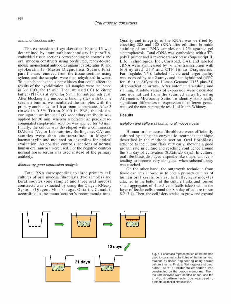
substitute was developed directly on the porousmembrane of the culture inserts. Twenty-four hours afterthe stromal matrix substitute had solidified, human oralkeratinocyte cells were seeded on top of the constructedstroma (approximately 1,000,000 keratinocytes per 25ml construct), and cultured for 10 days submerged inkeratinocytes culture medium. When keratinocyte cellsreached confluence, the air-liquid culture technique wasused for 21 more days. The whole process isschematically shown in Figure 1.Microscopic evaluation of artificial oral mucosa
Samples for scanning electron microscopy werefixed in cacodylate-buffered 2.5% glutaraldehyde andpostfixed in 1% osmium tetroxide for 90 minutes. Afterfixation, the samples were dehydrated in increasingconcentrations of acetone (30%, 50%, 70%, 95% and100%), critical point dried, mounted on aluminum stubs,sputter coated with gold according to routine procedures(Sanchez-Quevedo et al., 1994), and examined in ascanning electron microscope (Quanta 200; FEI,Eindhoven, The Netherlands) using a high vacuummode. For transmission electron microscopy, sampleswere fixed, postfixed and dehydrated as described abovefor scanning electron microscopy, and then embedded inSpurr’s resin and cut into ultrathin sections with anultramicrotrome. For analysis, the sections were stainedwith aqueous uranyl acetate and lead citrate andexamined with a transmission electron microscope(EM902; Carl Zeiss Meditec, Inc., Oberkochen,Germany).
633Oral mucosa constructs
Immunohistochemistry
The expression of cytokeratins 10 and 13 wasdetermined by immunohistochemistry in paraffin-embedded tissue sections corresponding to controls andoral mucosa constructs using prediluted, ready-to-use,mouse monoclonal antibodies against cytokeratin 10 andcytokeratin 13 (Master Diagnostica, Spain). First,paraffin was removed from the tissue sections usingxylene, and the samples were then rehydrated in water.To quench endogenous peroxidases that could affect theresults of the hybridization, all samples were incubatedin 3% H2O2 for 15 min. Then, we used 0.01 M citratebuffer (PH 6.0) at 98°C for 5 min for antigen retrieval.After blocking any unspecific binding sites with bovineserum albumin, we incubated the samples with theprimary antibodies for 1 h at room temperature. After 3rinses in 0.5% Triton-X100 in PBS, the biotin-conjugated antimouse IgG secondary antibody wasapplied for 30 min, whereas a horseradish peroxidase-conjugated streptavidin solution was applied for 40 min.Finally, the colour was developed with a commercialDAB kit (Vector Laboratories, Burlingame, CA) andsamples were then counterstained in Mayer ’shaematoxylin and mounted on coverslips for opticalevaluation. As positive controls, sections of normalhuman oral mucosa were used. For the negative controlsnormal horse serum was used instead of the primaryantibody.Microarray gene-expression analysis
Total RNA corresponding to three primary cellcultures of oral mucosa fibroblasts (two samples) andkeratinocytes (one sample) and three oral mucosaconstructs was extracted by using the Qiagen RNeasySystem (Qiagen, Mississauga, Ontario, Canada),according to the manufacturer’s recommendations.
Quality and integrity of the RNAs was verified bychecking 28S and 18S rRNA after ethidium bromidestaining of total RNA samples on 1.2% agarose gelelectrophoresis. Total cDNA was synthesized with a T7-polyT primer and a reverse transcriptase (Superscript II,Life Technologies, Inc., Carlsbad, CA), and labeledcRNA were synthesized by in vitro transcription withbiotinylated UTP and CTP (Enzo Diagnostics,Farmingdale, NY). Labeled nucleic acid target qualitywas assessed by test-2 arrays and then hybridized (45°Cfor 16 h) to Affymetrix Human Genome U133 plus 2.0oligonucleotide arrays. After automated washing andstaining, absolute values of expression were calculatedand normalized from the scanned array by usingAffymetrix Microarray Suite. To identify statisticallysignificant differences of expression of different genes,we used the non-parametric test U of Mann-Whitney.Results
Isolation and culture of human oral mucosa cells
Human oral mucosa fibroblasts were efficientlycultured by using the enzymatic treatment techniquedescribed in the methods section. Oral fibroblastsattached to the culture flask very early, showing a goodgrowth rate in culture and reaching confluence aroundthe 8th day of cultivation (8.32±3.23 days). In culture,oral fibroblasts displayed a spindle-like shape, with cellstending to become very elongated when subconfluencywas reached.On the other hand, the outgrowth technique from
tissue explants allowed us to obtain primary cultures ofhuman oral keratinocytes. Initially, keratinocytesattached to the bottom of the culture flasks and formedsmall aggregates of 4 to 5 cells (cells islets) within thelayer of feeder cells around the 8th day of culture (mean8.2±3.1). Then, the cell islets tended to grow and expand
634Oral mucosa constructs
Fig. 1. Schematic representation of the methodused to construct substitutes of the human oralmucosa by tissue engineering using porousculture inserts. First, a fibrin-agarose stromalsubstitute with fibroblasts embedded wasconstructed on the porous membrane. Then,the keratinocytes were seeded on top, and theair-l iquid culture technique was used topromote epithelial stratification.
centrifugally and substitute the 3T3 feeder cells. After28 days (28±13.1), all feeder cells had been substitutedby the keratinocytes. On confluency, oral keratinocytesshowed a polygonal shape with a pavement-likeappearance.Development of three-dimensional artificial oral mucosa
Construction of oral mucosa substitutes by tissueengineering was efficiently carried out by using porousculture inserts. First, we developed a stromal substitutecomposed of human fibrin and 0.1% agarose. Embeddedin these scaffolds, human oral mucosa fibroblastsdisplayed a rapid proliferation rate, becoming elongatedand spreading out in the stromal lattice after 1 to 3 daysof culture. No contraction of the fibrin-agarose gels wasobserved in any case. Once the stromal substitute hadbeen constructed, keratinocytes cells were seeded on topof the chorial matrix, and a cell monolayer ofkeratinocytes was observed after 7 to 10 days ofsubmersed culture. Stratification of the oral mucosaepithelial layer was observed after 2 weeks of exposureof keratinocytes cells to air in the culture inserts (air-liquid technique). Histological evaluation of the artificial oral mucosa
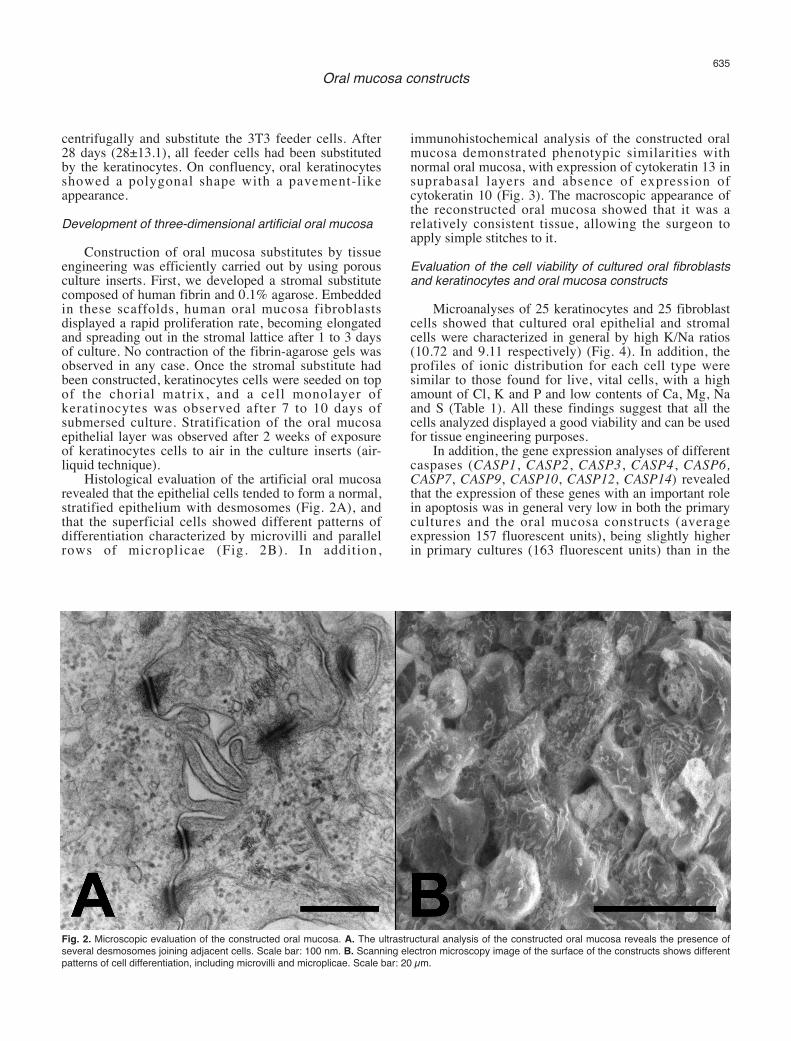
revealed that the epithelial cells tended to form a normal,stratified epithelium with desmosomes (Fig. 2A), andthat the superficial cells showed different patterns ofdifferentiation characterized by microvilli and parallelrows of microplicae (Fig. 2B). In addition,
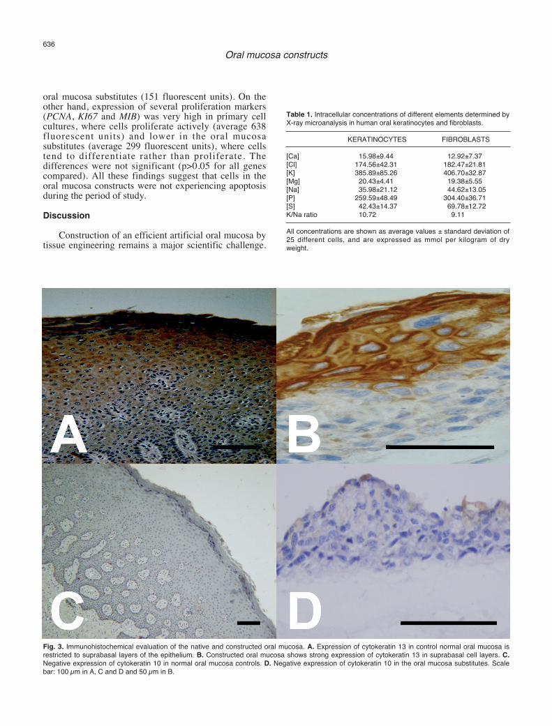
immunohistochemical analysis of the constructed oralmucosa demonstrated phenotypic similarities withnormal oral mucosa, with expression of cytokeratin 13 insuprabasal layers and absence of expression ofcytokeratin 10 (Fig. 3). The macroscopic appearance ofthe reconstructed oral mucosa showed that it was arelatively consistent tissue, allowing the surgeon toapply simple stitches to it.Evaluation of the cell viability of cultured oral fibroblastsand keratinocytes and oral mucosa constructs
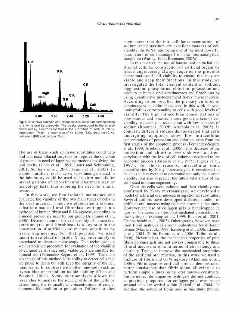
Microanalyses of 25 keratinocytes and 25 fibroblastcells showed that cultured oral epithelial and stromalcells were characterized in general by high K/Na ratios(10.72 and 9.11 respectively) (Fig. 4). In addition, theprofiles of ionic distribution for each cell type weresimilar to those found for live, vital cells, with a highamount of Cl, K and P and low contents of Ca, Mg, Naand S (Table 1). All these findings suggest that all thecells analyzed displayed a good viability and can be usedfor tissue engineering purposes. In addition, the gene expression analyses of different
caspases (CASP1, CASP2, CASP3, CASP4, CASP6,CASP7, CASP9, CASP10, CASP12, CASP14) revealedthat the expression of these genes with an important rolein apoptosis was in general very low in both the primarycultures and the oral mucosa constructs (averageexpression 157 fluorescent units), being slightly higherin primary cultures (163 fluorescent units) than in the
635Oral mucosa constructs
Fig. 2. Microscopic evaluation of the constructed oral mucosa. A. The ultrastructural analysis of the constructed oral mucosa reveals the presence ofseveral desmosomes joining adjacent cells. Scale bar: 100 nm. B. Scanning electron microscopy image of the surface of the constructs shows differentpatterns of cell differentiation, including microvilli and microplicae. Scale bar: 20 µm.
oral mucosa substitutes (151 fluorescent units). On theother hand, expression of several proliferation markers(PCNA, KI67 and MIB) was very high in primary cellcultures, where cells proliferate actively (average 638fluorescent units) and lower in the oral mucosasubstitutes (average 299 fluorescent units), where cellstend to differentiate rather than proliferate. Thedifferences were not significant (p>0.05 for all genescompared). All these findings suggest that cells in theoral mucosa constructs were not experiencing apoptosisduring the period of study.Discussion
Construction of an efficient artificial oral mucosa bytissue engineering remains a major scientific challenge.
636Oral mucosa constructs
Table 1. Intracellular concentrations of different elements determined byX-ray microanalysis in human oral keratinocytes and fibroblasts.
KERATINOCYTES FIBROBLASTS
[Ca] 15.98±9.44 12.92±7.37[Cl] 174.56±42.31 182.47±21.81[K] 385.89±85.26 406.70±32.87[Mg] 20.43±4.41 19.38±5.55[Na] 35.98±21.12 44.62±13.05[P] 259.59±48.49 304.40±36.71[S] 42.43±14.37 69.78±12.72K/Na ratio 10.72 9.11
All concentrations are shown as average values ± standard deviation of25 different cells, and are expressed as mmol per kilogram of dryweight.
Fig. 3. Immunohistochemical evaluation of the native and constructed oral mucosa. A. Expression of cytokeratin 13 in control normal oral mucosa isrestricted to suprabasal layers of the epithelium. B. Constructed oral mucosa shows strong expression of cytokeratin 13 in suprabasal cell layers. C.Negative expression of cytokeratin 10 in normal oral mucosa controls. D. Negative expression of cytokeratin 10 in the oral mucosa substitutes. Scalebar: 100 µm in A, C and D and 50 µm in B.
The use of these kinds of tissue substitutes could helporal and maxillofacial surgeons to improve the outcomeof patients in need of large reconstructions involving theoral cavity (Ueda et al., 1991; Lauer and Schimming,2001; Schlenz et al., 2001; Izumi et al., 2003). Inaddition, artificial oral mucosa substitutes generated inthe laboratory could be used as in vitro models forinvestigations of experimental pharmacology ortoxicology tests, thus avoiding the need for animalresearch.In this work, we first isolated, maintained and
evaluated the viability of the two main types of cells inthe oral mucosa. Then, we elaborated a stromalsubstitute made of oral fibroblasts entrapped in ahydrogel of human fibrin and 0.1% agarose, according toa model previously used by our group (Alaminos et al.,2006). Determination of the cell viability of human oralkeratinocytes and fibroblasts is a key step in theconstruction of artificial oral mucosa substitutes bytissue engineering. For that purpose, we usedquantitative electron probe X-ray microanalysisassociated to electron microscopy. This technique is awell established procedure for evaluation of the viabilityof cultured cells, since only viable cells are suitable forclinical use (Fernandez-Segura et al., 1999). The mainadvantage of this method is its ability to detect cells thatare prone to death but still keep the integrity of the cellmembrane. In contrast with other methods such astrypan blue or propidium iodide staining (Chen andWagner, 2001), X-ray microanalysis allows theresearcher to analyze the ionic profile of the cells bydetermining the intracellular concentrations of crucialelements like sodium or potassium. Different studies
have shown that the intracellular concentrations ofsodium and potassium are excellent markers of cellviability, the K/Na ratio being one of the most powerfulparameters of cell damage from the microanalyticalstandpoint (Warley, 1994; Roomans, 2002a).In this context, the use of human oral epithelial and
stromal cells for construction of artificial organs bytissue engineering always requires the previousdetermination of cell viability to ensure that they areviable and keep their functions. In this study, weinvestigated the total element content of sodium,magnesium, phosphorus, chlorine, potassium andcalcium in human oral keratinocytes and fibroblasts byusing quantitative histochemical X-ray microanalysis.According to our results, the primary cultures ofkeratinocytes and fibroblasts used in this work showedionic profiles corresponding to cells with good levels ofviability. The high intracellular concentrations ofphosphorous and potassium were good markers of cellviability, especially in association with low contents ofsodium (Roomans, 2002b; Arrebola et al., 2005). Incontrast, different studies demonstrated that cellsundergoing apoptosis show low intracellularconcentrations of potassium and chlorine, even from thefirst stages of the apoptotic process (Fernandez-Seguraet al., 1999; Arrebola et al., 2005). This decrease of thepotassium and chlorine levels showed a directcorrelation with the loss of cell volume associated to theapoptotic process (Barbiero et al., 1995; Hughes et al.,1997). For those reasons, intracellular ionicquantification by X-ray microanalysis is considered tobe an excellent method to determine not only the currentviability, but also to predict the future vital status of thecells used in tissue engineering.Once the cells were cultured and their viability was
confirmed by X-ray microanalysis, we developed amodel of artificial oral mucosa using stromal substitutes.Several authors have developed different models ofartificial oral mucosa using collagen stromal substitutes.However, the use of collagen gels is handicapped inmost of the cases by fibroblast-mediated contraction ofthe hydrogels (Schoop et al., 1999; Bach et al., 2001;Chinnathambi et al., 2003). Other groups, however, haveused fibrin matrices as stromal substitutes for differenttissues (Meana et al., 1998; Isenberg et al., 2006; Llameset al., 2004, 2006; Peretti et al., 2006; Talbot et al.,2006). Nevertheless, the mechanical properties of purefibrin polymer gels are not always comparable to thoseof oral mucosa stroma in terms of consistency andelasticity. Trying to improve the mechanical propertiesof the artificial oral mucosa, in this work we used amixture of fibrin and 0.1% agarose (Alaminos et al.,2006). Fibrin-agarose artificial stromas demonstratedbetter consistency than fibrin alone, allowing us toperform simple sutures on the oral mucosa constructs.Furthermore, fibrin-agarose hydrogels did not contract,as previously reported for collagen gels, even whenstromal cells are seeded within (Reichl et al., 2004). Inaddition, the source of fibrin used in this study (human
637Oral mucosa constructs
Fig. 4. Illustrative example of a microanalytical spectrum correspondingto a living oral keratinocyte. The peaks correspond to the energydispersed by electrons located in the k orbitals of sodium (NaK),magnesium (MgK), phosphorus (PK), sulfur (SK), chlorine (ClK),potassium (KK) and calcium (CaK).
plasma) has several advantages, especially the presenceof whole-blood sets of cytokines, attachment factors, andplatelet-derived growth factors. All these proteins arepresent and functional in the plasma-based scaffold,offering a highly proliferative environment forkeratinocytes (Marx et al., 1998). To develop an efficient substitute for the human oral
mucosa, we used a sequential culture technique usingcommercially available culture inserts (Reichl andMuller-Goymann, 2003). These culture inserts were usedfor a two reasons. First, they have a porous membranethat allows the culture media to cross over and reach allthe cells in the system. Second, the design of thesedevices allowed us to use an air-liquid culture techniqueto promote stratification of the engineered epithelium.Culture inserts of different types have been used to dateto construct various types of tissues by tissueengineering (Richard et al., 1991; Geroski and Hadley,1992; Hutak et al., 2002; Limat et al., 2003; Reichl andMuller-Goymann, 2003; Dai et al., 2005; Iida et al.,2005) as an efficient way to promote epithelialstratification (Casasco et al., 2001; Izumi et al., 2003). On the other hand, microscopic evaluation of the
artificial oral mucosa revealed the presence of astratified oral epithelium on the surface of the stromalsubstitutes. The use of fibrin-agarose scaffolds allowedstromal fibroblasts to grow and proliferate within theextracellular matrix and, at the same time, induced theadherence and maturation of a stratified epithelium ontop. Fibrin, the major component of the engineeredstroma, is a natural protein, very abundant in circulatingblood, which is physically recognised by stromalfibroblasts, and it is likely that oral fibroblasts cansynthesize collagen fibers and progressively reorganizethe stromal mesh. Moreover, our ultrastructural analysisof the artificial tissues showed that oral keratinocytes areable to form a multilayered epithelium with numerousdesmosomes and join complexes. Strikingly, differentpatterns of epithelial cell differentiation were detected onthe surface of the engineered epithelium, includingmicrovilli and parallel rows of microplicae. Thepresence of these kinds of cell surface specializationshas previously been associated with the normaldifferentiation process of oral keratinocytes (Moreu etal., 1993; Sanchez-Quevedo et al., 1994), and suggeststhat the oral epithelium generated by tissue engineeringis very similar to the native oral mucosa epithelium.Furthermore, Immunohistochemistry showed similaritiesbetween the native oral mucosa used as control and theconstructed epithelium, with a high expression ofcytokeratin 13 in suprabasal layers of the epithelium,whereas the microarray analysis demonstrated that thecells in the oral mucosa constructs were not experiencinga process of cell death by apoptosis and still had thecapability to proliferate. These results are in agreementwith those previously reported by other authors usingdifferent types of extracellular matrices (Cho et al.,2000; Igarashi et al., 2003). Altogether, our resultsindicate that cultured oral keratinocytes are able to
physically integrate with the fibrin-agarose lattice usedin this work, adhering to it and differentiating on top ofthe artificial stroma. These results suggest that the oralmucosa constructs based on fibrin-agarose scaffoldsprogressively acquire histolological and key cytokeratinexpression patterns similar to human oral mucosacontrols. Therefore, the fibrin-agarose complexesdescribed in this study appear to satisfy the criteria forbiomaterials used in tissue engineering of the human oralmucosa.Acknowledgements. This work was supported by the grants FIS03/0141 and FIS 04/1306 from the Spanish National Ministry of Health(Instituto de Salud Carlos III) and by CM 2005/011 from Junta deAndalucia.
References
Abraham E.H., Breslow J.L., Epstein J., Chang-Sing P. and Lechene C.(1985). Preparation of individual human diploid fibroblasts and studyof ion transport. Am. J. Physiol. 248, 154-164.
Alaminos M., Sanchez-Quevedo M.C., Muñoz-Avila J.I., Serrano D.,Medialdea S., Carreras I. and Campos A. (2006). Construction of acomplete rabbit cornea substitute using a fibrin-agarose scaffold.Invest. Ophthalmol. Vis. Sci. 47, 3311-3317.
Arrebola F., Zabiti S., Canizares F.J., Cubero M.A., Crespo P.V. andFernandez-Segura E. (2005). Changes in intracellular sodium,chlorine, and potassium concentrations in staurosporine-inducedapoptosis. J. Cell Physiol. 204, 500-507.
Aufderheide A.C. and Athanasiou K.A. (2005). Comparison of scaffoldsand culture conditions for tissue engineering of the knee meniscus.Tissue Eng. 11, 1095-1104.
Bach A.D., Bannasch H., Galla T.J., Bittner K.M. and Stark G.B. (2001).Fibrin glue as matrix for cultured autologous urothelial cells inurethral reconstruction. Tissue Eng. 7, 45-53.
Barbiero G., Duranti F., Bonelli G., Amenta J.S. and Baccino F.M.(1995). Intracellular ionic variations in the apoptotic death of L cellsby inhibitors of cell cycle progression. Exp. Cell Res. 217, 410-418.
Baumann I., Greschniok A., Bootz F. and Kaiserling E. (1996). Freetransplanted, microvascular reanastomosed forearm flap forreconstruction of the mouth cavity and oropharynx. Clinical andmorphologic findings with special reference to reinnervation. HNO44, 616–623.
Bergert H., Knoch K.P., Meisterfeld R., Jager M., Ouwendijk J., KerstingS., Saeger HD. and Solimena M. (2005). Effect of oxygenatedperfluorocarbons on isolated rat pancreatic islets in culture. CellTransplant 14, 441-448.
Boekestein A., Thiel F., Stols A.L.H., Bouw E. and Stadhouders A.(1984). Surfaces roughness and the use of peak to background inthe X-ray microanalysis of bulk bio-organic sample. J. Microsc. 134,327-334.
Casasco A., Casasco M., Zerbinati N., Icaro Cornaglia A. and CalligaroA. (2001). Cell proliferation and differentiation in a model of humanskin equivalent. Anat. Rec. 264, 261-272.
Chen J. and Wagner M.C. (2001). Altered membrane-cytoskeletonlinkage and membrane blebbing in energy-depleted renal proximaltubular cells. Am. J. Physiol. Renal Physiol. 280, 619-627.
Chen J., Li Q., Xu J., Huang Y., Ding Y., Deng H., Zhao S. and Chen R.
638Oral mucosa constructs
(2005). Study on biocompatibility of complexes of collagen-chitosan-sodium hyaluronate and cornea. Artificial Organs 29, 104-113.
Chinnathambi S., Tomanek-Chalkley A., Ludwig N., King E., DeWaardR., Johnson G., Wertz P.W. and Bickenbach J.R. (2003).Recapitulation of oral mucosal tissues in long-term organotypicculture. Anat. Rec. A Discov. Mol. Cell Evol. Biol. 270, 162-174.
Cho K.H., Ahn H.T., Park K.C., Chung J.H., Kim S.W., Sung M.W., KimK.H., Chung P.H., Eun H.C. and Youn J.I. (2000). Reconstruction ofhuman hard-palate mucosal epithelium on de-epidermized dermis. J.Dermatol. Sci. 22, 117-24.
Dai N.T., Yeh M.K., Liu D.D., Adams E.F., Chiang C.C., Yeng C.Y., ShihC.M., Sytwu H.K., Chen T.M., Wang H.J., Williamson M.R. andCoombre A.G.A. (2005). A co-cultured skin model based on cellsupport membranes. Biochem. Biophy. Res. Commun. 329, 905-908.
Fernandez-Segura E., Cañizares FJ., Cubero M.A., Revelles F. andCampos A. (1997). Electron probe X-ray microanalysis of culturedepithelial tumour cells with scanning electron microscopy. Cell Biol.Int. 21, 665-669.
Fernandez-Segura E., Cañizares F.J., Cubero M.A., Warley A. andCampos A. (1999). Changes in elemental content during apoptoticcell death studied by electron probe X-ray microanálisis. Exp. CellRes. 253, 454-462.
Geroski D.H. and Hadley A. (1992). Characterization of cornealendothelium cell cultured on microporous membrane filters. Curr.Eye Res.11, 61-72.
Hughes F.M., Bortner C.D., Purdy G.D. and Cidlowski J.A. (1997).Intracellular K+ suppresses the activation of apoptosis inlymphocytes. J. Biol. Chem. 272, 30567-30576.
Hutak C.M., Kavanagh M.E. and Reddy I.K. (2002). Comparativedevelopment of SIRC rabbit corneal cells grown on polycarbonate-and polyester-based filtres. Skin Pharmacol. Appl. Skin Physiol. 15,133-138.
Igarashi M., Irwin C.R., Locke M. and Mackenzie I.C. (2003).Construction of large area organotypical cultures of oral mucosa andskin. J. Oral Pathol. Med. 32, 422-430.
Iida T., Takami Y., Yamaguchi R., Shimazaki A. and Harii K. (2005).Development of a tissue-engineered human oral mucosa equivalentbased on an acellular allogenic dermal matrix: a preliminary report ofclinical application to burn wounds. Scand. J. Plasr. Reconstr. Surg.Hand Surg. 39, 138-146.
Isenberg B.C., Williams C. and Tranquillo R.T. (2006). Endothelializationand flow conditioning of fibrin-based media-equivalents. Ann.Biomed. Eng. 34, 971-985.
Izumi K., Feinberg S.E., Iida A. and Yoshizawa M. (2003). Intraoralgrafting of an ex vivo produced oral mucosa equivalent: apreliminary report. Int. J. Oral Maxillofac. Surg. 32, 188-197.
Krep H., Lefurgey A., Graves S.W., Hockett D., Ingram P. andHollenberg N.K. (1996). Elemental composition of Na pump inhibitedrabbit aorta VSM cells by electron probe X-ray microanalysis. Am. J.Physiol. 271, 514-520.
Lauer G. and Schimming R. (2001). Tissue-engineered mucosa graft forreconstruction of the intraoral lining after freeing of the tongue: aclinical and immunohistologic study. J. Oral Maxillofac. Surg. 59,169-175.
Limat A., French L.E., Blal L., Saurat J.H., Hunziker T. and Salomon D.(2003). Organotypic cultures of autologous hair follicle keratinocytesfor the treatment of recurrent leg ulcers. J. Am. Acad. Dermatol. 48,207-214.
Llames S.G., Del Rio M., Larcher F., Garcia E., Garcia M., EscamezM.J., Jorcano J.L., Holguin P. and Meana A. (2004). Human plasmaas a dermal scaffold for the generation of a completely autologousbioengineered skin. Transplantation 77, 350-355.
Llames S., Garcia E., Garcia V., del Rio M., Larcher F., Jorcano J.L.,Lopez E., Holguin P., Miralles F., Otero J. and Meana A. (2006).Clinical results of an autologous engineered skin. Cell Tissue Bank7, 47-53.
Marx R.E, Carlson E.R., Eichstaedt R.M., Schimmele S.R., Strauss J.E.and Georgeff K.R. (1998). Platelet-rich plasma: Growth factorenhancement for bone grafts. Oral Surg. Oral Med. Oral Pathol. OralRadiol. Endod. 85, 638-646.
Meana A., Iglesias J., Del Rio M., Larcher F., Madrigal B., Fresno MF.,Martin C., San Roman F. and Tevar F. (1998). Large surface ofcultured human epithelium obtained on a dermal matrix based onlive fibroblast-containing fibrin gels. Burns 24, 621-630.
Moreu G., Sanchez-Quevedo M.C., López-Escamez J.A., Gonzalez-Jaranay M. and Campos A. (1993). Cell surface patterns in normalhuman oral gingival epithelium. A quantitative scanning electronmicroscopy approach. Histol. Histopathol. 8, 47-50.
Nishida K. (2003). Tissue engineering of the cornea. Cornea 22, 28-34.Orwin E.J. and Hubel A. (2000). In vitro culture characteristics of corneal
epithelial, endothelial, and keratocyte cells in a native collagenmatrix. Tissue Eng. 6, 307-319.
Pascual G., Rodriguez M., Corrales C., Turegano F., Garcia-HonduvillaN., Bellon J.M. and Bujan J. (2004). New approach to improvingendothelial preservation in cryopreserved arterial substitutes.Cryobiology 48, 62-71.
Peretti G.M., Xu J.W., Bonassar L.J., Kirchhoff C.H., Yaremchuk M.J.and Randolph M.A. (2006). Review of injectable carti lageengineering using fibrin gel in mice and swine models. Tissue Eng.12, 1151-1168.
Reichl S. and Muller-Goymann C.C. (2003). The use of a porcineorganotypic cornea construct for permeation studies fromformulations containing befunolol hydrochloride. Int. J. Pharm. 250,191-201.
Reichl S., Bednarz J. and Muller-Goymann C.C. (2004). Human cornealequivalent as cell culture model for in vitro drug permeation studies.Br. J. Ophthalmol. 88, 560-565.
Rheinwald J. and Green H. (1975). Serial cultivation of stains of humanepidermal keratinocytes: the formation of keratinizing colonies fromsingle cells. Cell 6, 331–344.
Richard N.R., Anderson J.A., Weiss J.L. and Binder P.S. (1991).Air/liquid corneal organ culture: a light microscopic study. Curr. EyeRes. 10, 739-749.
Roomans G.M. (2002a). Application of X-ray microanalysis to the studyof cell physiology in cells attached to biomaterials. Eur. Cell Mater.18, 1-8.
Roomans G.M. (2002b). X-ray microanalysis of epithelial cell sinculture.Methods Mol. Biol. 188, 273-289.
Sanchez-Quevedo M.C., Moreu G., Campos A., García J.M. andGonzález-Jaranay M. (1994). Regional differences in cell surfacepatterns in normal human sulcular epithelium. Histol. Histopathol. 9,149-153.
Schlenz I., Korak K.J., Kunstfeld R., Vinzenz K., Plenk H.Jr. and Holle J.(2001). The dermis-prelaminated scapula flap for reconstructions ofthe hard palate and the alveolar ridge: a clinical and histologicevaluation. Plast. Reconstr. Surg. 108, 1519-1524.
Schoop V.M., Mirancea N. and Fusenig N.E. (1999). Epidermal
639Oral mucosa constructs
organization and differentiation of HaCaT keratinocytes inorganotypic coculture with human dermal fibroblasts. J. Invest.Dermatol. 112, 343-353.
Schultze-Mosgau S., Lee B.K., Ries J., Amann K. and Wiltfang J.(2004). In vitro cultured autologous pre-confluent oral keratinocytesfor experimental prefabrication of oral mucosa. Int. J. OralMaxillofac. Surg. 33, 476-485.
Somlyo A.V., Shuman H. and Somlyo A.P. (1989). Electron probe X-raymicroanalysis of Ca2+, Mg2+, and other ions in rapidly frozen cells.Methods Enzymol. 172, 203-229.
Song J., Izumi K., Lanigan T. and Feinberg S.E. (2004). Developmentand characterization of a canine oral mucosa equivalent in a serum-free environment. J. Biomed. Mater. Res.71, 143-53.
Staham P.J. and Pawley J.B. (1978). A new method for particle X-raymicroanalysis on peak to background measurements. ScanningElectron Microsc. 1, 469-478.
Talbot M., Carrier P., Giasson C.J., Deschambeault A., Guerin S.L.,Auger F.A., Bazin R. and Germain L. (2006). Autologoustransplantation of rabbit limbal epithelia cultured on fibrin gels forocular surface reconstruction. Mol. Vis.12, 65-75.
Tegtmeyer S., Papantoniou I. and Muller-Goymann C.C. (2001).Reconstruction of an in vitro cornea and its use for drug permeationstudies from different formulations containing pilocarpinehydrochloride. Eur. J. Pharm. Biopharm. 51, 19-125.
Toft K., Keller G.S. and Blackwell K.E. (2000). Ectopic hair growth afterflap reconstruction of the head and neck. Arch. Facial Plast. Surg. 2,148-150.
Ueda M., Ebata K. and Kaneda T. (1991). In Vitro fabrication ofbioartificial mucosa for reconstruction of oral mucosa: Basicresearch and clinical application. Ann. Plast. Surg. 27, 540-549.
Warley A. (1990). Standards for the application of X-ray microanalysis tobiological specimens. J. Microsc. 157, 129-138.
Warley A. (1994). The preparation of cultured cells for X-raymicroanalysis. Scanning Microsc. 8, 129-137.
Wunsch L., Ehlers E.M. and Russlies M. (2005). Matrix testing forurothelial t issue engineering. Eur. J. Pediatr. Surg. 15, 164-169.
Accepted December 22, 2006
640Oral mucosa constructs