Histamine release and surface CD200R1 staining as sensitive methods for assessing murine mast cell...
8
Research paper Histamine release and surface CD200R1 staining as sensitive methods for assessing murine mast cell activation David Larson, Edward Mitre ⁎ Department of Microbiology and Immunology, Uniformed Services University, 4301 Jones Bridge Road, Bethesda, MD, USA article info abstract Article history: Received 15 June 2011 Received in revised form 18 February 2012 Accepted 21 February 2012 Available online 28 February 2012 Mast cells are important effector cells of allergy and are involved in the pathology of many other diseases. Measurement of β-hexosaminidase activity, the most commonly used method for evaluation of murine mast cell activity, requires a large number of cells and thus is of limited utility for studying mast cells in mouse models of disease. In this study we evaluated the sensitivity of histamine release as compared to β-hexosaminidase activity in the measure- ment of mast cell activation. Whereas a minimum of 6 × 10 4 mast cells per ml were required to detect slight increases in β-hexosaminidase activity after anti-IgE and ionomycin stimulation, substantial increases in histamine release could be detected under the same activating condi- tions with as few as 480 mast cells per ml. These findings demonstrate that measurement of histamine release is substantially more sensitive than assessment of β-hexosaminidase activity for detecting mast cell activation. Additionally, we describe a novel flow cytometric method for detecting murine mast cell activation. When using 7.5 × 10 5 peritoneal cells per condition and gating on IgE + c-kit + cells, mast cell expression of surface CD200R1 increased after both IgE and non IgE-mediated activation. This flow cytometric procedure was uncomplicated and rapid, with increases in surface CD200R1 expression appearing after as little as 30 min of stimulation time. Measuring histamine release and surface CD200R1 expression are sensitive approaches for detection of murine mast cell activation. Further, both approaches can be done on unpurified peritoneal cell populations. By requiring low numbers of cells, these approaches are ideal for investigating mast cell activation in murine models of disease. Published by Elsevier B.V. Keywords: Mast cell Flow cytometry CD200R1 Histamine β-Hexosaminidase IgE 1. Introduction Mast cells, originally identified by Paul Ehrlich in 1878, are bone marrow-derived cells that reside in tissues throughout the body with skin, airways, and intestines being the most common sites (Metcalfe et al., 1997; Marshall, 2004). They typically become activated after surface bound IgE is cross- linked by multivalent antigen, which results in rapid release of inflammatory mediators such as histamine and leukotriene C4 and induces classical allergy symptoms (Metcalfe et al., 1997; Galli, 2000; Bischoff, 2007). Mast cells can also become activated by pathways that do not involve IgE and have the ability to release a wide range of cytokines and inflammatory molecules. In addition to being key effector cells of allergy, mast cells have become increasingly recognized as playing important roles in helminth infections, bacterial infections, autoimmunity, neurologic disorders, and vascular diseases (Marshall, 2004; Bischoff, 2007). Identifying easy and rapid techniques to measure activation of murine mast cells could be of substantial benefit for investigations involving disease models in mice. Currently, the most common approach taken to assess mu- rine mast cell activation is measurement of β-hexosaminidase activity. In this assay, purified mast cells are stimulated in vitro, supernatants are collected, and colored product is mea- sured after addition of a nominally-priced β-hexosaminidase substrate. Although this approach is relatively simple and in- expensive, a major drawback is that it requires high numbers Journal of Immunological Methods 379 (2012) 15–22 ⁎ Corresponding author. Tel.: + 1 301 295 1958; fax: + 1 301 295 1545. E-mail addresses: [email protected] (D. Larson), [email protected] (E. Mitre). 0022-1759/$ – see front matter. Published by Elsevier B.V. doi:10.1016/j.jim.2012.02.014 Contents lists available at SciVerse ScienceDirect Journal of Immunological Methods journal homepage: www.elsevier.com/locate/jim
-
Upload
david-larson -
Category
Documents
-
view
213 -
download
0
Transcript of Histamine release and surface CD200R1 staining as sensitive methods for assessing murine mast cell...
Journal of Immunological Methods 379 (2012) 15–22
Contents lists available at SciVerse ScienceDirect
Journal of Immunological Methods
j ourna l homepage: www.e lsev ie r .com/ locate / j im
Research paper
Histamine release and surface CD200R1 staining as sensitive methods forassessing murine mast cell activation
David Larson, Edward Mitre⁎Department of Microbiology and Immunology, Uniformed Services University, 4301 Jones Bridge Road, Bethesda, MD, USA
a r t i c l e i n f o
⁎ Corresponding author. Tel.: +1 301 295 1958; faxE-mail addresses: [email protected] (D. Larson), e
(E. Mitre).
0022-1759/$ – see front matter. Published by Elsevierdoi:10.1016/j.jim.2012.02.014
a b s t r a c t
Article history:Received 15 June 2011Received in revised form 18 February 2012Accepted 21 February 2012Available online 28 February 2012
Mast cells are important effector cells of allergy and are involved in the pathology of manyother diseases. Measurement of β-hexosaminidase activity, the most commonly used methodfor evaluation of murine mast cell activity, requires a large number of cells and thus is oflimited utility for studying mast cells in mouse models of disease. In this study we evaluatedthe sensitivity of histamine release as compared to β-hexosaminidase activity in the measure-ment of mast cell activation. Whereas a minimum of 6×10⁴ mast cells per ml were required todetect slight increases in β-hexosaminidase activity after anti-IgE and ionomycin stimulation,substantial increases in histamine release could be detected under the same activating condi-tions with as few as 480 mast cells per ml. These findings demonstrate that measurement ofhistamine release is substantially more sensitive than assessment of β-hexosaminidase activityfor detecting mast cell activation. Additionally, we describe a novel flow cytometric method fordetecting murine mast cell activation. When using 7.5×10⁵ peritoneal cells per conditionand gating on IgE+c-kit+cells, mast cell expression of surface CD200R1 increased afterboth IgE and non IgE-mediated activation. This flow cytometric procedure was uncomplicatedand rapid, with increases in surface CD200R1 expression appearing after as little as 30 min ofstimulation time. Measuring histamine release and surface CD200R1 expression are sensitiveapproaches for detection of murine mast cell activation. Further, both approaches can bedone on unpurified peritoneal cell populations. By requiring low numbers of cells, theseapproaches are ideal for investigating mast cell activation in murine models of disease.
Published by Elsevier B.V.
Keywords:Mast cellFlow cytometryCD200R1Histamineβ-HexosaminidaseIgE
1. Introduction
Mast cells, originally identified by Paul Ehrlich in 1878, arebone marrow-derived cells that reside in tissues throughoutthe body with skin, airways, and intestines being the mostcommon sites (Metcalfe et al., 1997; Marshall, 2004). Theytypically become activated after surface bound IgE is cross-linked by multivalent antigen, which results in rapid releaseof inflammatory mediators such as histamine and leukotrieneC4 and induces classical allergy symptoms (Metcalfe et al.,1997; Galli, 2000; Bischoff, 2007). Mast cells can also becomeactivated by pathways that do not involve IgE and have the
: +1 301 295 [email protected]
B.V.
ability to release a wide range of cytokines and inflammatorymolecules. In addition to being key effector cells of allergy,mast cells have become increasingly recognized as playingimportant roles in helminth infections, bacterial infections,autoimmunity, neurologic disorders, and vascular diseases(Marshall, 2004; Bischoff, 2007). Identifying easy and rapidtechniques to measure activation of murine mast cells couldbe of substantial benefit for investigations involving diseasemodels in mice.
Currently, themost common approach taken to assessmu-rinemast cell activation ismeasurement of β-hexosaminidaseactivity. In this assay, purified mast cells are stimulated invitro, supernatants are collected, and colored product is mea-sured after addition of a nominally-priced β-hexosaminidasesubstrate. Although this approach is relatively simple and in-expensive, a major drawback is that it requires high numbers
16 D. Larson, E. Mitre / Journal of Immunological Methods 379 (2012) 15–22
of mast cells. Typical protocols for the measurement of β-hexosaminidase activity require concentrations of 6×10⁵
mast cells/ml per condition tested (Karimi et al., 2000;Mortaz et al., 2005a, 2005b). While obtaining such numbersof mast cells is not an issue for in vitro experiments utilizingcells lines or bone-marrow derived mast cells, it is limitingfor experiments evaluating the function ofmast cells obtaineddirectly frommice. In this study, we testedwhether histaminerelease was a more sensitive measure of murine mast cellactivation than β-hexosaminidase assay.
Additionally, we developed a flow cytometric methodfor measuring murine mast cell activation. While there arenumerous flow cytometric methods for detecting activatedhuman mast cells, including measuring surface expressionof CD35, CD63, CD69, LAMP-1, and LAMP-2 (Diaz-Agustinet al., 1999; Escribano et al., 2002; Grutzkau et al., 2004),there is no widely used flow cytometric assay in place fordetection of murine mast cell activation. Recently, we dem-onstrated that surface expression of CD200R1, an inhibitoryreceptor belonging to the immunoglobulin superfamily,can be utilized as an activation marker of basophils (Torreroet al., 2009). As both basophils andmast cells can be activatedthrough cross-linking of surface IgE and as a prior studydemonstrated that murine mast cells express CD200R1(Cherwinski et al., 2005), in this study we evaluated whetherCD200R1 can be utilized for detection of murine mast cellactivation.
Our results demonstrate that histamine is a far moresensitive measure of murine mast cell activation than β-hexosaminidase and that CD200R1 can be used as a markerof mast cell activation.
2. Materials and methods
2.1. Animals
Female BALB/c mice were obtained from the NCI MouseRepository (Frederick, MD) and housed at the UniformedServices University (USU) Center for Laboratory Medicine.At study endpoints, mice were euthanized with carbon diox-ide and cells used for mast cell activation studies obtained byperitoneal lavage. All experiments were performed in accor-dance with protocols approved by the USU InstitutionalAnimal Care and Use Committee.
2.2. Infection of mice with Litomosoides sigmodontis
BALB/c mice were infected with L. sigmodontis, a filarialroundworm infection of rodents, by intraperitoneal injectionof 40 L3 stage larvae. These worms migrate to the pleuralcavity over the first few days of infection, where they subse-quently reside and develop into adult worms by approxi-mately 4 weeks post-infection. This infection results in theproduction of large quantities of parasite-specific IgE begin-ning at 4 weeks post-infection (Torrero et al., 2010), resultingin sensitization of both mast cells and basophils to parasiteantigen (LsAg) through binding of specific IgE to FcER1. L3stage larvae for infection studies were obtained from thepleural cavity of Mongolian jirds (Meriones unguiculatus,TRS Laboratory Inc., Athens, GA) that had been infected by
the bite of infectious mites 4 days earlier as previouslydescribed (Hubner et al., 2009).
2.3. L. sigmodontis worm antigen (LsAg) preparation
After harvesting male and female adult worms frominfected animals, worms were mechanically homogenized.PBS-soluble fractions were then collected and sterile filteredas previously described (Torrero et al., 2010).
2.4. Collection of peritoneal cells
Peritoneal cells were collected as previously described(Jensen et al., 2006; de Almeida Buranello et al., 2010). Brief-ly, after soaking the ventral surface with 70% ethanol, fur-bearing skin and subcutaneous tissue was dissected away.Approximately 5 ml 1×HBSS (Invitrogen, CA) was injectedinto the peritoneal cavity and the abdomen was manuallyagitated for 1 minute. After carefully cutting a 5 mm hole inthe peritoneum, 1×HBSS and cells were removed with atransfer pipette. The peritoneal cavity was then washed 2–3more times with 1×HBSS. If significant red blood cell con-tamination existed, red blood cells were lysed with 0.8%ammonium chloride (NH4Cl).
2.5. Purification of peritoneal mast cells
After centrifugation of peritoneal cells at 400×g for10 min, supernatant was thoroughly removed and the cellpellet resuspended in 70% percoll (GE Healthcare) solutionand overlayed with RPMI (Mediatech Inc., VA), as previouslydescribed (Jensen et al., 2006; Gri et al., 2008). Cells werecentrifuged for 15 min at 580×g. Non mast cells were care-fully removed from the percoll/RPMI interface along withmost of the remaining supernatant without disturbing themast cell pellet, which was then resuspended in RPMI.
2.6. Determining mast cell purity
2.6.1. Flow cytometric enumeration of mast cellsTotal numbers of peritoneal cells were enumerated by
counting non red cells using a hemocytometer. Cells werethen fixed with fixative solution from a whole blood lysingreagent kit (Beckman Coulter, CA) containing 9.25% formal-dehyde and 3.75% methanol. Cells were washed and resus-pended in 250 μl of 1% bovine serum albumin (BSA, Cohn Vfraction, Sigma-Aldrich, MO) in 1× PBS and incubated for1 h or overnight at 4 °C. Cells were surface stained withanti-CD117 (c-kit) APC (2B8, BD Bioscience, CA) and anti-IgE FITC (R35-72, BD Biosciences) and incubated at 4 °C for30 min. After washing cells twice with 1× PBS, cells wereresuspended in a final volume of 250 μl 1× PBS. Cell werethen analyzed using a BD LSRII flow cytometer and FACSDivasoftware (BD Biosciences) and the percentage of peritonealcells that were mast cells determined by measuring thepercentage of total cells that were c-kit+IgE+. For all flowcytometry experiments, optimal concentrations of antibodyused for staining were determined by individual antibodytitration experiments.
17D. Larson, E. Mitre / Journal of Immunological Methods 379 (2012) 15–22
2.6.2. CytospinsThirty thousand to seventy-five thousand cells were
centrifuged at 500 rpm for 5 min onto slides using a cytofun-nel and then allowed to air dry.
For May Grünwald/Giemsa staining, slides were placedin May Grünwald stain (Sigma-Aldrich) for 5 min, then1×PBS, pH 7.2 for 2 min, followed by Giemsa stain(Sigma-Aldrich) diluted 1/20 in distilled, deionized waterfor 18 min. Slides were removed from 1/20 diluted Giemsastain and briefly dipped in distilled, deionized water andallowed to air dry.
Toluidine blue staining was conducted as previouslydescribed (Kulka and Metcalfe, 2010). Briefly, slides wereplaced in Mota's fixative for 10 min, fixative was removedby rinsing with 66% ethanol, and then slides were washedby dipping in distilled, deionized water. Slides were stainedwith acid toluidine solution for 10 min followed by dippingin distilled, deionized water. Slides were rinsed with 66%ethanol and dipped in distilled, deionized water and thenrinsed with 100% ethanol and dipped in distilled, deionizedwater.
2.7. Measuring histamine concentration and β-hexosaminidaseactivity
Peritoneal cells and purified mast cells were adjusted tocorrect starting concentrations using Histamine ReleaseBuffer (Beckman Coulter). Cells were incubated at 37 °C for
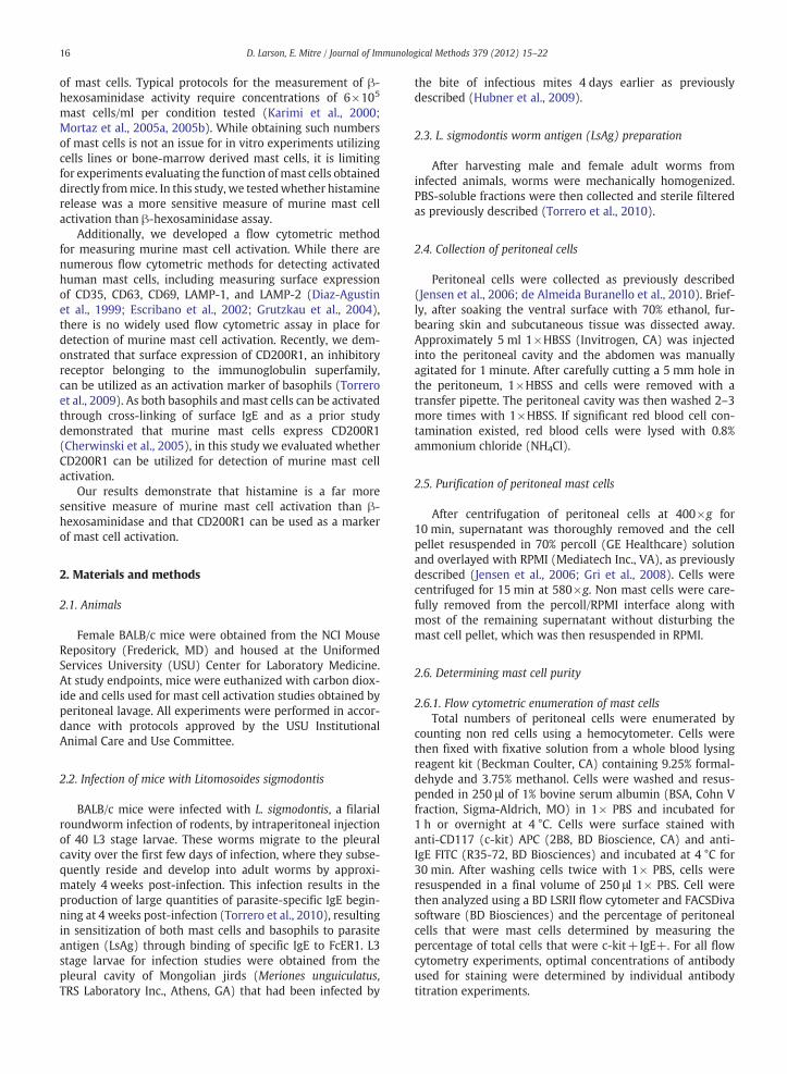
Fig. 1. Identification and enumeration of peritoneal cells and peritoneal mast cells. Aobtained by lavage and counted with a hemocytometer. B. Gating strategy to identifywere identified as IgE+and c-kit+cells (right panel). Mast cells shown in blue, othmast cells determined by flow cytometry. D. Total calculated number of peritoneal
15 min and then stimulated for 30 min with media, 1 μg/mlionomycin (CalBiochem, CA), and 0.5 μg/ml anti-IgE at37 °C. At the end of the incubation period, cells were centri-fuged at 400×g for 10 min.
For histamine measurement, 150 μl supernatant was firstacylated in order to prevent the breakdown of histamine.Histamine concentrations were then measured using acompetitive ELISA (EIA Histamine assay, Beckman Coulter)per the manufacturer's directions. Concentrations of hista-mine in acylated supernatants were determined from a stan-dard curve after reading absorbances at 405 nm using aVictor3 V plate reader (PerkinElmer, MA).
For β-hexosaminidase activity determination, 50 μl of su-pernatant was incubated with 200 μl of 1 mM 4-nitrophenylN-acetyl-β-D-glucosaminide enzyme substrate in 0.05 Mcitrate acid buffer (pH 4.6) for 1 h at 37 °C as previouslydescribed (Cohen and Brown, 2001). Reactions were termi-nated with 0.1 M sodium carbonate buffer. Activity of β-hexosaminidase was determined from a standard curve gen-erated with the enzyme product 4-nitrophenol in 0.05 Mcitrate acid buffer after measuring absorbances at 405 nmusing Victor3 V plate reader.
Fold increases in histamine release and 4-nitrophenolproduction were calculated by dividing media stimulationlevels.
Percentage of total histamine was calculated by dividingthe amount of histamine in supernatant after stimulationwith media, anti-IgE, ionomycin, or LsAg by the amount of
. Numbers of total peritoneal cells from individual mice. Peritoneal cells weremast cells. After initial forward and side scatter gating (left panel), mast cellser peritoneal cells shown in red. C. Percentages of peritoneal cells that weremast cells per mouse. For all panels, n=10.
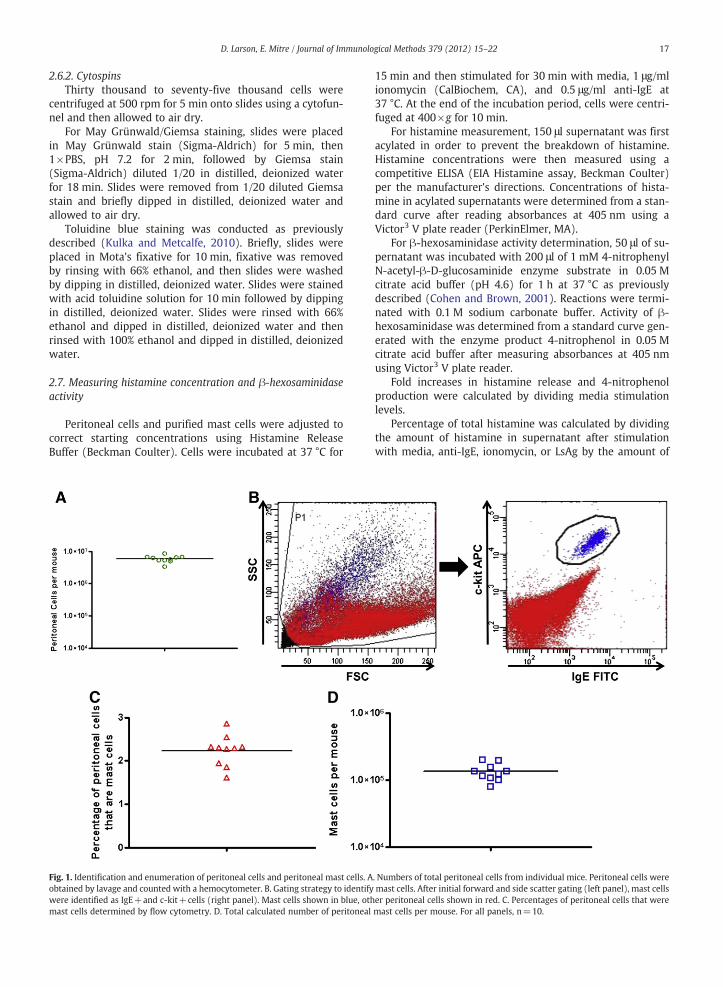
Fig. 2. A 70% percoll gradient results in a highly purified population of perito-neal mast cells. A. Cytospin of total peritoneal cells stained with MayGrünwald/Giemsa stain. Mast cells determined to be 2.5% of all peritonealcells. B. Cytospin of May Grünwald/Giemsa stained mast cells purified fromthe peritoneal cavity with percoll. Mast cells determined to be >98% of allcells. C. Additional staining of purified mast cells with Toluidine Blue Stain.Percentage of cells that were mast cells was identical to May Grünwald/Giemsa stained cells. Magnification=400×.
18 D. Larson, E. Mitre / Journal of Immunological Methods 379 (2012) 15–22
histamine in a lysed aliquot of cells. Percentage of total β-hexosaminidase activity was calculated by dividing theamount of 4-nitrophenol produced in supernatant after stim-ulation with media, anti-IgE, or ionomycin by the amount of4-nitrophenol produced in a lysed aliquot of cells.
2.8. Measuring mast cell activation by flow cytometry
After counting total numbers of peritoneal cells using ahemocytometer, peritoneal cells were diluted with RPMI toa concentration of 3.75×10⁶/ml and 200 μl of cells were ali-quoted per tube (750,000 cells/tube). Cells were incubatedfor 15 min at 37 °C and then stimulated with either severalconcentrations of anti-IgE (0.0078, 0.031, 0.125, 0.5, 2 μg/ml, R35-72, BD Biosciences), 0.5 μg/ml ionomycin, or 0.5 μMfMLP. Cells were incubated at 37 °C for 0.5, 1, 2, 3 or 4 h fortimecourse experiments and for 3 h for all other experiments.At the end of the incubation period, cells were fixed with100 μl of fixative solution from a whole blood lysing reagentkit (Beckman Coulter, CA), containing 9.25% formaldehydeand 3.75% methanol. Cells were washed and resuspendedin 250 μl 1% bovine serum albumin (BSA, Cohn V fraction,Sigma-Aldrich) in 1×PBS and blocked for 1 h or overnightat 4 °C. After incubation with 1% BSA, cells were surfacestained with anti-CD117 APC, anti-IgE FITC, and anti-CD200R1 (OX-110, AbdSerotec) for 30 min at 4 °C. Afterwashing cells twice with 1×PBS, cells were resuspended toa final volume of 250 μl with 1×PBS and analyzed using aBD LSRII flow cytometer and FACSDiva software (BD Biosci-ences). Gates for CD200R1 positivity were established usingthe fluorescence minus 1 (FMO) technique (Baumgarth andRoederer, 2000).
2.9. Statistical analyses
Two-tailed, paired Student's t test was used to determinestatistical significance between two groups. All statisticswere performed using GraphPad Prism 4.03. P-values lessthan 0.05 were considered significant.
3. Results
3.1. Identification and enumeration of peritoneal cells andperitoneal mast cells
Peritoneal cells were obtained from individual BALB/cmice by lavage of the peritoneal cavity with cold 1×HBSS.Total peritoneal cells from each mouse were enumeratedusing a hemocytometer and averaged 5.95×10⁶ per mouse(Fig. 1A). Mast cells were identified as IgE+c-kit+cells byflow cytometry (Fig. 1B). The mean percentage of peritonealcells that were mast cells was 2.23% (Fig. 1C), resulting in atotal of 132,685 peritoneal mast cells on average per mouse(Fig. 1D).
3.2. 70% percoll gradient results in a highly purified populationof peritoneal mast cells
Use of a percoll gradient to purify peritoneal mast cellshas been previously described (Jensen et al., 2006; Gri et al.,2008). As seen in Fig. 2, this approach results in a highly
purified population of mast cells. Peritoneal cells wereobtained by lavage of the peritoneal cavity of BALB/c mice.Mast cells were determined to be 2.5% of all peritoneal cellsby May Grünwald/Giemsa staining of cytospins (Fig. 2A). A70% percoll gradient was used to isolate mast cells fromother peritoneal cells. Mast cell purity was measured asgreater than 98% by both May Grünwald/Giemsa staining(Fig. 2B) and toluidine blue staining (Fig. 2C).
19D. Larson, E. Mitre / Journal of Immunological Methods 379 (2012) 15–22
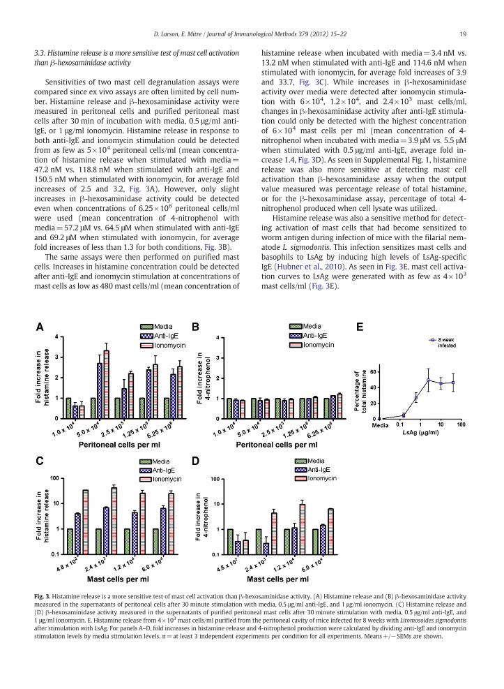
3.3. Histamine release is a more sensitive test of mast cell activationthan β-hexosaminidase activity
Sensitivities of two mast cell degranulation assays werecompared since ex vivo assays are often limited by cell num-ber. Histamine release and β-hexosaminidase activity weremeasured in peritoneal cells and purified peritoneal mastcells after 30 min of incubation with media, 0.5 μg/ml anti-IgE, or 1 μg/ml ionomycin. Histamine release in response toboth anti-IgE and ionomycin stimulation could be detectedfrom as few as 5×10⁴ peritoneal cells/ml (mean concentra-tion of histamine release when stimulated with media=47.2 nM vs. 118.8 nM when stimulated with anti-IgE and150.5 nM when stimulated with ionomycin, for average foldincreases of 2.5 and 3.2, Fig. 3A). However, only slightincreases in β-hexosaminidase activity could be detectedeven when concentrations of 6.25×10⁶ peritoneal cells/mlwere used (mean concentration of 4-nitrophenol withmedia=57.2 μM vs. 64.5 μM when stimulated with anti-IgEand 69.2 μM when stimulated with ionomycin, for averagefold increases of less than 1.3 for both conditions, Fig. 3B).
The same assays were then performed on purified mastcells. Increases in histamine concentration could be detectedafter anti-IgE and ionomycin stimulation at concentrations ofmast cells as low as 480 mast cells/ml (mean concentration of
Fig. 3. Histamine release is a more sensitive test of mast cell activation than β-hexosmeasured in the supernatants of peritoneal cells after 30 minute stimulation with m(D) β-hexosaminidase activity measured in the supernatants of purified peritonea1 μg/ml ionomycin. E. Histamine release from 4×10³ mast cells/ml purified from theafter stimulation with LsAg. For panels A–D, fold increases in histamine release and 4stimulation levels by media stimulation levels. n=at least 3 independent experime
histamine release when incubated with media=3.4 nM vs.13.2 nM when stimulated with anti-IgE and 114.6 nM whenstimulated with ionomycin, for average fold increases of 3.9and 33.7, Fig. 3C). While increases in β-hexosaminidaseactivity over media were detected after ionomycin stimula-tion with 6×10⁴, 1.2×10⁴, and 2.4×10³ mast cells/ml,changes in β-hexosaminidase activity after anti-IgE stimula-tion could only be detected with the highest concentrationof 6×10⁴ mast cells per ml (mean concentration of 4-nitrophenol when incubated with media=3.9 μM vs. 5.5 μMwhen stimulated with 0.5 μg/ml anti-IgE, average fold in-crease 1.4, Fig. 3D). As seen in Supplemental Fig. 1, histaminerelease was also more sensitive at detecting mast cellactivation than β-hexosaminidase assay when the outputvalue measured was percentage release of total histamine,or for the β-hexosaminidase assay, percentage of total 4-nitrophenol produced when cell lysate was utilized.
Histamine release was also a sensitive method for detect-ing activation of mast cells that had become sensitized toworm antigen during infection of mice with the filarial nem-atode L. sigmodontis. This infection sensitizes mast cells andbasophils to LsAg by inducing high levels of LsAg-specificIgE (Hubner et al., 2010). As seen in Fig. 3E, mast cell activa-tion curves to LsAg were generated with as few as 4×10³
mast cells/ml (Fig. 3E).
aminidase activity. (A) Histamine release and (B) β-hexosaminidase activityedia, 0.5 μg/ml anti-IgE, and 1 μg/ml ionomycin. (C) Histamine release and
l mast cells after 30 minute stimulation with media, 0.5 μg/ml anti-IgE, andperitoneal cavity of mice infected for 8 weeks with Litomosoides sigmodontis-nitrophenol production were calculated by dividing anti-IgE and ionomycinnts per condition for all experiments. Means+/−SEMs are shown.
20 D. Larson, E. Mitre / Journal of Immunological Methods 379 (2012) 15–22
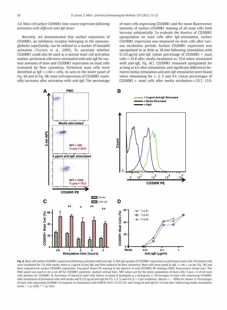
3.4. Mast cell surface CD200R1 time-course expression followingactivation with different anti-IgE doses
Recently, we demonstrated that surface expression ofCD200R1, an inhibitory receptor belonging to the immuno-globulin superfamily, can be utilized as a marker of basophilactivation (Torrero et al., 2009). To ascertain whetherCD200R1 could also be used as a murine mast cell activationmarker, peritoneal cells were stimulatedwith anti-IgE for var-ious amounts of time and CD200R1 expression on mast cellsevaluated by flow cytometry. Peritoneal mast cells wereidentified as IgE+c-kit+cells. As seen in the lower panel ofFig. 4A and in Fig. 4B, mast cell expression of CD200R1 mark-edly increases after activation with anti-IgE. The percentage
Fig. 4.Mast cell surface CD200R1 expression following activation with anti-IgE. A. Dowere incubated for 3 h with media alone or 2 μg/ml of anti-IgE and then analyzed bthen evaluated for surface CD200R1 expression. Top panel shows PE staining in thFMO panel was used to set a cut-off for CD200R1 positivity (dashed vertical line).cells positive for CD200R1. B. Activation of identical mast cells shown in panel Aafter stimulation of peritoneal cells with media and 0.125 μg/ml anti-IgE for 0.5, 1, 2,of mast cells expressing CD200R1 in response to stimulation with 0.0078, 0.031, 0.1levels. *=pb0.05, **=pb0.01.
of mast cells expressing CD200R1 and the mean fluorescenceintensity of surface CD200R1 staining of all mast cells bothincrease substantially. To evaluate the kinetics of CD200R1upregulation on mast cells after IgE-stimulation, surfaceCD200R1 expression was measured on mast cells after vari-ous incubation periods. Surface CD200R1 expression wasupregulated in as little as 30 min following stimulation with0.125 μg/ml anti-IgE (mean percentage of CD200R1+ mastcells=25.8 after media incubation vs. 53.0 when stimulatedwith anti-IgE, Fig. 4C). CD200R1 remained upregulated foras long as 4 h after stimulation, and significant differences be-tweenmedia stimulation and anti-IgE stimulationwere foundwhen stimulating for 1, 2, 3 and 4 h (mean percentages ofCD200R1+ mast cells after media incubation=23.7, 13.9,
t plot graphs of CD200R1 expression on peritoneal mast cells. Peritoneal cellsy flow cytometry. Mast cells were gated as IgE+c-kit+(as per Fig. 1B) ande absence of anti-CD200R1-PE staining (FMO, fluorescence minus one). TheMFI values are for the entire population of mast cells. % pos=% of all masdisplayed as a histogram. C. Percentages of mast cells expressing CD200R13, and 4 h (n=3 per condition). Means+/−SEMs are shown. D. Percentages25, 0.5, and 2.0 μg/ml anti-IgE for 3 h and after subtracting media stimulation
t
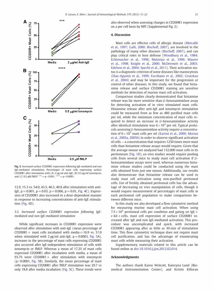
Fig. 5. Increased surface CD200R1 expression following IgE-mediated and nonIgE-mediated stimulation. Percentages of mast cells expressing surfaceCD200R1 after stimulation with (A) 2 μg/ml anti-IgE, (B) 0.5 μg/ml ionomycin,and (C) 0.5 μM fMLP. ***=pb0.001, ****=pb0.0001.
21D. Larson, E. Mitre / Journal of Immunological Methods 379 (2012) 15–22
12.9, 15.3 vs. 54.0, 43.5, 46.5, 40.8 after stimulation with anti-IgE, p=0.001, p=0.03, p=0.006, p=0.01, Fig. 4C). Expres-sion of CD200R1 also increased in a dose-dependent mannerin response to increasing concentrations of anti-IgE stimula-tion (Fig. 4D).
3.5. Increased surface CD200R1 expression following IgE-mediated and non IgE-mediated stimulation
While significant increases in CD200R1 expression wereobserved after stimulation with anti-IgE (mean percentage ofCD200R1+ mast cells incubated with media=16.9 vs. 57.0when stimulated with 2 μg/ml anti-IgE, p=0.0003, Fig. 5A),increases in the percentage of mast cells expressing CD200R1also occurred after IgE-independent stimulation of cells withionomycin or fMLP. Whereas a mean of 17.2% of mast cellsexpressed CD200R1 after incubation with media, a mean of93.7% were CD200R1+ after stimulation with ionomycin(pb0.0001, Fig. 5B). Similarly, the mean percentage of mastcells expressing CD200R1 after fMLP stimulation was 29.7 vsonly 18.8 after media incubation (Fig. 5C). These trends were
also observed when assessing changes in CD200R1 expressionon a per cell basis by MFI (Supplemental Fig. 2).
4. Discussion
Mast cells are effector cells of allergic disease (Metcalfeet al., 1997; Galli, 2000; Bischoff, 2007), are involved in thepathology of many other diseases (Bischoff, 2007), and canplay critical roles in host defense (Woodbury et al., 1984;Echtenacher et al., 1996; Malaviya et al., 1996; Maureret al., 1998; Knight et al., 2000; McDermott et al., 2003;Edelson et al., 2004; Specht et al., 2011). Their activation sta-tus is a diagnostic criterion of some diseases like mastocytosis(Diaz-Agustin et al., 1999; Escribano et al., 2002; Grutzkauet al., 2004) and may be important for the progression orcontrol of other diseases. In this study, we found that hista-mine release and surface CD200R1 staining are sensitivemethods for detection of murine mast cell activation.
Comparison studies clearly demonstrated that histaminerelease was far more sensitive than β-hexosaminidase assayfor detecting activation of in vitro stimulated mast cells.Histamine release after anti-IgE and ionomycin stimulationcould be measured from as few as 480 purified mast cellsper ml, while the minimum concentration of mast cells re-quired to detect an increase in β-hexosaminidase activityafter identical stimulation was 6×10⁴ per ml. Typical proto-cols assessing β-hexosaminidase activity require a concentra-tion of 6×10⁵ mast cells per ml (Karimi et al., 2000; Mortazet al., 2005a, 2005b) in order to observe significant activationof cells— a concentration that requires 1250 times more mastcells than histamine release assays would require. Given thatthe average mouse we analyzed had 132,000 mast cells in itsperitoneum (Fig. 1D), ex-vivo studies would require poolingcells from several mice to study mast cell activation if β-hexosaminidase assays were used, whereas numerous hista-mine release studies could be conducted using the mastcells obtained from just one mouse. Additionally, our resultsalso demonstrate that histamine release can be used tostudy mast cell activation using non-purified peritonealcells. Use of freshly obtained peritoneal cells has the advan-tage of decreasing ex vivo manipulation of cells, though itwould require measurement of percentages of mast cells ineach peritoneal cell population to make comparisons be-tween different mice.
In this study we also developed a flow cytometric methodfor measuring murine mast cell activation. When using7.5×105 peritoneal cells per condition and gating on IgE+c-kit+cells, mast cell expression of surface CD200R1 in-creased after IgE and non IgE-mediated activation. This pro-cedure was uncomplicated and quick, with increasedCD200R1 appearing after as little as 30 min of stimulationtime. This flow cytometric technique does not require mastcell purification, and has the advantage of enumeratingmast cells while measuring their activation.
Supplementary materials related to this article can befound online at doi:10.1016/j.jim.2012.02.014.
Acknowledgments
The authors thank Karen Wolcott, Kateryna Lund (Bio-medical Instrumentation Center), and Kristin Killoran
22 D. Larson, E. Mitre / Journal of Immunological Methods 379 (2012) 15–22
(Department of Microbiology) at the Uniformed ServicesUniversity for assistance with flow cytometry.
This work was supported by National Institutes of Allergyand Infectious Diseases, National Institutes of Health GrantR01AI076522.
References
Baumgarth, N., Roederer, M., 2000. A practical approach to multicolor flowcytometry for immunophenotyping. J. Immunol. Methods 243, 77.
Bischoff, S.C., 2007. Role of mast cells in allergic and non-allergic immuneresponses: comparison of human and murine data. Nat. Rev. Immunol.7, 93.
Cherwinski, H.M., Murphy, C.A., Joyce, B.L., Bigler, M.E., Song, Y.S., Zurawski,S.M., Moshrefi, M.M., Gorman, D.M., Miller, K.L., Zhang, S., Sedgwick, J.D.,Phillips, J.H., 2005. The CD200 receptor is a novel and potent regulator ofmurine and human mast cell function. J. Immunol. 174, 1348.
Cohen, J.S., Brown, H.A., 2001. Phospholipases stimulate secretion in RBLmast cells. Biochemistry 40, 6589.
de Almeida Buranello, P.A., Moulin, M.R., Souza, D.A., Jamur, M.C., Roque-Barreira, M.C., Oliver, C., 2010. The lectin ArtinM induces recruitmentof rat mast cells from the bone marrow to the peritoneal cavity. PLoSOne 5, e9776.
Diaz-Agustin, B., Escribano, L., Bravo, P., Herrero, S., Nunez, R., Navalon, R.,Navarro, L., Torrelo, A., Cantalapiedra, A., Del Castillo, L., Villarrubia, J.,Navarro, J.L., San Miguel, J.F., Orfao, A., 1999. The CD69 early activationmolecule is overexpressed in human bone marrow mast cells fromadults with indolent systemic mast cell disease. Br. J. Haematol. 106, 400.
Echtenacher, B., Mannel, D.N., Hultner, L., 1996. Critical protective role ofmast cells in a model of acute septic peritonitis. Nature 381, 75.
Edelson, B.T., Li, Z., Pappan, L.K., Zutter, M.M., 2004. Mast cell-mediated in-flammatory responses require the alpha 2 beta 1 integrin. Blood 103,2214.
Escribano, L., Diaz-Agustin, B., Nunez, R., Prados, A., Rodriguez, R., Orfao, A.,2002. Abnormal expression of CD antigens in mastocytosis. Int. Arch.Allergy Immunol. 127, 127.
Galli, S.J., 2000. Mast cells and basophils. Curr. Opin. Hematol. 7, 32.Gri, G., Piconese, S., Frossi, B., Manfroi, V., Merluzzi, S., Tripodo, C., Viola, A.,
Odom, S., Rivera, J., Colombo, M.P., Pucillo, C.E., 2008. CD4+CD25+ regu-latory T cells suppress mast cell degranulation and allergic responsesthrough OX40-OX40L interaction. Immunity 29, 771.
Grutzkau, A., Smorodchenko, A., Lippert, U., Kirchhof, L., Artuc, M., Henz,B.M., 2004. LAMP-1 and LAMP-2, but not LAMP-3, are reliable markersfor activation-induced secretion of human mast cells. Cytometry A 61,62.
Hubner, M.P., Torrero, M.N., McCall, J.W., Mitre, E., 2009. Litomosoides sigmo-dontis: a simple method to infect mice with L3 larvae obtained fromthe pleural space of recently infected jirds (Meriones unguiculatus).Exp. Parasitol. 123, 95.
Hubner, M.P., Torrero, M.N., Mitre, E., 2010. Type 2 immune-inducinghelminth vaccination maintains protective efficacy in the setting of re-peated parasite exposures. Vaccine 28, 1746.
Jensen, B.M., Swindle, E.J., Iwaki, S., Gilfillan, A.M., 2006. Generation, isola-tion, and maintenance of rodent mast cells and mast cell lines. Curr.Protoc. Immunol. Chapter 3, Unit 3 23.
Karimi, K., Redegeld, F.A., Blom, R., Nijkamp, F.P., 2000. Stem cell factor andinterleukin-4 increase responsiveness of mast cells to substance P. Exp.Hematol. 28, 626.
Knight, P.A., Wright, S.H., Lawrence, C.E., Paterson, Y.Y., Miller, H.R., 2000.Delayed expulsion of the nematode Trichinella spiralis in mice lackingthe mucosal mast cell-specific granule chymase, mouse mast cellprotease-1. J. Exp. Med. 192, 1849.
Kulka, M., Metcalfe, D.D., 2010. Isolation of tissue mast cells. Curr. Protoc.Immunol. Chapter 7, Unit 7 25.
Malaviya, R., Ikeda, T., Ross, E., Abraham, S.N., 1996. Mast cell modulation ofneutrophil influx and bacterial clearance at sites of infection throughTNF-alpha. Nature 381, 77.
Marshall, J.S., 2004. Mast-cell responses to pathogens. Nat. Rev. Immunol. 4,787.
Maurer, M., Echtenacher, B., Hultner, L., Kollias, G., Mannel, D.N., Langley,K.E., Galli, S.J., 1998. The c-kit ligand, stem cell factor, can enhance innateimmunity through effects on mast cells. J. Exp. Med. 188, 2343.
McDermott, J.R., Bartram, R.E., Knight, P.A., Miller, H.R., Garrod, D.R., Grencis,R.K., 2003. Mast cells disrupt epithelial barrier function during entericnematode infection. Proc. Natl. Acad. Sci. U. S. A. 100, 7761.
Metcalfe, D.D., Baram, D., Mekori, Y.A., 1997. Mast cells. Physiol. Rev. 77,1033.
Mortaz, E., Redegeld, F.A., Nijkamp, F.P., Engels, F., 2005a. Dual effects of acet-ylsalicylic acid onmast cell degranulation, expression of cyclooxygenase-2 and release of pro-inflammatory cytokines. Biochem. Pharmacol. 69,1049.
Mortaz, E., Redegeld, F.A., van der Heijden, M.W., Wong, H.R., Nijkamp, F.P.,Engels, F., 2005b. Mast cell activation is differentially affected by heatshock. Exp. Hematol. 33, 944.
Specht, S., Frank, J.K., Alferink, J., Dubben, B., Layland, L.E., Denece, G., Bain,O., Forster, I., Kirschning, C.J., Martin, C., Hoerauf, A., 2011. CCL17 con-trols mast cells for the defense against filarial larval entry. J. Immunol.186, 4845.
Torrero, M.N., Larson, D., Hubner, M.P., Mitre, E., 2009. CD200R surface ex-pression as a marker of murine basophil activation. Clin. Exp. Allergy39, 361.
Torrero, M.N., Hubner, M.P., Larson, D., Karasuyama, H., Mitre, E., 2010. Baso-phils amplify type 2 immune responses, but do not serve a protectiverole, during chronic infection of mice with the filarial nematode Litomo-soides sigmodontis. J. Immunol. 185, 7426.
Woodbury, R.G., Miller, H.R., Huntley, J.F., Newlands, G.F., Palliser, A.C.,Wakelin, D., 1984. Mucosal mast cells are functionally active duringspontaneous expulsion of intestinal nematode infections in rat. Nature312, 450.