Hippocampal Astrocytes in Migrating and Wintering ...
20
Western University Western University Scholarship@Western Scholarship@Western Brain and Mind Institute Researchers' Publications Brain and Mind Institute 1-1-2017 Hippocampal Astrocytes in Migrating and Wintering Hippocampal Astrocytes in Migrating and Wintering Semipalmated Sandpiper Semipalmated Sandpiper Dario Carvalho-Paulo Nara G de Morais Magalhães Diego de Almeida Miranda Daniel G Diniz Ediely P Henrique See next page for additional authors Follow this and additional works at: https://ir.lib.uwo.ca/brainpub Part of the Neurosciences Commons, and the Psychology Commons Citation of this paper: Citation of this paper: Carvalho-Paulo, Dario; de Morais Magalhães, Nara G; de Almeida Miranda, Diego; Diniz, Daniel G; Henrique, Ediely P; Moraes, Isis A M; Pereira, Patrick D C; de Melo, Mauro A D; de Lima, Camila M; de Oliveira, Marcus A; Guerreiro-Diniz, Cristovam; Sherry, David F; and Diniz, Cristovam W P, "Hippocampal Astrocytes in Migrating and Wintering Semipalmated Sandpiper" (2017). Brain and Mind Institute Researchers' Publications. 31. https://ir.lib.uwo.ca/brainpub/31
Transcript of Hippocampal Astrocytes in Migrating and Wintering ...
Western University Western University
Scholarship@Western Scholarship@Western
Brain and Mind Institute Researchers' Publications Brain and Mind Institute
1-1-2017
Hippocampal Astrocytes in Migrating and Wintering Hippocampal Astrocytes in Migrating and Wintering
Semipalmated Sandpiper Semipalmated Sandpiper
Dario Carvalho-Paulo
Nara G de Morais Magalhães
Diego de Almeida Miranda
Daniel G Diniz
Ediely P Henrique
See next page for additional authors
Follow this and additional works at: https://ir.lib.uwo.ca/brainpub
Part of the Neurosciences Commons, and the Psychology Commons
Citation of this paper: Citation of this paper: Carvalho-Paulo, Dario; de Morais Magalhães, Nara G; de Almeida Miranda, Diego; Diniz, Daniel G; Henrique, Ediely P; Moraes, Isis A M; Pereira, Patrick D C; de Melo, Mauro A D; de Lima, Camila M; de Oliveira, Marcus A; Guerreiro-Diniz, Cristovam; Sherry, David F; and Diniz, Cristovam W P, "Hippocampal Astrocytes in Migrating and Wintering Semipalmated Sandpiper" (2017). Brain and Mind Institute Researchers' Publications. 31. https://ir.lib.uwo.ca/brainpub/31

Authors Authors Dario Carvalho-Paulo, Nara G de Morais Magalhães, Diego de Almeida Miranda, Daniel G Diniz, Ediely P Henrique, Isis A M Moraes, Patrick D C Pereira, Mauro A D de Melo, Camila M de Lima, Marcus A de Oliveira, Cristovam Guerreiro-Diniz, David F Sherry, and Cristovam W P Diniz
This article is available at Scholarship@Western: https://ir.lib.uwo.ca/brainpub/31

fnana-11-00126 December 23, 2017 Time: 18:38 # 1
ORIGINAL RESEARCHpublished: 04 January 2018
doi: 10.3389/fnana.2017.00126
Edited by:Laurent Gautron,
University of Texas SouthwesternMedical Center, United States
Reviewed by:Vern P. Bingman,
Bowling Green State University,United States
Yanjun Sun,Stanford University, United States
*Correspondence:Cristovam Guerreiro-Diniz
Received: 26 September 2017Accepted: 05 December 2017
Published: 04 January 2018
Citation:Carvalho-Paulo D,
de Morais Magalhães NG,de Almeida Miranda D, Diniz DG,
Henrique EP, Moraes IAM,Pereira PDC, de Melo MAD,
de Lima CM, de Oliveira MA,Guerreiro-Diniz C, Sherry DF and
Diniz CWP (2018) HippocampalAstrocytes in Migrating and Wintering
Semipalmated Sandpiper Calidrispusilla. Front. Neuroanat. 11:126.
doi: 10.3389/fnana.2017.00126
Hippocampal Astrocytes in Migratingand Wintering SemipalmatedSandpiper Calidris pusillaDario Carvalho-Paulo1, Nara G. de Morais Magalhães1, Diego de Almeida Miranda2,Daniel G. Diniz1, Ediely P. Henrique2, Isis A. M. Moraes2, Patrick D. C. Pereira2,Mauro A. D. de Melo2, Camila M. de Lima1, Marcus A. de Oliveira1,Cristovam Guerreiro-Diniz2* , David F. Sherry3,4 and Cristovam W. P. Diniz1
1 Laboratório de Investigações em Neurodegeneração e Infecção no Hospital Universitário João de Barros Barreto, Institutode Ciências Biológicas, Universidade Federal do Pará, Belém, Brazil, 2 Laboratório de Biologia Molecular e Neuroecologia,Instituto Federal de Educação Ciência e Tecnologia do Pará, Bragança, Brazil, 3 Department of Psychology, University ofWestern Ontario, London, ON, Canada, 4 Advanced Facility for Avian Research, University of Western Ontario, London, ON,Canada
Seasonal migratory birds return to the same breeding and wintering grounds yearafter year, and migratory long-distance shorebirds are good examples of this. Thesetasks require learning and long-term spatial memory abilities that are integrated intoa navigational system for repeatedly locating breeding, wintering, and stopover sites.Previous investigations focused on the neurobiological basis of hippocampal plasticityand numerical estimates of hippocampal neurogenesis in birds but only a few studiesinvestigated potential contributions of glial cells to hippocampal-dependent tasks relatedto migration. Here we hypothesized that the astrocytes of migrating and winteringbirds may exhibit significant morphological and numerical differences connected tothe long-distance flight. We used as a model the semipalmated sandpiper Calidrispusilla, that migrates from northern Canada and Alaska to South America. Before thetransatlantic non-stop long-distance component of their flight, the birds make a stopoverat the Bay of Fundy in Canada. To test our hypothesis, we estimated total numbersand compared the three-dimensional (3-D) morphological features of adult C. pusillaastrocytes captured in the Bay of Fundy (n = 249 cells) with those from birds captured inthe coastal region of Bragança, Brazil, during the wintering period (n = 250 cells). Opticalfractionator was used to estimate the number of astrocytes and for 3-D reconstructionswe used hierarchical cluster analysis. Both morphological phenotypes showed reducedmorphological complexity after the long-distance non-stop flight, but the reduction incomplexity was much greater in Type I than in Type II astrocytes. Coherently, we alsofound a significant reduction in the total number of astrocytes after the transatlantic flight.Taken together these findings suggest that the long-distance non-stop flight alteredsignificantly the astrocytes population and that morphologically distinct astrocytes mayplay different physiological roles during migration.
Keywords: 3-D glial cell, Nearctic bird, non-stop flight, Bay of Fundy, Canela, stereology
Frontiers in Neuroanatomy | www.frontiersin.org 1 January 2018 | Volume 11 | Article 126

fnana-11-00126 December 23, 2017 Time: 18:38 # 2
Carvalho-Paulo et al. Hippocampal Astrocytes in C. pusilla
INTRODUCTION
Many migratory birds return to their breeding territory yearafter year, which requires a long-lasting memory (Sherry, 2006;Mettke-Hofmann, 2017). The hippocampus seems to be essentialin birds for recalling landmarks and migratory routes inlong-distance navigation (Mettke-Hofmann and Gwinner, 2003;Mouritsen et al., 2016). The involvement of the hippocampusin these tasks is reflected in the neuroanatomical differencesbetween the hippocampal formation in migratory versus non-migratory bird species (Krebs et al., 1989; Sherry and Vaccarino,1989; Jacobs et al., 1990; Healy and Krebs, 1996; Garamszegiand Eens, 2004; Lucas et al., 2004; Roth and Pravosudov, 2009;LaDage et al., 2010, 2011; Diniz C.G. et al., 2016). In addition,de Morais Magalhães et al. (2017) demonstrated that the long-distance migratory species Calidris pusilla has a significantdifference in the number of newborn neurons in migratingand wintering individuals. However, most studies of the roleof the hippocampal plastic response in memory formationhave focused on the number of neurons and on changes inhippocampal volume, with few reports investigating the role ofglial cells and the hippocampus in migration (Healy et al., 1996;Roth et al., 2013). A recent study investigated two sandpiperspecies with contrasting demands on visuospatial learning duringtheir migrations. One species, Actitis macularia, relies moreon remembering visual cues during overland migration andhas a larger hippocampus and more microglial cells than theother species, C. pusilla, which migrates via a long-distancenon-stop flight over the Atlantic Ocean (Diniz C.G. et al.,2016). In addition to these indirect findings, Gibbs et al. (2011)demonstrated that glial cells may more directly contribute tolong-term memory formation. Indeed, they demonstrated thatmemory consolidation in chicks is modulated by endogenoushippocampal adenosine triphosphate (ATP) from astrocytes,revealing that astrocytes are important players in memoryformation and recollection in birds. In astrocytes, GFAP, togetherwith lesser amounts of vimentin (Eliasson et al., 1999), nestin(Thomsen et al., 2013), and synemin (Jing et al., 2007), are themajor intermediate filament proteins that constitute the glialfilaments. Antibodies specific to GFAP are important tools tofacilitate studies of the normal biology of GFAP (Lin et al., 2017).
Metabolically, long-distance migratory flights require lipidreserves (Landys et al., 2005). In the brain, vast demands areplaced on astrocyte metabolism. Accordingly, we hypothesizedthat long flights may affect the morphology and physiology ofastrocytes and that this may be detectable by morphometry.To our knowledge, there are no detailed three-dimensional (3-D) morphological or stereological studies of astrocytes in thehippocampal formation of migratory birds, and no studies haveinvestigated the impact of uninterrupted long-distance migratoryflights on astrocytes in the hippocampal formation.
In the present work, we investigated changes in astrocytes inthe C. pusilla hippocampal formation. Specifically, we comparedthe morphology and number of these cells in the hippocampalformation of birds captured before and after the transatlanticmigratory flight to the northeast coast of South America. Wepredicted that the astrocytes in the hippocampi of the sandpipers
would show clear morphological and numerical differences thatmight be related to the intense metabolic demands imposed bythe non-stop long-distance flight.
MATERIALS AND METHODS
Five migrating C. pusilla were collected in August 2012 ata stopover site in the Bay of Fundy, Canada (45◦50′19.3′′ Nand 64◦31′5.39′′ W), and another five were captured in thewintering period, between September and March, on Isla Canela,in the tropical coastal zone of northern Brazil (00◦47′09.07′′S and 46◦43′11.29′′ W). Birds were captured under licenseN◦ 44551-2 from the Chico Mendes Institute for BiodiversityConservation (ICMBio) and Scientific Capture permit ST2783from the Canadian Wildlife Service. All procedures were carriedout in accordance with the Association for the Study of AnimalBehavior/Animal Behavior Society Guidelines for the Use ofAnimals in Research and with approval of the Animal UsersSubcommittee of the University of Western Ontario. All effortswere made to minimize the number of animals used and the stressand discomfort to animals.
Semipalmated sandpipers reach the coastal zone of northernBrazil in August and September and begin their migration to thearctic between May and July.
Perfusion and HistologyUnder deep isoflurane anesthesia, the birds were perfusedtranscardially with 0.1 M phosphate-buffered saline (PBS)followed by aldehyde fixative (4% paraformaldehyde in 0.1 Mphosphate buffer, pH 7.2–7.4). The brains were dissected, post-fixed in 4% paraformaldehyde, stored in 0.05 M PBS and slicedusing a Vibratome (Leica VT1000S) in the coronal plane into60-µm thick sections to obtain six anatomical serial sections.The free-floating sections were immunolabeled with anti-GFAPantibody (SC-6170, Santa Cruz Biotechnology) and mounted onglass slides coated with an aqueous solution of gelatin (10%) andchromium potassium sulfate (0.5%). The sections were air-driedat room temperature, dehydrated and cleared using an alcoholand xylene series.
ImmunohistochemistryFree-floating sections were subjected to antigenic retrieval byincubation in 0.2 M boric acid (pH 9) at 70◦C for 60 min,washed in PBS-Triton (PBST; 0.1% Triton) and washed 3× 2 minin PBS. The sections were then immersed for 12 h inPBST plus 5% normal horse serum and incubated for 12 hat 4◦C with the anti-GFAP antibody (SC-6170, Santa CruzBiotechnology) diluted 1:500 in PBST (0.3% Triton) with gentleand continuous agitation. After washing in PBST (0.1% Triton),the sections were incubated overnight with horse anti-goatsecondary antibody (Vector Laboratories, Inc.) diluted 1:400 inPBST (0.3% Triton); incubated in 0.3% hydrogen peroxide for15 min; washed 3 × 2 min in PBST; and then incubated for60 min in avidin–biotin–peroxidase complex solution [VectorLaboratories, Burlingame, CA, United States; 37.5 µl A+ 37.5 µlB in 13.12 ml PBST (0.3% Triton)]. After a 2-min wash
Frontiers in Neuroanatomy | www.frontiersin.org 2 January 2018 | Volume 11 | Article 126

fnana-11-00126 December 23, 2017 Time: 18:38 # 3
Carvalho-Paulo et al. Hippocampal Astrocytes in C. pusilla
in PBS, the glucose-oxidase-DAB-nickel method (Shu et al.,1988) was used to visualize GFAP-immunolabeled astrocytes.The reaction was stopped after fine astrocytic branches weredetected under the microscope. Sections were rinsed 4 × 5 minin 0.1 M PBS, mounted on gelatinized slides, dehydrated inan alcohol and xylene series, and placed on a coverslip withEntellan (Merck). Five animals from each group with completeGFAP immunohistochemistry slide series that contained clearmorphological details of astrocytes were used for the 3-Dreconstruction and morphometric analyses. We confirmed thespecificity of the immunohistochemical pattern using a negativecontrol reaction that omitted the primary antibody (Saper andSawchenko, 2003). This negative control showed no astrocyteimmunolabeling.
Three-Dimensional AstrocyteReconstruction and QuantitativeMorphologyBrain sections were analyzed with a NIKON Eclipse 80imicroscope (Nikon, Japan) equipped with a motorizedstage (MAC6000, Ludl Electronic Products, Hawthorne,NY, United States). The astrocytes were imaged with a high-resolution, 100× oil immersion plan fluoride objective (Nikon,NA 1.3, DF = 0.19 µm). Images were acquired and analyzed withNeurolucida Neuron Tracing Software (Neurolucida 11.03; MBFBioscience, Williston, VT, United States). Although shrinkagein the z-axis is not a linear event, we corrected the shrinkagein the z-axis based on previous evidence of 75% shrinkage(Carlo and Stevens, 2011). Without correction, this shrinkagewould significantly distort the length measurements alongthis axis. Only cells with processes that were unequivocallycomplete were included in the 3-D analysis; cells were discardedwhen the branches appeared to be artificially cut or not fullyimmunolabeled. The terminal branches were typically thin.
Morphometric Analysis and StatisticsThe morphometric analysis was performed on 10 birds,including all five from each group. We performed digital 3-Dreconstruction on a total of 499 astrocytes that were selectedusing a systematic random unbiased sampling approach (Glaserand Wilson, 1998; Glaser and Glaser, 2000) (Figure 1). Wealso estimate the number of total astrocytes in the hippocampalformation in migrating and wintering birds using opticalfractionator (West et al., 1991).
Based on previous descriptions (Atoji and Wild, 2006; Atojiet al., 2016), we defined the sandpiper hippocampal formation ascomprising the hippocampus proper and the parahippocampalarea. The lateral and ventral limits of the hippocampus weredefined by the lateral ventricle, the dorsal and caudal limitscorresponded to the cerebral surface, the medial limit wasdefined by the interhemispheric fissure and the inferior limitwas defined by a marked change in cell density in the dorsal-most hippocampal “V” region near the septal area (Figure 1).The parahippocampal area was located dorsal and lateral to thehippocampus, as defined medially by the paraventricular sulcus(Atoji and Wild, 2006; Atoji et al., 2016). To generate unbiased
and statistically valid results, we used systematic randomsampling to ensure that all regions of the hippocampal formationwould contribute to the sample with the same probability. Thus,systematic random samples were taken from a series of sectionscontaining dorsal and ventral portions of the hippocampalformation. Each box in the outlined hippocampal formation(Figure 1) indicates a site from which we selected a singleastrocyte for 3-D reconstruction. We used multivariate statisticalanalyses to compare morphological features of astrocytes inmigrating and wintering birds.
We first investigated shared morphological features in theastrocytes in the two groups. We selected all morphometricquantitative variables with multimodality indices (MMIs) higherthan 0.55 for an initial cluster analysis (Ward’s hierarchicalclustering method) that included all animals from each group.We estimate the MMI based on the skewness and kurtosis ofour sample for each morphometric variable as previously definedelsewhere: MMI = (M32
+ 1)/[M4+ 3 (n− 1)2/(n− 2) (n− 3)],where M3 is skewness and M4 is kurtosis and n is the sample size(Kolb et al., 1994; Schweitzer and Renehan, 1997). Kurtosis andskewness describe the shape of the data distribution and allowus to distinguish between unimodal and multimodal curves.Multimodal data sets are essential for separating a population ofcells into cell types (Kolb et al., 1994; Schweitzer and Renehan,1997). The multimodal index of each variable was estimatedbased on the measurements of 20 morphometric features ofastrocyte branches (Figure 2).
We found that a few morphological features of the astrocytesshowed an MMI greater than 0.55, indicating that the distributionwas at least bimodal and might be multimodal. These featureswere selected for cluster analysis as described previously(Kolb et al., 1994; Schweitzer and Renehan, 1997). We usedWard’s method with standardized variables and a tree diagram(dendrogram) to illustrate the classification generated by clusteranalysis.
Astrocyte classification as suggested by cluster analysiswas assessed using a forward stepwise discriminant functionanalysis performed with Statistica 12.0 (Statsoft, Tulsa, OK,United States). Discriminant function analysis was used todetermine which variables discriminate between two or morenaturally occurring groups. The purpose of this procedureis to determine whether the groups differ in the meanvalue of a variable, and then to use that variable to predictgroup membership. We used Statistica software to performcomparisons between the matrices of total variances and co-variances. These matrices were compared using multivariateF-tests to determine whether there were any significancebetween-group differences (for all variables). In the step-forwarddiscriminant function analysis, the program builds a modelof discrimination step-by-step. In this model, at each step, allvariables are reviewed and evaluated to determine which variablecontributes the most to the discrimination between groups.We used this procedure to identify the morphometric variablesthat provided the best separation between the astroglial classesthat were suggested by the cluster analysis. In addition, wecalculated the arithmetic means and standard deviations forthe variables chosen as the best predictors for the astroglial
Frontiers in Neuroanatomy | www.frontiersin.org 3 January 2018 | Volume 11 | Article 126

fnana-11-00126 December 23, 2017 Time: 18:38 # 4
Carvalho-Paulo et al. Hippocampal Astrocytes in C. pusilla
FIGURE 1 | (A,D,G) Low-power photomicrographs of the C. pusilla hippocampal formation from rostral (A), medial (D), and caudal (G) sections that wereimmunolabeled with anti-GFAP antibody to define the limits of the area of interest and the sampling strategy (C,F,I). The hippocampal formation comprises thehippocampus proper (Hp) and the parahippocampal area (PHA). The hippocampal formation is shown in blue. Intense GFAP immunostaining clearly shows the Vregion of the hippocampus proper. (B,E,H) The red grid establishes the intervals between the square green boxes and illustrates the systematic random samplingapproach. (C,F,I) The number of boxes in each section was proportional to the area covered by the hippocampal formation. A single astrocyte located inside everybox was selected for 3-D reconstruction. Scale bars: 250 µm.
groups. We performed a normality test (Shapiro–Wilk) to verifyif the data follows a normal distribution. Parametric and non-parametric statistical analyses using t-tests (normal distribution)and Mann–Whitney (non-normal distribution) were used tocompare groups of astrocytes within each group and to detectpossible morphological differences between the average valuesof morphometric features of our sample of astrocytes from thehippocampal formation of migrating versus wintering groups.All astrocytes from the hippocampal formation were measuredmultiple times, and dedicated software (Neurolucida Explorer11.03, MBF Bioscience, Williston, VT, United States) was used toprocess the data. We applied these procedures to our sample ofastrocytes to search for potential astroglial morphological classeswithin each experimental group.
Mechanical factors associated with vibratome sectioningand the dehydration procedure can affect microscopic 3-Dreconstructions and can induce non-uniform shrinkage in thez-axis of the sections (Hosseini-Sharifabad and Nyengaard,2007). Because shrinkage after dehydration in the x- and y-axisdirections is minimal compared with shrinkage in the z-axisdirection, morphological changes in the x- and y-axis directions
cannot be linearly extrapolated to the z dimension. However, thecurling of branches is a reliable indication of severe shrinkage inthe z-axis, as this indicates that the individual processes did notshrink at the same rate as the slice in which they were located.This effect seems greatest at the surface of the slice, decreasingalong the z-axis. To minimize this differential shrinkage effect,we selected samples from the middle region of the z-axis wherethe impact of these changes is expected to be lower. Recently itwas demonstrated that in the z-axis (i.e., perpendicular to thecutting surface), the sections shrink by approximately 75% of thecut thickness after dehydration and clearing (Carlo and Stevens,2011). Based on these findings, all astrocyte reconstructionsin our report were corrected for z-axis shrinkage of 75% ofthe original value. Since we expected that the x–y dimensionswould not change substantially after dehydration and clearing, nocorrections were applied to these dimensions.
StereologyWe delineated at all levels in the histological sections the regionof hippocampal formation, digitizing directly from sections usinglow power 4× objective on a NIKON Eclipse 80i microscope
Frontiers in Neuroanatomy | www.frontiersin.org 4 January 2018 | Volume 11 | Article 126

fnana-11-00126 December 23, 2017 Time: 18:38 # 5
Carvalho-Paulo et al. Hippocampal Astrocytes in C. pusilla
FIGURE 2 | Schematic representation of the morphometric features obtained from the three-dimensional reconstructions. Comparisons of 20 morphometricvariables (1–20) revealed significant differences between astrocyte types (Type I versus Type II) and between the experimental groups (migrating versus winteringbirds). Scale bars = 10 µm. Green numbers have drawings for better definitions of the variables.
(Nikon, Japan), equipped with a motorized stage (MAC6000,Ludl Electronic Products, Hawthorne, NY, United States). Thissystem was coupled to a computer running Stereo Investigator2017.02 (MBF Bioscience, Williston, VT, United States) used tostore and analyzed x, y, and z coordinates of digitized points.In order to detect and count unambiguously the objects ofinterest in the dissector probe, low power objective was replacedby a plan fluoride objective (Nikon, NA 1.3, DF = 0.19 µm)to count GFAP astrocytes. Thus, all stereological estimationsstarted with the delimitation of the region of interest incoronal sections where the limits of hippocampal formationwere unambiguously identified and outlined. At each countingsite, the thickness of the section was carefully assessed usingthe high-power objective and the fine focus of the microscopeto define the immediate defocus above (top of section) andbelow (bottom). Because both the thickness and the distributionof cells in the section were uneven, we estimated the totalnumber of objects of interest based on the number weightedsection thickness. We have used selective GFAP marker ofastrocytes in anatomical serial of sections to unambiguouslydistinguish all objects of interest. All sampled objects that cameinto focus inside the counting frame were counted and addedto the total marker sample, provided they are entirely within
the counting frame or intersects the acceptance lines withouttouching the rejection lines (Gundersen and Jensen, 1987).The counting boxes were random and systematically placedwithin a grid previously defined. Supplementary Tables S1–S4show experimental parameters from the optical fractionator ofGFAP immunolabeled astrocytes of migrating and winteringC. pusilla. These grid sizes were adopted to achieve an acceptablecoefficient of error (CE). The calculation of the CE for thetotal cell counts of each subject in the present study adoptedthe one-stage systematic sampling procedure (Scheaffer CE) thathas been used previously and validated elsewhere (Glaser andWilson, 1998). The level of acceptable errors of the stereologicalestimations was defined by the ratio between the intrinsicerror introduced by the methodology and the coefficient of thevariation (Glaser and Wilson, 1998; Slomianka and West, 2005).The CE expresses the accuracy of the cell number estimates,and a value of CE ≤ 0.05 was deemed appropriate for thepresent study because variance introduced by the estimationprocedure contributes little to the observed group variance(Slomianka and West, 2005). The experimental parametersfor each cell marker and regions were established in pilotexperiments and uniformly applied to all animals for eachmarker.
Frontiers in Neuroanatomy | www.frontiersin.org 5 January 2018 | Volume 11 | Article 126

fnana-11-00126 December 23, 2017 Time: 18:38 # 6
Carvalho-Paulo et al. Hippocampal Astrocytes in C. pusilla
The determination of cell number in the optical fractionatormethod is based on a random and systematic distribution ofcounting blocks in a series of section containing the regionof interest, all of them with the same probability of beensampled. The optical fractionator determines the number ofcells multiplying the number of objects identified inside eachcounting box by the values of three ratios: (i) the ratio betweenthe number of sections sampled and the total number of sections(section sampling fraction, ssf); (ii) the ratio of the countingbox and the area of the grid (area sampling fraction, asf); and(iii) the ratio between the height of the counting frame andthe section thickness after histological procedures (thicknesssampling fraction, tsf). Thus, the total number of cells for eachmarker was obtained by the following equation:
N = 6 Q × 1/ssf × 1/asf × 1/tsf
Where, N is the total number of cells and 6Q is the number ofcounted objects (West et al., 1991).
RESULTS
Morphology of C. pusilla Hippocampaland Parahippocampal Astrocytes:Qualitative AnalysisFigure 3 shows different magnifications of a GFAP-immunolabeled stellate astrocyte from the hippocampusof C. pusilla to illustrate the morphological features ofthe astrocyte. Under a 100× oil immersion objective, allmorphological astrocyte details that could aid in 3-D microscopicreconstructions were digitized and stored as x, y, and zcoordinates. This procedure identified two other distinct glial celltypes, namely radial glia and blood vessel-associated astrocytes,which were GFAP-positive in the C. pusilla hippocampalformation (Figure 4).
Figure 4 shows the 3-D reconstructions of astrocyteswith three different morphologies associated with distinctphysiological roles: radial astrocytes are associated withneurogenesis and neuronal migration (Kempermann et al.,2015); vascular astrocytes are associated with the blood–brainbarrier (BBB) inside the neurovascular unit; and stellateastrocytes are more connected to the astrocyte network and lessconnected to blood vessels (Matyash and Kettenmann, 2010;McConnell et al., 2017).
Quantitative Analysis ofThree-Dimensional Hippocampal andParahippocampal ReconstructedAstrocytesWe performed 3-D reconstructions of cells from the hippocampaland parahippocampal regions of C. pusilla but radial astrocyteswere not included in our analysis. Based on bimodal ormultimodal 3-D morphological features (MMI > 0.55),we searched for morphological families of astrocytes usinghierarchical cluster analysis. Independent of the origin of
the sample (migrating Bay of Fundy, Canada; winteringBragança, Brazil), the results showed two families of astrocytesthat we designated Type I and Type II (Figures 5, 6) thathad remarkable differences in morphological complexity.Astrocytes morphological classification using Type I and Type IIdesignations was based only on their morphological complexities.We designated as Type I astrocytes of the group of cells withhigher complexity mean values than that of Type II group(lower complexities). Thus, Type I astrocytes had processes withsignificantly higher complexity values and more branches thanType II astrocytes. Type I astrocytes also had arbors with morenodes; a higher density of segments/mm; larger branch volumes,branch angles, and tree surface area; and longer total branchlength. Type I astrocytes were morphologically more complexthan Type II astrocytes in both migrating and wintering birds.
There were other significant differences in morphologicalparameters in type I astrocytes in wintering and migrating birdssampled in Canada and Brazil (Figure 7) and between astrocytetypes I and II of migrating individuals (Figure 8). Indeed, thearbors of migrating animal’s astrocytes had significantly highervalues for their total branch length, branch volumes, numberof segments, surface areas, complexity, convex-hull volumes,area, surface area, and perimeter and vertexes (Figure 7). Inaddition, total branch length, tortuosity, branch volume, numberof segments, surface area, complexity, planar angle, convex-hullvolume, surface area, area, and perimeter as well as the vertexesof type I astrocytes have significant higher values (Figure 8).
Both Type I and Type II astrocytes from the hippocampalformations of migrating birds showed greater morphologicalcomplexity than the corresponding Type I and Type II astrocytesfrom wintering birds (Figure 9). Indeed, the Type I astrocytesof migrating birds were, on average, 2.4 times more complexthan the Type I astrocytes from wintering birds, and the TypeII astrocytes of migrating birds were, on average, 1.4 times morecomplex than the Type II astrocytes from wintering birds.
Interestingly, the proportion of Type II astrocytes relative tothe total number of reconstructed astrocytes was decreased from76.7% before the transatlantic flight, as seen in migrating birds to63.6% after the long flight as seen in wintering birds; however, thenumber of Type I astrocytes increased from 23.3% in migratingbirds to 36.4% in wintering birds (Figure 9).
Notably, the transatlantic long-distance non-stop flight hada differential effect on the complexity of Type I and Type IIastrocytes. Indeed, the complexity of Type I astrocytes seemedto be much more affected by the transatlantic journey than thecomplexity of Type II astrocytes, and many other morphologicalfeatures were also more affected in Type I astrocytes than in TypeII astrocytes of wintering birds (Figures 9B, 10).
Graphical representation of the differences in themorphological features of Type I and Type II astrocytesfrom wintering birds (Figure 10) and in type II astrocytes frommigrating and wintering birds (Figure 11). Note that after thelong-distance flight, both types of astrocytes decreased in size.Indeed, wintering birds showed astrocytes with shorter branches,number of segments, segments/mm, smaller areas and volumes,complexity, convex-hull measurements, and fewer vertices.Exceptions to this rule were that, compared with migrating
Frontiers in Neuroanatomy | www.frontiersin.org 6 January 2018 | Volume 11 | Article 126

fnana-11-00126 December 23, 2017 Time: 18:38 # 7
Carvalho-Paulo et al. Hippocampal Astrocytes in C. pusilla
FIGURE 3 | Hippocampal formation brain section photo from a C. pusilla, captured on the coast of Bragança, Pará, Brazil, show a stellate astrocyte from the graymatter of the hippocampal V region. Scale bars: (A) 250 µm, (B) 250 µm, (C) 120 µm, (D) 60 µm, and (E) 25 µm.
FIGURE 4 | Three-dimensional reconstructions and the respective dendrograms of stellate (A,B), vascular (C,D), and radial (E,F) astrocytes. Radial astrocytes werenot included in our analysis.
FIGURE 5 | The morphological phenotypes of astrocytes in the hippocampal formation of C. pusilla migrating birds. Cluster discriminant analysis (Ward’s method)was performed after three-dimensional reconstruction of astrocytes from five birds. (A) Dendrogram groupings of 249 astrocytes identified two main morphologicalphenotypes, Type I and Type II. (B) Graphic representation of complexity mean values and corresponding standard errors illustrates the significant differencesbetween Type I and Type II astrocytes. ∗ means there is a significant difference between Type I and Type II astrocytes. (C) Graphic representation of discriminantanalysis. Note higher dispersion of red filled square corresponding to Type I astrocytes. (D) Discriminant statistical analysis results. The variable that contributed themost to cluster formation was complexity (p < 0.000). Type I astrocytes (red filled square) showed higher x–y dispersion than Type II astrocytes (green filled circles).Astrocytes were reconstructed from the rostral to the caudal regions of the hippocampal formation; cluster analysis was based on multimodal or at least bi-modalmorphometric features of the astrocytes (MMI > 0.55).
Frontiers in Neuroanatomy | www.frontiersin.org 7 January 2018 | Volume 11 | Article 126

fnana-11-00126 December 23, 2017 Time: 18:38 # 8
Carvalho-Paulo et al. Hippocampal Astrocytes in C. pusilla
FIGURE 6 | The morphological phenotypes of astrocytes in the hippocampal formation of C. pusilla wintering birds. Cluster discriminant analysis (Ward’s method)was performed after three-dimensional reconstructions of astrocytes from five birds. (A) Dendrogram groupings of 231 astrocytes identified two main morphologicalphenotypes, Type I and Type II. (B,C) Graphic representation of complexity and branch volume mean values and corresponding standard errors illustrate thesignificant differences between Type I and Type II astrocytes. ∗ means significant statistical diferences. (D) Graphic representation of the discriminant analysis. Thevariable that contributed the most to cluster formation was complexity (p < 0.000). Type I astrocytes (red filled square) showed higher x–y dispersion than Type IIastrocytes (green filled circles). Astrocytes were reconstructed from both the rostral and caudal regions of the hippocampal formation; cluster analysis was based onmultimodal or at least bi-modal morphometric features of astrocytes (MMI > 0.55). (E) Discriminant statistical analysis results.
FIGURE 7 | Morphometry of astrocytes that were three-dimensionally reconstructed from the hippocampal formation of five C. pusilla individuals captured on theBay of Fundy, Canada (migrating birds). (A–L) Graphic representations show the mean values and standard errors for 12 morphological parameters in Type Iastrocytes. Note that after the transatlantic flight, the astrocytes in wintering birds showed shorter total length; lower branch volume; a reduced number of segmentsand branch surfaces; were less complex; had reduced volume, surface, area, and perimeter of convex-hull and had fewer vertices (Va, Vb, and Vc). Asterisk “∗”indicates statistical significant differences.
Frontiers in Neuroanatomy | www.frontiersin.org 8 January 2018 | Volume 11 | Article 126

fnana-11-00126 December 23, 2017 Time: 18:38 # 9
Carvalho-Paulo et al. Hippocampal Astrocytes in C. pusilla
FIGURE 8 | Morphometry of astrocytes that were three-dimensionally reconstructed from the hippocampal formation of 5 C. pusilla individuals captured on the Bayof Fundy, Canada (migrating birds). (A–N) Graphic representations show the mean values and standard errors for 14 morphological parameters in Type I and IIastrocytes. Asterisk “∗” indicates statistical significant differences.
FIGURE 9 | (A) Percentage and number of Type I and Type II astrocytes in C. pusilla migrating (Canada) and wintering (Brazil) birds and (B) astrocyte complexity. Thebars in (B) show the means and standard error values. Note that Type II astrocytes were less complex than Type I astrocytes, were less affected by the long-distanceflight and accounted for a higher proportion of the astrocyte population both in migrating (Canada) and wintering (Brazil) birds. ∗ means significant statisticaldiferences.
birds, the wintering birds showed higher mean values for Type IIastrocyte branch length and planar angle (Figure 11B).
We validated these cluster analyses results usingPERMANOVA, a multivariate statistical analysis designedfor samples without normal distributions. This test applied toall morphological variables comparing astrocyte types and themigrating windows confirmed significant differences betweenastrocytes for migrating and wintering birds (p = 0.001), aswell as between astrocytes types I and II (p = 0.001) (please seeSupplementary Data Sheet S2).
Morphological Distinction ofHippocampal and ParahippocampalAstrocytes in Migrating and WinteringBirdsFigure 12 shows 3-D reconstructions from microscopic imagesthat illustrate the impact of the transatlantic flight on astrocyte
morphology. The 3-D reconstructed cells were selected to showmorphometric features that are typical (in terms of meanvalues) of the Type I and Type II astrocytes we observedin migrating and wintering birds. The 3-D reconstructionsand corresponding dendrograms show that in general, beforethe long-distance non-stop flight, astrocytes are on averagemuch more ramified than astrocytes after migratory flight.Figure 12 also shows that long-distance flight affected themorphology of Type I astrocytes much more than Type IIastrocytes.
To further investigate these changes, we estimated how manyof the reconstructed astrocytes from birds captured in Brazilunequivocally exhibited blood vessel connections and identifiedtheir morphological class (Type I or Type II). We found that mostType II astrocytes from birds captured after the long flight wereconnected to blood vessels and that a greater percentage of themhad preserved their original (pre-flight) morphology comparedto Type I astrocytes.
Frontiers in Neuroanatomy | www.frontiersin.org 9 January 2018 | Volume 11 | Article 126

fnana-11-00126 December 23, 2017 Time: 18:38 # 10
Carvalho-Paulo et al. Hippocampal Astrocytes in C. pusilla
FIGURE 10 | Astrocytes Types I and II morphometry that were three-dimensionally reconstructed from the hippocampal formation of five C. pusilla birds captured onIsla Canela, Bragança, Brazil (wintering birds). (A–L) Graphic representation of the mean and standard error values of 12 morphological parameters of Type I and IIastrocytes. Asterisk “∗” indicates statistical significant differences.
FIGURE 11 | Morphometry of Type II astrocytes that were three-dimensionally reconstructed from the hippocampal formation of five C. pusilla birds captured on theBay of Fundy, Canada and on Isla Canela, Bragança, Brazil. (A–O) Graphic representation of the mean and standard error values of 15 morphological parameters ofType II astrocytes. Apart from the mean branch length and planar angle values, which tended to increase after the long flight, the mean values of all other featurestended to be reduced in wintering birds. Asterisk “∗” indicates statistical significant differences.
In addition, to further investigate whether all Type I astrocyteshad complexity reduced after the long flight, we appliedhierarchical cluster analysis to the entire sample of reconstructedastrocytes from birds captured in Brazil, including outliers thatwere previously removed. We found a small population ofastrocytes (less than 8%, N = 19; “Type III” astrocytes) inbirds captured in Brazil that, on average, showed morphological
complexity that was like that of Type I astrocytes from birdscaptured before the transatlantic flight (Figure 13). See Table 1for numerical details.
Similarly, the mean branch volume in Type III astrocytesfrom the hippocampal formation in wintering birds resembledthe mean branch volumes of Type I astrocytes in migratingbirds.
Frontiers in Neuroanatomy | www.frontiersin.org 10 January 2018 | Volume 11 | Article 126

fnana-11-00126 December 23, 2017 Time: 18:38 # 11
Carvalho-Paulo et al. Hippocampal Astrocytes in C. pusilla
FIGURE 12 | Three-dimensional reconstructions and correspondent dendrograms of Type I (A,B) and Type II astrocytes (C,D) from migrating birds and Type I (E,F)and Type II astrocytes (G,H) from wintering birds. Dendrogram were plotted and analyzed with Neurolucida Explorer (MBF Bioscience, Williston, VT United States).Branches of the same parental (primary branch) trunk are shown in one color. As compared to migrating, wintering birds show significant shrinkage of astrocytesbranches. Scale bar are the same for (A–H) 10 µm.
Number of Astrocytes and Volumes ofHippocampal and ParahippocampalRegions and Telencephalon of Migratingand Wintering C. pusillaSupplementary Tables S1–S4 exhibit all stereological parametersused to count astrocytes with optical fractionator. Table 2 showsthe total number of GFAP positive hippocampal astrocytes ofmigrating and wintering birds and Table 3 shows volumesof the hippocampal formation and telencephalon in bothhemispheres. We found a significant reduction in the totalnumber of wintering bird’s astrocytes compared to migratingbirds in both hemispheres (RHF migrating, n = 150,217± 30,164versus RHF wintering n = 50,423 ± 14,203; LHF migrating,n = 152,237± 39,004 versus LHF wintering, n = 49,614± 17,624).
Indeed, migrating birds had 2.76 (LHF) to 2.98 (RHF) times,more astrocytes than wintering birds (two tail t-tests, p = 0.0007;t = 5.36 for LHF and p = 0.0002; t = 6.69 for RHF), andthese changes were not accompanied by changes in hippocampalvolume that remained unaltered. As expected, similar differenceswere found for cell density estimations of hippocampal GFAPimmunolabeled astrocytes in both hemispheres, in migrating andwintering bird’s hippocampus.
DISCUSSION
The avian hippocampus seems to be essential for the integrationof multisensory spatial information for navigation, and astrocytesmay participate in this task by responding to synaptic
Frontiers in Neuroanatomy | www.frontiersin.org 11 January 2018 | Volume 11 | Article 126

fnana-11-00126 December 23, 2017 Time: 18:38 # 12
Carvalho-Paulo et al. Hippocampal Astrocytes in C. pusilla
FIGURE 13 | Morphological phenotypes of astrocytes in the hippocampal formation of C. pusilla wintering birds. Cluster discriminant analysis (Ward’s method) wasapplied to three-dimensional reconstructions of astrocytes from five birds. (A) Dendrogram groupings of 250 astrocytes show three main morphological phenotypes(Type I, Type II, and Type III). (B,C) Graphic representation of the mean complexity and branch volume values and standard errors to illustrate the significantmorphological differences between the morphological phenotypes of birds captured before (Canada) and after (Brazil) a long-distance transatlantic flight. ∗ meanssignificant statistical diferences. (D) Graphic representation of the discriminant analysis. Complexity was the variable that contributed most to cluster formation(p < 0.000). Type I astrocytes (red filled square) showed higher x–y dispersion than Type II astrocytes (green filled circles). The astrocytes were reconstructed fromboth the rostral and caudal regions of the hippocampal formation; the cluster analysis was based on multimodal or at least bi-modal morphometric features of theastrocytes (MMI > 0.55). Note that the small cluster (19 cells) of Type III astrocytes was not distinguishable when the outliers were removed from the sample.
activity and to neuronal metabolism. Here we measured theimpact of a non-stop flight over the Atlantic Ocean on themorphology C. pusilla astrocytes. Specifically, we compared 3-Dreconstructions of astrocytes from the hippocampal formationsof migrating versus wintering birds. Based on hierarchical clusteranalysis of astrocyte morphological features, we categorizedthe astrocytes into two groups, designated Type I andType II astrocytes, and analyses showed that complexity wasthe morphometric feature that best distinguished these twogroups. We also found that Type I astrocytes were moreaffected than Type II astrocytes by the transatlantic flight,suggesting that these populations may have distinct physiologicalroles that make them more susceptible to the effects of along flight. Because most Type II astrocytes were physicallycloser to cerebral blood vessels than Type I astrocytes, wesuggest that Type II astrocytes may be more involved inthe function of the neurovascular unit and, as such, theirbioenergetics and redox activity may protect them so that theycan survive and function during the transatlantic non-stopflight.
The Morphology of Type II Astrocytes IsLess Affected than the Morphology ofType I Astrocytes by the Non-stopTransatlantic FlightHierarchical cluster analysis was applied to the morphologicalfeatures of astrocytes in the hippocampal formation before andafter the long-distance flight. Statistical comparative analysisof astrocyte morphologies showed that after transatlantic flight
TABLE 1 | The number and percentage of astrocytes interacting with bloodvessels in the hippocampal formation of C. pusilla.
Migrating birds Wintering birds
N % N %
Type I 21 20 27 35
Type II 84 80 41 53
Type III – – 9 12
Total 105 100 77 100
Frontiers in Neuroanatomy | www.frontiersin.org 12 January 2018 | Volume 11 | Article 126

fnana-11-00126 December 23, 2017 Time: 18:38 # 13
Carvalho-Paulo et al. Hippocampal Astrocytes in C. pusilla
occurred a reduction of the morphological complexities of TypeI and Type II astrocytes but to a very different extent. Wespeculated that the less complex morphological phenotypesobserved after the long flight, were due to alterations inthe phenotypes of the pre-flight astrocyte families found inindividuals captured on Bay of Fundy.
Possible Implications of the Effects ofNon-stop Transatlantic Flight on C.pusilla Hippocampal AstrocytePhysiologyNo previous reports have described astrocytes changes inshorebirds after long-distance non-stop flights, and never have3-D reconstructions of astrocytes and a stereological unbiasedsampling approach been used to quantify such changes. Here wecompared the 3-D morphology of astrocytes in migrating versuswintering birds using hierarchical cluster analysis to classify thecells. We discovered two types of astrocytes with morphologicalphenotypes that were differentially affected by the long-distancenon-stop transatlantic flight. The branches of the two typesof astrocytes shrunk to different extents after the long flight,and the two types also showed differences in terms of the
percentage of cells that were connected to blood vessels. Becausea greater percentage of Type II astrocytes (72.5%) interacted withblood vessels compared to Type I astrocytes (27.5%), both inmigrating and in wintering birds, and because this interactionmay reflect their relative contribution to the neurovascularunit, we hypothesize that Type II astrocytes may be moreinvolved in the BBB than Type I astrocytes. The BBB includesbrain microvascular endothelial cells, astrocytes, neurons, andpericytes, all of which form the neurovascular unit (Weiss et al.,2009; Almutairi et al., 2016; Canfield et al., 2016; McConnellet al., 2017). Because the long flight differentially affected TypeI versus Type II astrocytes, we suggest that these cells may havedistinct physiological roles. If more Type II astrocytes are indeed,more involved in the neurovascular unit than Type I astrocytes,then the integrity of Type II astrocytes are likely be essentialto the integrity of the BBB, and their morphology should beconserved even in adverse conditions. The BBB forms a physicaland metabolic barrier between the blood and the brain and helpsdetermine the polarity of astrocyte control of blood flow overarterioles in response to bioenergetic demands (Gordon et al.,2008). To guarantee the integrity of the BBB, we suggest that TypeII astrocytes may increase redox activity levels via alternativemetabolic pathways to control blood flow and to ensure neuronal
TABLE 2 | Stereological results of GFAP positive astrocytes on the left hippocampal formation (LHF) and right hippocampal formation (RHF) of migrating and wintering C.pusilla.
Capture date GFAP LHF SCE LHF Thickness GFAP/mm3 GFAP RHF SCE RHF Thickness GFAP/mm3
(µm) LHF LHF (µm) RHF RHF
Migrating
C. pusilla 10 August 04, 2012 135,733.81 0.03 26.4 22,126.66 134,630.95 0.03 26.7 23,028.01
C. pusilla 12 August 04, 2012 101,521.00 0.03 28.1 17,364.70 121,941.35 0.04 29.3 13,680.37
C. pusilla 14 August 04, 2012 186,233.47 0.02 26.9 29,326.26 200,939.44 0.02 27,6 36,721.39
C. pusilla 16 August 04, 2012 141,162.76 0.04 27.8 29,616.22 147,468.72 0.03 29,4 28,368.10
C. pusilla 21 August 06, 2012 196,536.56 0.02 27.4 31,303.61 146,110.63 0.04 26,9 26,913.98
Mean 152,237.52 0.03 27.3 25,947.49 150,218.22 0.037 28.0 25,742.37
SD 39,004.22 0.007 0.68 5,954.55 30,164.63 0.01 1.29 8,391.45
CV2 0.065641 0.040322
CE2 0.001239 0.001369
CVB2 0.064402 0.038953
CVB2/CV2 0.981124 0.966048
CVB2(%CV2) 98.11241 96.60489
Wintering
C. pusilla 82 March 03, 2009 46,428.60 0.03 27.20 14,069.27 56,020.57 0.03 26.80 11,719.78
C. pusilla 100 November 10, 2014 55,787.59 0.04 25.40 10,198.83 53,856.90 0.04 26.30 10,880.18
C. pusilla 102 November 10, 2014 49,026.91 0.03 26.00 7,708.63 52,460.95 0.06 27.50 10,266.33
C. pusilla 105 September 12, 2015 72,817.91 0.03 25.30 14,084.70 63,578.57 0.03 24.70 16,428.57
C. pusilla 89 January 14, 2014 24,010.78 0.05 27.10 3,141.95 26,202.79 0.04 25.70 3,738.98
Mean 49,614.36 0.04 26.20 9,840.68 50,423.96 0.04 26.20 10,606.77
SD 17,624.92 0.01 0.91 4,621.68 14,203.57 0.01 1.07 4,541.18
CV2 0.126194 0.079345
CE2 0.001600 0.001600
CVB2 0.124594 0.077745
CVB2/CV2 0.987321 0.979835
CVB2(%CV2) 98.732113 97.983497
SCE, Scheaffer coefficient of error; SD, standard deviation.
Frontiers in Neuroanatomy | www.frontiersin.org 13 January 2018 | Volume 11 | Article 126

fnana-11-00126 December 23, 2017 Time: 18:38 # 14
Carvalho-Paulo et al. Hippocampal Astrocytes in C. pusilla
TAB
LE3
|The
volu
me
estim
ates
for
the
left
hipp
ocam
palf
orm
atio
n(L
HF)
,rig
hthi
ppoc
ampa
lfor
mat
ion
(RH
F),t
elen
ceph
alon
,and
the
ratio
betw
een
them
for
mig
ratin
gan
dw
inte
ring
C.p
usilla
.
Cap
ture
dat
eE
stim
ated
CE
Gun
der
sen
Est
imat
edC
EG
und
erse
nVo
l.LH
F/E
stim
ated
CE
Gun
der
sen
Est
imat
edC
EG
und
erse
nVo
l.R
HF/
Vol.
(mm
ł)LH
Fm
=1
LHF
Vol.
(mm
ł)LT
m=
1LT
Vol.
LT(m
mł)
RH
Fm
=1
RH
FVo
l.(m
mł)
RT
m=
1R
TVo
l.R
T
Mig
rati
ng
C.p
usilla
10A
ugus
t04,
2012
6.13
0.01
695
.40.
004
0.06
45.
850.
015
101.
320.
006
0.05
8
C.p
usilla
12A
ugus
t04,
2012
5.85
0.01
410
3.45
0.00
50.
057
8.91
0.01
210
2.24
0.00
50.
087
C.p
usilla
14A
ugus
t12,
2012
6.35
0.01
594
.28
0.00
20.
067
5.47
0.01
699
.88
0.00
50.
055
C.p
usilla
16A
ugus
t07,
2012
4.77
0.01
985
.90
0.00
50.
055
5.20
0.01
780
.44
0.00
40.
065
C.p
usilla
05A
ugus
t07,
2012
6.28
0.01
571
.37
0.00
80.
088
5.43
0.01
679
.36
0.00
60.
068
Mea
n5.
880.
016
90.0
80.
005
0.06
66.
170.
015
92.6
50.
005
0.06
7
SD
0.65
0.00
212
.17
0.00
20.
013
1.55
0.00
211
.67
0.00
10.
013
Win
teri
ng
C.p
usilla
82M
arch
03,2
009
3.3
0.02
991
.70.
008
0.03
64.
780.
023
76.3
0.00
80.
063
C.p
usilla
100
Nov
embe
r10
,201
45.
470.
023
77.5
30.
017
0.07
14.
950.
019
103.
140.
012
0.04
8
C.p
usilla
102
Nov
embe
r10
,201
46.
360.
018
111.
630.
006
0.05
75.
110.
023
108.
080.
009
0.04
7
C.p
usilla
105
Sep
tem
ber
12,2
015
5.17
0.01
397
.42
0.00
50.
053
3.87
0.01
688
.98
0.00
60.
043
C.p
usilla
89Ja
nuar
y14
,201
47.
640.
016
131.
480.
004
0.05
87.
008
0.01
812
6.98
90.
004
0.05
5
Mea
n5.
588
0.02
010
1.95
0.00
80.
055
5.14
40.
020
100.
698
0.00
80.
051
SD
1.60
00.
006
20.5
400.
005
0.01
21.
148
0.00
319
.263
0.00
30.
008
RT,
right
tele
ncep
halo
n;LT
,lef
ttel
ence
phal
on;C
E,co
effic
ient
ofer
ror;
Vol.,
volu
me.
activity and survival during the transatlantic non-stop flight. Thebirds must fast for 6 days during the long flight (Brown, 2014),and when glucose is in short supply, the brain increases ketonebody metabolism. This may lead to a high demand for astrocytesto take up, synthesize and release fatty acids, which are alternativesources of energy that can be released as β-hydroxybutyrate,a ketone body that can fuel brain cells, including astrocytes,neurons, and oligodendrocytes (Achanta and Rae, 2017). Indeed,it has been demonstrated that higher levels βOHB in the blood,as is expected during the fasting period associated with thetransatlantic flight, can meet all basal requirements and aroundhalf of all necessary energy for neuronal activity (Chowdhuryet al., 2014). Notably, βOHB reduces the levels of reactiveoxygen species associated with mitochondrial bioenergetic, wesuggest that there may be an alternative pathway in Type IIastrocytes using ketone bodies in association with the imposedfasting period of the long flight. However, because this alternativepathway does not support synaptic activity (Arakawa et al.,1991) we suggest that synaptic activity would depend on TypeI astrocytes. Indeed, during neurotransmission, glutamate in thesynaptic cleft activates its receptors on neurons and astrocytesbefore reuptake by astrocytes; this requires energy. Although thedetailed glutamate uptake mechanism used by astrocytes remainscontroversial, astrocytes are known to store glycogen, particularlyin areas with high synaptic activity, and glycogen-derived lactatecan sustain neuronal activity, especially during hypoglycemia(Bolaños, 2016). Taken together, these findings support the ideathat these distinct astrocyte metabolic pathways are differentiallyactivated in Type I versus Type II astrocytes, inducing differentlevels of oxidative stress. Interestingly, more than 50% of TypeIII astrocytes (9 of 19) in wintering birds showed a complexitythat was like that of Type I astrocytes in migrating birds andwere unequivocally connected to blood vessels (Table 1). Thesecells seemed to have preserved their original morphology afterthe long-distance flight.
Potential Limitations and AlternativeInterpretationsHowever, it is not clear why hippocampal astrocytes morphologyand numbers might differ between wintering and activelymigrating birds.
Indeed, we must keep in mind that the glial morphologies ofbirds collected in August in the Bay of Fundy and Septemberto March on Isla Canela are clearly different, but our discussionand interpretation so far, say that these differences are dueto the long transatlantic flight, which occurred for these birdsbetween August and September. Because the birds had spent atleast 1 month (August to September) and perhaps as many asseven months (August to March) in Brazil, is the transatlanticflight the only possible cause of the differences that were foundin glial morphology? To measure possible influence of capturedates on hippocampal astrocytes morphological complexities ofwintering birds, we selected birds with closer capture dates andsubmit the sample of three or four animals captured in Bragança(Brazil) to the same statistical analysis. We found smaller changesin the mean values of hippocampal astrocytes morphological
Frontiers in Neuroanatomy | www.frontiersin.org 14 January 2018 | Volume 11 | Article 126

fnana-11-00126 December 23, 2017 Time: 18:38 # 15
Carvalho-Paulo et al. Hippocampal Astrocytes in C. pusilla
complexity which did not affect significantly the remarkabledifferences between hippocampal astrocytes morphologies ofmigrating and wintering birds detected with the whole sample(five birds). For some birds, however, the transatlantic flightwas a long time in the past, and many other differences inthe environment of Bay of Fundy and Isla Canela may changebirds brain during this period. For example: (1) a differentdiet in Brazil versus the Bay of Fundy; (2) post-breedingphysiological condition in the Bay of Fundy versus winteringor even pre-breeding physiological condition in Brazil; (3) ashortest flight from arctic Canada to the Bay of Fundy versusa very long flight from the Bay of Fundy to Brazil; and (4)very long days in the arctic summer versus 12-h days at theequator.
Thus, the evidence necessarily does not show the differences inglial cell numbers and morphologies are due to the transatlanticflight. Indeed, because cognitive activity, environmentalenrichment, diet and metabolic stress are known to affectastrocytes morphophysiology, at least in mammals (Soffie et al.,1999; Diniz et al., 2010, 2012; Diniz D.G. et al., 2016; Rodríguezet al., 2014; Yeh et al., 2015; Tsai et al., 2016; Verkhratsky et al.,2016) it is difficult to distinguish their relative contributions tothe morphological changes.
For example, there may be diet changes and glucocorticoidseffects that may contribute to reduce potential environmentalenrichment effects which may affect differentially,migrating and wintering bird’s metabolic pathways, withsignificant influences on astrocytes morphologies andnumber.
In addition, since the breeding range of semipalmatedsandpipers is extensive, there is no reason to assume that thebirds captured in Canada are from the same population as thebirds captured in Brazil. Thus, another possibility to interpretour findings could be that the reported differences could also berelated to potential breeding differences.
It is important to keep in mind that, based on morphologicalanalysis of three selective astroglial markers (GFAP, glutaminesynthetase, and S-100β), there is an emerging view that“astrocytes” comprise a heterogeneous population even withina given region (Rodríguez et al., 2014). Our findings limitedto GFAP-immunolabeled astrocytes, may show only some ofthe morphological and numerical changes of astrocytes of thehippocampal formation of C. pusilla in response to a variety ofenvironmental stimuli along the migratory route including thatrelated to the intense exercise associated with the transatlanticflight.
Finally, it is important to recognize that because individualastrocytes were selected using systematic random sampling andthe number of elements selected for reconstruction was ratherlarge (499 in total; 250 in wintering birds and 249 in migratingbirds), we expect no a priori sampling bias. Therefore, it isreasonable to propose that the proportions of reconstructedcells reflect the quantitative distribution of Type I and Type IIastrocytes in the whole hippocampal formation of C. pusilla.Because we knew the total number of astrocytes from each animal(from stereological analysis) and also knew the proportion ofType I and Type II astrocytes expressed as percentage values of
reconstructed cells in each group suggested by cluster analysis,we simply multiply the mean percentage values of each groupby the total number of astrocytes to obtain the total numbers ofType I and Type II astrocytes in both wintering and migratingbirds.
If this assumption is correct the total number of TypeI and Type II astrocytes before (migrating birds) and after(wintering birds) the long-distance transatlantic flight are asfollows: migrating sandpipers showed 118,621 Type II (76.7%)and 36,035 (23.3%) Type I astrocytes, whereas wintering birdsshowed 44,924 (63.6%) and 25,711 (36.4%) Type II and TypeI astrocytes respectively. Taken together, these findings showthat both Type I and Type II numbers were affected bythe long journey but Type II astrocytes were more severelyimpacted.
Because we have no information in the literature aboutpotential influence of sex and age on hippocampal astrocytesmorphology in long-distance migratory birds, and we did notmeasure the age of individuals in our sample due to technicallimitations, it is difficult to discuss these potential influences indetail. However, experience and sex are important variables thathave been previously demonstrated to influence hippocampal-dependent tasks in birds (Astié et al., 2015; Rensel et al., 2015;Guigueno et al., 2016; Bingman and MacDougall-Shackleton,2017), and migratory behavior is accompanied by hippocampalmorphological changes including volume and neurogenesis(Barkan et al., 2016, 2017; de Morais Magalhães et al., 2017).Thus, these variables should be considered in future studies ofhippocampal astrocytes morphologies in long-distance migratorybirds.
Due to the correlational nature of the analysis, the differencesin the morphologies and number of astrocytes, were associatedto the role of the hippocampus as a center of multisensoryintegration using celestial and magnetic compasses fornavigation. However, the story could be different if anotherhigher order brain area, less involved in migration, exhibitedsimilar astrocytes differences and this is a potential limitationof the present study, that could be avoided if another area wasexplored.
It is important to note that in previous work we detectedan increase in volume in wintering C. pusilla when comparedto migrating (de Morais Magalhães et al., 2017). In the presentresults this reduction in volume of hippocampal formation wasnot detected, suggesting that other cells of the hippocampalformation and associated neuropil may have effect over thevolume and more data is necessary to clarify what generates theobserved volume fluctuation.
Environmental Changes and AstrocytesMorphologyThe fall migration of the semipalmated sandpiper includescontinental stopover sites and a multi-day non-stop flightacross the Atlantic Ocean from northeastern North Americato northeastern South America. The environment throughwhich the birds fly, changes dramatically during this flight andbirds probably integrate global cues, learned local gradient
Frontiers in Neuroanatomy | www.frontiersin.org 15 January 2018 | Volume 11 | Article 126

fnana-11-00126 December 23, 2017 Time: 18:38 # 16
Carvalho-Paulo et al. Hippocampal Astrocytes in C. pusilla
maps, and local landmark information in order to successfullycomplete migration (Bingman and Cheng, 2005; Mouritsenet al., 2016). Trans-oceanic and trans-continental long-distance navigation makes use of celestial and geomagneticinformation (Frost and Mouritsen, 2006; Thorup and Holland,2009) whereas learned gradient maps and local landmarks invarious sensory modalities are associated with short distanceoverland migratory behavior (Biro et al., 2004; Frost andMouritsen, 2006). Thus, migration may be considered as akind of environmental enrichment which should contributeto increase astrocytes complexity (Ramírez-Rodríguez et al.,2014; Diniz D.G. et al., 2016). On the other hand, it has beendemonstrated in mammals, that intense exercise, may causeexcessive apoptosis and synapse plasticity damage throughCa2+ overload and endoplasmic reticulum stress-inducedapoptosis pathway (Ding et al., 2014). These findings alsodemonstrated astrocytes activation after intense exercisewhich would explain astrocytes shrinkage we have found inwintering birds. Although these effects need to be confirmedin birds, it is a temptation to speculate that the intenseexercise during the long-distance transatlantic non-stop flightcould contribute to reduced morphological complexity andnumber of astrocytes. Because Type I astrocytes of migratingbirds were affected in higher proportion than Type II, wesuggested that in this subpopulation the beneficial effect ofthe environmental enrichment of migratory route may notcompensate the negative influences induced by the intenseexercise of the non-stop flight. In agreement our findings onCharadrius semipalmatus a shorebird which migrates overland with many stopovers, revealed an inverse correlationwhere wintering birds showed higher values of hippocampalastrocytes morphological complexities than that of migratingbirds (unpublished results).
Thus, our findings from the hippocampus astrocytes ofC. pusilla demonstrated a reduction in both morphologicaland number of GFAP immunolabeled astrocytes in winteringbirds, suggesting that if the environmental enrichment alongthe journey does act to increase morphological complexity andnumber of astrocytes, its effects were not vigorous enough to beseen during the wintering period.
CONCLUSION
Although our findings did not explain the genesis of themorphological and numerical astrocytes changes, many otherstudies have demonstrated that the form-function model is agood starting point for investigating the possible influences ofmultivariate factors on cell morphology and function. Becausethe evidence that Type I and Type II astrocytes may havedistinct roles in the brain is indirect, and this study is explicitlycorrelational, the results simply raise important questions aboutthe relationships between morphological and numerical changesin astrocytes and long-distance migration in shorebirds. Theclear morphological and numerical differences we observedbetween migrating and wintering birds indicate that long-distance shorebird migrants may provide several opportunities
to investigate a variety of questions about hippocampal astrocytesand migration.
AUTHOR CONTRIBUTIONS
All authors listed executed substantial contributions to theconception or design of the work; or the acquisition, analysis,or interpretation of data for the work; and drafting the work orrevising it critically for important intellectual content; and finalapproval of the version to be published; agreed to be accountablefor all aspects of the work in ensuring that questions related tothe accuracy or integrity of any part of the work are appropriatelyinvestigated and resolved.
FUNDING
This research was supported by: Coordenação deAperfeiçoamento de Pessoal de Nível Superior (CAPES);Programa Ciências do Mar II; The Canadian Bureau forInternational Education (CBIE); the Brazilian Research Council(CNPq) Edital Universal Grant number 440722/2014-4;Fundação Amazônia Paraense de Amparo à Pesquisa (FAPESPA);Programa de Apoio a Núcleos Emergentes and Financiadora deEstudos e Projetos (FINEP); Instituto Brasileiro de Neurociências(IBNnet); and the Natural Sciences and Engineering ResearchCouncil of Canada (NSERC).
SUPPLEMENTARY MATERIAL
The Supplementary Material for this article can be found onlineat: https://www.frontiersin.org/articles/10.3389/fnana.2017.00126/full#supplementary-material
TABLE S1 | Stereological parameters for the GFAP positive neurons in the lefthippocampal formation of Calidris pusilla. 6Q− = equals the total number ofobjects of interest counted using the optical dissector, SSF = section samplingfraction, ASF = area sampling fraction, LHF = left hippocampal formation.
TABLE S2 | Stereological parameters for the GFAP positive neurons in the righthippocampal formation of C. pusilla. 6Q− = equals the total number of objects ofinterest counted using the optical dissector, SSF = section sampling fraction,ASF = area sampling fraction, TSF = thickness sampling fraction, a(frame) = areaof counting frame, A(x,y step) = grid size, RHF = right hippocampal formation.
TABLE S3 | Stereological parameters for the GFAP positive neurons in the lefthippocampal formation of C. pusilla. 6Q− = equals the total number of objects ofinterest counted using the optical dissector, SSF = section sampling fraction,ASF = area sampling fraction, TSF = thickness sampling fraction, a(frame) = areaof counting frame, A(x,y step) = grid size, LHF = left hippocampal formation.
TABLE S4 | Stereological parameters for the GFAP positive neurons in the righthippocampal formation of C. pusilla. 6Q− = equals the total number of objects ofinterest counted using the optical dissector, SSF = section sampling fraction,ASF = area sampling fraction, TSF = thickness sampling fraction, a(frame) = areaof counting frame, A(x,y step) = grid size, RHF = right hippocampal formation.
DATA SHEET S2 | Excel file shows all data related to astrocytes morphometricfeatures with MMI > 0.55 and correspondent frequency distribution histograms foreach morphometric variable. Because hierarchical cluster analysis was based on
Frontiers in Neuroanatomy | www.frontiersin.org 16 January 2018 | Volume 11 | Article 126

fnana-11-00126 December 23, 2017 Time: 18:38 # 17
Carvalho-Paulo et al. Hippocampal Astrocytes in C. pusilla
morphometric variables with multimodality index higher than 0.55, it is expected tofind multimodal or at least bimodal distribution of morphometric features whichwere used to perform cluster analysis. Computationally, discriminant functionanalysis is very similar to multivariate analysis of variance (MANOVA). Thisprocedure is used to determine whether groups differ with regard to the mean of avariable, and then to use that variable to predict group membership. The softwareused in the present report (Statistica 12) performed comparisons between amatrix of total variances and covariances, as well as between matrices of pooledwithin-group variances and covariances. These matrices were compared via
multivariate F-tests in order to determine whether or not there are any significantdifferences (with regard to all variables) between groups. In addition, arithmeticmean and standard deviation were calculated for the variables chosen as bestpredictors for groups. Parametric statistical analysis using t-tests andnon-parametric analysis using the Mann–Whitney U-test were applied forcomparison between astrocytes morphological groups suggested by multivariatedata analysis. Statistical significance was accepted at the 95% confidence level(p < 0.05). We further assessed our morphological classification usingPERMANOVA and found similar results as described for cluster analysis.
REFERENCESAchanta, L. B., and Rae, C. D. (2017). β-hydroxybutyrate in the brain: one molecule,
multiple mechanisms. Neurochem. Res. 42, 35–49. doi: 10.1007/s11064-016-2099-2
Almutairi, M. M., Gong, C., Xu, Y. G., Chang, Y., and Shi, H. (2016). Factorscontrolling permeability of the blood-brain barrier. Cell Mol. Life. Sci 73, 57–77.doi: 10.1007/s00018-015-2050-8
Arakawa, T., Goto, T., and Okada, Y. (1991). Effect of ketone body (D-3-hydroxybutyrate) on neural activity and energy metabolism in hippocampalslices of the adult guinea pig. Neurosci. Lett. 130, 53–56. doi: 10.1016/0304-3940(91)90225-I
Astié, A. A., Scardamaglia, R. C., Muzio, R. N., and Reboreda, J. C. (2015). Sexdifferences in retention after a visual or a spatial discrimination learning taskin brood parasitic shiny cowbirds. Behav. Processes 119, 99–104. doi: 10.1016/j.beproc.2015.07.016
Atoji, Y., Sarkar, S., and Wild, J. M. (2016). Proposed homology of the dorsomedialsubdivision and V-shaped layer of the avian hippocampus to Ammon’s hornand dentate gyrus, respectively. Hippocampus 26, 1608–1617. doi: 10.1002/hipo.22660
Atoji, Y., and Wild, J. M. (2006). Anatomy of the avian hippocampal formation.Rev. Neurosci. 17, 3–15. doi: 10.1515/REVNEURO.2006.17.1-2.3
Barkan, S., Roll, U., Yom-Tov, Y., Wassenaar, L. I., and Barnea, A. (2016). Possiblelinkage between neuronal recruitment and flight distance in migratory birds.Sci. Rep. 6:21983. doi: 10.1038/srep21983
Barkan, S., Yom-Tov, Y., and Barnea, A. (2017). Exploring the relationship betweenbrain plasticity, migratory lifestyle, and social structure in birds. Front. Neurosci.11:139. doi: 10.3389/fnins.2017.00139
Bingman, V. P., and Cheng, K. (2005). Mechanisms of animal global navigation:comparative perspectives and enduring challenges. Ethol. Ecol. Evolut. 17,295–318. doi: 10.1080/08927014.2005.9522584
Bingman, V. P., and MacDougall-Shackleton, S. A. (2017). The avian hippocampusand the hypothetical maps used by navigating migratory birds (with somereflection on compasses and migratory restlessness). J. Comp. Physiol.A Neuroethol. Sens. Neural. Behav. Physiol. 203, 465–474. doi: 10.1007/s00359-017-1161-0
Biro, D., Meade, J., and Guilford, T. (2004). Familiar route loyalty implies visualpilotage in the homing pigeon. Proc. Natl. Acad. Sci. U.S.A. 101, 17440–17443.doi: 10.1073/pnas.0406984101
Bolaños, J. P. (2016). Bioenergetics and redox adaptations of astrocytes to neuronalactivity. J. Neurochem. 139(Suppl. 2), 115–125. doi: 10.1111/jnc.13486
Brown, S. (2014). The Remarkable Odyssey of a Semipalmated Sandpiper. Availableat: http://shorebirdscience.org/
Canfield, S. G., Stebbins, M. J., Morales, B. S., Asai, S. W., Vatine, G. D., Svendsen,C. N., et al. (2016). An isogenic blood-brain barrier model comprisingbrain endothelial cells, astrocytes, and neurons derived from human inducedpluripotent stem cells. J. Neurochem. 140, 874–888. doi: 10.1111/jnc.13923
Carlo, C. N., and Stevens, C. F. (2011). Analysis of differential shrinkage in frozenbrain sections and its implications for the use of guard zones in stereology.J. Comp. Neurol. 519, 2803–2810. doi: 10.1002/cne.22652
Chowdhury, G. M., Jiang, L., Rothman, D. L., and Behar, K. L. (2014). Thecontribution of ketone bodies to basal and activity-dependent neuronaloxidation in vivo. J. Cereb. Blood Flow Metab. 34, 1233–1242. doi: 10.1038/jcbfm.2014.77
de Morais Magalhães, N. G., Guerreiro Diniz, C., Guerreiro Diniz, D.,Pereira Henrique, E., Correa Pereira, P. D., Matos Moraes, I. A., et al.(2017). Hippocampal neurogenesis and volume in migrating and wintering
semipalmated sandpipers (Calidris pusilla). PLOS ONE 12:e0179134. doi: 10.1371/journal.pone.0179134
Ding, Y., Chang, C., Xie, L., Chen, Z., and Ai, H. (2014). Intense exercise can causeexcessive apoptosis and synapse plasticity damage in rat hippocampus throughCa2+ overload and endoplasmic reticulum stress-induced apoptosis pathway.Chin. Med. J. 127, 3265–3271.
Diniz, C. G., Magalhães, N. G., Sousa, A. A., Santos Filho, C., Diniz, D. G., Lima,C. M., et al. (2016). Microglia and neurons in the hippocampus of migratorysandpipers. Braz. J. Med. Biol. Res. 49:e5005. doi: 10.1590/1414-431X20155005
Diniz, D., Foro, C., Bento-Torres, J., Vasconcelos, P., and Cw, P.-D. (2012).“Aging, environmental enrichment, object recognition and astrocyte plasticityin dentate gyrus,” in Astrocytes: Structure, Functions and Role in Disease, ed. O.Gonzalez-Perez (New York, NY: Nova Science Publishers).
Diniz, D. G., De Oliveira, M. A., De Lima, C. M., Fôro, C. A., Sosthenes,M. C., Bento-Torres, J., et al. (2016). Age, environment, object recognitionand morphological diversity of GFAP-immunolabeled astrocytes. Behav. BrainFunct. 12:28. doi: 10.1186/s12993-016-0111-2
Diniz, D. G., Foro, C. A., Rego, C. M., Gloria, D. A., De Oliveira, F. R., Paes, J. M.,et al. (2010). Environmental impoverishment and aging alter object recognition,spatial learning, and dentate gyrus astrocytes. Eur. J. Neurosci. 32, 509–519.doi: 10.1111/j.1460-9568.2010.07296.x
Eliasson, C., Sahlgren, C., Berthold, C. H., Stakeberg, J., Celis, J. E., Betsholtz, C.,et al. (1999). Intermediate filament protein partnership in astrocytes. J. Biol.Chem. 274, 23996–24006. doi: 10.1074/jbc.274.34.23996
Frost, B. J., and Mouritsen, H. (2006). The neural mechanisms of long distanceanimal navigation. Curr. Opin. Neurobiol. 16, 481–488. doi: 10.1016/j.conb.2006.06.005
Garamszegi, L. Z., and Eens, M. (2004). The evolution of hippocampus volumeand brain size in relation to food hoarding in birds. Ecol. Lett. 7, 1216–1224.doi: 10.1111/j.1461-0248.2004.00685.x
Gibbs, M. E., Shleper, M., Mustafa, T., Burnstock, G., and Bowser, D. N. (2011).ATP derived from astrocytes modulates memory in the chick. Neuron Glia Biol.7, 177–186. doi: 10.1017/S1740925X12000117
Glaser, E. M., and Wilson, P. D. (1998). The coefficient of error of opticalfractionator population size estimates: a computer simulation comparing threeestimators. J. Microsc. 192, 163–171. doi: 10.1046/j.1365-2818.1998.00417.x
Glaser, J. R., and Glaser, E. M. (2000). Stereology, morphometry, and mapping: thewhole is greater than the sum of its parts. J. Chem. Neuroanat. 20, 115–126.doi: 10.1016/S0891-0618(00)00073-9
Gordon, G. R., Choi, H. B., Rungta, R. L., Ellis-Davies, G. C., and Macvicar,B. A. (2008). Brain metabolism dictates the polarity of astrocyte control overarterioles. Nature 456, 745–749. doi: 10.1038/nature07525
Guigueno, M. F., Macdougall-Shackleton, S. A., and Sherry, D. F. (2016).Sex and seasonal differences in hippocampal volume and neurogenesis inbrood-parasitic brown-headed cowbirds (Molothrus ater). Dev. Neurobiol. 76,1275–1290. doi: 10.1002/dneu.22421
Gundersen, H., and Jensen, E. (1987). The efficiency of systematic sampling instereology and its prediction. J. Microsc. 147, 229–263. doi: 10.1111/j.1365-2818.1987.tb02837.x
Healy, S. D., Gwinner, E., and Krebs, J. R. (1996). Hippocampal volume inmigratory and non-migratory warblers: effects of age and experience. Behav.Brain Res. 81, 61–68. doi: 10.1016/S0166-4328(96)00044-7
Healy, S. D., and Krebs, J. R. (1996). Food storing and the hippocampus in Paridae.Brain Behav. Evol. 47, 195–199. doi: 10.1159/000113239
Hosseini-Sharifabad, M., and Nyengaard, J. R. (2007). Design-based estimationof neuronal number and individual neuronal volume in the rat hippocampus.J. Neurosci. Methods 162, 206–214. doi: 10.1016/j.jneumeth.2007.01.009
Frontiers in Neuroanatomy | www.frontiersin.org 17 January 2018 | Volume 11 | Article 126
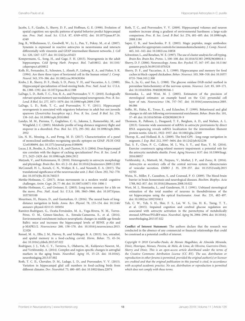
fnana-11-00126 December 23, 2017 Time: 18:38 # 18
Carvalho-Paulo et al. Hippocampal Astrocytes in C. pusilla
Jacobs, L. F., Gaulin, S., Sherry, D. F., and Hoffman, G. E. (1990). Evolution ofspatial cognition: sex-specific patterns of spatial behavior predict hippocampalsize. Proc. Natl. Acad. Sci. U.S.A. 87, 6349–6352. doi: 10.1073/pnas.87.16.6349
Jing, R., Wilhelmsson, U., Goodwill, W., Li, L., Pan, Y., Pekny, M., et al. (2007).Synemin is expressed in reactive astrocytes in neurotrauma and interactsdifferentially with vimentin and GFAP intermediate filament networks. J. CellSci. 120, 1267–1277. doi: 10.1242/jcs.03423
Kempermann, G., Song, H., and Gage, F. H. (2015). Neurogenesis in the adulthippocampus. Cold Spring Harb. Perspect. Biol. 7:a018812. doi: 10.1101/cshperspect.a018812
Kolb, H., Fernandez, E., Schouten, J., Ahnelt, P., Linberg, K. A., and Fisher, S. K.(1994). Are there three types of horizontal cell in the human retina? J. Comp.Neurol. 343, 370–386. doi: 10.1002/cne.903430304
Krebs, J. R., Sherry, D. F., Healy, S. D., Perry, V. H., and Vaccarino, A. L. (1989).Hippocampal specialization of food-storing birds. Proc. Natl. Acad. Sci. U.S.A.86, 1388–1392. doi: 10.1073/pnas.86.4.1388
LaDage, L. D., Roth, T. C., Fox, R. A., and Pravosudov, V. V. (2010). Ecologicallyrelevant spatial memory use modulates hippocampal neurogenesis. Proc. R. Soc.Lond. B Biol. Sci. 277, 1071–1079. doi: 10.1098/rspb.2009.1769
LaDage, L. D., Roth, T. C., and Pravosudov, V. V. (2011). Hippocampalneurogenesis is associated with migratory behaviour in adult but not juvenilesparrows (Zonotrichia leucophrys ssp.). Proc. R. Soc. Lond. B Biol. Sci. 278,138–143. doi: 10.1098/rspb.2010.0861
Landys, M. M., Piersma, T., Guglielmo, C. G., Jukema, J., Ramenofsky, M., andWingfield, J. C. (2005). Metabolic profile of long-distance migratory flight andstopover in a shorebird. Proc. Biol. Sci. 272, 295–302. doi: 10.1098/rspb.2004.2952
Lin, N. H., Messing, A., and Perng, M. D. (2017). Characterization of a panelof monoclonal antibodies recognizing specific epitopes on GFAP. PLOS ONE12:e0180694. doi: 10.1371/journal.pone.0180694
Lucas, J. R., Brodin, A., De Kort, S. R., and Clayton, N. S. (2004). Does hippocampalsize correlate with the degree of caching specialization? Proc. R. Soc. Lond. B271, 2423–2429. doi: 10.1098/rspb.2004.2912
Matyash, V., and Kettenmann, H. (2010). Heterogeneity in astrocyte morphologyand physiology. Brain Res. Rev. 63, 2–10. doi: 10.1016/j.brainresrev.2009.12.001
McConnell, H. L., Kersch, C. N., Woltjer, R. L., and Neuwelt, E. A. (2017). Thetranslational significance of the neurovascular unit. J. Biol. Chem. 292, 762–770.doi: 10.1074/jbc.R116.760215
Mettke-Hofmann, C. (2017). Avian movements in a modern world: cognitivechallenges. Anim. Cogn. 20, 77–86. doi: 10.1007/s10071-016-1006-1
Mettke-Hofmann, C., and Gwinner, E. (2003). Long-term memory for a life onthe move. Proc. Natl. Acad. Sci. U.S.A. 100, 5863–5866. doi: 10.1073/pnas.1037505100
Mouritsen, H., Heyers, D., and Gunturkun, O. (2016). The neural basis of long-distance navigation in birds. Annu. Rev. Physiol. 78, 133–154. doi: 10.1146/annurev-physiol-021115-105054
Ramírez-Rodríguez, G., Ocaña-Fernández, M. A., Vega-Rivera, N. M., Torres-Pérez, O. M., Gómez-Sánchez, A., Estrada-Camarena, E., et al. (2014).Environmental enrichment induces neuroplastic changes in middle age femaleBalb/c mice and increases the hippocampal levels of BDNF, p-Akt andp-MAPK1/2. Neuroscience 260, 158–170. doi: 10.1016/j.neuroscience.2013.12.026
Rensel, M. A., Ellis, J. M., Harvey, B., and Schlinger, B. A. (2015). Sex, estradiol,and spatial memory in a food-caching corvid. Horm. Behav. 75, 45–54.doi: 10.1016/j.yhbeh.2015.07.022
Rodríguez, J. J., Yeh, C. Y., Terzieva, S., Olabarria, M., Kulijewicz-Nawrot, M.,and Verkhratsky, A. (2014). Complex and region-specific changes in astroglialmarkers in the aging brain. Neurobiol. Aging 35, 15–23. doi: 10.1016/j.neurobiolaging.2013.07.002
Roth, T. C. II., Chevalier, D. M., Ladage, L. D., and Pravosudov, V. V. (2013).Variation in hippocampal glial cell numbers in food-caching birds fromdifferent climates. Dev. Neurobiol. 73, 480–485. doi: 10.1002/dneu.22074
Roth, T. C., and Pravosudov, V. V. (2009). Hippocampal volumes and neuronnumbers increase along a gradient of environmental harshness: a large-scalecomparison. Proc. R. Soc. Lond. B Biol. Sci. 276, 401–405. doi: 10.1098/rspb.2008.1184
Saper, C. B., and Sawchenko, P. E. (2003). Magic peptides, magic antibodies:guidelines for appropriate controls for immunohistochemistry. J. Comp. Neurol.465, 161–163. doi: 10.1002/cne.10858
Schweitzer, L., and Renehan, W. E. (1997). The use of cluster analysis for cell typing.Brain Res. Brain Res. Protoc. 1, 100–108. doi: 10.1016/S1385-299X(96)00014-1
Sherry, D. F. (2006). Neuroecology. Annu. Rev. Psychol. 57, 167–197. doi: 10.1146/annurev.psych.56.091103.070324
Sherry, D. F., and Vaccarino, A. L. (1989). Hippocampus and memory for foodcaches in black-capped chickadees. Behav. Neurosci. 103, 308–318. doi: 10.1037/0735-7044.103.2.308
Shu, S., Ju, G., and Fan, L. (1988). The glucose oxidase-DAB-nickel method inperoxidase histochemistry of the nervous system. Neurosci. Lett. 85, 169–171.doi: 10.1016/0304-3940(88)90346-1
Slomianka, L., and West, M. J. (2005). Estimators of the precision ofstereological estimates: an example based on the CA1 pyramidal celllayer of rats. Neuroscience 136, 757–767. doi: 10.1016/j.neuroscience.2005.06.086
Soffie, M., Hahn, K., Terao, E., and Eclancher, F. (1999). Behavioural and glialchanges in old rats following environmental enrichment. Behav. Brain Res. 101,37–49. doi: 10.1016/S0166-4328(98)00139-9
Thomsen, R., Pallesen, J., Daugaard, T. F., Børglum, A. D., and Nielsen, A. L.(2013). Genome wide assessment of mRNA in astrocyte protrusions by directRNA sequencing reveals mRNA localization for the intermediate filamentprotein nestin. Glia 61, 1922–1937. doi: 10.1002/glia.22569
Thorup, K., and Holland, R. A. (2009). The bird GPS - long-range navigation inmigrants. J. Exp. Biol. 212, 3597–3604. doi: 10.1242/jeb.021238
Tsai, S. F., Chen, P. C., Calkins, M. J., Wu, S. Y., and Kuo, Y. M. (2016).Exercise counteracts aging-related memory impairment: a potential role forthe astrocytic metabolic shuttle. Front. Aging Neurosci. 8:57. doi: 10.3389/fnagi.2016.00057
Verkhratsky, A., Matteoli, M., Parpura, V., Mothet, J. P., and Zorec, R. (2016).Astrocytes as secretory cells of the central nervous system: idiosyncrasiesof vesicular secretion. EMBO J. 35, 239–257. doi: 10.15252/embj.201592705
Weiss, N., Miller, F., Cazaubon, S., and Couraud, P. O. (2009). The blood-brainbarrier in brain homeostasis and neurological diseases. Biochim. Biophys. Acta1788, 842–857. doi: 10.1016/j.bbamem.2008.10.022
West, M. J., Slomianka, L., and Gundersen, H. J. (1991). Unbiased stereologicalestimation of the total number of neurons in thesubdivisions of therat hippocampus using the optical fractionator. Anat. Rec. 231, 482–497.doi: 10.1002/ar.1092310411
Yeh, C. W., Yeh, S. H., Shie, F. S., Lai, W. S., Liu, H. K., Tzeng, T. T.,et al. (2015). Impaired cognition and cerebral glucose regulation areassociated with astrocyte activation in the parenchyma of metabolicallystressed APPswe/PS1dE9 mice. Neurobiol. Aging 36, 2984–2994. doi: 10.1016/j.neurobiolaging.2015.07.022
Conflict of Interest Statement: The authors declare that the research wasconducted in the absence of any commercial or financial relationships that couldbe construed as a potential conflict of interest.
Copyright © 2018 Carvalho-Paulo, de Morais Magalhães, de Almeida Miranda,Diniz, Henrique, Moraes, Pereira, de Melo, de Lima, de Oliveira, Guerreiro-Diniz,Sherry and Diniz. This is an open-access article distributed under the terms ofthe Creative Commons Attribution License (CC BY). The use, distribution orreproduction in other forums is permitted, provided the original author(s) or licensorare credited and that the original publication in this journal is cited, in accordancewith accepted academic practice. No use, distribution or reproduction is permittedwhich does not comply with these terms.
Frontiers in Neuroanatomy | www.frontiersin.org 18 January 2018 | Volume 11 | Article 126