Himi Tripathi 1, Deepika Gupta 2, Sujata Mohanty 3, Seema Sen 4, Seema Kashyap 4, Radhika Tandon 1 1...
12
Himi Tripathi 1 , Deepika Gupta 2 , Sujata Mohanty 3 , Seema Sen 4 , Seema Kashyap 4 , Radhika Tandon 1 1 Department of Ophthalmology, Dr Rajendra Prasad Centre for Ophthalmic Sciences; 2 SMITA Research Labs , Department of Textile Technology, Indian Institute of Technology, New Delhi ; 3 Stem Cell Facility, All India Institute of Medical Sciences; 4 Department of Ocular Pathology, Dr Rajendra Prasad Centre for Ophthalmic Sciences, All India Institute of Medical Sciences Submission ID: 14408 The authors have no financial interests to disclose Tissue Engineered Decellularized Human Corneal Stroma for Reconstructing the Corneal Endothelium in-Vitro
-
Upload
jayce-orcutt -
Category
Documents
-
view
224 -
download
3
Transcript of Himi Tripathi 1, Deepika Gupta 2, Sujata Mohanty 3, Seema Sen 4, Seema Kashyap 4, Radhika Tandon 1 1...

Himi Tripathi1, Deepika Gupta2, Sujata Mohanty 3, Seema Sen4, Seema Kashyap4, Radhika Tandon1
1Department of Ophthalmology, Dr Rajendra Prasad Centre for Ophthalmic Sciences; 2SMITA Research Labs , Department of Textile Technology, Indian Institute of Technology, New Delhi ; 3Stem Cell Facility, All India Institute of Medical Sciences; 4Department of Ocular Pathology, Dr Rajendra Prasad Centre for Ophthalmic Sciences, All India Institute of Medical Sciences
Submission ID: 14408
The authors have no financial interests to disclose
Tissue Engineered Decellularized Human Corneal Stroma for Reconstructing the Corneal Endothelium in-Vitro

Introduction The lack of an intact endothelium of sufficient cell density and functionality
results in vision loss Pseudophakic bullous keratopathy, aphakic bullous keratopathy, corneal
endotheliopathy and Fuchs’ dystrophy are the diseases in which endothelium loss occurs and require only corneal endothelium replacement1 .
The approach to corneal transplantation has changed as the concept of customized corneal component replacement and therapy is advocated corneal tissue engineering emerges as a new frontier
Biological scaffolds are difficult to handle during the transplantation surgery and synthetic scaffolds often integrate poorly with host tissues 2
Besides synthetic polymers and biomaterials, naturally occurring biomaterials like cornea after decellularization, itself can be more promising as it has a unique extracellular matrix(ECM) organization3Use of different methods using mechanical, chemical and enzymatic protocols for decellularization of corneal stroma has been reported4,5
Aim of our study was to evaluate decellularized corneal stromal scaffolds for cultivating human corneal endothelial cells (HCECs)

Materials and MethodsHuman donor corneas not suitable for surgery were used in the study
After removing epithelium and endothelium mechanically, stromas were decellularized using six different protocols (mechanical, chemical and enzymatic using different concentrations of detergents, alcohol and trypsin EDTA)
Those decellularized corneas were subjected to histological analysis to visualize cellular remnants and the general histoarchitecture of tissues; DNA extraction to estimate DNA concentration in untreated and decellularized tissues; scanning electron microscopy (SEM) for visualization of cellular morphology; alcian blue staining to preserve ECM immuhistochemistry for major structural proteins collagen I, II, IV, fibronectin; tensile strength to estimate the mechanical strength using uniaxial load testing equipment & optical transparency via UV-visible recording spectrophotometer to check the transparency of decellularized tissues
10,000 HCECs were seeded on the decellularized corneal tissue and placed in culture medium for 14 days and then analyzed for recellularization by histology SEM, live-dead staining and immunocytochemistry for endothelial specific markers ZO-1, Na+ /K+-ATPase, VE-Cadherin & Connexin-43

Table 1: Protocols Used for Decellularization
S.No. Protocols Reagents used Time @ temperature
1. Protocol I A (PIA) 0.1%SDS&75% ethanol 27 hrs; 20+ 2°C
2. Protocol I B (PIB) 0.5%SDS&75% ethanol 27 hrs; 20+ 2°C
3. Protocol II (PII) 75% ethanol& trypsin-EDTA (0.05%) 109 hrs;20+ 2°C
4. Protocol III (PIII) 2% Triton X-100& 0.1% NH4OH 72 hrs; 4°C
5. Protocol IV A (PIV A) 0.5% trypsin 0.05%EDTA, 0.02mg/ml DNAse, 100 U/ml penicillin & 100μg/ml streptomycin
24 hrs; 37 °C
6. Protocol IV B (PIV B) 0.25% trypsin 0.05%EDTA, 0.02mg/ml DNAse, 100 U/ml penicillin & 100μg/ml streptomycin
24 hrs; 37 °C
Results
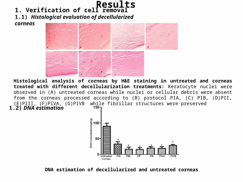
Histological analysis of corneas by H&E staining in untreated and corneas treated with different decellularization treatments: Keratocyte nuclei were observed in (A) untreated corneas while nuclei or cellular debris were absent from the corneas processed according to (B) protocol PIA, (C) PIB, (D)PII, (E)PIII, (F)PIVA, (G)PIVB while fibrillar structures were preserved
DNA estimation of decellularized and untreated corneas
1. Verification of cell removal1.1) Histological evaluation of decellularized corneas
1.2) DNA estimation

2. Characterizations of Decellularized for Extra Cellular Matrix (ECM) Architecture
The alcian blue staining confirmed an intact and well organized extracellular matrix in (A) untreated cornea and approximately unchanged stromal matrix in the decellularized corneas processed according to (B) protocol PIA, (C) PIB, (E)PIII, (F)PIVA, (G) PIVB except in PII (D)
2.1 Alcian blue staining
2.2 Collagen I staining

2.3 Collagen II Staining 2.4 Collagen IV Staining
Immunohistological characterization of decellularized human corneal stromas for various ECM proteins : (A) Untreated and decellularized corneal stromas treated according to protocol (B) PIA, (C) PIB, (D) PII, (E) PIII, (F) PIVA and (G) PIVB were immunostained with collagen I, collagen II, collagen IV & fibronectin; the decellularized corneal stromas showing similar staining as untreated cornea for various ECM
2.5Fibronectin Staining

Scanning electron micrographs showing images of untreated and decellularized corneal stromas. The collagen fibrils appear to be interconnected and they formed collagen bundles regular and parallel to corneal surface. The results were similar in both the groups;(A)untreated & decellularized corneal stromas treated according to protocol (B) protocol PIA, (C) PIB, (D) PII, (E) PIII, (F) PIVA and (G) PIVB except in PII
3. Scanning Electron Micrographs Showing Morphology of Collagen Fibrils in Untreated & Decellularized Corneal Stromas
5. Mechanical Strength of Decellularized Corneal Stromas
4. Transmittance Assay for Optical Transparency
Stress-Strain curve of decellularized corneal stroma
Representative plot showing light transmittance percentage of all the decellularized &untreated corneas

6. Repopulation of Decellularized Human Corneas
HCECs cultured on decellularized corneal stroma: H&E images of decellularized tissue (A) unseeded & (B) HCECs repopulated on decelluarized corneal human stromas showed monolayer of cultured HCECs on decellularized corneal stroma; Cell viability staining of cultured human corneal endothelial cells over decellularized corneal stroma PIVB for two weeks: Viable monolayers was determined by staining with Live/Dead Viability Kit in which the live cells fluoresce green 6.3 SEM of Repopulated Decellularized Corneas
Scanning electron microscopy images of HCECs cultured over decellularized stroma PIVB to evaluate the attachment ability at different time intervals for 2 weeks: (A) after 1 day, (B) after 3 days, (C) after 7 days and (D) after 14 days; (E) image of decellularized stroma (F) HCECs adhered to decellularized stroma and showing smooth cell sheet and close association with neighbourhood cells (G) Cultured cells showing numerous microvili at higher magnification.
6.1 Hematoxylin &Eosin Staining 6.2 Live/Dead Cell Viability Staining
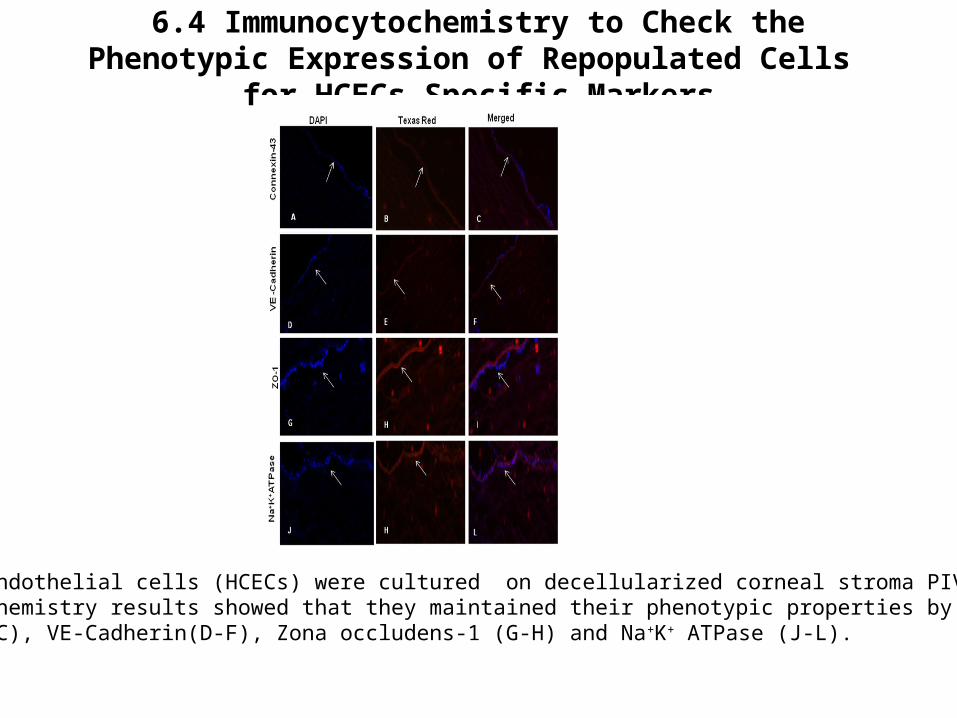
6.4 Immunocytochemistry to Check the Phenotypic Expression of Repopulated Cells for HCECs Specific Markers
Human corneal endothelial cells (HCECs) were cultured on decellularized corneal stroma PIVB for 2 weeks and immunocytochemistry results showed that they maintained their phenotypic properties by expressing Connexin-43 (A-C), VE-Cadherin(D-F), Zona occludens-1 (G-H) and Na+K+ ATPase (J-L).
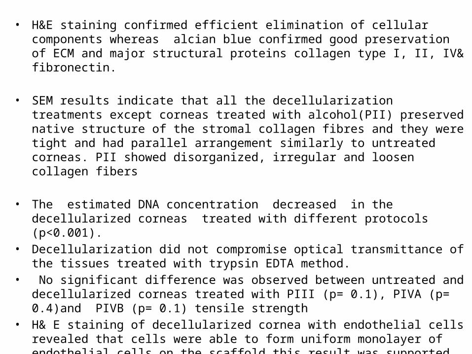
• H&E staining confirmed efficient elimination of cellular components whereas alcian blue confirmed good preservation of ECM and major structural proteins collagen type I, II, IV& fibronectin.
• SEM results indicate that all the decellularization treatments except corneas treated with alcohol(PII) preserved native structure of the stromal collagen fibres and they were tight and had parallel arrangement similarly to untreated corneas. PII showed disorganized, irregular and loosen collagen fibers
• The estimated DNA concentration decreased in the decellularized corneas treated with different protocols (p<0.001).
• Decellularization did not compromise optical transmittance of the tissues treated with trypsin EDTA method.
• No significant difference was observed between untreated and decellularized corneas treated with PIII (p= 0.1), PIVA (p= 0.4)and PIVB (p= 0.1) tensile strength
• H& E staining of decellularized cornea with endothelial cells revealed that cells were able to form uniform monolayer of endothelial cells on the scaffold this result was supported by SEM . Cell viability staining revealed that decellularized corneas were biocompatible and support expansion of HCECs
• Moreover, they also expressed the endothelial specific markers Na+ /K+-ATPase, ZO-1 , VE-Cadherin& Connexin-43
•
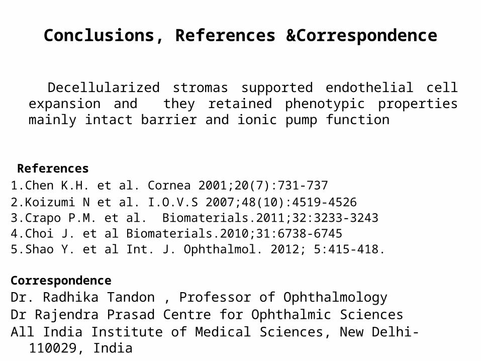
Conclusions, References &Correspondence
Decellularized stromas supported endothelial cell expansion and they retained phenotypic properties mainly intact barrier and ionic pump function
References 1.Chen K.H. et al. Cornea 2001;20(7):731-7372.Koizumi N et al. I.O.V.S 2007;48(10):4519-45263.Crapo P.M. et al. Biomaterials.2011;32:3233-32434.Choi J. et al Biomaterials.2010;31:6738-67455.Shao Y. et al Int. J. Ophthalmol. 2012; 5:415-418.
CorrespondenceDr. Radhika Tandon , Professor of OphthalmologyDr Rajendra Prasad Centre for Ophthalmic Sciences All India Institute of Medical Sciences, New Delhi-110029, Indiaemail id : [email protected]