Higher State Educational Establishment of Ukraine ... · Benign tumors and cysts of salivary glands...
17
Ministry of Public Health of Ukraine Higher State Educational Establishment of Ukraine “Ukrainian Medical Stomatological Academy” “APPROVED” at the meeting of the Department of surgical stomatology and maxillofacial surgery with plastic and reconstructive surgery of the head and neck Minutes No.1 Head of the Department D.Med.Sci., Prof. __________D.S. Avetikov METHODICAL GUIDANCE for students’ self-directed work when preparing for and during the practical session Academic subject Surgical stomatology Module No. 3 Content module No. 3 Topic of practical session Tumors of blood and lymphatic vessels of soft tissues of maxillofacial area and jaws. Tumors and tumor- like formations of peripheral nerves of the face. Clinic, diagnostics, differential diagnostics, treatment. Epithelial and non-epithelial tumors of salivary glands. Cysts of salivary glands. Clinic, diagnostics, differential diagnostics, treatment Year of study IV Faculty Foreign Students Training (Stomatology) Poltava
Transcript of Higher State Educational Establishment of Ukraine ... · Benign tumors and cysts of salivary glands...
Ministry of Public Health of Ukraine
Higher State Educational Establishment of Ukraine
“Ukrainian Medical Stomatological Academy”
“APPROVED”
at the meeting of the Department
of surgical stomatology
and maxillofacial surgery with plastic and
reconstructive surgery of the head and neck
Minutes No.1
Head of the Department
D.Med.Sci., Prof. __________D.S. Avetikov
METHODICAL GUIDANCE
for students’ self-directed work when preparing for
and during the practical session
Academic subject Surgical stomatology
Module No. 3
Content module No. 3
Topic of practical session Tumors of blood and lymphatic vessels of soft tissues
of maxillofacial area and jaws. Tumors and tumor-
like formations of peripheral nerves of the face.
Clinic, diagnostics, differential diagnostics,
treatment.
Epithelial and non-epithelial tumors of salivary
glands. Cysts of salivary glands. Clinic, diagnostics,
differential diagnostics, treatment
Year of study IV
Faculty Foreign Students Training (Stomatology)
Poltava
1. Relevance of the topic.
Stomatologists of any profile sometimes find the necessity of inspection and
treatment of patients with the tumors of blood and lymphatic vessels of soft tissues
and jaws. An important moment is realization of differential diagnostics between
benign and malignant tumors, that requires from the doctor of theoretical
knowledge and practical abilities of clinical and paraclinical examination of
patients, abilities to interpret data of additional methods of examinations and
appoint adequate treatment to every concrete patient, in fact quality of life of
patient depends in future on it.
Benign tumors and cysts of salivary glands are rather difficult pathology of a
maxillofacial site which equally often meets at men and women of different age.
This group of diseases has a various, often similar, clinical picture which needs
detailed knowledge at future doctors concerning an etiology, clinic, diagnostics
and treatment.
2. Specific aims:
2.1.To analyse etiology and pathogeny of tumors of blood and lymphatic vessels
and peripheral nerves of maxillofacial area.
2.2.To explain factors which provoke the origin of tumors of blood and lymphatic
vessels and peripheral nerves of maxillofacial region.
2.3.To offer new approaches in diagnostics of tumors of blood and lymphatic
vessels and peripheral nerves of maxillofacial area.
2.4.To classify the tumors of blood and lymphatic vessels of maxillofacial area.
2.5.To interpret the additional methods of examination at diagnostics of tumors of
blood and lymphatic vessels and peripheral nerves of maxillofacial area.
2.6.To draw the charts of roentgenologic picture of tumors of blood and lymphatic
vessels and peripheral nerves of bones of facial skeleton.
2.7.To analyse data of additional methods of examination of patients with tumors
of blood and lymphatic vessels and peripheral nerves of maxillofacial area.
2.8.To lay down the plan of treatment of patients with the tumors of blood and
lymphatic vessels and peripheral nerves of maxillofacial area.
2.9.To analyse clinical manifestations of epithelial and non-epithelial tumors and
cysts of salivary glands.
2.10.Ethiological and pathogenetical factors of development of tumor-like
formations of soft tissues.
2.11.The main and additional investigations of patients with epithelial and non-
epithelial tumors and cysts of salivary glands.
2.12.To treat emergence and development of epithelial tumors and cysts of salivary
glands.
2.13.To draw schemes, graphs of classification of epithelial tumors and cysts of
salivary glands.
2.14.To analyse classification of epithelial tumors and cysts of salivary glands.
2.15.To make the plan of treatment of patients with epithelial tumors and cysts of
salivary glands.

3. Basic knowledge and skills necessary to study the topic
(interdisciplinary integration).
The preceding subjects The obtained skills
1. Normal anatomy. To define localization of tumors of blood and
lymphatic vessels of soft tissues and jaws.
2. Operative surgery and
topographical anatomy.
To conduct cuts depending on the place of
location of tumor.
To lay on the different types of guy-sutures that
is used for moving away of tumors of blood and
lymphatic vessels.
3. Internal illnesses. To conduct the inspections of patient.
To interpret data of laboratory investigations.
4. Pathological
morphology.
To know the histological structure of tumors of
blood and lymphatic vessels of soft tissues and
jaws.
5. Pathological physiology. To know the etiological and pathological aspects
of origin of tumors of peripheral nerves blood
and lymphatic vessels of soft tissues and jaws.
6. Propaedeutics of internal
illnesses.
To adhere to the deontology at
commonunication with patients.
The preceding subjects The obtained skills
1. Normal anatomy. To be guided in the main questions of a
structure of salivary glands.
2. Histology. To prepare a material for histological or
cytological research of tumors of salivary
glands.
3. Pathological anatomy. To make the exact diagnosis.
4. Pathological physiology. Etiology and pathogenesis of developments of
benign tumors of salivary glands.
5. Topographical anatomy and
operative surgery.
To be guided in a choice of operative accesses
at treatment of benign tumors of salivary
glands.
6. General oncology. To make a prognosis at treatment of patients
with epithelial and non-epithelial tumors of
salivary glands.
7. Surgical stomatology and
maxillo-facial surgery.
Treatment of benign tumors of salivary glands.
4. The tasks for students’ self-directed work during preparation for the class.
4.1. The list of basic terms, parameters and characteristics which students
should master while preparing for the class:
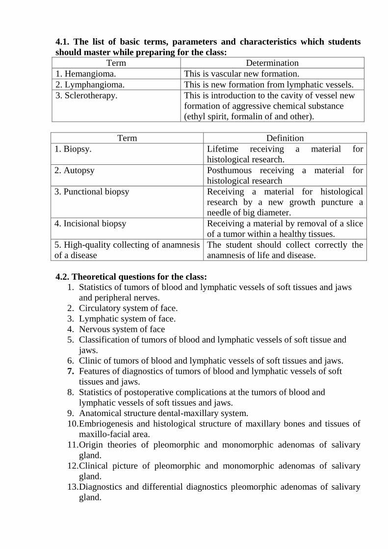
Term Determination
1. Hemangioma. This is vascular new formation.
2. Lymphangioma. This is new formation from lymphatic vessels.
3. Sclerotherapy. This is introduction to the cavity of vessel new
formation of aggressive chemical substance
(ethyl spirit, formalin of and other).
Term Definition
1. Biopsy. Lifetime receiving a material for
histological research.
2. Autopsy Posthumous receiving a material for
histological research
3. Punctional biopsy Receiving a material for histological
research by a new growth puncture a
needle of big diameter.
4. Incisional biopsy Receiving a material by removal of a slice
of a tumor within a healthy tissues.
5. High-quality collecting of anamnesis
of a disease
The student should collect correctly the
anamnesis of life and disease.
4.2. Theoretical questions for the class:
1. Statistics of tumors of blood and lymphatic vessels of soft tissues and jaws
and peripheral nerves.
2. Circulatory system of face.
3. Lymphatic system of face.
4. Nervous system of face
5. Classification of tumors of blood and lymphatic vessels of soft tissue and
jaws.
6. Clinic of tumors of blood and lymphatic vessels of soft tissues and jaws.
7. Features of diagnostics of tumors of blood and lymphatic vessels of soft
tissues and jaws.
8. Statistics of postoperative complications at the tumors of blood and
lymphatic vessels of soft tissues and jaws.
9. Anatomical structure dental-maxillary system.
10. Embriogenesis and histological structure of maxillary bones and tissues of
maxillo-facial area.
11. Origin theories of pleomorphic and monomorphic adenomas of salivary
gland.
12. Clinical picture of pleomorphic and monomorphic adenomas of salivary
gland.
13. Diagnostics and differential diagnostics pleomorphic adenomas of salivary
gland.

14. Treatment methods of pleomorphic and monomorphic adenomas of salivary
gland.
4.3. Practical tasks to be completed during the class:
1. To conduct palpation of tumor maxillofacial region.
2. To conduct diagnostic puncture.
3. Working off the algorithm of examination and treatment of the patients with
the vascular tumors of face.
4. To seize a technique of inspection of the patient with pleomorphic adenomas
of salivary gland.
5. To carry out a curation of the patient with pleomorphic adenomas of salivary
gland.
6. To fill an out-patient medical card of the patient from pleomorphic
adenomas of salivary gland.
5. The content of the topic:
Haemangioma Haemangioma is generally accepted to be hamartomatous lesion rather than
true neoplasm.
Clinical features
Common age: At birth, or early after birth, some lesions may not be noted until
later.
Common sex: Female more than male.
Common site: Head and neck region more than other part of the body. Intraorally
common on the lip, tongue, cheek and palate.
Intraoral mucosal haemangioma appear as flat or nodular, soft dark reddish or
purplish pinkish lesion, that may bleed following mild trauma. Haemangioma
characteristically blanch under pressure, this may help lo differentiate it from other
soft tissue hemorrhagic lesion e.g. ecechymosis.
Haemangioma of the cheek
Haemangioma may occur inside jaw bone (intraosseously), which appear
radiographically as irregulars radiolucent area or multilocular or honey comb or
soap bubble appearance. Haemangioma is usually solitary lesion but multiple
lesions may occur rarely as part of some generalized angiomatous syndrome such
as (Sturge- Weber syndrome) (Encephatotrigaminal angiomatosis) and (Hereditary
haemorrhagic telangectasi,) (Rendu-Osler- Weber syndrome).

Sturge-Weber syndrome (Encephalotrigaminal angiomatosis)
Sturge-Weber syndrome is a congenital disorder characterized by the following
main features:
1. Ipsilateral haemangiomotous lesion of the face following the distribution of one or
more of thebranches of the trigeminal nerve, (port-wine staine). The facial lesion may
extend intraorally involving the buccal mucosa and gingiva.
2. Calcification of leptomeninges over the cerebral cortex.
3. Convulsion of the limb, on the opposite side of the body.
4. Ocular lesion also may be present.
Sturge-Weber syndrome (port-wine stain)
Hereditary hemorrhagic telangectasia (Rendu-Osler-Weber Syndrome)
Hereditary hemorrhagic telangectasia ii a hereditary disorder transmitted as
autosomal dominant and characterized by:
- Multiple knots of dilated malformed capillares (telangectasia) present in skin,
mucous membrane of oral cavity and nasal cavity Fig.
- Frequent nose bleeding is the commonest presenting sign of Rendu -Osler-
Weber Syndrome.
Hereditary hemorrhagic telangectasia of toungue
Histopathological features
Histologically haemangioma is classified into capillary, or cavernous or
mixed, depending upon the size of the vascular spaces, these spaces are lincd by
endothelium without muscular support. Clinically there is no significant difference
between capillary and cavernous haemangioma.

Capillary haemangioma (histological features)
Diagnosis of haemangioma
• Clinical examination shows congenital vascular lesion that blanch on pressure.
• The boney lesion shows honey comb appearance radiographically.
Treatment - Surgical excision, following localization of the lesion through selective
arteriole embolization.
- Sclerosant therapy or cryosurgery.
- The laser therapy is now used as a primary treatment of selected vascular
lesion.
Lymphangioma Lymphangioma is considered as a hamartomatous, rather than being true neoplasm
and it is less common than haemangioma.
Clinical features Common ages: At birth (Congenital) or shortly after birth,
Common site: Tongue leading to macroglossia and the lip leading to macropheilia.
The superficially located lesion appears as painless nodular or papillary
swelling with the same color or slightly redder than the surrouding mucosa. The
deeper lesions show diffuse nodules without significant changes in the surface texture or
color. On palpation it may produce a crepitant sound due to pushing of lymphatic
fluid form one area to another.
Limphangiomatous malformation may occur in the neck region in early
development leading to cystic hygroma which presents as a large fluctuant
swellings up to 10 cm in diameter and may extend to involve vital structure in the
neck with respiratory distress, hemorrhage and disfigurement.
Histopathological features

Microscopically lymphagioma composed of thin-walled vascular spaces, containing
pinkish amorphous material as the result of fixation of limph.
Treatment - Surgical excision with reccurence, because of lack of encapsulation.
- The treatment of cystic hygroma may need stages surgical procedures to
control the lesion.
Neurofibroma is uncommon benign tumor of nerve sheath origin. It occurs
in two forms: either as single lesion, which is true neoplasm or as multiple lesions
which considered as hamartomas rather than neoplasms. The multiple lesions are
also known as neurofibromatosis or Von Recklinghausen’s disease of skin.
The single neurofibroma commonly occurs in the tongue and buccal mucosa
and appears as smooth painless fusiform nodules, covered by normal mucosa.
Neurofibroma also may occur intraosseously. The multiple or neurofibromatosis
appear as a numerous discrete or separated nodules on the skin and oral mucosa.
The skin lesions may be so numerous and prominent that they become
cosmetically significant with cutaneous melanin pigmentation described as café-
au-lait spots which precede the neural lesions. Malignant transformation may be
seen in 5-15% of cases of neurofibromatosis.
Microscopically neurofibroma shows a mixture of Shwann cells and
fibroblast cells, with varying amount of collagen and mucinous material.
The differential diagnosis of neurofibroma includes: traumatic neuroma,
traumatic fibroma, granular cell tumor, some other connective tissues lesions.
Treatment: surgical excision for single neurofibroma, for surgical excision is
impractical for neurofibromatosis.
Neurilemmoma (schwannona) is uncommon benign neoplasm of axon
sheath origin (Schwann’s sheath), but it is more common than neurofibroma.
Clinically neurilemmomaappears as painless slowly growing, submucosal mass;
the tongue is favored site, also may occur in bone, producing radiolucent areas and
may associate with pain or parestesia.
Histologically neurilemmoma shows two characteristic patterns:
Antony A, which consists of parallel rows or whorls of collagen fibers and
spindle with palisaded nuclei.
Antony B, which consists of disorderly arranged cells and collagen fibers in
a mucinous microcystic stroma.
The different diagnosis of neurilemmoma includes: traumatic neuroma,
salivary gland tumors, some other benign mesenchymal tumors.
Treatment: surgical excision with excellent prognosis.
Traumatic neuroma is a non-neoplastic, but considered as a reactive
hyperplasia of peripheral nerve fibers as a result of traumatic injury. Traumatic
neuroma is common in areas of previous surgery. Traumatic neuroma is
uncommon lesion in the oral cavity and appears as small rounded nodules, which
can give rise to pain on pressure.
Microscopically traumatic neuroma shows nerve fibers, Shwann cells, and
connective tissue stroma.

The differential diagnosis of traumatic neuroma includes: any small mass
painful on pressure (a typical facial pain).
Treatment: surgical excision.
The non-neoplastic salivary gland diseases represent a disparate group of
disorders affecting both the major and minor salivary glands. These conditions
range from inflammatory disorders of infectious, granulomatous, or autoimmune
etiology to obstructive, developmental, and idiopathic disorders. The major
salivary glands are most often involved, and many of these salivary gland disorders
are associated with the presence of other systemic illnesses. A thorough history and
physical examination is typically adequate to recognize and differentiate this group
of conditions, and an elaborate diagnostic evaluation is usually not required to
manage these illnesses.
This presentation will attempt to achieve the following objectives. The first
is to provide a useful scheme for classifying these hetergeneous disorders so that
they may be approached in a systematic manner. The second is to enumerate the
distinguishing features of these entities to aid in their appropriate recognition.
Finally, general management principles for the various conditions will be
discussed.
Benign epithelial tumors
Pleomorphic adenomas (benign mixed tumors) are the most common
tumors of the salivary gland and are most often located in the tail of the parotid
gland. When found in the minor salivary glands, the hard palate is the site most
frequently involved, followed by the upper lip.
These tumors were termed pleomorphic because of the epithelial and
connective tissue components that compose them in varying degrees. Their gross
appearance is a round, smooth mass with a thin, delicate, incomplete capsule. Of
note, pleomorphic adenomas that arise in the minor salivary glands usually lack a
capsule. These tumors grow slowly, although they may become larger than other
SGTs. The thin, delicate capsule may have projections into the surrounding parotid
tissue. This is of particular clinical significance because obtaining clean margins
and avoiding spillage are mandatory to minimize recurrence.
Microscopically, benign mixed tumors are characterized by variable,
diverse, structural histologic patterns. Frequently, they have growth patterns of
sheets, strands, or islands of spindle and stellate cells, with a myxoid configuration
occasionally predominating. Treatment of benign neoplasms involves the complete
surgical excision of the affected gland. If the parotid gland is involved, superficial
parotidectomy with standard facial nerve dissection and preservation is the
procedure of choice. Enucleation is contraindicated because of the tendency
towards tumor spillage that can lead to tumor recurrence.
Warthin tumor (papillary cystadenoma lymphomatosum, cystic
papillary adenoma, adenolymphoma)
Albrecht first recognized this tumor in 1910, and Warthin later described it
in 1929. In gross appearance, it is a smooth, soft, parotid mass. It is well
encapsulated when located in the parotid gland and contains multiple cysts.
Histologically, the Warthin tumor has a heavy lymphoid stroma and aciniform
epithelial cells that line the cystic areas with papillary projections. Malignant
transformation has not been observed. All patients with this tumor survive, and the
recurrence rate is 5%. The Warthin tumor tends to be bilateral (10% of cases) and
is usually found in the major glands, as are most other types.
Intraductal papilloma
IDP is a small, tan, fairly smooth lesion that is usually found in the
submucosal layer. Microscopically, IDP consists of a cystically dilated duct
partially lined with a cuboidal epithelium with complex anastomosing papillary
fronds of variable size filling the cystic area. IDP of the minor salivary gland is a
rare lesion that has been described only in various case reports.
Histologically, the differential diagnosis of IDP includes papillary
cystadenoma, which is commonly but erroneously diagnosed as IDP. In papillary
cystadenoma, intraductal hyperplasia occurs and the dilated duct contains some
papillary folds and projections. However, this occurs much less frequently than in
IDP.
Oxyphil adenoma (oncocytoma)
Duplay first described the oncocytic tumor in 1875. Oncocytomas of the
salivary glands are very uncommon. Such neoplasms occur more often in women
than in men, with a female-to-male ratio of 2:1. Patients are older than 50 years,
and the superficial lobe of the parotid gland is the most commonly reported
location. Oncocytomas rarely, if ever, occur in the minor salivary glands.
Oncocytomas manifest as small (< 5 cm in diameter), firm, slow-growing,
spherical masses. Bilateral oncocytomas of the parotid glands have been described.
Histologically, they are large and spherical and have a distinct capsule. Uniform
cells are arranged in solid sheets. These tumors recur if excision is incomplete.
Benign nonepithelial tumors
Hemangiomas are the most common SGTs in children and usually involve
the parotid gland. Less often, they may involve the submandibular gland. These
vascular tumors may be distinguished from vascular malformations by their
presence early in life, rapid growth phase in children aged approximately 1-6
months, and gradual involution over 1-12 years. The typical presentation is an
asymptomatic, unilateral, compressible mass. Gross examination reveals a dark
red, lobulated, unencapsulated mass. Microscopically, hemangiomas are composed
of solid masses of cells and multiple anastomosing capillaries that replace the
acinar structure of the gland. Because they lack a capsule, they tend to infiltrate
neighboring structures.
Treatment should initially consist of steroids administered 2-4 mg/kg/d.
Although the response may be immediate, only 40-60% of hemangiomas exhibit a
response to steroids. Despite the tendency toward spontaneous involution, specific
conditions may warrant surgical excision.
Lymphangioma (cystic hygroma) are most commonly located in the head
and neck region of infants and children. They are believed to be due to lymphatic
sequestration of primitive embryonic lymph ducts that undergo irregular growth
and canalization. They are spongy, multiloculated masses with a yellowish or

bluish surface and are formed by endothelial-lined spaces. More than 50%
manifest at birth, and 80% manifest by age 2 years. Usually, they manifest as
painless masses that may involve parotid glands, submandibular glands, or both.
Diagnosis is made based on clinical findings. Surgical excision with preservation
of the vital structures is the treatment of choice. Lymphangiomas rarely cause
symptoms of airway obstruction, and excision is usually for cosmetic reasons.
Lipoma tumors are relatively uncommon in a major salivary gland. They
derive from fat cells and appear grossly as smooth, well-demarcated, bright-yellow
masses. Histologically, the tumor consists of mature adipose cells with uniform
nuclei.
These tumors manifest as soft, mobile, painful masses and peak in the fifth
and sixth decades of life, with a male-to-female ratio of 10:1. They are slow-
growing tumors with an average diameter of 3 cm. Treatment is surgical excision.
Cysts are common, membrane-covered, sac-like structures that can grow
anywhere in the body. Cysts can be filled with fluid, gas or semi-solid material.
Hundreds of kinds of cysts exist, and they can be microscopically small or big
enough to crowd other organs. Most cysts are harmless.
Different kinds of salivary gland cysts exist as well. Salivary gland cysts can
develop in the parotid glands, which are found in your upper cheeks, near your
ears. You can also find salivary gland cysts in the submandibular and sublingual
glands, the other major salivary glands, as well as in the 600 to 1,000 minor
salivary glands scattered throughout the oral cavity.
Most cysts aren’t cancerous, but even a benign cyst can cause problems. If
you have cysts, mouth problems can result, since even a painless, benign cyst can
grow large enough to interfere with eating, speaking or swallowing.
Development Of Salivary Cysts: Mouth Injuries And More
Salivary gland cysts are sometimes found in newborns, due to problems with
ear development during gestation.
After birth, salivary gland cysts can be the result of salivary gland stones or
infection. Although typically benign, these cysts can interfere with saliva flow.
With other cysts, mouth injury may be the cause. Frequently found inside
the lower lip, this type of benign cyst is called a mucocele, and is filled with
mucus.
Salivary Gland Cysts And HIV
Salivary gland cysts can form in the parotid gland. People with HIV
infection have a tendency to develop salivary gland cysts in this area. In fact, the
connection between HIV infection and salivary gland cysts in the parotid gland is
so strong that if doctors have a patient with persistent parotid cysts, an HIV test is
often recommended.
Treating Salivary Gland Cysts
If you discover a new lump or swelling in your mouth that doesn’t go away,
contact your doctor or dentist, even if the bump causes no pain. Most salivary
gland cysts can be treated without complication, but it is helpful to rule out other
problems.

Some salivary gland cysts, particularly a small, benign cyst, will drain and
disappear on their own. Mucoceles can release a straw-colored liquid.
Larger salivary gland cysts, even a benign cyst, can grow big enough to
require treatment. Treatment options for salivary gland cysts include:
Compresses
Drawing off fluid with a needle
Laser surgery
Medication to reduce saliva flow
Traditional surgery.
Salivary gland Lymphoepithelial cysts are benign, slowly growing
unilocular or multilocular lesions that may appear in the head and neck. Among the
reported head and neck sites are the salivary glands (typically the parotid gland)
and the oral cavity (usually the floor of the mouth). These cysts are usually seen in
adults and only occasionally in children. They range in size from 0.5 to 5.0 cm, and
they can cause considerable cosmetic deformity and physical discomfort.
Lymphoepithelial cysts have been associated with human immunodeficiency
virus (HIV) infection as part of a diffuse infiltrative lymphocytosis syndrome.
They can also arise in HIV-negative patients who have Sjogren's syndrome,
Mikulicz's disease, and myoepithelial sialadenitis. HIV infection should be
suspected in a patient who has multiple bilateral lymphoepithelial cysts of the
major salivary glands, especially the parotid glands. Ultrasound imaging is a good
diagnostic modality. Fine-needle aspiration can be both diagnostic and therapeutic.
These cysts are lined with a squamous or glandular epithelium, and they are
surrounded by dense polymorphous (polyclonal) lymphoid tissue. Prominent
epithelial infiltration by lymphocytes is characteristic, as is the presence of
epimyoepithelial islands, which are epithelial cell nests extensively infiltrated by
lymphocytes.
Salivary glande (retention cyst) A Ranula is a transparent retention cyst in
the floor of mouth arising from the sublingual salivary glands. Ranula means a
small frog and the cyst is so-called because of a supposed resemblance to a small
frog. The cest enlarges slowly, penetration the deep structures of the floor of the
mouth above the mylohyoid muscle. It is more common in neonates and children
and potentially can cause respiratory embarrassment. It is appears as blau-grey,
dome-like swelling beneath the tongue. It is highly transluminable. It may burst
spontaneously, discharging its contens and collapsing, but almost invariably
recurring.
Treatment depends on size. Small ranulae are excised, larger ones are
marsupialized, i. e. de-roofed so thet the cyst opens into the floor or the mouth.
Congenital Sublingual Cyst
Congenital sublingual cysts are usually rare. A neonate presented in our
facility with feeding difficulties caused by a large sublingual cyst. Excision of the
cyst was done and post operative period was uneventful. In this case we discuss the
common presentation of such rare cyst and their management.
A ranula is a pseudocyst that is caused by the extravasation of mucus from
the sublingual gland. It occurs specifically in the floor of the mouth associated with
the sublingual or submandibular salivary glands as a fluctuant, unilateral, bluish
soft tissue mass. They are cystic and are frequently blue owing to the Tyndall
effect, whereby blue light is reflected more than red light at the interface of soft
tissue and cyst. Ranula could occur following trauma or obstruction of the outlet of
the glands. Similar cysts could arise from; an imperforate submandibular or
sublingual duct orifice. This may result in a cystic swelling with a characteristic
bluish, translucent appearance though it occurs rarely. Unlike ranula cyst these
have epithelial lining. Other differential diagnoses include dermoid cysts,
lymphatic or vascular malformations, minor salivary gland tumors, and mucous
retention cysts. Dermoid cyst usually occurs in the midline
Patients usually present with non painful swelling noticed incidentally or by
displacement of the tongue. It may also be suspected in a neonate with feeding
difficulties. Interference with feeding may lead to failure to thrive if not
addressed5. The methods, and the age of intervention are diverse in literature.
Observation for 6 months has been suggested as is aspiration upto removal of
ipsilateral sublingual gland. We present a case of a neonate with a congenital
sublingual cyst who presented with feeding difficulties at birth.
Types of individual work of students.
A. To study the following questions:
1. X-ray pictures of patients with hemangioma of bones of facial skeleton.
2. Photos of patients with the vascular tumors of face.
3. Anatomy and physiology of salivary glands of human.
4. Embriogenesis and histologic structure of big and small salivary glands.
5. Theories of an origin of benign tumors of salivary glands.
6. Histologic and clinical classifications of benign tumors of salivary glands.
7. Clinical picture of benign tumors of salivary glands.
8. Diagnostics and differential diagnostics of benign tumors of salivary glands.
9. Methods of treatment of benign tumors of salivary glands.
10. A prognosis and complication in time and after treatment of benign tumors
of salivary glands.
B. Situational tasks for self-check:
1. Patient, 20 years old, has asymmetry of face due to the tumor of upper lip on
left. Skin under neoplasm is blue, positive symptoms of "compression" and
"filling" are determined. What most credible diagnosis?
(Answer: Cavernous hemangioma).
2. A patient, 17 years old, complains of the presence of tumor on a low lip.
Anamnesis of disease: tumor was determined after birth, slowly increasing, but for
the last year acute increased in a size. At a review on a low lip there is a tumor of
red color, soft, at pressure becomes empty and pale. What previous diagnosis?
(Answer: hemangioma of low lip).
3. A patient, 20 years old, appealed to the surgeon with a tumor on the left cheek.
The presence of neoplasm marks from birth. At a review on the skin of the left

cheek flat rounded form spot, to 1,5 cm in a diameter, pink-red color. At pressure
on a tumor by a finger or instrument neoplasm turns pale. Define a diagnosis.
(Answer: hemangioma).
4. The patient of 65 years came to the stomatologist-surgeon with complaints to a
swelling in the left half of the face addressed. At the review of the patient it was
established that: in the left parotid site the new growth in the size 3х 4 cm. At a
palpation the new growth has dense character, painless, to surrounding tissues isn't
soldered. After carrying out contrast radiological research on sialogramme is
revealed that normal drawing of branchings excretory canal the passage sharply
breaks on a limit with pathological process. For what disease characteristic such
sialografichesky picture? What else additional methods of inspection need to be
carried out?
(Answer: tumor of salivary gland).
5. The patient of 57 years came to the surgeon-stomatologist with the complaint to
asymmetry of the face. Objectively: in a site of the right parotid salivary gland the
tumor of densely elastic consistence is found painless, mobile, a roundish form
with a hilly surface. The tumor tends to slow increase. It is possible to establish
what previous diagnosis in this case?
(Answer: the mixed tumor).
C. Materials for test control. Test tasks with the single correct answer (α=2):
1. The basic clinical sign of branched hemangioma is:
А. Pulsation.
В. Presence of phlebolith.
С. Pain at palpation.
D. Sickly infiltration.
Е. Vensan’s symptom.
(Right answer: А).
2. The basic methods of treatment of capillary hemangioma is:
A. Sclerotherapy.
B. Sewing.
C. Electro-coagulation.
D. Radial therapy.
Е. Excision of tumor.
(Right Answer: А).
3. Clinically a lymphangioma is characterized by varieties:
A. Porous, cystic, lytic.
B. Polymorphic, monomorphic, proliferative.
C. Capillary, cystic, cavitary.
D. Dissipated, local.
Е. Solid, monomorphic.
(Answer: С).
4. Benign tumors doesn't treat:
A. Adenoma;
B. Adenolimfoma;
C. The mixed tumor;
D. Mucoepidermal tumor;
E. Cyllindroma;
(Right answer: D).
5. Clinical picture of adenoma of salivary glands
A. Tumor painless dense, with the smooth surface, not soldered to surrounding
tissues.
B. Tumor painless, mobile, the round or extended form, a chardelastic consistence.
C. The tumor grows is quickly soldered to surrounding tissues.
D. Tumor painless, not mobile, the correct form, a plotnoelastichny consistence
grown together with skin.
(Right answer: А).
6. What method of treatment belongs to surgical tumors of salivary glands.
A. Across Lviv.
B. On Kovtunovich-Mukhin.
C. According to Hitrov.
D. According to Gorbushenina.
E. According to Limberg.
(Right answer: В).
7. Ranula is more often located:
A. In submaxillary salivary gland.
B. In parotid salivary gland.
C. To hypoglossal salivary gland.
(Right answer: С).
D. Educational tasks of 3th levels (atypical tasks):
1. A patient appealed to the clinic with complaints of the considerable increasing
of low lip. There is increasing of low lip, pinky color, tissues of lips painless and
densely-elastic at palpation. Define a diagnosis, conduct differential diagnostics,
appoint treatment.
(Answer: lymphangioma of low lip, operative treatment).
2. A patient, 25 years old, appealed to the policlinic with complaints of he presence
of the swelling in the left parotid-masticatory area. At inclination of head the
symptom of "filling" is determined in the left parotid-masticatory area. In the
cavity of mouth the mucous membrane of this area in a color is not changed.
Define a diagnosis, conduct differential diagnostics, appoint treatment.
(Answer: hemangioma of parotid-masticatory area, sclerotherapy with next
operative intervention).
3. The patient of 65 years addressed to the stomatologist-surgeon with a swelling
in a parotid site. After carrying out contrast radiological research on sialogramme
seen that normal drawing of branchings of a channel sharply breaks on a border
with pathological process. At a puncture received transparent viscous liquid. What
diagnosis?
(Right answer: Cyst of parotid salivary gland).

4. The patient of 57 years addressed to the surgeon-stomatologist with the
complaint to asymmetry of a face. Objectively: in a site of parotid salivary gland is
formation painless, mobile, a roundish form softelastic consistences. The tumor
tends to slow increase. Function of gland isn't broken. It is possible to establish
what preliminary diagnosis in this case?
(Right answer: Cyst of parotid salivary gland).
5. The patient of 35 years addressed to the doctor stomatologist with complaints to
a swelling under language.There is the new growth in hypoglossal area in the size
of 3х4 cm transparent to appearance, at a palpation painless. It is possible to
establish what preliminary diagnosis:
(Answer: Ranula | hypoglossal salivary gland).
6. The patient address to the surgeon-stomatologist with complaints to a new
growth on the bottom jaw at the left. Objectively skin over a new growth isn't
changed, at a palpation the tumor isn't soldered to surrounding fabrics is painless.
To what expert is need to direct the patient for diagnosis specification?
(Right answer: oncostomatologist).
References
Basic:
1. Peterson’s Principle of oral and maxillofacial surgery. 3rd Edition / M. Miloro,
G.E. Ghali, P.E. Larsen, P.D. Waite. – Hamilton London, BC Decker Inc, 2012. –
1664 p.
2. Oral and maxillofacial surgery : textbook / Ed. by prof. V. Malanchuk / part one.
– Vinnytsia : Nova Knyha Publishers, 2011. – 424 p.
3. Oral and Maxillofacial Surgery / J. Pedlar, J. Frame. – Edinburg, London, New
York, Philadelphia, Sydney, Toronto: Churchill Livingstone, 2003. – 325 p.
4. Principle of oral and maxillofacial surgery / Ed. by U.J. Moore. – Blackwell
Science, 2001. – 276 p.
5. Skikevich M.G. Benign tumors and tumor-like formations of maxilla-facial
region / M.G. Skikevich, V.N. Gavrilyev. – Poltava : ASMI, 2008. – 132 p.
6. Textbook of general and oral surgery / D. Wray, D. Stenhouse, D. Lee, A. Clark.
– Edinburg, London, New York, Philadelphia, St Louis, Toronto, Churchill
Livingstone, 2003. – 322 p.
Additional:
1. Mitchell D. An Introduction to Oral and Maxillofacial Surgery / D. Mitchell. –
Oxford University Press, Jan, 2006. – 356 p.
2. Coulthard P. Master dentistry / P. Coulthard, K. Horner PH. Sloan, E. Theaker.
– Edinburg, London, New York, Philadelphia, St Louis, Toronto, Churchill
Livingstone, 2003. – 267 p.
3. Tkachenko P.I. Propaedeutics of surgical stomatology and inflammatory
diseases of maxillofacial region / P.I. Tkachenko, A.I. Pan’kevich, K.Yu. Rezvina.
– Poltava : ASMI, 2011. – 283 p.
4. Web source:
[http://www.sciencedirect.com/science/article/pii/S1010518289800988], 2016
6. Web source:

[http://www.sciencedirect.com/science/article/pii/S0022347663803303], 2015
The content of the topic:
References:
Basic:
1. Peterson’s Principle of oral and maxillofacial surgery. 3rd Edition / M. Miloro,
G.E. Ghali, P.E. Larsen, P.D. Waite. – Hamilton London, BC Decker Inc, 2012. –
1664 p.
2. Oral and maxillofacial surgery : textbook / Ed. by prof. V. Malanchuk / part one.
– Vinnytsia : Nova Knyha Publishers, 2011. – 424 p.
3. Oral and Maxillofacial Surgery / J. Pedlar, J. Frame. – Edinburg, London, New
York, Philadelphia, Sydney, Toronto: Churchill Livingstone, 2003. – 325 p.
4. Principle of oral and maxillofacial surgery / Ed. by U.J. Moore. – Blackwell
Science, 2001. – 276 p.
5. Skikevich M.G. Benign tumors and tumor-like formations of maxilla-facial
region / M.G. Skikevich, V.N. Gavrilyev. – Poltava : ASMI, 2008. – 132 p.
6. Textbook of general and oral surgery / D. Wray, D. Stenhouse, D. Lee, A. Clark.
– Edinburg, London, New York, Philadelphia, St Louis, Toronto, Churchill
Livingstone, 2003. – 322 p.
Additional:
1. Mitchell D. An Introduction to Oral and Maxillofacial Surgery / D. Mitchell. –
Oxford University Press, Jan, 2006. – 356 p.
2. Coulthard P. Master dentistry / P. Coulthard, K. Horner PH. Sloan, E. Theaker.
– Edinburg, London, New York, Philadelphia, St Louis, Toronto, Churchill
Livingstone, 2003. – 267 p.
3. Tkachenko P.I. Propaedeutics of surgical stomatology and inflammatory
diseases of maxillofacial region / P.I. Tkachenko, A.I. Pan’kevich, K.Yu. Rezvina.
– Poltava : ASMI, 2011. – 283 p.
4. Web source:
[http://www.sciencedirect.com/science/article/pii/S0344033811802207], 2016
5. Web source:
[https://link.springer.com/chapter/10.1007/978-1-60327-173-8_17#page-1], 2015