High-Velocity Nasal Insufflation in the Treatment of ... · Failure of the assigned noninvasive...
16
High-Velocity Nasal Insufflation in the Treatment of Respiratory Failure: A Randomized Clinical Trial Pratik Doshi, MD*; Jessica S. Whittle, MD; Michael Bublewicz, MD; Joseph Kearney, MD; Terrell Ashe, RRT; Russell Graham, RRT; Suesann Salazar, RRT; Terry W. Ellis, Jr, RRT; Dianna Maynard, RRT; Rose Dennis, RRT; April Tillotson, RRT; Mandy Hill, DrPH; Misha Granado, MPH; Nancy Gordon, MS; Charles Dunlap, RRT; Sheldon Spivey, RRT; Thomas L. Miller, PhD *Corresponding Author. E-mail: [email protected]. Study objective: We compare high-velocity nasal insufflation, a form of high-flow nasal cannula, with noninvasive positive-pressure ventilation in the treatment of undifferentiated respiratory failure with respect to therapy failure, as indicated by requirement for endotracheal intubation or cross over to the alternative therapy. Methods: This was a multicenter, randomized trial of adults presenting to the emergency department (ED) with respiratory failure requiring noninvasive positive-pressure ventilation. Patients were randomly assigned to high-velocity nasal insufflation (initial flow 35 L/min; temperature 35 C (95 F) to 37 C (98.6 F); FiO 2 1.0) or noninvasive positive- pressure ventilation using an oronasal mask (inspiratory positive airway pressure 10 cm H 2 O; expiratory positive airway pressure 5 cm H 2 O). The primary outcome was therapy failure at 72 hours after enrollment. A subjective outcome of crossover was allowed as a risk mitigation to support deferment of informed consent. Noninferiority margins were set at 15 and 20 percentage points, respectively. Results: A total of 204 patients were enrolled and included in the analysis, randomized to high-velocity nasal insufflation (104) and noninvasive positive-pressure ventilation (100). The intubation rate (high-velocity nasal insufflation¼7%; noninvasive positive-pressure ventilation¼13%; risk difference¼–6%; 95% confidence interval –14% to 2%) and any failure of the assigned arm (high-velocity nasal insufflation¼26%; noninvasive positive-pressure ventilation¼17%; risk difference 9%; confidence interval –2% to 20%) at 72 hours met noninferiority. The effect on PCO 2 over time was similar in the entire study population and in patients with baseline hypercapnia. Vital signs and blood gas analyses improved similarly over time. The primary limitation was the technical inability to blind the clinical team. Conclusion: High-velocity nasal insufflation is noninferior to noninvasive positive-pressure ventilation for the treatment of undifferentiated respiratory failure in adult patients presenting to the ED. [Ann Emerg Med. 2017;-:1-11.] Please see page XX for the Editor’s Capsule Summary of this article. 0196-0644/$-see front matter Copyright © 2017 by the American College of Emergency Physicians. https://doi.org/10.1016/j.annemergmed.2017.12.006 INTRODUCTION Background Dyspnea and acute respiratory failure are among the top 5 reasons for patients to present to the emergency department (ED). 1 Tools available to emergency physicians for respiratory support include oxygen therapy, noninvasive positive-pressure ventilation, and mechanical ventilation. More recently, oxygen through a high-flow nasal cannula has been used to provide respiratory support as an escalation from simple oxygen therapy. In contrast to traditional nasal cannula therapy, a high-flow nasal cannula can deliver up to 100% oxygen by nasal cannula. 2,3 Additionally, it has been shown to induce a mild distending pressure 4 and improve ventilation efficiency by way of extrathoracic dead-space clearance. 5-7 High-velocity nasal insufflation, a form of high-flow nasal cannula, focuses on optimum efficiency of the dead-space purge to augment ventilation (removal of carbon dioxide from the dead space between breaths), in addition to providing other effects of high-flow nasal cannula. 6,8 This is accomplished by use of small-bore nasal cannulae (typically 2.7-mm internal diameter for adult patients) to produce high velocity flow that is approximately 360% greater than that of the larger- bore cannulae used in previous studies. According to flow analyses 8 and clinical experience, 9 high- velocity nasal insufflation typically requires a flow of 25 to 35 L/min in adults to accomplish a complete purge of the extrathoracic anatomic reservoir between breaths. Volume -, no. - : - 2017 Annals of Emergency Medicine 1 PULMONARY/ORIGINAL RESEARCH
Transcript of High-Velocity Nasal Insufflation in the Treatment of ... · Failure of the assigned noninvasive...
PULMONARY/ORIGINAL RESEARCH
Volume -, no.
High-Velocity Nasal Insufflation in the Treatmentof Respiratory Failure: A Randomized Clinical Trial
Pratik Doshi, MD*; Jessica S. Whittle, MD; Michael Bublewicz, MD; Joseph Kearney, MD;Terrell Ashe, RRT; Russell Graham, RRT; Suesann Salazar, RRT; Terry W. Ellis, Jr, RRT; Dianna Maynard, RRT;
Rose Dennis, RRT; April Tillotson, RRT; Mandy Hill, DrPH; Misha Granado, MPH; Nancy Gordon, MS;Charles Dunlap, RRT; Sheldon Spivey, RRT; Thomas L. Miller, PhD
*Corresponding Author. E-mail: [email protected].
Study objective: We compare high-velocity nasal insufflation, a form of high-flow nasal cannula, with noninvasivepositive-pressure ventilation in the treatment of undifferentiated respiratory failure with respect to therapy failure, asindicated by requirement for endotracheal intubation or cross over to the alternative therapy.
Methods: This was a multicenter, randomized trial of adults presenting to the emergency department (ED) withrespiratory failure requiring noninvasive positive-pressure ventilation. Patients were randomly assigned to high-velocitynasal insufflation (initial flow 35 L/min; temperature 35�C (95�F) to 37�C (98.6�F); FiO2 1.0) or noninvasive positive-pressure ventilation using an oronasal mask (inspiratory positive airway pressure 10 cm H2O; expiratory positive airwaypressure 5 cm H2O). The primary outcome was therapy failure at 72 hours after enrollment. A subjective outcome ofcrossover was allowed as a risk mitigation to support deferment of informed consent. Noninferiority margins were set at15 and 20 percentage points, respectively.
Results: A total of 204 patients were enrolled and included in the analysis, randomized to high-velocity nasalinsufflation (104) and noninvasive positive-pressure ventilation (100). The intubation rate (high-velocity nasalinsufflation¼7%; noninvasive positive-pressure ventilation¼13%; risk difference¼–6%; 95% confidence interval –14%to 2%) and any failure of the assigned arm (high-velocity nasal insufflation¼26%; noninvasive positive-pressureventilation¼17%; risk difference 9%; confidence interval –2% to 20%) at 72 hours met noninferiority. The effect on PCO2
over time was similar in the entire study population and in patients with baseline hypercapnia. Vital signs and blood gasanalyses improved similarly over time. The primary limitation was the technical inability to blind the clinical team.
Conclusion: High-velocity nasal insufflation is noninferior to noninvasive positive-pressure ventilation for the treatmentof undifferentiated respiratory failure in adult patients presenting to the ED. [Ann Emerg Med. 2017;-:1-11.]
Please see page XX for the Editor’s Capsule Summary of this article.
0196-0644/$-see front matterCopyright © 2017 by the American College of Emergency Physicians.https://doi.org/10.1016/j.annemergmed.2017.12.006
INTRODUCTIONBackground
Dyspnea and acute respiratory failure are among thetop 5 reasons for patients to present to the emergencydepartment (ED).1 Tools available to emergencyphysicians for respiratory support include oxygen therapy,noninvasive positive-pressure ventilation, and mechanicalventilation. More recently, oxygen through a high-flownasal cannula has been used to provide respiratorysupport as an escalation from simple oxygen therapy. Incontrast to traditional nasal cannula therapy, a high-flownasal cannula can deliver up to 100% oxygen by nasalcannula.2,3 Additionally, it has been shown to induce amild distending pressure4 and improve ventilationefficiency by way of extrathoracic dead-space clearance.5-7
- : - 2017
High-velocity nasal insufflation, a form of high-flownasal cannula, focuses on optimum efficiency of thedead-space purge to augment ventilation (removal ofcarbon dioxide from the dead space between breaths), inaddition to providing other effects of high-flow nasalcannula.6,8 This is accomplished by use of small-borenasal cannulae (typically 2.7-mm internal diameter foradult patients) to produce high velocity flow that isapproximately 360% greater than that of the larger-bore cannulae used in previous studies. Accordingto flow analyses8 and clinical experience,9 high-velocity nasal insufflation typically requires a flow of25 to 35 L/min in adults to accomplish a completepurge of the extrathoracic anatomic reservoir betweenbreaths.
Annals of Emergency Medicine 1

2
High-Velocity Nasal Insufflation in the Treatment of Respiratory Failure Doshi et al
Editor’s Capsule Summary
What is already known on this topicNoninvasive positive-pressure ventilation is anestablished emergency department (ED) treatmentfor patients requiring respiratory support. High-velocity nasal insufflation by nasal cannula might beeasier to apply but is less studied.
What question this study addressedThis randomized, nonblinded, noninferiority trialcompared high-velocity nasal insufflation withnoninvasive positive-pressure ventilation in 204 EDpatients with respiratory distress. Treatment failurewas defined as intubation or crossover to alternatetherapy.
What this study adds to our knowledgeHigh-velocity nasal insufflation had a treatmentfailure rate that was noninferior to that ofnoninvasive positive-pressure ventilation.
How this is relevant to clinical practiceHigh-velocity nasal insufflation may be a reasonabletreatment option for select ED patients withrespiratory distress.
ImportanceThe application of high-flow nasal cannula in the
ED has not been well studied, and when it has, thefocus has been on oxygen delivery.10,11 Patientspresenting to the ED with respiratory distress oftenrequire interventions before determination of theunderlying pathology, and can be hypoxic, hypercapnic,or both. Conventionally, noninvasive positive-pressureventilation is used in this setting because of itsability to support both type 1 (hypoxic) and type 2(hypercapnic) respiratory failure, and has been wellestablished for the treatment of chronic obstructivepulmonary disease and cardiogenic pulmonary edema.12
Several trials have demonstrated high-flow nasalcannula to be efficacious as a means of supportinghypoxic patients who are not hypercarbic.13-16
Experience9 and preclinical data6,8 suggest that high-velocity nasal insufflation may be effective in patientsrequiring ventilatory support as well. Therefore, it isimportant to assess whether high-velocity nasalinsufflation can be used in the early management ofrespiratory distress patients in the same manner asnoninvasive positive-pressure ventilation.
Annals of Emergency Medicine
Goals of This InvestigationThe goal of this study was to assess the ability of
high-velocity nasal insufflation to support patients withundifferentiated respiratory failure in the ED who requiredventilatory support. The hypothesis of this trial wasthat high-velocity nasal insufflation is noninferior tononinvasive positive-pressure ventilation in treatment ofundifferentiated respiratory failure with respect to therapyfailure, as indicated by the requirement for intubation orcrossover to the alternate therapy.
MATERIALS AND METHODSStudy Design and Setting
This study was a prospective, multicenter, parallel-group, randomized controlled trial of 2 noninvasiveventilatory support modalities, high-velocity nasalinsufflation and noninvasive positive-pressure ventilation,using a noninferiority model. The trial was conducted at5 centers across the southeastern United States, 2 academicand 3 community centers (Table E1, available online athttp://www.annemergmed.com). Clinical managementindependent of the study interventions was conductedaccording to standard care in each facility. All respiratoryinterventions were tracked for 72 hours afterrandomization; beyond 72 hours, patients requiringventilatory support were reasoned to be in a long-term orprogressive condition.
The study was approved by the institutional reviewboard at each of the centers, and safety was monitored byan independent data and safety monitoring board. Thenature of the study required a mitigation of risk owing tothe state of duress at the point of randomization. Hence,the study design necessitated the a priori option to crossover to the alternate therapy (high-velocity nasalinsufflation or noninvasive positive-pressure ventilation)at the request of the treating physician. Although escalationto intubation was the intended primary endpoint, asubjective crossover was treated as a failure of the assignedtherapy if the patient was not in need of immediateintubation.
Data were collected by research teams at each site andplaced in a database. Data management and analysis wereperformed by third-party data capture and managementproviders who were not the sponsor. The full trial protocolis included in Appendix E1, available online at http://www.annemergmed.com.
Selection of ParticipantsPatients presenting to the EDwith respiratory compromise
were screened for eligibility. Each site screened consecutive
Volume -, no. - : - 2017
Doshi et al High-Velocity Nasal Insufflation in the Treatment of Respiratory Failure
patients during the study period according to the site-specificprocess that was based on available resources at the sites.For example, the primary site enrolled patients between 7 AM
and 5 PM, whereas other sites enrolled patients during theirpeak volume times when others enrolled 24 hours a day.Patients were randomized to either high-velocity nasalinsufflation or noninvasive positive-pressure ventilationtherapy and enrolled if they met the inclusion criteria. Ifexclusion criteria were identified as laboratory or diagnosticresults became available, the patient was withdrawn from thestudy. Patients assented to the trial at randomization andinformed consent was obtained when they were medicallystable.
Criteria for inclusion were older than 18 years, withclinical judgment of the treating clinician of acuterespiratory failure requiring escalation to noninvasivepositive-pressure ventilation or to maintain noninvasivepositive-pressure ventilation if the patient was delivered tothe ED while receiving either type of ventilation from theout-of-hospital setting. Exclusion criteria were suspecteddrug overdose, cardiovascular instability (hypotensionrequiring immediate intervention), end-stage cancer, lifeexpectancy less than 6 months, significant respiratorydepression on presentation (eg, drug overdose), GlasgowComa Scale score less than 9, cardiac or respiratory arreston presentation, need for emergency intubation, known orsuspected cerebrovascular accident, known or suspectedST-segment elevation myocardial infarction, and patientswith increased risk of pulmonary aspiration, agitation, oruncooperativeness.
A computer-generated block-randomization schedule wasused to produce the randomization sequence. Sealed,sequentially numbered envelopes were prepared to provide a1:1 randomization ratio for each center in the study and wereopened when the decision was made to randomize a patient.
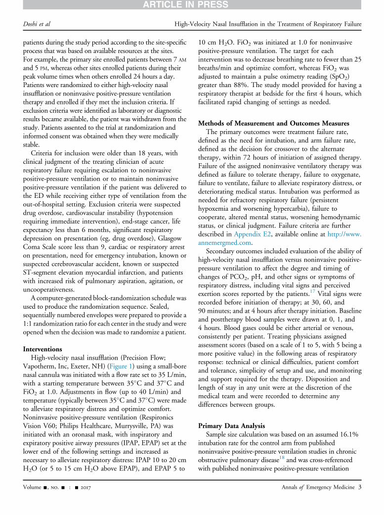
InterventionsHigh-velocity nasal insufflation (Precision Flow;
Vapotherm, Inc, Exeter, NH) (Figure 1) using a small-borenasal cannula was initiated with a flow rate set to 35 L/min,with a starting temperature between 35�C and 37�C andFiO2 at 1.0. Adjustments in flow (up to 40 L/min) andtemperature (typically between 35�C and 37�C) were madeto alleviate respiratory distress and optimize comfort.Noninvasive positive-pressure ventilation (RespironicsVision V60; Philips Healthcare, Murrysville, PA) wasinitiated with an oronasal mask, with inspiratory andexpiratory positive airway pressures (IPAP, EPAP) set at thelower end of the following settings and increased asnecessary to alleviate respiratory distress: IPAP 10 to 20 cmH2O (or 5 to 15 cm H2O above EPAP), and EPAP 5 to
Volume -, no. - : - 2017
10 cm H2O. FiO2 was initiated at 1.0 for noninvasivepositive-pressure ventilation. The target for eachintervention was to decrease breathing rate to fewer than 25breaths/min and optimize comfort, whereas FiO2 wasadjusted to maintain a pulse oximetry reading (SpO2)greater than 88%. The study model provided for having arespiratory therapist at bedside for the first 4 hours, whichfacilitated rapid changing of settings as needed.
Methods of Measurement and Outcomes MeasuresThe primary outcomes were treatment failure rate,
defined as the need for intubation, and arm failure rate,defined as the decision for crossover to the alternatetherapy, within 72 hours of initiation of assigned therapy.Failure of the assigned noninvasive ventilatory therapy wasdefined as failure to tolerate therapy, failure to oxygenate,failure to ventilate, failure to alleviate respiratory distress, ordeteriorating medical status. Intubation was performed asneeded for refractory respiratory failure (persistenthypoxemia and worsening hypercarbia), failure tocooperate, altered mental status, worsening hemodynamicstatus, or clinical judgment. Failure criteria are furtherdescribed in Appendix E2, available online at http://www.annemergmed.com.
Secondary outcomes included evaluation of the ability ofhigh-velocity nasal insufflation versus noninvasive positive-pressure ventilation to affect the degree and timing ofchanges of PCO2, pH, and other signs or symptoms ofrespiratory distress, including vital signs and perceivedexertion scores reported by the patients.17 Vital signs wererecorded before initiation of therapy; at 30, 60, and90 minutes; and at 4 hours after therapy initiation. Baselineand posttherapy blood samples were drawn at 0, 1, and4 hours. Blood gases could be either arterial or venous,consistently per patient. Treating physicians assignedassessment scores (based on a scale of 1 to 5, with 5 being amore positive value) in the following areas of respiratoryresponse: technical or clinical difficulties, patient comfortand tolerance, simplicity of setup and use, and monitoringand support required for the therapy. Disposition andlength of stay in any unit were at the discretion of themedical team and were recorded to determine anydifferences between groups.
Primary Data AnalysisSample size calculation was based on an assumed 16.1%
intubation rate for the control arm from publishednoninvasive positive-pressure ventilation studies in chronicobstructive pulmonary disease18 and was cross-referencedwith published noninvasive positive-pressure ventilation
Annals of Emergency Medicine 3
Figure 1. High-velocity nasal insufflation device platform. A, The main device unit (Precision Flow; Vapotherm, Inc) allows theclinician to set the flow rate (liters/minute), FiO2 (percentage of oxygen), and temperature (degrees Celsius). The unit connectsdirectly to oxygen and compressed air inputs (B), and delivers high-velocity flow through a modified nasal cannula (C).
High-Velocity Nasal Insufflation in the Treatment of Respiratory Failure Doshi et al
studies in cardiogenic pulmonary edema in which theintubation rate was 16.7% in studies in which the sample sizewas greater than or equal to 20.19 A sample size of 204 patients(102 in each arm) was calculated such that a test ofproportions with a .05 significance level and 90% power witha noninferiority margin for intubation of 15 percentagepoints. TheWald test for noninferiority was used for primaryoutcomes of intubation and treatment arm failure. Theprespecified noninferiority margins of 15 and 20 percentagepoints for differences in intubation and failure rates,respectively, were selected owing to the substantial variabilityin intubation rates from the literature, and the anticipatedincrease associated with the subjective decision for crossover.The 15 percentage points are the result of wide confidenceintervals (CIs) in rates of intubation in the literature;CIs in theCochrane review assessing noninvasive positive-pressureventilation in chronic obstructive pulmonary disease andcongestive heart failure were 7% and 9%, respectively. Thus,for the purposes of power analysis, a 10% difference was usedto incorporate the limits of the CI, and an additional 5% wasconsidered the accepted difference in the outcome ofintubation for high-velocity nasal insufflation to be considerednoninferior to noninvasive positive-pressure ventilation.
All analyses were based on an intention-to-treat modeldefined according to the protocol. Subanalyses of
4 Annals of Emergency Medicine
intubation or failure rates within the ED and within 4hours, as well as differences between treatment arm andreason for intubation or failure, are presented as point andinterval estimates of effect magnitude. Rates of intubationand failure were also described with Kaplan-Meier plots.Baseline demographic factors were summarized by studyarm. For secondary outcomes, data for physiologicparameters were summaries by group, with point andinterval estimates of effect magnitude when applicable. Allanalyses were performed with SAS (version 9.3; SASInstitute, Inc., Cary, NC).
RESULTSCharacteristics of Study Subjects
Patients were recruited from October 2014 toSeptember 2016. During this period, 228 patients wererandomized and 204 were enrolled in the trial (Figure 2).The 24 patients randomized but not enrolled wereexcluded for meeting exclusion criteria (10), consent notobtained or withdrawn (6), bedside clinician notcomfortable with enrollment after randomization (2), andpatient identified to not need noninvasive positive-pressureventilation after initial evaluation, thus failing to meetinclusion criteria (6). A total of 104 enrolled patients
Volume -, no. - : - 2017

Analyzed Analyzed
e
ie, study
Figure 2. Screening, randomization, and enrollment of study participants. From October 2014 to September 2016, patientspresenting to the ED with respiratory failure were screened according to the clinical need for advancement to noninvasiveventilatory support. Patients meeting eligibility were randomized to either high-velocity nasal insufflation through high-flow nasalcannula (HVNI) or NIPPV. If exclusion criteria were observed after randomization, patients were not subsequently enrolled. The largenumber of screen failures because of logistic reasons represents patients who presented and began receiving noninvasive supportbut who could not be enrolled because of resources and activity level in the units. HVNI, High-velocity nasal insufflation; NIPPV,noninvasive positive-pressure ventilation.
Doshi et al High-Velocity Nasal Insufflation in the Treatment of Respiratory Failure
Volume -, no. - : - 2017 Annals of Emergency Medicine 5
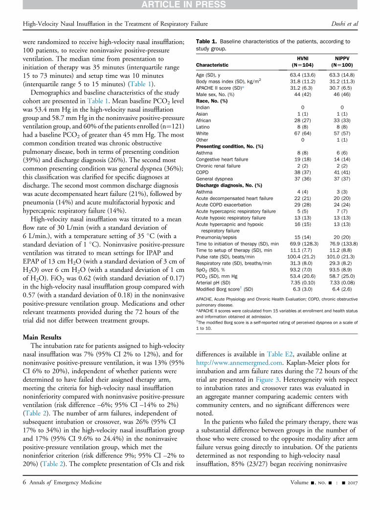
Table 1. Baseline characteristics of the patients, according tostudy group.
CharacteristicHVNI
(N[104)NIPPV
(N[100)
Age (SD), y 63.4 (13.6) 63.3 (14.8)Body mass index (SD), kg/m2 31.8 (11.2) 31.2 (11.3)APACHE II score (SD)* 31.2 (6.3) 30.7 (6.5)Male sex, No. (%) 44 (42) 46 (46)Race, No. (%)Indian 0 0Asian 1 (1) 1 (1)African 28 (27) 33 (33)Latino 8 (8) 8 (8)White 67 (64) 57 (57)Other 0 1 (1)Presenting condition, No. (%)Asthma 8 (8) 6 (6)Congestive heart failure 19 (18) 14 (14)Chronic renal failure 2 (2) 2 (2)COPD 38 (37) 41 (41)General dyspnea 37 (36) 37 (37)Discharge diagnosis, No. (%)Asthma 4 (4) 3 (3)Acute decompensated heart failure 22 (21) 20 (20)Acute COPD exacerbation 29 (28) 24 (24)Acute hypercapnic respiratory failure 5 (5) 7 (7)Acute hypoxic respiratory failure 13 (13) 13 (13)Acute hypercapnic and hypoxicrespiratory failure
16 (15) 13 (13)
Pneumonia/sepsis 15 (14) 20 (20)Time to initiation of therapy (SD), min 69.9 (128.3) 76.9 (133.8)Time to setup of therapy (SD), min 11.1 (7.7) 11.2 (8.8)Pulse rate (SD), beats/min 100.4 (21.2) 101.0 (21.3)Respiratory rate (SD), breaths/min 31.3 (8.0) 29.3 (8.2)SpO2 (SD), % 93.2 (7.0) 93.5 (8.9)PCO2 (SD), mm Hg 53.4 (20.6) 58.7 (25.0)Arterial pH (SD) 7.35 (0.10) 7.33 (0.08)Modified Borg score† (SD) 6.3 (3.0) 6.4 (2.6)
APACHE, Acute Physiology and Chronic Health Evaluation; COPD, chronic obstructivepulmonary disease.*APACHE II scores were calculated from 15 variables at enrollment and health statusand information obtained at admission.†The modified Borg score is a self-reported rating of perceived dyspnea on a scale of1 to 10.
High-Velocity Nasal Insufflation in the Treatment of Respiratory Failure Doshi et al
were randomized to receive high-velocity nasal insufflation;100 patients, to receive noninvasive positive-pressureventilation. The median time from presentation toinitiation of therapy was 35 minutes (interquartile range15 to 73 minutes) and setup time was 10 minutes(interquartile range 5 to 15 minutes) (Table 1).
Demographics and baseline characteristics of the studycohort are presented in Table 1. Mean baseline PCO2 levelwas 53.4 mm Hg in the high-velocity nasal insufflationgroup and 58.7 mmHg in the noninvasive positive-pressureventilation group, and 60%of the patients enrolled (n¼121)had a baseline PCO2 of greater than 45 mm Hg. The mostcommon condition treated was chronic obstructivepulmonary disease, both in terms of presenting condition(39%) and discharge diagnosis (26%). The second mostcommon presenting condition was general dyspnea (36%);this classification was clarified for specific diagnoses atdischarge. The second most common discharge diagnosiswas acute decompensated heart failure (21%), followed bypneumonia (14%) and acute multifactorial hypoxic andhypercapnic respiratory failure (14%).
High-velocity nasal insufflation was titrated to a meanflow rate of 30 L/min (with a standard deviation of6 L/min.), with a temperature setting of 35 �C (with astandard deviation of 1 �C). Noninvasive positive-pressureventilation was titrated to mean settings for IPAP andEPAP of 13 cm H2O (with a standard deviation of 3 cm ofH2O) over 6 cm H2O (with a standard deviation of 1 cmof H2O). FiO2 was 0.62 (with standard deviation of 0.17)in the high-velocity nasal insufflation group compared with0.57 (with a standard deviation of 0.18) in the noninvasivepositive-pressure ventilation group. Medications and otherrelevant treatments provided during the 72 hours of thetrial did not differ between treatment groups.
Main ResultsThe intubation rate for patients assigned to high-velocity
nasal insufflation was 7% (95% CI 2% to 12%), and fornoninvasive positive-pressure ventilation, it was 13% (95%CI 6% to 20%), independent of whether patients weredetermined to have failed their assigned therapy arm,meeting the criteria for high-velocity nasal insufflationnoninferiority compared with noninvasive positive-pressureventilation (risk difference –6%; 95% CI –14% to 2%)(Table 2). The number of arm failures, independent ofsubsequent intubation or crossover, was 26% (95% CI17% to 34%) in the high-velocity nasal insufflation groupand 17% (95% CI 9.6% to 24.4%) in the noninvasivepositive-pressure ventilation group, which met thenoninferior criterion (risk difference 9%; 95% CI –2% to20%) (Table 2). The complete presentation of CIs and risk
6 Annals of Emergency Medicine
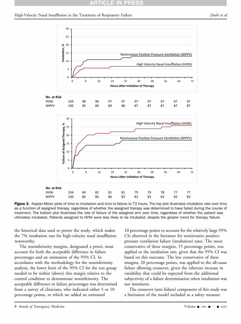
differences is available in Table E2, available online athttp://www.annemergmed.com. Kaplan-Meier plots forintubation and arm failure rates during the 72 hours of thetrial are presented in Figure 3. Heterogeneity with respectto intubation rates and crossover rates was evaluated inan aggregate manner comparing academic centers withcommunity centers, and no significant differences werenoted.
In the patients who failed the primary therapy, there wasa substantial difference between groups in the number ofthose who were crossed to the opposite modality after armfailure versus going directly to intubation. Of the patientsdetermined as not responding to high-velocity nasalinsufflation, 85% (23/27) began receiving noninvasive
Volume -, no. - : - 2017
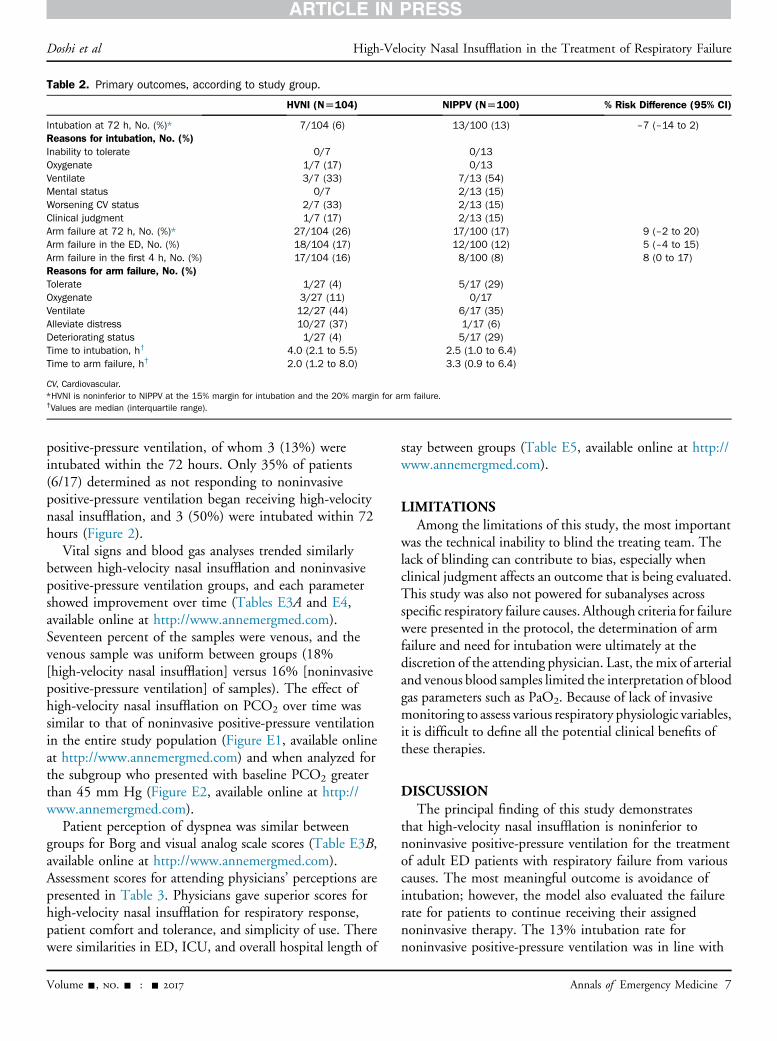
Table 2. Primary outcomes, according to study group.
HVNI (N[104) NIPPV (N[100) % Risk Difference (95% CI)
Intubation at 72 h, No. (%)* 7/104 (6) 13/100 (13) –7 (–14 to 2)Reasons for intubation, No. (%)Inability to tolerate 0/7 0/13Oxygenate 1/7 (17) 0/13Ventilate 3/7 (33) 7/13 (54)Mental status 0/7 2/13 (15)Worsening CV status 2/7 (33) 2/13 (15)Clinical judgment 1/7 (17) 2/13 (15)Arm failure at 72 h, No. (%)* 27/104 (26) 17/100 (17) 9 (–2 to 20)Arm failure in the ED, No. (%) 18/104 (17) 12/100 (12) 5 (–4 to 15)Arm failure in the first 4 h, No. (%) 17/104 (16) 8/100 (8) 8 (0 to 17)Reasons for arm failure, No. (%)Tolerate 1/27 (4) 5/17 (29)Oxygenate 3/27 (11) 0/17Ventilate 12/27 (44) 6/17 (35)Alleviate distress 10/27 (37) 1/17 (6)Deteriorating status 1/27 (4) 5/17 (29)Time to intubation, h† 4.0 (2.1 to 5.5) 2.5 (1.0 to 6.4)Time to arm failure, h† 2.0 (1.2 to 8.0) 3.3 (0.9 to 6.4)
CV, Cardiovascular.*HVNI is noninferior to NIPPV at the 15% margin for intubation and the 20% margin for arm failure.†Values are median (interquartile range).
Doshi et al High-Velocity Nasal Insufflation in the Treatment of Respiratory Failure
positive-pressure ventilation, of whom 3 (13%) wereintubated within the 72 hours. Only 35% of patients(6/17) determined as not responding to noninvasivepositive-pressure ventilation began receiving high-velocitynasal insufflation, and 3 (50%) were intubated within 72hours (Figure 2).
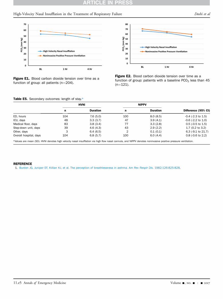
Vital signs and blood gas analyses trended similarlybetween high-velocity nasal insufflation and noninvasivepositive-pressure ventilation groups, and each parametershowed improvement over time (Tables E3A and E4,available online at http://www.annemergmed.com).Seventeen percent of the samples were venous, and thevenous sample was uniform between groups (18%[high-velocity nasal insufflation] versus 16% [noninvasivepositive-pressure ventilation] of samples). The effect ofhigh-velocity nasal insufflation on PCO2 over time wassimilar to that of noninvasive positive-pressure ventilationin the entire study population (Figure E1, available onlineat http://www.annemergmed.com) and when analyzed forthe subgroup who presented with baseline PCO2 greaterthan 45 mm Hg (Figure E2, available online at http://www.annemergmed.com).
Patient perception of dyspnea was similar betweengroups for Borg and visual analog scale scores (Table E3B,available online at http://www.annemergmed.com).Assessment scores for attending physicians’ perceptions arepresented in Table 3. Physicians gave superior scores forhigh-velocity nasal insufflation for respiratory response,patient comfort and tolerance, and simplicity of use. Therewere similarities in ED, ICU, and overall hospital length of
Volume -, no. - : - 2017
stay between groups (Table E5, available online at http://www.annemergmed.com).
LIMITATIONSAmong the limitations of this study, the most important
was the technical inability to blind the treating team. Thelack of blinding can contribute to bias, especially whenclinical judgment affects an outcome that is being evaluated.This study was also not powered for subanalyses acrossspecific respiratory failure causes. Although criteria for failurewere presented in the protocol, the determination of armfailure and need for intubation were ultimately at thediscretion of the attending physician. Last, the mix of arterialand venous blood samples limited the interpretation of bloodgas parameters such as PaO2. Because of lack of invasivemonitoring to assess various respiratory physiologic variables,it is difficult to define all the potential clinical benefits ofthese therapies.
DISCUSSIONThe principal finding of this study demonstrates
that high-velocity nasal insufflation is noninferior tononinvasive positive-pressure ventilation for the treatmentof adult ED patients with respiratory failure from variouscauses. The most meaningful outcome is avoidance ofintubation; however, the model also evaluated the failurerate for patients to continue receiving their assignednoninvasive therapy. The 13% intubation rate fornoninvasive positive-pressure ventilation was in line with
Annals of Emergency Medicine 7
No. at RiskHVNI 104 98 98 97 97 97 97 97 97 97NIPPV 100 90 89 89 88 87 87 87 87 87
No. at RiskHVNI 104 84 82 81 81 79 79 78 77 77NIPPV 100 86 86 86 83 83 83 83 83 83
0
5
10
15
20
25
30
0 8 16 24 32 40 48 56 64 72
Intu
baon
, %
Hours A er Ini a on of Therapy
0
5
10
15
20
25
30
0 8 16 24 32 40 48 56 64 72
Failu
re o
f Ass
igne
d Th
erap
y, %
Hours A er Ini a on of Therapy
High Velocity Nasal Insuffla on (HVNI)
High Velocity Nasal Insuffla on (HVNI)
Noninvasive Posi ve Pressure Ven la on (NIPPV)
Noninvasive Posi ve Pressure Ven la on (NIPPV)
Figure 3. Kaplan-Meier plots of time to intubation and time to failure to 72 hours. The top plot illustrates intubation rate over timeas a function of assigned therapy, regardless of whether the assigned therapy was determined to have failed during the course oftreatment. The bottom plot illustrates the rate of failure of the assigned arm over time, regardless of whether the patient wasultimately intubated. Patients assigned to HVNI were less likely to be intubated, despite the greater trend for therapy failure.
High-Velocity Nasal Insufflation in the Treatment of Respiratory Failure Doshi et al
the historical data used to power the study, which makesthe 7% intubation rate for high-velocity nasal insufflationnoteworthy.
The noninferiority margins, designated a priori, mustaccount for both the acceptable difference in failurepercentages and an estimation of the 95% CI. Inaccordance with the methodology for the noninferiorityanalysis, the lower limit of the 95% CI for the test groupneeded to be within (above) this margin relative to thecontrol condition to demonstrate noninferiority. Theacceptable difference in failure percentages was determinedfrom a survey of clinicians, who indicated either 5 or 10percentage points, to which we added an estimated
8 Annals of Emergency Medicine
10 percentage points to account for the relatively large 95%CIs observed in the literature for noninvasive positive-pressure ventilation failure (intubation) rates. The moreconservative of these margins, 15 percentage points, wasapplied to the intubation rate, given that the 95% CI wasbased on this outcome. The less conservative of thesemargins, 20 percentage points, was applied to the all-causefailure allowing crossover, given the inherent increase invariability that could be expected from the additionalsubjectivity of a failure determination when intubation wasnot imminent.
The crossover (arm failure) component of this study wasa limitation of the model included as a safety measure
Volume -, no. - : - 2017

Table 3. Attending physician perceptions, according to studygroup.
HVNI(N[104)
NIPPV(N[100)
Patients’ respiratory response, No. (%)1, insufficient 8/104 (8) 7/100 (7)2 2/104 (2) 6/100 (6)3, adequate 18/104 (17) 32/100 (32)4 15/104 (14) 11/100 (11)5, excellent 57/104 (55) 40/100 (40)Technical/clinical difficulties, No. (%)1, frequent 0/104 0/1002 1/104 (1) 0/1003, occasional 9/104 (9) 14/100 (14)4 8/104 (8) 6/100 (6)5, never 82/104 (79) 76/100 (76)Patients’ comfort/tolerance, No. (%)1, insufficient 2/104 (2) 4/100 (4)2 1/104 (1) 3/100 (3)3, adequate 16/104 (15) 45/100 (45)4 6/104 (6) 10/100 (10)5, excellent 75/104 (72) 34/100 (34)Simplicity of use, No. (%)1, complex 0/104 1/100 (1)2 0/104 0/1003, typical 27/104 (26) 48/100 (48)4 8/104 (8) 3/100 (3)5, simple 65/104 (63) 44/100 (44)Monitoring required, No. (%)1, frequent 1/104 (1) 2/100 (2)2 1/104 (1) 1/100 (1)3, typical 34/104 (33) 46/100 (46)4 9/104 (9) 1/100 (1)5, minimal 55/104 (53) 45/100 (45)
Doshi et al High-Velocity Nasal Insufflation in the Treatment of Respiratory Failure
associated with the need for deferred consent; therefore, thearm failure rate that induced an element of subjectivity wasexpected to be greater than the intubation rate. Despitethis, the arm failure rate met the a priori noninferioritycriteria. These data reveal a tendency to cease high-velocitynasal insufflation if patients are not immediately responsive,and as such, much of the observed difference in arm failurerates may be due to less clinical familiarity with high-velocity nasal insufflation in this diverse population. Asmentioned in the “Limitations,” because of a lack ofinvasive monitoring to assess for other physiologic variables,there may be benefits that are not accounted for in thisstudy.
Another important finding of this study is thedemonstration of the ventilatory effect with high-velocitynasal insufflation that is similar to that of noninvasivepositive-pressure ventilation, as evidenced by theimprovement in the PCO2 level over time in both arms, ata similar rate in all patients, as well as the subgroup withinitial PCO2 greater than 45. Recent studies of high-flownasal oxygen for adults in hypoxemic respiratory failureindicate that, generally, high-flow nasal cannula may be as
Volume -, no. - : - 2017
effective in the treatment of hypoxemia as noninvasivepositive-pressure ventilation.14-16 Although these studiesreport positive outcomes for oxygenation support, theyfocused specifically on patients with stable ventilatoryparameters and therefore did not show differences inventilatory indices. Of these trials, Stephan et al14
presented PCO2 and breathing frequency data withoutsignificant and clinically meaningful reductions.
These findings of our trial may be attributed to thephysiologic effects of small-bore cannulae used in high-velocity nasal insufflation. Mechanistic evidence suggeststhat high-flow nasal cannulae will purge the anatomic deadspace of expired carbon dioxide between breaths, thusproviding a ventilatory effect.7,20 Moreover, the ventilatoryeffect may be attributed to flow parameters generated by asmall-bore, high-velocity cannula. Frizzola et al6
demonstrated in an animal model of respiratory failure thata cannula design optimizing the potential to flushextrathoracic dead space results in improved physiologicventilation outcomes compared with one that was designedmore for airway-pressure generation. Subsequentcomputation fluid modeling work demonstrated thatcannula flow velocity plays the major role, in that greatervelocity adds turbulent energy and accelerates the vorticesformed on the nasal cavity.8 This trial translates the clinicaleffect of these theoretical flow properties to an adultrespiratory failure population.
In light of our findings, the nasal cannula applicationoffers several distinct advantages over noninvasive positive-pressure ventilation. High-flow nasal cannula systems, bynecessity, provide humidification superior to that ofnoninvasive positive-pressure ventilation systems andprovide support without the need to secure a seal on theface, which preserves the nasal and facial tissue fromlesions.21,22 In addition, our clinical experience has shownthat for many patients, the full face mask of somenoninvasive positive-pressure ventilation systems can beanxiety provoking, which may further exacerbate theadrenergic response often associated with respiratorydistress. Patients treated with high-velocity nasal insufflationcan more easily communicate, receive oral medications,and eat without interruption of therapy, which arelimitations of noninvasive positive-pressure ventilation.
Last, the findings of this trial are generalizable across caresettings. The trial was conducted across the southeasternUnited States in 5 centers with different characteristics.Two of the centers were academic centers, one a largeurban center and second a tertiary care center with largecatchment area. Three of the centers were communitycenters with volumes ranging from 50,000 to 70,000patient visits per year.
Annals of Emergency Medicine 9
High-Velocity Nasal Insufflation in the Treatment of Respiratory Failure Doshi et al
In summary, within the described limitations of this trial,specifically, being a nonblinded trial and not powered to assessperformance within any specific respiratory failure cause,high-velocity nasal insufflation is noninferior to noninvasivepositive-pressure ventilation for the treatment of respiratoryfailure from various causes in adult patients presenting to theED who do not require emergency intubation.
The authors acknowledge Daniel Ostermayer, MD, forproviding site supervision at McGovern Medical School in theabsence of a primary site principal investigator, and SamLuber, MD, Kimberly Chambers, MD, YashwantChathampally, MD, and George Dungan, MPhil, for theirhelp with review and editing of the article.
Supervising editor: Gregory W. Hendey, MD
Author affiliations: From the McGovern Medical School at TheUniversity of Texas Health Science Center at Houston, Houston, TX(Doshi, Hill, Granado); the Memorial Hermann Hospital–TexasMedical Center, Houston, TX (Doshi, Graham, Salazar); theUniversity of Tennessee, Chattanooga, TN (Whittle); the ErlangerHealth System, Chattanooga, TN (Whittle, Ellis); MemorialHermann The Woodlands Hospital, The Woodlands, TX (Bublewicz,Maynard); the McLeod Regional Medical Center, Florence, SC(Kearney, Tillotson); the Athens Regional Medical Center, Athens,GA (Ashe, Dennis, Spivey); Gordon and Associates, Berkeley, CA(Gordon); Vapotherm, Inc, Exeter, NH (Dunlap, Miller); and theSidney Kimmel Medical College, Philadelphia, PA (Miller).
Author contributions: PD led the trial. PD, JSW, MB, JK, TA, RG, S.Salazar, TEW, DM, RD, AT, MH, MG, and S. Spivey gathered thedata. PD, CD, and TLM were involved in the study design andprotocol development. NG performed the data management andstatistical analysis. PD, JSW, MB, JK, and TLM prepared the article.All authors vouch for the completeness and accuracy of the dataand analyses and for the fidelity of the trial to the protocol, andreviewed the article and agreed to the content. PD takesresponsibility for the paper as a whole.
All authors attest to meeting the four ICMJE.org authorship criteria:(1) Substantial contributions to the conception or design of thework; or the acquisition, analysis, or interpretation of data for thework; AND (2) Drafting the work or revising it critically for importantintellectual content; AND (3) Final approval of the version to bepublished; AND (4) Agreement to be accountable for all aspects ofthe work in ensuring that questions related to the accuracy orintegrity of any part of the work are appropriately investigated andresolved.
Funding and support: By Annals policy, all authors are required todisclose any and all commercial, financial, and other relationshipsin any way related to the subject of this article as per ICMJE conflictof interest guidelines (see www.icmje.org). Dr. Doshi reportsreceiving funding from Zoll Inc for coordination of the clinical trial.Dr. Miller and Mr. Dunlap were employed by Vapotherm. All authorsreport receiving study-related support from Vapotherm, Inc. Thetrial was sponsored by Vapotherm, which participated in the designof the protocol and the selection and management of the sites.
10 Annals of Emergency Medicine
The sponsor agreed a priori to allow publication of the findings ofthe trial at the discretion of the principal investigators. A steeringcommittee composed of the principal investigators oversaw theanalysis and interpretation of the data, wrote the first andsubsequent drafts of the article, and made the decision to submitthe article for publication.
Publication dates: Received for publication July 14, 2017.Revisions received September 21, 2017, and November 1, 2017.Accepted for publication November 30, 2017.
Trial registration number: NCT02236559
REFERENCES1. Weiss AJ, Wier LM, Stocks C, et al. Overview of Emergency Department
Visits in the United States, 2011. Agency for Healthcare Research &Quality. 2014.
2. Dysart K, Miller TL, Wolfson MR, et al. Research in high flow therapy:mechanisms of action. Respir Med. 2009;103:1400-1405.
3. Spoletini G, Alotaibi M, Blasi F, et al. Heated humidified high-flow nasaloxygen in adults: mechanisms of action and clinical implications.Chest. 2015;148:253-261.
4. Parke RL, McGuinness SP. Pressures delivered by nasal high flowoxygen during all phases of the respiratory cycle. Respir Care.2013;58:1621-1624.
5. Dewan NA, Bell CW. Effect of low flow and high flow oxygen delivery onexercise tolerance and sensation of dyspnea. A study comparing thetranstracheal catheter and nasal prongs. Chest.1994;105:1061-1065.
6. Frizzola M, Miller TL, Rodriguez ME, et al. High-flow nasal cannula:impact on oxygenation and ventilation in an acute lung injury model.Pediatr Pulmonol. 2011;46:67-74.
7. Moller W, Celik G, Feng S, et al. Nasal high flow clears anatomical deadspace in upper airway models. J Appl Physiol. 2015;118:1525-1532.
8. Miller TL, Saberi B, Saberi S. Computational fluid dynamics modelingof extrathoracic airway flush: evaluation of high flow nasal cannuladesign elements. J Pulmon Respir Med. 2016;6:376.
9. Spivey S, Ashe T, Dennis R, et al. Assessment of high flow nasal cannulatherapy use in the emergency department setting: observations ofpractice across four systems. Respir Ther. 2015;10:30-34.
10. Jones PG, Kamona S, Doran O, et al. Randomized controlled trial ofhumidified high-flow nasal oxygen for acute respiratory distress in theemergency department: the HOT-ER study. Respir Care. 2016;61:291-299.
11. Bell N, Hutchinson CL, Green TC, et al. Randomised control trial ofhumidified high flow nasal cannulae versus standard oxygen in theemergency department. Emerg Med Australas. 2015;27:537-541.
12. Bolton R, Bleetman A. Non-invasive ventilation and continuous positivepressure ventilation in emergency departments: where are we now?Emerg Med J. 2008;25:190-194.
13. Hernandez G, Vaquero C, Gonzalez P, et al. Effect of postextubationhigh-flow nasal cannula vs conventional oxygen therapy onreintubation in low-risk patients: a randomized clinical trial. JAMA.2016;315:1354-1361.
14. Stephan F, Barrucand B, Petit P, et al. High-flow nasal oxygen vsnoninvasive positive airway pressure in hypoxemic patients aftercardiothoracic surgery: a randomized clinical trial. JAMA.2015;313:2331-2339.
15. Frat JP, Thille AW, Mercat A, et al. High-flow oxygen through nasalcannula in acute hypoxemic respiratory failure. N Engl J Med.2015;372:2185-2196.
16. Hernandez G, Vaquero C, Colinas L, et al. Effect of postextubation high-flow nasal cannula vs noninvasive ventilation on reintubation andpostextubation respiratory failure in high-risk patients: a randomizedclinical trial. JAMA. 2016;316:1565-1574.
Volume -, no. - : - 2017
Doshi et al High-Velocity Nasal Insufflation in the Treatment of Respiratory Failure
17. Burdon JG, Juniper EF, Killian KJ, et al. The perception ofbreathlessness in asthma. Am Rev Respir Dis. 1982;126:825-828.
18. Ram FS, Picot J, Lightowler J, et al. Non-invasive positive pressureventilation for treatment of respiratory failure due to exacerbations ofchronic obstructive pulmonary disease. Cochrane Database Syst Rev.2004;(3)CD004104.
19. Vital FM, Ladeira MT, Atallah AN. Non-invasive positive pressureventilation (CPAP or bilevel NPPV) for cardiogenic pulmonary oedema.Cochrane Database Syst Rev. 2013;(5)CD005351.
Volume -, no. - : - 2017
20. Moller W, Feng S, Domanski U, et al. Nasal high flow reduces deadspace. J Appl Physiol. 2017;122:191-197.
21. Collins CL, Holberton JR, Barfield C, et al. A randomized controlled trialto compare heated humidified high-flow nasal cannulae with nasalcontinuous positive airway pressure postextubation in prematureinfants. J Pediatr. 2012.
22. Woodhead DD, Lambert DK, Clark JM, et al. Comparing two methodsof delivering high-flow gas therapy by nasal cannula followingendotracheal extubation: a prospective, randomized, masked,crossover trial. J Perinatol. 2006;26:481-485.
Annals of Emergency Medicine 11

High-Velocity Nasal Insufflation in the Treatment of Respiratory Failure Doshi et al
APPENDIX E2
Supplementary AppendixThis appendix has been provided by the authors to give
readers additional information about their work.Supplement to Doshi P, Whittle J, Bublewicz, et al.
High velocity nasal insufflation in the treatment ofrespiratory failure. Ann Emerg Med. 2017.
List of Investigators. Pratik Doshi, MD, MemorialHermann Hospital - Texas Medical Center, Houston TX
Russell Graham, BSRC, RRT, CPFT, RCP, FAARC,Memorial Hermann Hospital - Texas Medical Center,Houston TX
Suesann Salazar, RRT, Memorial Hermann Hospital -Texas Medical Center, Houston TX
Mandy Hill, DrPH, MPH, BS, McGovern MedicalSchool at UT Health Science Center at Houston, HoustonTX
Misha Granado, MPH, MS, BS, McGovern MedicalSchool at UT Health Science Center at Houston, HoustonTX
Jessica S. Whittle, MD, PhD, FACEP, University ofTennessee & Erlanger Health System, Chattanooga, TN
Terry W. Ellis, Jr., BS, RRT, Erlanger Health System,Chattanooga, TN
Joan C. Douglas, RRT, Erlanger Health System,Chattanooga, TN
Trisha C. McClellan, BS, RRT, Erlanger HealthSystem, Chattanooga, TN
Matthew McConnell, RRT, Erlanger Health System,Chattanooga, TN
Yvonne M. Jones, BS, RRT, Erlanger Health System,Chattanooga, TN
Leland R. Stewart, BA, RRT, Erlanger Health System,Chattanooga, TN
Donald K. Brooks, RRT-ACCS, Erlanger HealthSystem, Chattanooga, TN
Michael Bublewicz, MD, Memorial Hermann TheWoodlands Hospital, The Woodlands, TX
Dianna Maynard, Memorial Hermann The WoodlandsHospital, The Woodlands, TX
Jessica Traylor, RN, Memorial Hermann TheWoodlands Hospital, The Woodlands, TX
Tracy Green, Memorial Hermann The WoodlandsHospital, The Woodlands, TX
Donella Lavergne, Memorial Hermann The WoodlandsHospital, The Woodlands, TX
11.e1 Annals of Emergency Medicine
Danielle White, Memorial Hermann The WoodlandsHospital, The Woodlands, TX
Scott Walsh, Memorial Hermann The WoodlandsHospital, The Woodlands, TX
Rachael Cameron, Memorial Hermann The WoodlandsHospital, The Woodlands, TX
Terrell Ashe, RRT-NPS, RCP, Athens Regional MedicalCenter, Athens, GA
Rose Dennis MHS, BSN, BSRT, RRT, RN, CPFT,Athens Regional Medical Center, Athens, GA
Sheldon Spivey AS, RRT, Athens Regional MedicalCenter, Athens, GA
John McMullan, MD, Athens Regional Medical Center,Athens, GA
John Methe AS, RRT, Athens Regional Medical Center,Athens, GA
Joe Kearney, MD, McLeod Regional Medical Center,Florence, SC
April Tillotson, McLeod Regional Medical Center,Florence, SC
Contribution to the Study. This was a sponsoredmulticenter trial led by Pratik Doshi. All investigatorsmentioned as coauthors gathered the data except forCharles Dunlap, Tom Miller, and Nancy Gordon. CharlesDunlap and Tom Miller were involved in the study designand protocol development along with Pratik Doshi. NancyGordon, with Gordon and Associates, performed the datamanagement and statistical analysis. Data were checked bythe clinical research team and electronic data capturesystem was Imednet (Mednet system, Minnetonka, MN),and the database build was performed by Fuel Studios,which was financed by study funding.
The manuscript was prepared by Pratik Doshi, JessicaWhittle, Michael Bubblewicz, Joe Kearney, and TomMiller. The manuscript was reviewed by all the coauthorsand they agreed to the content and plan for submission toAnnals of Emergency Medicine.
Data Safety and Monitoring Board. Chad Cannon, MD(DSMB Chair), Vice Chair, Associate Professor & ResearchDirector, Emergency Medicine at University of KansasMedical Center
Joseph Miller, MD, Senior Staff Physician at HenryFord Hospital
Amy Kaji, MD, PhD, Associate Professor of EmergencyMedicine at UCLA-Harbor
Independent Statistician: Manya Harsch, MS,Technomics Research, LLC
Volume -, no. - : - 2017

Table E1. Study sites and enrollment periods.
Study Site Enrollment Open Enrollment Closed Total Enrolled
Memorial Hermann Hospital - TexasMedical Center, Houston, TX
Oct 2014 Sept 2016 71
Erlanger Health System,Chattanooga, TN
Feb 2015 Sept 2016 23
Memorial Hermann The WoodlandsHospital, The Woodlands, TX
Apr 2015 Sept 2016 56
Athens Regional Medical Center,Athens, GA*
Jun 2015 Apr 2016 25Aug 2016 Sept 2016 1
McLeod Regional Medical Center,Florence, SC
Mar 2016 Sept 2016 28
*Enrollment was paused at Athens Regional Medical Center for four months associated with a reorganization of the IRB; this was unrelated to the conduct of the trial.
Doshi et al High-Velocity Nasal Insufflation in the Treatment of Respiratory Failure
Methods: Failure Criteria and Study Interventions.Treatment Failure. Treatment failure will be defined as 1)
Failure to tolerate if the patient is unable to tolerate the mask,nasal prongs, air flow or pressure, has persisting asynchrony,and is unable to cooperate with the therapy; 2) Failure tooxygenate if the modality is unable to sustain an O2
sat > 88 - 92% or PaO2 > 60 – 65 mmHg despite maximaltreatment with FI02 100% and optimal manipulations offlow rate and/or airway pressures; 3) Failure to ventilate if thepatients remain acutely hypercarbic and academic with lackof reduction in PaCO2 or improvement in pH; 4) Failure toalleviate respiratory distress if the patient has no alleviation ofmoderate to severe dyspnea or tachypnea (RR remains >30/min) with inability to reduce work of breathing asmanifested by sustained increase in accessory muscle use;5) Deteriorating medical status manifested by worseningmental status or hemodynamics, manifested by hypotension(systolic P < 90 mm Hg), unremitting tachycardia (>140 orincrease by >20% during therapy), or other conditionsinterpreted by the patient’s clinicians as constituting evidenceof deterioration.Criteria for Intubation. Intubation will be undertaken for
unremitting respiratory failure despite maximal use of initialand/or crossover therapy as manifested by failure to maintainSaO2> 88% despite FIO2 1.0 and optimization of flow and/or PEEP settings, progressive increase > 10 mmHg inPaCO2 and concomitant drop in pH despite maximalattempts to enhance ventilation, inability to cooperate withtherapy in the face of persisting evidence of respiratory failure,unremitting agitation interfering with ability to cooperate andwith persisting evidence of respiratory failure, deterioratingmental status despite maximal therapy with HVNI and/orNIPPV, worsening hemodynamic status (systemic SBP <90mmHg or MAP < 60 mmHg despite fluid resuscitation anduse of low dose pressors), unremitting life-threateningarrhythmias, cardiac or respiratory arrest, or any other
Volume -, no. - : - 2017
condition, which, in the judgment of the clinical care team,warrants intubation.Crossover. Treatment failures on initial therapy wherein
immediate intubation is not required will be eligible forcrossover to the alternative therapy. The decision tointubate is entirely at the discretion of the clinical teamcaring for the patient without input from the investigators.Crossover will be offered to Treatment Failures only ifdeemed safe by the clinical care team.The Initial and Subsequent Settings for Application of
Each Therapeutic Arm. These initial settings in the twoarms are designed as a standardization of usual medicaltreatment for the respective therapies, and were devised toprovide critical intervention and rapid abatement of bothdyspnea and increased work of breathing. Once the patienthas been placed upon the initial settings, the medical staffmay, and should, manipulate and titrate the settings tooptimize effectiveness and subject’s tolerance.
HVNIFiO2 ¼ 1.0Flow ¼ 35 L/minTemperature ¼ 35 -37 CPatients will be fit with a Vapotherm adult nasalcannula that will be applied by a respiratory therapistor other clinician skilled in management of HVNI.Initial flow will be set to 35 L/min but can bedecreased or increased as rapidly as necessary toalleviate respiratory distress and optimize patientcomfort. Targets should be to lower respiratory rate tothe low 20s and with a HVNI flow rate between 20 to35 L/min. Starting temperature will be between 35�Cand 37�C; if patients find the gas temperature to beuncomfortable, it can be lowered as necessary down to33�C to enhance tolerance.FIO2 will be 1.0 initially to assure adequateoxygenation, but should be adjusted promptly to
Annals of Emergency Medicine 11.e2

High-Velocity Nasal Insufflation in the Treatment of Respiratory Failure Doshi et al
maintain an FIO2 of no greater than 0.6 to maintain aPaO2 > 88%.
NIPPVFiO2 ¼ 1.0IPAP ¼ 10 – 20 cmH2O (or 5 to 15 cmH20 pressuresupport above EPAP)EPAP ¼ 5-10 cmH2OBackup Ventilation Rate ¼ 0 - 4 breaths/min (lowestsetting)Applied with humidification per individual hospitalpracticePatients will be fit with an oronasal mask using afitting gauge that will be applied by a respiratorytherapist or other clinician skilled in management ofNIPPV. Initial pressures will be at low end of
Table E2. Primary outcomes.
Outcome
HVNI N
Risk 95% Confidence Limits Risk 95
Intubation 0.0673 0.0192 to 0.1155 0.1300 0.0Arm Failure 0.2596 0.1754 to 0.3468 0.1700 0.0
Table E3A. Secondary outcomes: vitals.*
Characteristic HVNI
Heart Rate (beats�min-1)Baseline (n¼204, HVNI¼104, NIPPV¼100) 100.4 (2130 min (n¼189, HVNI¼96, NIPPV¼93) 95.6 (2060 min (n¼180, HVNI¼92, NIPPV¼88) 94.0 (1890 min (n¼167, HVNI¼84, NIPPV¼83) 91.8 (17240 min (n¼149, HVNI¼73, NIPPV¼76) 92.1 (17Treatment Failure (n¼22, HVNI¼12, NIPPV¼10) 106.4 (29
Respiratory Rate (breaths�min-1)Baseline (n¼204, HVNI¼104, NIPPV¼100) 31.3 (8.30 min (n¼189, HVNI¼96, NIPPV¼93) 26.0 (6.60 min (n¼180, HVNI¼92, NIPPV¼88) 23.9 (5.90 min (n¼167, HVNI¼84, NIPPV¼83) 22.9 (5.240 min (n¼149, HVNI¼73, NIPPV¼76) 22.2 (4.Treatment Failure (n¼23, HVNI¼13, NIPPV¼10) 26.4 (11
SpO2 (%)Baseline (n¼204, HVNI¼104, NIPPV¼100) 93.2 (7.30 min (n¼189, HVNI¼96, NIPPV¼93) 97.5 (3.60 min (n¼180, HVNI¼92, NIPPV¼88) 97.6 (3.90 min (n¼167, HVNI¼84, NIPPV¼83) 97.8 (2.240 min (n¼148, HVNI¼73, NIPPV¼75) 96.8 (2.Treatment Failure (n¼23, HVNI¼13, NIPPV¼10) 93.3 (3.
*Values are mean (SD). HVNI denotes high velocity nasal insufflation via high flow nasal c
11.e3 Annals of Emergency Medicine
suggested range but can be increased as rapidly asnecessary to alleviate respiratory distress. Targetsshould be to lower respiratory rate to the low 20s andachieve tidal volumes of 6-8 ml/kg ideal body weight.If patients find pressures uncomfortably high, theycan be lowered as necessary by 1 to 2 cmH2Odecrements to enhance tolerance. EPAP (PEEP) canalso be adjusted upward as needed to reduce triggeringeffort (by counterbalancing auto-PEEP) or to improveoxygenation.FIO2 will be 1.0 initially to assure adequateoxygenation, but should be adjusted promptly tomaintain an FIO2 of no greater than 0.6 with anEPAP (PEEP) of not more than 10 cm H2O tomaintain a PaO2 > 88%.
IPPV (HVNI – NIPPV)
% Confidence Limits Risk Difference 95% Confidence Limits
641 to 0.1959 -0.0627 -0.1443 to 0.0189964 to 0.2436 0.0896 -0.0223 to 0.2015
NIPPV Difference (95% CI)
.2) 101.0 (21.3) -0.6 (-6.5 to 5.3)
.4) 96.4 (22.0) -0.8 (-6.9 to 5.3)
.4) 93.7 (20.4) 0.3 (-5.4 to 6.0).8) 92.2 (21.6) -0.4 (-6.4 to 5.6).4) 89.6 (18.2) 2.5 (-3.3 to 8.3).8) 108.9 (33.5) -2.5 (-30.6 to 25.6)
0) 29.3 (8.2) 2.0 (-0.2 to 4.2)1) 25.6 (7.6) 0.4 (-1.6 to 2.4)5) 23.4 (6.6) 0.5 (-1.3 to 2.3)8) 22.7 (6.4) 0.2 (-1.7 to 2.1)7) 22.1 (4.8) 0.1 (-1.4 to 1.6).4) 27.4 (10.2) -1.0 (-10.5 to 8.5)
0) 93.5 (8.9) -0.3 (-2.5 to 1.9)4) 97.8 (3.3) -0.3 (-1.3 to 0.7)0) 97.8 (3.0) -0.2 (-1.1 to 0.7)3) 97.7 (2.3) 0.1 (-0.6 to 0.8)8) 97.2 (2.3) -0.4 (-1.2 to 0.4)8) 91.4 (6.1) 1.9 (-2.4 to 6.2)
annula, and NIPPV denotes noninvasive positive pressure ventilation.
Volume -, no. - : - 2017
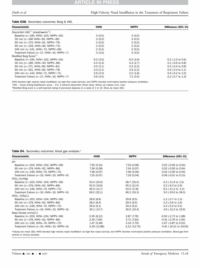
Table E3B. Secondary outcomes: Borg & VAS.
Characteristic HVNI NIPPV Difference (95% CI)
Discomfort VAS†1 (breaths�min-1)Baseline (n¼195, HVNI¼103, NIPPV¼92) 4 (0,5) 4 (0,5)30 min (n¼180 HVNI¼95, NIPPV¼85) 3 (0,5) 3 (0,5)60 min (n¼170, HVNI¼91, NIPPV¼79) 2 (0,5) 2 (0,5)90 min (n¼159, HVNI¼84, NIPPV¼75) 2 (0,5) 2 (0,5)240 min (n¼141, HVNI¼72, NIPPV¼69) 2 (0,4) 2 (0,5)Treatment Failure (n¼17, HVNI¼10, NIPPV¼7) 3 (2,5) 4 (3,5)
Modified Borg Score‡1
Baseline (n¼195, HVNI¼102, NIPPV¼93) 6.3 (3.0) 6.5 (2.6) -0.2 (-1.0 to 0.6)30 min (n¼180, HVNI¼94, NIPPV¼86) 4.4 (2.3) 4.3 (2.7) 0.1 (-0.6 to 0.8)60 min (n¼171, HVNI¼90, NIPPV¼81) 3.5 (2.1) 3.3 (2.2) 0.2 (-0.4 to 0.8)90 min (n¼161, HVNI¼83, NIPPV¼78) 3.3 (2.1) 2.9 (2.2) 0.4 (-0.3 to 1.1)240 min (n¼142, HVNI¼71, NIPPV¼71) 2.6 (2.0) 2.2 (1.8) 0.4 (-0.2 to 1.0)Treatment Failure (n¼17, HVNI¼10, NIPPV¼7) 4.9 (3.5) 7.1 (3.0) -2.2 (-5.7 to 1.3)
HVNI denotes high velocity nasal insufflation via high flow nasal cannula, and NIPPV denotes noninvasive positive pressure ventilation.†VAS – Visual Analog Satisfaction score – 0-5, 5 extreme discomfort (frown face). Shown as median (min, max)‡Modified Borg score is a self-reported rating of perceived dyspnea on a scale of 1 to 10. Show as mean (SD).
Table E4. Secondary outcomes: blood gas analysis.*
Characteristic HVNI NIPPV Difference (95% CI)
pHBaseline (n¼203, HVNI¼104, NIPPV¼99) 7.35 (0.10) 7.33 (0.08) 0.02 (-0.00 to 0.04)60 min (n¼178, HVNI¼92, NIPPV¼86) 7.36 (0.08) 7.34 (0.07) 0.02 (-0.00 to 0.04)240 min (n¼146, HVNI¼74, NIPPV¼72) 7.38 (0.07) 7.36 (0.06) 0.02 (-0.00 to 0.04)Treatment Failure (n¼16, HVNI¼10, NIPPV¼6) 7.25 (0.07) 7.19 (0.04) 0.06 (-0.01 to 0.13)
PCO2 (mmHg)Baseline (n¼203, HVNI¼104, NIPPV¼99) 53.4 (20.6) 58.7 (25.0) -5.3 (-11.6 to 1.0)60 min (n¼178, HVNI¼92, NIPPV¼86) 52.0 (19.6) 55.2 (21.5) -3.2 (-9.3 to 2.9)240 min (n¼146, HVNI¼74, NIPPV¼72) 46.3 (12.7) 52.5 (17.8) -6.2 (-11.2 to -1.2)Treatment Failure (n¼16, HVNI¼10, NIPPV¼6) 69.2 (32.1) 66.2 (33.3) 3.0 (-33.0 to 39.0)
HCO3- (mEq/L)
Baseline (n¼203, HVNI¼104, NIPPV¼99) 28.6 (8.6) 29.8 (9.5) -1.2 (-3.7 to 1.3)60 min (n¼178, HVNI¼92, NIPPV¼86) 28.4 (8.4) 29.4 (9.5) -1.0 (-3.6 to 1.6)240 min (n¼146, HVNI¼74, NIPPV¼72) 26.9 (6.1) 29.3 (9.2) -2.4 (-5.0 to 0.2)Treatment Failure (n¼16, HVNI¼10, NIPPV¼6) 30.1 (13.7) 26.5 (15.4) 3.6 (-12.3 to 19.5)
Base Excess (mmol/L)Baseline (n¼203, HVNI¼104, NIPPV¼99) 2.35 (8.12) 2.87 (7.76) -0.52 (-2.72 to 1.68)60 min (n¼178, HVNI¼92, NIPPV¼86) 2.30 (7.95) 2.71 (7.92) -0.41 (-2.76 to 1.94)240 min (n¼146, HVNI¼74, NIPPV¼72) 1.47 (5.48) 3.14 (7.79) -1.67 (-3.87 to 0.53)Treatment Failure (n¼16, HVNI¼10, NIPPV¼6) 2.29 (12.88) -2.12 (13.75) 4.41 (-10.21 to 19.03)
*Values are mean (SD). HVNI denotes high velocity nasal insufflation via high flow nasal cannula, and NIPPV denotes noninvasive positive pressure ventilation. Blood gas fromarterial or venous samples.
Doshi et al High-Velocity Nasal Insufflation in the Treatment of Respiratory Failure
Volume -, no. - : - 2017 Annals of Emergency Medicine 11.e4
0
10
20
30
40
50
60
70
80
BL 1 Hr 4 Hr
PCO
2(m
m H
g)
High Velocity Nasal Insuffla on
Noninvasive Posi ve Pressure Ven la on
Figure E2. Blood carbon dioxide tension over time as afunction of group: patients with a baseline PCO2 less than 45(n¼121).
0
10
20
30
40
50
60
70
BL 1 Hr 4 Hr
PCO
2(m
m H
g)
High Velocity Nasal Insuffla on
Noninvasive Posi ve Pressure Ven la on
Figure E1. Blood carbon dioxide tension over time as afunction of group: all patients (n¼204).
Table E5. Secondary outcomes: length of stay.*
HVNI NIPPV
Difference (95% CI)n Duration n Duration
ED, hours 104 7.6 (5.0) 100 8.0 (8.5) -0.4 (-2.3 to 1.5)ICU, days 48 3.3 (3.7) 47 3.9 (4.1) -0.6 (-2.2 to 1.0)Medical floor, days 83 3.8 (3.4) 77 3.3 (2.8) 0.5 (-0.5 to 1.5)Step-down unit, days 39 4.6 (4.3) 43 2.9 (2.2) 1.7 (0.2 to 3.2)Other, days 3 6.4 (6.5) 2 0.1 (0.1) 6.3 (-9.1 to 21.7)Overall hospital, days 104 6.8 (5.7) 100 6.0 (4.4) 0.8 (-0.6 to 2.2)
*Values are mean (SD). HVNI denotes high velocity nasal insufflation via high flow nasal cannula, and NIPPV denotes noninvasive positive pressure ventilation.
REFERENCE1. Burdon JG, Juniper EF, Killian KJ, et al. The perception of breathlessness in asthma. Am Rev Respir Dis. 1982;126:825-828.
High-Velocity Nasal Insufflation in the Treatment of Respiratory Failure Doshi et al
11.e5 Annals of Emergency Medicine Volume -, no. - : - 2017