High-throughput screening of natural compounds and ...
17
RESEARCH Open Access High-throughput screening of natural compounds and inhibition of a major therapeutic target HsGSK-3β for Alzheimer’s disease using computational approaches Rohit Shukla and Tiratha Raj Singh * Abstract Background: Alzheimer’s disease is a leading neurodegenerative disease worldwide and is the 6th leading cause of death in the USA. AD is a very complex disease and the drugs available in the market cannot fully cure it. The glycogen synthase kinase 3 beta plays a major role in the hyperphosphorylation of tau protein which forms the neurofibrillary tangles which is a major hallmark of AD. In this study, we have used a series of computational approaches to find novel inhibitors against GSK-3β to reduce the TAU hyperphosphorylation. Results: We have retrieved a set of compounds (n=167,741) and screened against GSK-3β in four sequential steps. The resulting analysis of virtual screening suggested that 404 compounds show good binding affinity and can be employed for pharmacokinetic analysis. From here, we have selected 20 compounds those were good in terms of pharmacokinetic parameters. All these compounds were re-docked by using Autodock Vina followed by Autodock. Four best compounds were employed for MDS and here predicted RMSD, RMSF, Rg, hydrogen bonds, SASA, PCA, and binding-free energy. From all these analyses, we have concluded that out of 167,741 compounds, the ZINC15968620, ZINC15968622, and ZINC70707119 can act as lead compounds against HsGSK-3β to reduce the hyperphosphorylation. Conclusion: The study suggested three compounds (ZINC15968620, ZINC15968622, and ZINC70707119) have great potential to be a drug candidate and can be tested using in vitro and in vivo experiments for further characterization and applications. Keywords: Alzheimer’s disease, Neurofibrillary tangles, Tauopathies, GSK-3β, Virtual screening, Molecular docking, Molecular dynamics simulation, Principal component analysis, MM-PBSA Background Alzheimer’s disease (AD) majorly contributes to demen- tia and is a lethal neurodegenerative disease. Worldwide approximately 50 million people are suffering from some form of dementia in which AD is the most contributing disease (60–70%). It is the major cause of death in the USA and is ranked 6th in number. According to the World Alzheimer Report of WHO 2018, this number (50 million) for dementia will be tripled (~152 million) by the year 2050, which reflects the seriousness of this disease for mankind. The disease showed a very high economic burden on the global society as 1 trillion US$ loss in 2018 was observed while it will double till 2030 [1]. The impact of the disease is reflected by its rise in incidence rate where one person develops dementia every 3 second globally. There is an urgent need to find treatment and cure for this disease. AD showed very complex disease etiology which is characterized by ma- jorly two hallmarks, first is the association of amyloid β © The Author(s). 2021 Open Access This article is licensed under a Creative Commons Attribution 4.0 International License, which permits use, sharing, adaptation, distribution and reproduction in any medium or format, as long as you give appropriate credit to the original author(s) and the source, provide a link to the Creative Commons licence, and indicate if changes were made. The images or other third party material in this article are included in the article's Creative Commons licence, unless indicated otherwise in a credit line to the material. If material is not included in the article's Creative Commons licence and your intended use is not permitted by statutory regulation or exceeds the permitted use, you will need to obtain permission directly from the copyright holder. To view a copy of this licence, visit http://creativecommons.org/licenses/by/4.0/. * Correspondence: [email protected]; [email protected] Department of Biotechnology and Bioinformatics, Jaypee University of Information Technology (JUIT), Waknaghat, Solan H.P., 173234, India Journal of Genetic Engineering and Biotechnology Shukla and Singh Journal of Genetic Engineering and Biotechnology (2021) 19:61 https://doi.org/10.1186/s43141-021-00163-w
Transcript of High-throughput screening of natural compounds and ...
RESEARCH Open Access
High-throughput screening of naturalcompounds and inhibition of a majortherapeutic target HsGSK-3β for Alzheimer’sdisease using computational approachesRohit Shukla and Tiratha Raj Singh*
Abstract
Background: Alzheimer’s disease is a leading neurodegenerative disease worldwide and is the 6th leading cause ofdeath in the USA. AD is a very complex disease and the drugs available in the market cannot fully cure it. Theglycogen synthase kinase 3 beta plays a major role in the hyperphosphorylation of tau protein which forms theneurofibrillary tangles which is a major hallmark of AD. In this study, we have used a series of computationalapproaches to find novel inhibitors against GSK-3β to reduce the TAU hyperphosphorylation.
Results: We have retrieved a set of compounds (n=167,741) and screened against GSK-3β in four sequential steps.The resulting analysis of virtual screening suggested that 404 compounds show good binding affinity and can beemployed for pharmacokinetic analysis. From here, we have selected 20 compounds those were good in terms ofpharmacokinetic parameters. All these compounds were re-docked by using Autodock Vina followed by Autodock.Four best compounds were employed for MDS and here predicted RMSD, RMSF, Rg, hydrogen bonds, SASA, PCA,and binding-free energy. From all these analyses, we have concluded that out of 167,741 compounds, theZINC15968620, ZINC15968622, and ZINC70707119 can act as lead compounds against HsGSK-3β to reduce thehyperphosphorylation.
Conclusion: The study suggested three compounds (ZINC15968620, ZINC15968622, and ZINC70707119) have greatpotential to be a drug candidate and can be tested using in vitro and in vivo experiments for furthercharacterization and applications.
Keywords: Alzheimer’s disease, Neurofibrillary tangles, Tauopathies, GSK-3β, Virtual screening, Molecular docking,Molecular dynamics simulation, Principal component analysis, MM-PBSA
BackgroundAlzheimer’s disease (AD) majorly contributes to demen-tia and is a lethal neurodegenerative disease. Worldwideapproximately 50 million people are suffering from someform of dementia in which AD is the most contributingdisease (60–70%). It is the major cause of death in theUSA and is ranked 6th in number. According to theWorld Alzheimer Report of WHO 2018, this number
(50 million) for dementia will be tripled (~152 million)by the year 2050, which reflects the seriousness of thisdisease for mankind. The disease showed a very higheconomic burden on the global society as 1 trillion US$loss in 2018 was observed while it will double till 2030[1]. The impact of the disease is reflected by its rise inincidence rate where one person develops dementiaevery 3 second globally. There is an urgent need to findtreatment and cure for this disease. AD showed verycomplex disease etiology which is characterized by ma-jorly two hallmarks, first is the association of amyloid β
© The Author(s). 2021 Open Access This article is licensed under a Creative Commons Attribution 4.0 International License,which permits use, sharing, adaptation, distribution and reproduction in any medium or format, as long as you giveappropriate credit to the original author(s) and the source, provide a link to the Creative Commons licence, and indicate ifchanges were made. The images or other third party material in this article are included in the article's Creative Commonslicence, unless indicated otherwise in a credit line to the material. If material is not included in the article's Creative Commonslicence and your intended use is not permitted by statutory regulation or exceeds the permitted use, you will need to obtainpermission directly from the copyright holder. To view a copy of this licence, visit http://creativecommons.org/licenses/by/4.0/.
* Correspondence: [email protected]; [email protected] of Biotechnology and Bioinformatics, Jaypee University ofInformation Technology (JUIT), Waknaghat, Solan H.P., 173234, India
Journal of Genetic Engineeringand Biotechnology
Shukla and Singh Journal of Genetic Engineering and Biotechnology (2021) 19:61 https://doi.org/10.1186/s43141-021-00163-w
plaques which is formed by the abnormal cutting of amyl-oid precursor protein (APP) while another hallmark is theneurofibrillary tangles (NFTs) being formed by the associ-ation of hyperphosphorylated microtubule-associatedbinding protein (MAPT) [2, 3]. AD is also linked with sev-eral autosomal mutations in the genes which are inheritedfrom parents encoding APP, tau protein, and presenilins 1and 2 (PSEN1 and PSEN2), and these mutations inducethe Aβ plaques and NFTs formation [4–7].For understanding the disease mechanism and inhibi-
tor identification, several studies have been done re-cently [8–10]. Quite a lot of efforts have been madetowards finding the cure for AD for the last 20 yearsagainst Aβ-based therapeutics including drug identifica-tion, and antibody generation but these are not success-ful [1]. Scientists also looked towards other tau-inducedtherapies for AD such as tauopathies. The tau is amicrotubule-stabilizing protein that stabilizes the micro-tubule and binds with α and β tubulin units of microtu-bules and forms the nerve cell cytoskeleton. During theAβ formation and several other conditions, various ki-nases like GSK-3β (glycogen synthase kinase 3 beta),CDK5 (cyclin-dependent kinase 5), DYRK1A (dual speci-ficity tyrosine-phosphorylation-regulated kinase 1A), andfew more enzymes hyperphosphorylate the tau protein[11]. Due to the hyperphosphorylation, tau detachedfrom the microtubule and aggregates in the form ofclumps of intracellular NFTs, which block the nerve cellcommunication and contributes towards AD progres-sion. The NFTs are the result of assembled tau proteinfragments, and they can also disrupt the nuclear-cytoplasmic transport [12]. Several compounds are in aclinical trial for reducing the NFTs aggregation. Butthere is an urgent need for inhibitors which can reducetau hyperphosphorylation by inhibiting the kinaseswhich are responsible for hyperphosphorylation [11].The GSK-3β is an enzyme belonging to the family of
proline-directed serine/threonine kinase and plays a vitalrole in the phosphorylation of various substrates in arange of pathways [13]. This enzyme is involved in theregulation of various cellular processes like metabolism,cardiac hypertrophy, cell proliferation, apoptosis, andoncogenesis [14]. It is known as a major therapeutic tar-get against various metabolic disorders like insulin re-sistance and type-2 diabetes because its function isassociated with glycogen metabolism. The GSK-3β is as-sociated with several central nervous systems (CNS) dis-eases like AD, stroke, and Huntington’s disease due toits overexpression in the brain [15, 16]. Various strongevidence in the literature showed that the GSK-3β co-localizes preferentially with neurofibrillary tangles [17].GSK-3β expressed actively in pre-tangle neurons andplays an active role in the formation of paired helical fil-aments (PHFs) or NFTs in the AD patient brain [18, 19].
Alteration in the GSK-3β function induces various neu-roinflammation and neurodegenerative disorders whichaffect the CNS. The hyperphosphorylation occurs in thePHFs due to the over activation of the GSK-3β, and it isrevealed both in transfected mammalian neuronal cellsand in vivo experiments [14]. The role of GSK-3β ismainly involved in the tau protein phosphorylation while itis also associated with some other AD-related mechanisms.Various strategies have been applied to find novel inhibitorsagainst GSK-3β to reduce the phosphorylation burden oftau and for the management of tauopathies [20, 21].Here we have applied the computational high through-
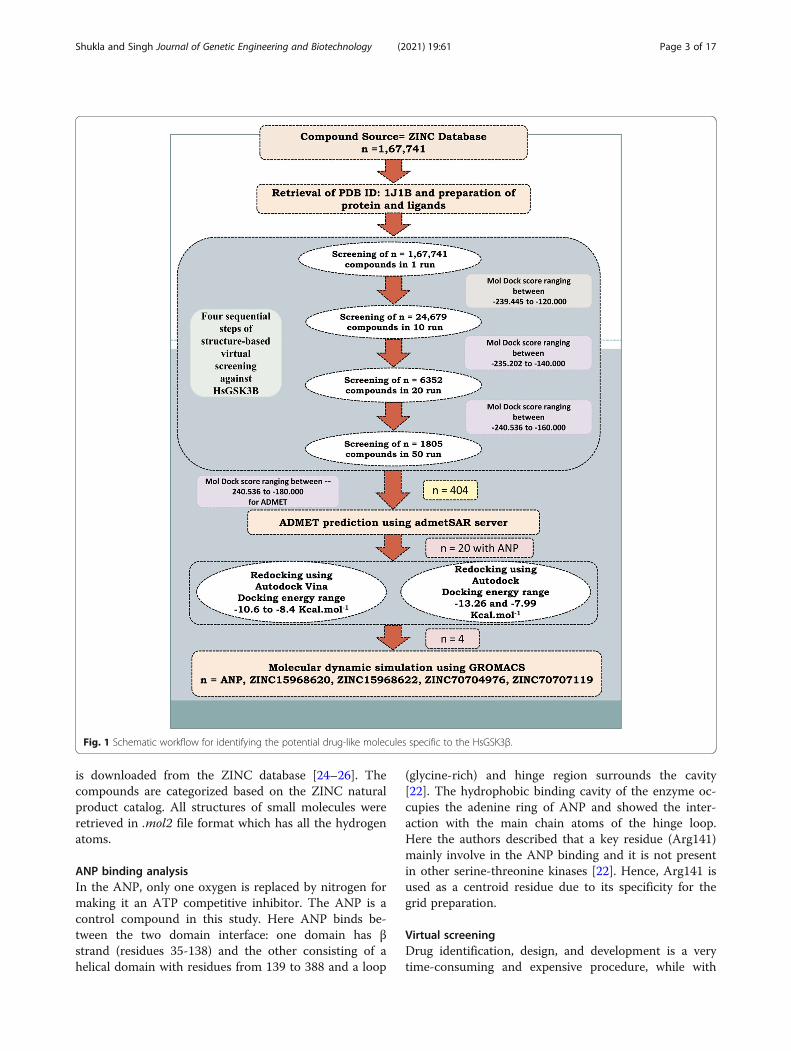
put screening method to predict the potential inhibitorsagainst GSK-3β. A natural subset (n=167,741) was re-trieved and screened in four steps of virtual screeningagainst GSK-3β. Based on the MolDock score the 404compounds were selected and employed for ADMETprediction. The 20 compounds were selected based onpharmacokinetics evaluations along with ATP analogANP (PhosphoAminophosphonic acid-adenylate ester)for re-docking by using Autodock Vina and Autodocksoftware. Based on re-docking, we have selected finallyfour compounds that showed a greater binding affinitythan the control compound and employed them for mo-lecular dynamics simulation (MDS) study. Various MDSresults were performed and analyzed like RMSD, RMSF,radius of gyration, number of hydrogen bonds, PCA, andbinding-free energy. The analysis suggests that threecompounds can act as best lead molecules against GSK-3β and can act as an anti-Alzheimer compound or couldbe proposed as potential lead molecules for tauopathies.The whole method which has been used in this studywas depicted in Fig. 1.
MethodsReceptor and ligand preparationThe 3D crystal structure of GSK-3β was retrieved fromthe RCSB protein databank. Various crystal structures(approximately >35) are available in PDB with goodresolution. From there, we have selected GSK-3β(RCSB-PDB ID: 1J1B, X-ray, 1.8 Å) [22] which is co-crystallized with ANP an analog ATP. Here we wantedto design the ATP competitive inhibitor therefore ANPwas a good control for this study. 1J1B was importedinto Chimera 1.13.2 [23], and all the hydrogens wereadded by using a structure preparation wizard. There areseveral open-source protein preparation tools availablewhile Chimera 1.13.2 has multiple advanced optionsthan other tools such as structure editing, multipleforce-fields. The structure minimization was performedusing Amber ff99SB force-field through the Amber toolof Chimera 1.13.2 [23]. This force-field is widely used toperform the MD simulation and energy minimization ofthe structure. The natural compound subset (n=167,741)
Shukla and Singh Journal of Genetic Engineering and Biotechnology (2021) 19:61 Page 2 of 17
is downloaded from the ZINC database [24–26]. Thecompounds are categorized based on the ZINC naturalproduct catalog. All structures of small molecules wereretrieved in .mol2 file format which has all the hydrogenatoms.
ANP binding analysisIn the ANP, only one oxygen is replaced by nitrogen formaking it an ATP competitive inhibitor. The ANP is acontrol compound in this study. Here ANP binds be-tween the two domain interface: one domain has βstrand (residues 35-138) and the other consisting of ahelical domain with residues from 139 to 388 and a loop
(glycine-rich) and hinge region surrounds the cavity[22]. The hydrophobic binding cavity of the enzyme oc-cupies the adenine ring of ANP and showed the inter-action with the main chain atoms of the hinge loop.Here the authors described that a key residue (Arg141)mainly involve in the ANP binding and it is not presentin other serine-threonine kinases [22]. Hence, Arg141 isused as a centroid residue due to its specificity for thegrid preparation.
Virtual screeningDrug identification, design, and development is a verytime-consuming and expensive procedure, while with
Fig. 1 Schematic workflow for identifying the potential drug-like molecules specific to the HsGSK3β.
Shukla and Singh Journal of Genetic Engineering and Biotechnology (2021) 19:61 Page 3 of 17
the advent of new computational technologies, the drugidentification becomes a little easy as well as less time-consuming. The in silico drug identification method canscreen the large databases based on binding site infor-mation or the basis of pharmacophore or QSAR modeland can predict the lead compound to reduce the timefor the drug identification [27]. This process is faster,has a low cost, and reduces the burden of unnecessaryexperiments [28]. The structure-based virtual screeningapproach is a popular and established method for thescreening of datasets retrieved from the ZINC database[24]. The ligands and receptors were prepared using theMolegro Virtual Docker (MVD) [29] and screened infour steps using MVD. The MVD is a protein-liganddocking software and used for the screening of largedatasets as mentioned earlier [30, 31]. The MolDockScore is provided in the form of a binding score. Thenfour subsequent runs were performed to find the novelinhibitors from the virtual screening. In the first run, atotal of n=167,741 natural ligands were used for virtualscreening in one run. Other parameters were set likepopulation size (50), maximum iteration (2000), scalingfactor (0.50), and crossover rate (0.90). From the firstround of docking, we have selected 24,689 compoundsfor the next round of virtual screening. The same par-ameter was used for the next round except for the num-ber of runs which was increased from 1 to 10. Thenfrom here 6352 compounds were selected and screenedin 20 runs. Finally, 1805 compounds were selected fromthe previous result and screened in 50 runs. Then finally,out of 1805 compounds, 404 compounds were selectedfor ADMET (Absorption, Distribution, Metabolism, Ex-cretion, and Toxicity) prediction. Although this is awell-proved [27, 30, 31] and a good method for virtualscreening where it has some drawbacks also, in the 1run, sometimes it can exclude the good compounds butthis error can also be removed by screening all the com-pounds in 100 runs with good computational facility.Some other methods are also available in which userscan screen a large library by using the Autodock Vinaand other software [32–34]. In the Autodock Vina, theuser can increase the exhaustiveness instead of the num-ber of runs and then can perform the screening. TheAutodock Vina is a command-line tool while MVD is aGUI-based tool where users can easily give a library ofcompounds and can screen in multiple steps. Hence,screening by using the MVD is better than AutodockVina and other command-line tools. We have also usedAutodock Vina for redocking in this study.
Pharmacokinetic evaluationThe ADMET parameters are very important when weare proposing the lead compound. The ADMET valuespredictions by using in silico tools can reduce the time
and give a proper estimation of the pharmacokinetic be-havior of a predicted small chemical molecule. Severaltools such as preADMET, pkCSM, and others are avail-able to predict the ADMET values but we have used theadmetSAR online server (http://lmmd.ecust.edu.cn/admetsar1/) [35] for the calculation of pharmacokineticparameters (ADMET). It predicts more and relevant de-scriptors as compare to other tools. Additionally, the de-velopers are regularly updating this server. It uses thedata of FDA approved drugs and predicts the values forthe newly given compound using the machine learningalgorithm. The data has also been taken from GoogleScholar (https://scholar.google.co.in/) and Pubmed(https://www.ncbi.nlm.nih.gov/pubmed/). We have pre-dicted various parameters like BBB (blood-brain barrier),Caco-2 cell permeability, HIA (Human Intestinal Ab-sorption), Pgp Substrate/Inhibitor identification, Car-cinogenicity, toxicity, Cyp450 metabolism, hERG geneinhibition, and Lethal dose. We have employed all theselected 404 compounds for ADMET prediction, andseveral descriptors were calculated. From this analysis,we have selected 20 compounds that were employed forre-docking studies.
Molecular dockingFor the prediction of better binding poses as well as forvalidating our selected ligands, we have re-docked all se-lected 20 compounds including ANP by using threewidely used software’s Molegro Virtual Docker [29],Autodock Vina [36], and Autodock [37]. The number ofruns is increased to 100 for MVD and a total of 5 posesis generated for each ligand. The grid box for the dock-ing with Autodock Vina, and Autodock was set based onco-crystallized ligand ANP and kept the same whichwere used in the MVD virtual screening process. Herewe have focused on Arg141 because this is not con-served in other kinases, as mentioned earlier [22]. TheMGL tools provide a complete suite to perform theprotein-ligand preparation, grid preparation, etc. Thenfor docking, we have used Autodock which is a freelyavailable and highly cited tool for protein-ligand dock-ing. The grid box is generated by using the grid gener-ation box of Autodock Tools. We used ANP inhibitor asa reference to construct the size of the grid (X, Y, andZ-coordinates as 50, 50, and 50 Å, respectively) with0.4Å grid spacing, where the incoming ligand binds dur-ing the docking process. First Autodock Vina was usedfor re-docking studies. It is a very efficient and less time-consuming software than Autodock. Then to crosscheckand finally select the best results, we used Autodock.The Autodock uses the Lamarckian genetic algorithmconformational search method for the generation ofbinding poses. The semi-empirical free energy force fieldwas used with all the default parameters except pose
Shukla and Singh Journal of Genetic Engineering and Biotechnology (2021) 19:61 Page 4 of 17
generation. The sum of the freedom of a torsional de-gree was used for the calculation of the conformationalentropy. The binding energy was evaluated in two stepsfirstly, the protein and ligand energy were calculated inthe unbound state, and then in the second step, theprotein-ligand complex energy was calculated [38]. Fi-nally, the difference between 1 and 2 was considered forthe result.
ΔG ¼ VL−Lbound−V
L−Lunbound
� �þ VP−Pbound−V
P−Punbound
� �
þ VP−Lbound−V
P−Lunbound þ ΔSconf
� � ð1ÞIn this equation, the protein is referred to by P, Ligand
is referred by L, pairwise evaluation is denoted by V, andΔSconf denotes the loss of conformational entropy duringbinding. For each ligand, 100 binding poses were gener-ated for result analysis. Then, these generated bindingposes were sort-listed based on binding affinity as wellas the number of hydrogen bonds.
Conformational stability analysisThe conformational changes and complex stability canbe best achieved by the MDS [39, 40]. It is a very usefuland widely accepted method to predict the accuracy ofdocking poses [41]. The complexes were selected basedon interaction and binding affinity analyses which werecarried forward for the MDS analysis. The apo-HsGSK3β and predicted complex (HsGSK3β-ANP (con-trol compound), HsGSK3β-ZINC15968620, HsGSK3β-ZINC15968622, HsGSK3β-ZINC70704976, andHsHSK3β-ZINC70707119) were employed for 60 ns mo-lecular dynamics simulation studies by using Gromacs5.1.2 [42] for analyzing the protein-ligand stability. TheGromacs is an open-source widely usable software forthe MDS. It has several force-fields such as Amber,GROMOS, and CHARMM. with regular updates. How-ever, it cannot generate the topology other than 20amino acids, hence we have to use third-party softwareto generate the ligand topology such as ProDRG. Thegeneration of each conformation with the time frame isbased on Newton’s law of molecular motions (Eq. 1).
Fi ¼ miai ¼ δV rNð Þδri
ð2Þ
Here i represents the mass of the atom while mi andai represent its acceleration. The force acting on i givenby the partial spatial derivative of the potential energyfunction V that is dependent on the positions rN = (r1,r2, … , rN) of all N particles in the system is representedby Fi. The protein topology and ligand topology weregenerated by using the GROMOS 9653A6 force field[43]. The ligand coordinates were generated by using theProDRG server [44]. A cubic box is created and thenHsGSK3β and HsGSK3β-ligand complexes were placed
in that box. Solvent molecules (no. 28,101) were addedand 8 Cl- ions were also added for neutralizing the sys-tems. Energy minimizations of all the systems were per-formed to remove the steric clashes of the system. Thenthe PBC (periodic boundary condition) was applied andthe Ewald summation method [45] was used for thelong-range electrostatic interaction calculation. Afterthat NVT (the constant number of particles, volume,and temperature), simulation was performed to fix thevolume and temperature (300K) of the systems. ThenNPT (the constant number of particles, pressure, andtemperature) simulation was carried out to fix the pres-sure of the system. Finally, all the systems wereemployed for the 60 ns MDS study, and the coordinateswere saved in 2fs. Then all the trajectories were analyzedwith various Gromacs utilities. The gmx rms, gmx rmsf,gmx gyrate, gmx sasa, and gmx hbond tools were usedfor the calculation of various parameters like RMSD,RMSF, Rg, SASA, and number of hydrogen bonds. Thetrajectories were visualized and analyzed by usingChimera 1.13.2 [23].
Binding-free energy calculationThe g_mmpbsa developed by Kumara et al. is a com-monly used tool to calculate the binding-free energyusing Gromacs as an interface [46]. This is a very easy-to-use tool as compare to others. The user can calculatethe binding free energy by using only two steps. The last10 ns MD trajectory snapshots were used for the calcu-lation of binding free energy. The ΔGbind is calculated bythe following equation:
ΔGbind ¼ ΔGmm þ ΔGsol - TΔS ð3Þ
The electrostatic and Van der Wall interactions werecomputed in the molecular mechanics energy (ΔGmm).The polar and non-polar contributions defined solvationfree energy (ΔGsol). The Solvent Accessible Surface Area(SASA) model was used for the determination of nonpo-lar solvation-free energy. In this method, the entropy(-TΔS) is not calculated due to the high computationalcost.
ResultsVirtual high-throughput screeningFor the identification of potential drug candidates thatcan inhibit the activity of HsGSK3β, a systematicstructure-based virtual screening approach was imple-mented. The crystal structure of HsGSK3β was used forvirtual screening. It is co-crystallized with the ANP. Sowe have set a binding site based on the control com-pound ANP and then docked this compound first. Thenall the retrieved ligand compounds (n=167,741) werescreened against this enzyme in the defined grid. From
Shukla and Singh Journal of Genetic Engineering and Biotechnology (2021) 19:61 Page 5 of 17

the first round of virtual screening, 24,689 compoundswere selected which showed the MolDock Score rangingbetween −239.45 and −120.00. These compounds wereagain screened in 10 runs. Increasing the number ofruns can remove false-positive binders [27]. The 6352compounds were selected between the MolDock Score−235.20 and −140.00 from this round of screening. Thenin the last run, we have selected 1805 compounds andscreened in 50 runs. These showed the MolDock Scoreof −240.54 to −160.00. Then from this round of virtualscreening, it was observed that many of these naturalcompounds reflect a good binding affinity score towardsthe binding pocket of HsGSK3β; therefore, these com-pounds (n=404) were selected for the pharmacokineticevaluation.
Pharmacokinetic evaluationThe natural compounds selected from the previous ana-lysis (404) were subjected to an admetSAR tool for theevaluation of various pharmacokinetic descriptors likeBBB, HIA, Caco-2 cell permeability, Cyp450 substrate/inhibition, Pgp-substrate/inhibition, carcinogenicity,hERG gene inhibition, and Lethal dose. As we are target-ing the central nervous system (CNS), so we have se-lected only those compounds for further study whichcan cross the BBB. We found that out of 404 com-pounds, 269 compounds were showing permeability to-wards the BBB. HIA describes the absorption of a smallmolecule in the large intestine. 367 compounds could beabsorbed from the HIA from our dataset of 404 com-pounds. In the Caco-2 parameter, we could not foundmore compounds that can pass this parameter like outof 404, 361 cannot pass from these criteria while 41compounds can pass from these criteria. In our study,only 43 compounds can pass from this filter. P-gp (P-glycoprotein) is a cell surface receptor for the efflux ofthe xenobiotics. Out of 404 compounds, the 315 and 89compounds acting as a substrate and non-substrate to-wards P-gp, respectively. Out of 404, the 168 and 236compounds acted as a substrate and non-substrate to-wards P-gp, respectively (Supplementary Table S1). TheCytochrome P450 is the key enzyme for the metaboliza-tion of the chemical molecule or xenobiotics. In thisstudy, we have taken 5 variants of Cyp450 and predictedthe inhibition and substrate probability. In our study,176 and 228 compounds act as high and low inhibitionpromiscuity against Cyp450, respectively (SupplementaryTable S2). Further, toxicity, carcinogenicity, and otherparameters filtering were also done. The toxic com-pound cannot be selected because it can cause adverseeffects when entering into the bloodstream. So we haveremoved those compounds which showed the toxicity.In this study, only 31 compounds act as toxic com-pounds out of 404 compounds. Carcinogenicity of the
compound described that if the compound can causethe mutation. So here only 2 compounds act as carcino-genic compounds while others are safe in this parameter.We have also predicted hERG gene inhibition becauseinhibition of this gene can cause long QT syndrome. Inour study, 269 compounds act as a non-inhibitor while135 compounds act as an inhibitor for this gene. Lastly,we have predicted the lethal dose and acute oral toxicityof all the compounds. The LD50 of maximum com-pounds came between 2 and 3 mol.Kg-1 (SupplementaryTable S3). From all these analyses, we have selected only20 compounds out of 404 compounds that passed all theparameters successfully and were subjected for re-docking studies.
Molecular docking analysisThe re-docking study is a good approach to predict thebinding poses from various tools and methods for valid-ating the study. Based upon the technical evaluationfrom the previous set of analyses, we have taken 20 com-pounds along with the control compound ANP for re-docking studies. We have used Autodock Vina, Auto-dock, and MVD for this purpose. The ANP showedbinding affinity through Autodock, Autodock Vina, andMVD and its scores are −7.8, −7.83, Kcal.mol-1, and−179.07 MolDock Score, respectively. From the Auto-dock, we have seen ZINC70707119 and ZINC95100194showed the highest and lowest binding affinity of −13.26and −7.99 Kcal.mol-1. The binding affinity for 20 com-pounds ranging between −13.26 and −7.99 Kcal.mol-1
from ADT. The ZINC15968620 and ZINC95100194showed the higher and lower binding affinity of −10.6and −8.4 Kcal.mol-1 from the Autodock Vina. The bind-ing affinity ranging between −10.6 and −8.4 Kcal.mol-1
for all the 20 compounds. The MVD, ZINC70704976,and ZINC70700682 showed the highest and lowest Mol-Dock Score of −207.04 and −181.85, respectively. In thecase of MVD, we have seen the MolDock Score rangingfrom −207.04 to −181.25. The binding affinity, numberof hydrogen bonds, and interacting residues for all the20 compounds with control compound ANP wereshown in Supplementary Table S4.
Structural analysis of selected hitsThe binding pattern of the four selected(ZINC15968620, ZINC15968622, ZINC70704976,ZINC70707119) hits is described below, which was se-lected by rounds of virtual screening as well as ADMETstudies from 167,741 compounds. The ANP binding pat-tern was also checked for validation of the dockingstudy. The chemical structure, binding affinity, interact-ing residues for four selected hits, and ANP were shownin Table 1 and Fig. 2.
Shukla and Singh Journal of Genetic Engineering and Biotechnology (2021) 19:61 Page 6 of 17
Docking protocol validationThe ANP is co-crystallized in the PDB structure (PDBID: 1J1B). The ANP is removed from the PDB and again
re-docked by all three docking tools. The binding resi-dues are found similar from all the three docking toolswhile binding affinity is different due to different
Table 1 Details of the four selected compounds with control compound ANP. ZINC ID, structures, binding affinity, and interactingresidues obtained after molecular docking are shown. The residues which are forming hydrogen bonds are shown in bold
Shukla and Singh Journal of Genetic Engineering and Biotechnology (2021) 19:61 Page 7 of 17
algorithms. The RMSD of the ligands from all the dock-ing tools is <2 Å from the experimentally solved 3D co-ordinates. Various residues that interact in the PDBstructure like Asp133 and Val135 are the same andforming the hydrogen bonds in re-docking studies. ThePDB structure authors resolved that Arg141 is a con-served residue, and it showed the interaction from allthree docking tools [22]. The binding affinity for ANPfrom all the three tools was −7.83, −7.8 Kcal.mol-1, and−179.077 MolDock Score. Table 1 and Fig. 2a show de-tailed analyses of interacting residues and binding affin-ity. The common residues which lie from all threesoftwares are Val70, Tyr134, and Arg141. In these threeresidues, the Val70 and Arg141 are also predicted by anexperimentally solved (PDB ID: 1J1B) structure.
ZINC15968620It is the predicted hit, which showed the highest bindingaffinity from Autodock VINA as compared to all otherligands. The binding affinity from Autodock Vina, Auto-dock, and MVD was shown −10.6, −11.5 Kcal.mol-1, and−184.884 MolDock Score. The binding affinity of thiscompound is greater than ANP. The binding score rep-resents that it has more affinity than ANP and can blockthe active site of GSK3β and inhibits the enzyme’s func-tion. It binds with the key catalytic residues of the ATPsite. Table 1 and Fig. 2b showed detailed analyses ofinteracting residues, binding affinity, and the number ofhydrogen bonds. Common interacting residues, which
have been predicted from all three docking tool’s areIle62, Asn64, Ala83, Val135, Thr138, Arg141, Gln185,Leu188, and Asp200. Here we have seen that most ofthe amino acid residues are hydrophobic and positivelycharged. It describes that the binding site has the cap-ability of hydrophobic interactions. Arg141 is also con-served and it is a key residue that is not conserved inother kinases and actively participating in GSK3β-ZINC15968620 complex stabilization. In these residuesVal135, Arg141, Gln185, and Asp200 are actively partici-pating in the ANP stabilization in the experimentally re-solved structure also. It represents that our ligand isinteracting with the key residues which are required forthe activity of the enzyme and the docking tools are giv-ing accurate binding poses. Additionally, newly identifiedresidues could also be checked experimentally and canprovide new substrate specificity.
ZINC15968622It is the derivative of ZINC15968620 with a bonding dif-ference. It showed approximately similar binding affinitywhich was shown by ZINC15968620. It showed thebinding affinity from Autodock Vina, Autodock, andMVD as −10.5, −11.53 Kcal.mol-1, and −188.439 Mol-Dock Score, respectively. It also showed a greater bind-ing affinity than control ligand ANP. Binding affinityproves that it can compete with the ANP and occupythe ATP binding site and can block the enzyme func-tion. It showed good hydrogen and other bonding
Fig. 2 Schematic representations of the binding interactions with HsGSK3β. a HsGSK3β-ANP. b HsGSK3β-ZINC15968620. c HsGSK3β-ZINC15968622. d HsGSK3β-ZINC70704976. e HsGSK3β-ZINC70707119
Shukla and Singh Journal of Genetic Engineering and Biotechnology (2021) 19:61 Page 8 of 17

interactions with the key catalytic residues. It showedthe binding with Arg141 through two of the three soft-ware used. A detailed description of the chemical struc-ture, hydrogen bonding, and binding affinity is given inTable 1 and Fig. 2c. The identical residues from all threedocking tools were predicted and compared with the X-ray crystallized structure. The residues Ile62, Asn64,Ala83, and Leu188 were found common from all threedocking tools.
ZINC70704976The ZINC70704976 also showed a good binding affinityand even greater than ANP from all the three dockingtools. The binding affinity of −10.0, −11.43 Kcal.mol-1,and −207.046 MolDock Score was observed from Auto-dock Vina, Autodock, and MVD tools, receptively. Itshowed the highest MolDock Score compared to allother compounds. The compounds showed the inter-action with the key catalytic residues. The number ofhydrogen bonds, binding affinity, and the chemicalstructure is shown in Table 1 and Fig. 2d. The residues,which were identified from all the docking tools, areAsn64, Ile62, Ala83, Tyr134, and Val135. These all resi-dues are catalytically important. The Val135 is a residuethat is also found in the stabilization of ANP withHsGSK3β in the experimentally resolved structure.
ZINC70707119The ZINC70707119 showed the highest binding affinityof −13.26 Kcal.mol-1 from the Autodock as compared toall other selected ligands. The ZINC70707119 alsoshowed a greater binding score than ANP. The bindingscore from Autodock Vina was −9.7 while the −200.768MolDock score was observed from the MVD. It interactswith the various key catalytic residues like Arg141 whichis a unique residue of GSK3β and participates in thehydrogen bonding during docking. A detailed descrip-tion of hydrogen bonds, hydrophobic interaction, andchemical structure is given in Table 1 and Fig. 2e. Theresidues found identical from all the docking tools areIle62, Asn64, Val70, Ala83, Tyr134, Val135, Thr138,Arg141, Gln185, Leu188, and Asp200. These are the keycatalytic residues and are also found in ANP interactionin the crystal structure. The residues Val70, Val135, andAsp200 are identical to the experimentally solved struc-ture. From this analysis, we have seen that all four li-gands have a higher binding affinity than the controlligand ANP. It represents that all selected hits can com-pete with the ANP and can occupy the binding cavity.These residues are interacting with the key catalytic resi-dues which are required for enzyme function. It revealsthat our predicted inhibitors may alter the enzyme func-tion. Further MDS study was performed for the
prediction of conformational dynamics of all the fourcompounds with ANP complex and apo-HsGSK3β.
Common scaffold for all four selected compoundsWe obtained a very interesting finding from this ana-lysis. We have worked on a structure-based virtualscreening approach with a set of compounds. Fromthese rounds of virtual screening, we have selected 4compounds for the MDS study. In these compounds, wehave seen that they belong to one scaffold (β-carboline-hydantoin derivative named “2,5-DIPHENYL-1,3-DIOXO-6H-1,2,3,5,11,11A-HEXAHYDROIMIDAZO-[1,5-B]-BETA-CARBOLINE”) [47] only and all the com-pounds are derivatives of this scaffold. The chemicalstructure is shown in Supplementary Figure 1. It repre-sents that this scaffold can play a key role in the design-ing of potent inhibitors against GSK3β for AD and othertauopathies.
Molecular dynamic simulationThe MDS was performed in two phases. First, the sys-tems are prepared to remove the steric clashes and thetemperature and volume of the system are fixed, thensecond the production run of 60 ns was carried out forpredicting the protein-ligand complex stability analysis.The MDS provides the deeper detail of the mechanismof the ligand-binding, and we can predict the changes atthe atomic level with the time-scale. We have predictedvarious values such as RMSD, RMSF, Rg, SASA, numberof hydrogen bonds, PCA, and binding-free energy forpredicting the protein-ligand complex stability. The tra-jectories are equilibrated after 20 ns, hence the analysiswas carried out from the last 40 ns trajectory.
RMSDThe RMSD is a widely used parameter to check the sta-bility of the system. Here the RMSD value is calculatedfor all the systems to predict the conformational changesafter ligand binding in the apo-protein and depicted inFig. 3a. The average RMSD value for apo-HsGSK3β,HsGSK3β-ANP, HsGSK3β-ZINC15968620, HsGSK3β-ZINC15968622, HsGSK3β-ZINC70704976, andHsGSK3β-ZINC70707119 were 0.38, 0.38, 0.37, 0.35,0.35, and, 0.34 nm, respectively, for the last 40 ns trajec-tory. From the RMSD value, we have analyzed that theligand-binding induces stability in the apo-HsGSK3β.The apo-HsGSK3β and HsGSK3β-ANP showed thesame RMSD value while HsGSK3β-ZINC70707119showed the least RMSD value as compared to all othercompounds. All the trajectories got the equilibrationstate after 20 ns while some extent in the RMSD peakwas observed in the HsGSK3β-ZINC70707119. Itshowed the lower and higher RMSD peak till 40 ns, andafter that, it got the equilibration state and showed a
Shukla and Singh Journal of Genetic Engineering and Biotechnology (2021) 19:61 Page 9 of 17

stable peak. From the RMSD, we have concluded that allthe trajectories are well stable and can be considered forfurther analysis.
Residue mobility analysisThe RMSF defines residual mobility after ligand binding.The rigid structure like helix and sheet showed lowerRMSF values while loosely folded structures like turnsand coils showed higher RMSF values. We have calcu-lated the RMSF value for all the ligand complexes andshown in Fig. 3b. The average RMSF value for apo-HsGSK3β, HsGSK3β-ANP, HsGSK3β-ZINC15968620,HsGSK3β-ZINC15968622, HsGSK3β-ZINC70704976,and HsGSK3β-ZINC70707119 were 0.11, 0.13, 0.15,0.15, 0.13, and 0.14 nm, respectively. Figure 3b showedthat the ligand-binding induces conformational changes.It showed the major changes in the residues between64–70, 90–95, 119–126, 140–154, and 202–216. Wehave observed a higher peak in the C-terminal region.From the RMSF analysis, we have seen that ligand bind-ing altering the geometry of the protein structure and
for performing the native function of any protein aproper conformation is required. The HsGSK3β-ZINC70704976 and HsGSK3β-ZINC70707119 showedless fluctuation as compared to other predicted ligands.
Radius of gyrationThe radius of gyration analysis of the protein-ligandcomplex is done by the radius of gyration (Rg) parameteranalysis. The less Rg value is observed in the compactlyfolded protein while the higher Rg value is generallyshown by the loosely folded protein structure. The Rgvalue for all the systems was calculated for the last 40 nstrajectory and plotted in Fig. 4a. The average Rg valuewere recorded 2.13, 2.15, 2.15, 2.16, 2.16, and 2.17 nmfor apo-HsGSK3β, HsGSK3β-ANP, HsGSK3β-ZINC15968620, HsGSK3β-ZINC15968622, HsGSK3β-ZINC70704976, and HsGSK3β-ZINC70707119, respect-ively. The result showed that the least Rg value was ob-served for the apo-HsGSK3β as compared to othercompounds and control compounds. It represents thatafter ligand binding, some conformational changes have
Fig. 3 Molecular dynamic simulation. a Root mean square deviation (RMSD) values of Cα backbone atoms for 60 ns. b Root mean squarefluctuation (RMSF) values of Cα atoms for the last 40 ns. The color code for all panels are apo-HsGSK3β (black), HsGSK3β-ANP (red), HsGSK3β-ZINC15968620 (green), HsGSK3β-ZINC15968622 (blue), HsGSK3β-ZINC70704976 (cyan), and HsGSK3β-ZINC70707119 (magenta)
Fig. 4 Rg and hydrogen bond. a The radius of gyration value for backbone at 300 K for last 40 ns trajectory. b Number of hydrogen bondsbetween protein and ligand for last 40 ns trajectory. The color code for all panels are apo-HsGSK3β (black), HsGSK3β-ANP (red), HsGSK3β-ZINC15968620 (green), HsGSK3β-ZINC15968622 (blue), HsGSK3β-ZINC70704976 (cyan), and HsGSK3β-ZINC70707119 (magenta)
Shukla and Singh Journal of Genetic Engineering and Biotechnology (2021) 19:61 Page 10 of 17
occurred. The HsGSK3β-ZINC15968620 showed thesame average Rg value as compared to the control com-pound. The HsGSK3β-ZINC15968622 and HsGSK3β-ZINC70704976 showed the same Rg value and higherthan control while less than HsGSK3β-ZINC70704976.All the complexes showed the stable peak from 20 to 40ns while the value for HsGSK3β-ZINC15968620 is de-creasing from higher to lower with the timescale. Fromhere, we have concluded that all predicted hits are goodand showed the protein-ligand complex stability.
Interaction analysisVarious interactions between protein and ligand play akey role in protein-ligand stabilization. The hydrogenbonds are the key interaction between protein and lig-and. They provide interaction specificity and directional-ity which is a key step in molecular recognition. Wehave calculated the number of hydrogen bonds vs. timeand plotted it in Fig. 4b. All the complexes showed goodbonding between protein and ligand. The average num-ber of hydrogen bonds for HsGSK3β-ANP, HsGSK3β-ZINC15968620, HsGSK3β-ZINC15968622, HsGSK3β-ZINC70704976, and HsGSK3β-ZINC70707119 was 0–1for all the ligand complexes. The HsGSK3β-ZINC15968620 and HsGSK3β-ZINC15968622 showed0–3 hydrogen bonds respected time. The control com-pound also showed 3 hydrogen bonds in some timesteps. It represents that these compounds stably inter-acted with the HsGSK3β binding cavity and providesstable interaction.
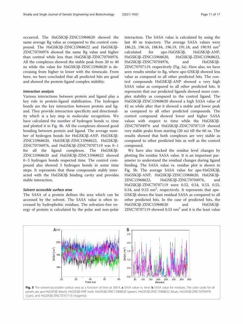
Solvent accessible surface areaThe SASA of a protein defines the area which can beaccessed by the solvent. The SASA value is often in-creased by hydrophobic residues. The solvation-free en-ergy of protein is calculated by the polar and non-polar
interaction. The SASA value is calculated by using thelast 40 ns trajectory. The average SASA values were186.23, 196.16, 188.84, 196.19, 191.18, and 190.93 nm2
calculated for apo-HsGSK3β, HsGSK3β-ANP,HsGSK3β-ZINC15968620, HsGSK3β-ZINC15968622,HsGSK3β-ZINC70704976, and HsGSK3β-ZINC70707119, respectively (Fig. 5a). Here also, we haveseen results similar to Rg, where apo-GSK3β showed lessvalue as compared to all other predicted hits. The con-trol compounds HsGSK3β-ANP showed a very highSASA value as compared to all other predicted hits. Itrepresents that our predicted ligands showed more com-plex stability as compared to the control ligand. TheHsGSK3β-ZINC15968620 showed a high SASA value of42 ns while after that it showed a stable and lower peakas compared to all other predicted compounds. Thecontrol compound showed lower and higher SASAvalues with respect to time while the HsGSK3β-ZINC70704976 and HsGSK3β-ZINC70707119 showedvery stable peaks from starting (20 ns) till the 60 ns. Theresults showed that both complexes are very stable ascompared to other predicted hits as well as the controlcompound.We have also tracked the residue level changes by
plotting the residue SASA value. It is an important par-ameter to understand the residual changes during ligandbinding. The SASA value vs. residue plot is shown inFig. 5b. The average SASA value for apo-HsGSK3β,HsGSK3β-ANP, HsGSK3β-ZINC15968620, HsGSK3β-ZINC15968622, HsGSK3β-ZINC70704976, andHsGSK3β-ZINC70707119 were 0.52, 0.54, 0.53, 0.55,0.54, and 0.53 nm2, respectively. It represents that apo-GSK3β shows the least residual SASA as compared to allother predicted hits. In the case of predicted hits, theHsGSK3β-ZINC15968620 and HsGSK3β-ZINC70707119 showed 0.53 nm2 and it is the least value
Fig. 5 The solvent-accessible surface area as a function of time at 300 K. a SASA value vs. time. b SASA value for residues. The color code for allpanels are apo-HsGSK3β (black), HsGSK3β-ANP (red), HsGSK3β-ZINC15968620 (green), HsGSK3β-ZINC15968622 (blue), HsGSK3β-ZINC70704976(cyan), and HsGSK3β-ZINC70707119 (magenta)
Shukla and Singh Journal of Genetic Engineering and Biotechnology (2021) 19:61 Page 11 of 17

as compared to other predicted hits. It is confirmedfrom this finding that these compounds showed a stablecomplex.
Principal component analysisThe PCA or essential dynamics (ED) are used to analyzethe correlated motions in the protein after ligand bind-ing. The total motility in the system is equivalent to thesum of the eigenvalues. It can be used to compare theflexibility of a protein under different conditions. TheGromacs provide the facility to calculate the eigenvec-tors for characterizing the protein motions. Here wehave considered the first fifty eigenvectors for result ana-lysis because it is a well-known fact that the first fewPCs describe the overall dynamics of the system. Wehave calculated the percent wise motions for the firstfive eigenvectors. The first five eigenvectors accountedfor 52.49%, 70.70%, 72.35%, 65.83%, 69.84%, and 66.59%of the motions, recorded for last 40 ns trajectory forapo-HsGSK3β, HsGSK3β-ANP, HsGSK3β-ZINC15968620, HsGSK3β-ZINC15968622, HsGSK3β-ZINC70704976, and HsGSK3β-ZINC70707119, respect-ively (Fig. 6a). The apo-HsGSK3β showed very less mo-tions as compared to ligand-bound complexes. Itindicates that ligand binding induces structural changesand motions in the protein. In the case of the ligand-
protein complex the HsGSK3β-ZINC15968622,HsGSK3β-ZINC70704976, and HsGSK3β-ZINC70707119 showed less motions as compared to thecontrol compound. So from here, we have concludedthat these three complexes can act as lead compounds.In this result, we have observed that the first few ei-
genvectors were describing the overall dynamics of theprotein. So in the next analysis, we have considered thefirst two eigenvectors, and a 2D projection plot was plot-ted to achieve the phase space behavior of the protein-ligand complex (Fig. 6b). In this plot, the clear clusterdescribes the well stable complex while the non-stablecluster defines the non-stable complex. In Fig. 6b, wehave seen that apo-HsGSK3β showed a well stable clus-ter. In the case of the protein-ligand complex, theHsGSK3β-ZINC15968622, HsGSK3β-ZINC70704976,and HsGSK3β-ZINC70707119 showed well stable clusteras compared to the control compound. This result isalso consistent with the above-stated analysis.After that, we have calculated the eigRMSF for the one
eigenvector vs. residues and plotted it in Fig. 6c. It describesthe motions based on residues that were affected after ligandbinding. We have calculated the eigRMSF value for all theprotein-ligand complexes and shown in Fig. 6c. The averagevalue for apo-HsGSK3β, HsGSK3β-ANP, HsGSK3β-ZINC15968620, HsGSK3β-ZINC15968622, HsGSK3β-
Fig. 6 Principal component analysis. a The plot of eigenvalues vs. first 50 eigenvectors. b First two eigenvectors describing the projection ofprotein motion in phase space for all the systems. c eigRMSF obtained from the first PC during PCA calculations. The color code for all panels isapo-HsGSK3β (black), HsGSK3β-ANP (red), HsGSK3β-ZINC15968620 (green), HsGSK3β-ZINC15968622 (blue), HsGSK3β-ZINC70704976 (cyan), andHsGSK3β-ZINC70707119 (magenta)
Shukla and Singh Journal of Genetic Engineering and Biotechnology (2021) 19:61 Page 12 of 17

ZINC70704976, and HsGSK3β-ZINC70707119 were 0.03,0.05, 0.09, 0.08, 0.06, and 0.06 nm, respectively. The averagevalue suggested that apo-protein showed very less motionsas compare to all other protein-ligand complexes includingthe control compound. The HsGSK3β-ZINC70704976 andHsGSK3β-ZINC70707119 showed the least value as com-pared to other ligands but more than the control compound.The overall patterns were found similar to the RMSFanalysis.
Gibbs free energy landscapeThe gmx sham tool was used for the calculation of theGibbs free energy landscape. The projection of the firsttwo principal components PC1 and PC2 was done forthe prediction of Gibbs free energy landscape. Thecolor-coded representation of the Gibbs free energylandscape for all the systems was shown in Fig. 7. Thedirection of fluctuation for all the Cα atoms wasinspected for apo-HsGSK3β, HsGSK3β-ANP, HsGSK3β-ZINC15968620, HsGSK3β-ZINC15968622, HsGSK3β-ZINC70704976, and HsGSK3β-ZINC70707119 from thelast 40 ns trajectory. The deeper blue color on the con-tour map represents the lower energy for all the systems.A higher blue color was observed for the control com-pound and HsGSK3β-ZINC15968620. It represents thatthese complexes have only one minimum state. Theapo-HsGSK3β showed a very stable cluster with bluecolor, and it also represents only one stable
conformation. In the case of HsGSK3β-ZINC15968622,HsGSK3β-ZINC70704976, and HsGSK3β-ZINC70707119, we have observed the two to four con-formational states so it represents that these complexeshave many energy minima. We have concluded fromoverall MD analysis that our all protein-ligand com-plexes showed robust stability.
Binding free energy analysisFor the investigation of the protein-ligand complex sta-bility and binding of selected hits after MD simulation,binding free energy of the lead compounds was calcu-lated by using Molecular-Mechanics Poisson BoltzmannSurface Area (MM-PBSA) method. We have selected thelast five ns trajectory for the calculation of binding freeenergy. It calculated the polar and non-polar solvationenergy in energetic terms like electrostatic interaction,Van der Walls energy, and SASA energy. The averagebinding affinity for all the protein-ligand complexes wassummarized in Table 2. The binding affinity forHsGSK3β-ANP, HsGSK3β-ZINC15968620, HsGSK3β-ZINC15968622, HsGSK3β-ZINC70704976, andHsGSK3β-ZINC70707119 were −150.49, −159.86,−161.42, −143.55, and −159.54 kJ.mol-1, respectively. Allthe compounds showed greater binding affinity thancontrol compound ANP except HsGSK3β-ZINC70704976 while it showed higher Van der Wallsenergy, electrostatic energy, and polar solvation energy
Fig. 7 Gibbs free energy landscape obtained from first two PCs at 300 K. a HsGSK3β, b HsGSK3β-ANP, c HsGSK3β-ZINC15968620, d HsGSK3β-ZINC15968622, e HsGSK3β-ZINC70704976, and f HsGSK3β-ZINC70707119
Shukla and Singh Journal of Genetic Engineering and Biotechnology (2021) 19:61 Page 13 of 17
than the control compound. It represents that this com-pound also acts as an inhibitor against HsGSK3β. TheHsGSK3β-ZINC15968622 showed the highest bindingaffinity than all other selected hits. From here, we haveconcluded that these complexes are energetically favor-able and can act as a novel compound against HsGSK3β.In structure-based drug designing, the residual contri-
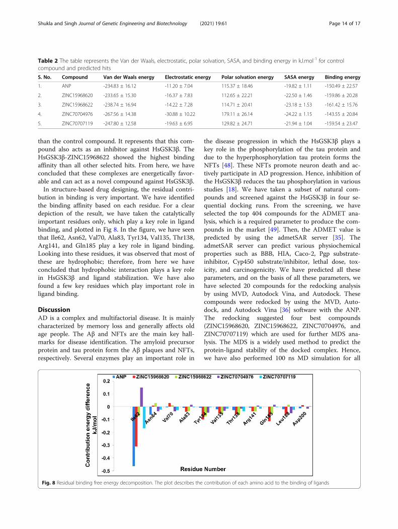
bution in binding is very important. We have identifiedthe binding affinity based on each residue. For a cleardepiction of the result, we have taken the catalyticallyimportant residues only, which play a key role in ligandbinding, and plotted in Fig 8. In the figure, we have seenthat Ile62, Asn62, Val70, Ala83, Tyr134, Val135, Thr138,Arg141, and Gln185 play a key role in ligand binding.Looking into these residues, it was observed that most ofthese are hydrophobic; therefore, from here we haveconcluded that hydrophobic interaction plays a key rolein HsGSK3β and ligand stabilization. We have alsofound a few key residues which play important role inligand binding.
DiscussionAD is a complex and multifactorial disease. It is mainlycharacterized by memory loss and generally affects oldage people. The Aβ and NFTs are the main key hall-marks for disease identification. The amyloid precursorprotein and tau protein form the Aβ plaques and NFTs,respectively. Several enzymes play an important role in
the disease progression in which the HsGSK3β plays akey role in the phosphorylation of the tau protein anddue to the hyperphosphorylation tau protein forms theNFTs [48]. These NFTs promote neuron death and ac-tively participate in AD progression. Hence, inhibition ofthe HsGSK3β reduces the tau phosphorylation in variousstudies [18]. We have taken a subset of natural com-pounds and screened against the HsGSK3β in four se-quential docking runs. From the screening, we haveselected the top 404 compounds for the ADMET ana-lysis, which is a required parameter to produce the com-pounds in the market [49]. Then, the ADMET value ispredicted by using the admetSAR server [35]. TheadmetSAR server can predict various physiochemicalproperties such as BBB, HIA, Caco-2, Pgp substrate-inhibitor, Cyp450 substrate/inhibitor, lethal dose, tox-icity, and carcinogenicity. We have predicted all theseparameters, and on the basis of all these parameters, wehave selected 20 compounds for the redocking analysisby using MVD, Autodock Vina, and Autodock. Thesecompounds were redocked by using the MVD, Auto-dock, and Autodock Vina [36] software with the ANP.The redocking suggested four best compounds(ZINC15968620, ZINC15968622, ZINC70704976, andZINC70707119) which are used for further MDS ana-lysis. The MDS is a widely used method to predict theprotein-ligand stability of the docked complex. Hence,we have also performed 100 ns MD simulation for all
Table 2 The table represents the Van der Waals, electrostatic, polar solvation, SASA, and binding energy in kJ.mol-1 for controlcompound and predicted hits
S. No. Compound Van der Waals energy Electrostatic energy Polar solvation energy SASA energy Binding energy
1. ANP -234.83 ± 16.12 -11.20 ± 7.04 115.37 ± 18.46 -19.82 ± 1.11 -150.49 ± 22.57
2. ZINC15968620 -233.65 ± 15.30 -16.37 ± 7.83 112.65 ± 22.21 -22.50 ± 1.46 -159.86 ± 20.28
3. ZINC15968622 -238.74 ± 16.94 -14.22 ± 7.28 114.71 ± 20.41 -23.18 ± 1.53 -161.42 ± 15.76
4. ZINC70704976 -267.56 ± 14.38 -30.88 ± 10.22 179.11 ± 26.14 -24.22 ± 1.15 -143.55 ± 20.84
5. ZINC70707119 -247.80 ± 12.58 -19.63 ± 6.95 129.82 ± 24.71 -21.94 ± 1.04 -159.54 ± 23.47
Fig. 8 Residual binding free energy decomposition. The plot describes the contribution of each amino acid to the binding of ligands
Shukla and Singh Journal of Genetic Engineering and Biotechnology (2021) 19:61 Page 14 of 17

the complexes. Several structural parameters such asRMSD, RMSF, Rg, SASA, PCA, and binding free energyanalysis were carried out. The RMSD value suggested thatall the complexes are stable and producing the equilibratedtrajectory for further analysis. Hence, we have calculatedvarious other structural parameters such as RMSF, Rg, PCA,Gibbs free energy, and binding free energy analysis. Thehydrogen bonds analysis suggest that all the complexes arestable and showing the interaction with the key catalytic resi-dues of the binding site. The essential dynamics analysis alsoagreed with this result. Then we have also carried out theGibbs free energy landscape analysis. The analysis suggestedthat some complexes followed the stable state with the meta-stable state. The binding free energy analysis showed thatout of four compounds three compounds (ZINC15968620,ZINC15968622, and ZINC70707119) are better thanZNC70704976. Hence from all these results we have selectedZINC15968620, ZINC15968622, and ZINC70707119 thatcan act as a lead compound against the HsGSK3β to reducethe NFTs burden from the cell. We have proposed thesecompounds to the global scientific community as they canfurther evaluate these compounds by using the in vitro andin vivo techniques.
ConclusionAD is a major problem for our society at a global level,and there is a need to look for possible treatment strat-egies. The HsGSK3β is an important protein kinase in-volved in tau hyperphosphorylation in AD includingseveral other diseases. So in this study, we have targetedthe HsGSK3β to reduce the hyperphosphorylation of tauprotein. We have used various computational ap-proaches for predicting the small molecules againstGSK3β which can bind to the active site and can blockthe enzyme activity. We have retrieved the natural com-pound library (n=167,741) from the ZINC database andscreened it against HsGSK3β in various steps by usingMVD. From the virtual screening, we have selected 404compounds. These compounds were further employedfor the pharmacokinetic analysis. From this analysis, 20compounds were selected and employed for the redock-ing. Then selected 4 plausible compounds from dockingthat were employed for MDS. From MDS, finally, threecompounds ZINC15968620, ZINC15968622, andZINC70707119 were selected and proposed as potentlead compounds against HsGSK3β. These compoundscan be further evaluated through in vitro and in vivo ex-periments. These compounds will serve as an initialpoint to design the novel inhibitors against HsGSK3βand can act as a novel therapeutic compound for AD. Itis anticipated that proposed compounds will provideready to use input for the experimental scientists andafter their respective verifications could help in the man-agement of tauopathies and AD.
AbbreviationsAD: Alzheimer’s disease; NFTs: Neurofibrillary tangles; GSK-3β: Glycogensynthase kinase 3 beta; MDS: Molecular dynamics simulation; PCA: Principalcomponent analysis; MM-PBSA: Molecular Machenics – Poisson BoltzmannSurface Area
Supplementary InformationThe online version contains supplementary material available at https://doi.org/10.1186/s43141-021-00163-w.
Additional file 1 Figure 1. The figure represents the common ring in allthe four compounds. The red circle represents the different group in thecompounds.s. Supplementary Table S1. In-silico absorption anddistribution profile obtained from admetSAR server for selected 404compounds. Selected compounds (20) for redocking were highlighted inbold. Supplementary Table S2. In-silico Cyp450 enzyme metabolismprofile was obtained from admetSAR server for selected 404 compounds.Selected compounds (20) for redocking were highlighted in bold.Supplementary Table S3. In-silico toxicity, carcinogenicity and LD50
profile obtained from admetSAR server for selected 404 compounds.Selected compounds (20) for redocking were highlighted in bold.Supplementary Table S4. Summary of binding affinity with interactingresidues of the top 20 compounds with control compound ANPobtained from molecular docking studies by three docking tools:Autodock Tools, AutodockVina and Molegro Virtual Docker. The Residueswhich involved in hydrogen bonding were highlighted in bold as well asselected hits for MDS are also highlighted in bold.
AcknowledgementsRS wants to thanks the Indian Council of Medical Research (ICMR) forproviding the Senior Research Fellowship ((ISRM/11(53)/2019)).
Authors’ contributionsTRS conceived the study. RS carried out all the experiments and the dataanalysis. RS and TRS participated in its overall design and coordination of thestudy. The first draft of the manuscript was prepared by RS. Both authorsread and approved the final manuscript.
FundingAuthors want to thanks the Indian Council of Medical Research (ICMR) (ISRM/11(53)/2019) for providing the Senior Research Fellowship (SRF) to RohitShukla.
Availability of data and materialsProvided in the form of the supplementary table.
Declarations
Ethics approval and consent to participateNot applicable
Consent for publicationNot applicable
Competing interestsThe authors declare that they have no competing interests.
Received: 12 January 2021 Accepted: 15 April 2021
References1. Patterson C (2018) World Alzheimer Report 2018, The state of the art of
dementia research: new frontiers. Alzheimer’s Disease International (ADI),London
2. Gold G, Bouras C, Kovari E, Canuto A, Glaria BG, Malky A et al (2000) Clinicalvalidity of Braak neuropathological staging in the oldest-old. ActaNeuropathol 99:579–582 discussion 583-584
Shukla and Singh Journal of Genetic Engineering and Biotechnology (2021) 19:61 Page 15 of 17
3. Tripathi T, Kalita P (2019) Synergistic effect of amyloid-β and tau disruptsneural circuits. ACS Chem Neurosci 10(3):1129–1130. https://doi.org/10.1021/acschemneuro.9b00037
4. Goate A, Chartier-Harlin MC, Mullan M, Brown J, Crawford F, Fidani L, GiuffraL, Haynes A, Irving N, James L, Mant R, Newton P, Rooke K, Roques P, TalbotC, Pericak-Vance M, Roses A, Williamson R, Rossor M, Owen M, Hardy J(1991) Segregation of a missense mutation in the amyloid precursor proteingene with familial Alzheimer’s disease. Nature 349(6311):704–706. https://doi.org/10.1038/349704a0
5. Levy-Lahad E, Wasco W, Poorkaj P, Romano DM, Oshima J, Pettingell WH,Yu C, Jondro P, Schmidt S, Wang K, al (1995) Candidate gene for thechromosome 1 familial Alzheimer’s disease locus. Science 269(5226):973–977. https://doi.org/10.1126/science.7638622
6. Rogaev EI, Sherrington R, Rogaeva EA, Levesque G, Ikeda M, Liang Y, Chi H,Lin C, Holman K, Tsuda T, Mar L, Sorbi S, Nacmias B, Piacentini S, AmaducciL, Chumakov I, Cohen D, Lannfelt L, Fraser PE, Rommens JM, George-HyslopPHS (1995) Familial Alzheimer’s disease in kindreds with missense mutationsin a gene on chromosome 1 related to the Alzheimer’s disease type 3 gene.Nature 376(6543):775–778. https://doi.org/10.1038/376775a0
7. Sherrington R, Rogaev EI, Liang Y, Rogaeva EA, Levesque G, Ikeda M, Chi H,Lin C, Li G, Holman K, Tsuda T, Mar L, Foncin JF, Bruni AC, Montesi MP,Sorbi S, Rainero I, Pinessi L, Nee L, Chumakov I, Pollen D, Brookes A,Sanseau P, Polinsky RJ, Wasco W, da Silva HAR, Haines JL, Pericak-Vance MA,Tanzi RE, Roses AD, Fraser PE, Rommens JM, St George-Hyslop PH (1995)Cloning of a gene bearing missense mutations in early-onset familialAlzheimer’s disease. Nature 375(6534):754–760. https://doi.org/10.1038/375754a0
8. Kumar A, Singh TR (2017) A new decision tree to solve the puzzle ofAlzheimer’s disease pathogenesis through standard diagnosis scoringsystem. Interdiscip Sci 9(1):107–115. https://doi.org/10.1007/s12539-016-0144-0
9. Verma S, Kumar A, Tripathi T, Kumar A (2018) Muscarinic and nicotinicacetylcholine receptor agonists: current scenario in Alzheimer’s diseasetherapy. J. Pharm. Pharmacol. 70(8):985–993. https://doi.org/10.1111/jphp.12919
10. Shukla R, Singh TR (2019) Virtual screening, pharmacokinetics, moleculardynamics and binding free energy analysis for small natural moleculesagainst cyclin-dependent kinase 5 for Alzheimer’s disease. J Biomol StructDyn 38(1):1–22. https://doi.org/10.1080/07391102.2019.1571947
11. Li C, Gotz J (2017) Tau-based therapies in neurodegeneration: opportunitiesand challenges. Nat Rev Drug Discov 16(12):863–883. https://doi.org/10.1038/nrd.2017.155
12. Tripathi T, Prakash J, Shav-Tal Y (2018) Phospho-tau impairs nuclear-cytoplasmic transport. ACS Chem Neurosci 10(1):36–38. https://doi.org/10.1021/acschemneuro.8b00632
13. Cohen P, Yellowlees D, Aitken A, Donella-Deana A, Hemmings BA, Parker PJ(1982) Separation and characterisation of glycogen synthase kinase 3,glycogen synthase kinase 4 and glycogen synthase kinase 5 from rabbitskeletal muscle. Eur J Biochem 124(1):21–35. https://doi.org/10.1111/j.1432-1033.1982.tb05902.x
14. Xie H, Wen H, Zhang D, Liu L, Liu B, Liu Q, Jin Q, Ke K, Hu M, Chen X (2017)Designing of dual inhibitors for GSK-3β and CDK5: virtual screening andin vitro biological activities study. Oncotarget 8(11):18118–18128. https://doi.org/10.18632/oncotarget.15085
15. Kannoji A, Phukan S, Sudher Babu V, Balaji VN (2008) GSK3beta: a masterswitch and a promising target. Expert Opin Ther Targets 12(11):1443–1455.https://doi.org/10.1517/14728222.12.11.1443
16. Martinez A, Gil C, Perez DI (2011) Glycogen synthase kinase 3 inhibitors inthe next horizon for Alzheimer’s disease treatment. Int J Alzheimers Dis2011:280502–280507. https://doi.org/10.4061/2011/280502
17. Ishizawa T, Sahara N, Ishiguro K, Kersh J, McGowan E, Lewis J, Hutton M, DicksonDW, Yen SH (2003) Co-localization of glycogen synthase kinase-3 with neurofibrillarytangles and granulovacuolar degeneration in transgenic mice. Am J Pathol 163(3):1057–1067. https://doi.org/10.1016/S0002-9440(10)63465-7
18. Pandey MK, DeGrado TR (2016) Glycogen synthase kinase-3 (GSK-3)-targetedtherapy and imaging. Theranostics 6(4):571–593. https://doi.org/10.7150/thno.14334
19. Hur E-M, Zhou F-Q (2010) GSK3 signalling in neural development. Nat RevNeurosci 11(8):539–551. https://doi.org/10.1038/nrn2870
20. Daggupati T, Pamanji R, Yeguvapalli S (2018) In silico screening andidentification of potential GSK3β inhibitors. J Recept Signal Transduct Res38(4):279–289. https://doi.org/10.1080/10799893.2018.1478854
21. Rampa A, Gobbi S, Concetta Di Martino RM, Belluti F, Bisi A (2017) DualBACE-1/GSK-3β inhibitors to combat Alzheimer’s disease: a focused review.Curr Top Med Chem 17(31):3361–3369. https://doi.org/10.2174/1568026618666180112161406
22. Aoki M, Yokota T, Sugiura I, Sasaki C, Hasegawa T, Okumura C, Ishiguro K,Kohno T, Sugio S, Matsuzaki T (2004) Structural insight into nucleotiderecognition in tau-protein kinase I/glycogen synthase kinase 3 beta. ActaCrystallogr D Biol Crystallogr 60(3):439–446. https://doi.org/10.1107/S090744490302938X
23. Pettersen EF, Goddard TD, Huang CC, Couch GS, Greenblatt DM, Meng EC,Ferrin TE (2004) UCSF Chimera--a visualization system for exploratoryresearch and analysis. J Comput Chem 25(13):1605–1612. https://doi.org/10.1002/jcc.20084
24. Irwin JJ, Shoichet BK (2005) ZINC – a free database of commerciallyavailable compounds for virtual screening. J Chem Inf Model 45(1):177–182.https://doi.org/10.1021/ci049714
25. Sterling T, Irwin JJ (2015) ZINC 15 – ligand discovery for everyone. J ChemInf Model 55(11):2324–2337. https://doi.org/10.1021/acs.jcim.5b00559
26. Irwin JJ, Sterling T, Mysinger MM, Bolstad ES, Coleman RG (2012) ZINC: afree tool to discover chemistry for biology. J Chem Inf Model 52(7):1757–1768. https://doi.org/10.1021/ci3001277
27. Shukla R, Shukla H, Tripathi T (2018) Structural and energetic understandingof novel natural inhibitors of Mycobacterium tuberculosis malate synthase. JCell Biochem 120(2):2469–2482. https://doi.org/10.1002/jcb.27538
28. Andrade EL, Bento AF, Cavalli J, Oliveira SK, Freitas CS, Marcon R, SchwankeRC, Siqueira JM, Calixto JB (2016) Non-clinical studies required for new drugdevelopment - part I: early in silico and in vitro studies, new targetdiscovery and validation, proof of principles and robustness of animalstudies. Braz J Med Biol Res 49(11):e5644. https://doi.org/10.1590/1414-431X20165644
29. Thomsen R, Christensen MH (2006) MolDock: a new technique for high-accuracy molecular docking. J Med Chem 49(11):3315–3321. https://doi.org/10.1021/jm051197e
30. Shukla R, Chetri PB, Sonkar A, Pakharukova MY, Mordvinov VA, Tripathi T(2017) Identification of novel natural inhibitors of Opisthorchis felineuscytochrome P450 using structure-based screening and molecular dynamicsimulation. J Biomol Struct Dyn 36(13):1–16. https://doi.org/10.1080/07391102.2017.1392897
31. Shukla R, Shukla H, Kalita P, Sonkar A, Pandey T, Singh DB, Kumar A, TripathiT (2018) Identification of potential inhibitors of Fasciola giganticathioredoxin1: computational screening, molecular dynamics simulation, andbinding free energy studies. J Biomol Struct Dyn 36(8):2147–2162. https://doi.org/10.1080/07391102.2017.1344141
32. Chandra A, Gurjar V, Ahmed MZ, Alqahtani AS, Qamar I, Singh N (2021)Exploring potential inhibitor of SARS-CoV2 replicase from FDA approveddrugs using insilico drug discovery methods. J Biomol Struct Dyn:1–8.https://doi.org/10.1080/07391102.2020.1871416
33. Chandra A, Ananda H, Singh N, Qamar I (2020) Identification of a novel andpotent small molecule inhibitor of SRPK1: mechanism of dual inhibition ofSRPK1 for the inhibition of cancer progression. Aging (Albany NY) 13(1):163–180. https://doi.org/10.18632/aging.202301
34. Chandra A, Chaudhary M, Qamar I, Singh N, Nain V. In silico identificationand validation of natural antiviral compounds as potential inhibitors ofSARS-CoV-2 methyltransferase. J Biomol Struct Dyn 2021;0:1–11. doi: https://doi.org/10.1080/07391102.2021.1886174.
35. Cheng F, Li W, Zhou Y, Shen J, Wu Z, Liu G, Lee PW, Tang Y (2012)admetSAR: a comprehensive source and free tool for assessment ofchemical ADMET properties. J Chem Inf Model 52(11):3099–3105. https://doi.org/10.1021/ci300367a
36. Trott O, Olson AJ (2009) AutoDock Vina: improving the speed and accuracy ofdocking with a new scoring function, efficient optimization, andmultithreading. J Comput Chem 31:455–461. https://doi.org/10.1002/jcc.21334
37. Morris GM, Huey R, Lindstrom W, Sanner MF, Belew RK, Goodsell DS, OlsonAJ (2009) AutoDock4 and AutoDockTools4: automated docking withselective receptor flexibility. J Comput Chem 30(16):2785–2791. https://doi.org/10.1002/jcc.21256
38. Huey R, Morris GM, Olson AJ, Goodsell DS (2007) A semiempirical freeenergy force field with charge-based desolvation. J Comput Chem 28(6):1145–1152. https://doi.org/10.1002/jcc.20634
39. Shukla H, Shukla R, Sonkar A, Tripathi T (2017) Alterations in conformationaltopology and interaction dynamics caused by L418A mutation leads to
Shukla and Singh Journal of Genetic Engineering and Biotechnology (2021) 19:61 Page 16 of 17
activity loss of Mycobacterium tuberculosis isocitrate lyase. Biochem.Biophys. Res. Commun. 490(2):276–282. https://doi.org/10.1016/j.bbrc.2017.06.036
40. Shukla R, Shukla H, Tripathi T (2018) Activity loss by H46A mutation inMycobacterium tuberculosis isocitrate lyase is due to decrease in structuralplasticity and collective motions of the active site. Tuberculosis (Edinb) 108:143–150. https://doi.org/10.1016/j.tube.2017.11.013
41. Shukla R, Shukla H, Sonkar A, Pandey T, Tripathi T (2018) Structure-basedscreening and molecular dynamics simulations offer novel naturalcompounds as potential inhibitors of Mycobacterium tuberculosis isocitratelyase. J Biomol Struct Dyn 36(8):2045–2057. https://doi.org/10.1080/07391102.2017.1341337
42. Abraham MJ, Murtola T, Schulz R, Pall S, Smith JC, Hess B et al (2015)GROMACS: High performance molecular simulations through multi-levelparallelism from laptops to supercomputers. SoftwareX 1–2:19–25. https://doi.org/10.1016/j.softx.2015.06.001
43. Oostenbrink C, Villa A, Mark AE, van Gunsteren WF. A biomolecular forcefield based on the free enthalpy of hydration and solvation: the GROMOSforce-field parameter sets 53A5 and 53A6. J Comput Chem 2004;25:1656–1676. doi: https://doi.org/10.1002/jcc.20090, 13.
44. Schuittelkopf AW, van Aalten DMF (2004) PRODRG: a tool for high-throughput crystallography of protein-ligand complexes. Acta Crystallogr DBiol Crystallogr 60(8):1355–1363. https://doi.org/10.1107/S0907444904011679
45. Darden T, York D, Pedersen L (1993) Particle mesh Ewald: An N·log(N)method for Ewald sums in large systems. J Chem Phys 98(12):10089–10092.https://doi.org/10.1063/1.464397
46. Kumari R, Kumar R, Lynn A (2014) g_mmpbsa--a GROMACS tool for high-throughput MM-PBSA calculations. J Chem Inf Model 54(7):1951–1962.https://doi.org/10.1021/ci500020m
47. Brana MF, Garrido M, Rodriguez MLL, Miguel P, Morcillo MJ, Riano A (1990)Synthesis of new derivatives of β-carboline-hydantoin. J. Heterocycl Chem27(3):703–706. https://doi.org/10.1002/jhet.5570270342
48. Cao J, Hou J, Ping J, Cai D (2018) Advances in developing novel therapeuticstrategies for Alzheimer’s disease. Mol. Neurodegener. 13(1):64. https://doi.org/10.1186/s13024-018-0299-8
49. Lucas AJ, Sproston JL, Barton P, Riley RJ (2019) Estimating human ADMEproperties, pharmacokinetic parameters and likely clinical dose in drugdiscovery. Expert Opin Drug Discov 14(12):1313–1327. https://doi.org/10.1080/17460441.2019.1660642
Publisher’s NoteSpringer Nature remains neutral with regard to jurisdictional claims inpublished maps and institutional affiliations.
Shukla and Singh Journal of Genetic Engineering and Biotechnology (2021) 19:61 Page 17 of 17