SPONDYLOLISTHESIS: DIAGNOSIS, CLASSIFICATION, EVALUATION AND MANAGEMENT

Case Report eISSN2465-891X The Nerve.2015.1(1):37-39http://dx.doi.org/10.21129/nerve.2015.1.1.37
www.thenerve.net
Journal of the Korean Society of Peripheral Nervous System 37
High-grade Traumatic Spondylolisthesis of C7 on T1 with No Neurological Deficit
Tae-Keun Ahn1*, Young-Sun Chung2*, Min-Soo Kim3, Inbo Han3
1Departments of Orthopedics, CHA Bundang Medical Center, CHA University College of Medicine, Seongnam, 2Department of Neurosurgery, Konkuk University Hospital, Konkuk University College of Medicine, Chungju,
3Departments of Neurosurgery, CHA Bundang Medical Center, CHA University College of Medicine, Seongnam, Korea
High-grade spondylolisthesis at the cervicothoracic junction is rare and can cause devastating spinal cord injuries. Here, the authors describe two patients who presented with high-grade spondylolisthesis without neurological deficit at the level C7/T1. Cervicothoracic junction instability was stabilized with the circumferential fusion with no further neurological deficits.
Key Words: TraumaㆍSpondylolisthesisㆍCervicothoracic junction
Corresponding author: Inbo Han, MDDepartment of Neurosurgery, CHA Bundang Medical Center, CHA Uni- versity College of Medicine, 59 Yatap-ro Bundang-gu, Seongnam-si, Gyeunggi-do 13496, KoreaTel: +82-31-780-1924, Fax: +82-31-780-5269E-mail: [email protected]*equal contribution.
INTRODUCTION
The cervicothoracic junction (CVJ) is a potentially unstable site due to an abrupt transition from the lordotic mobile cer- vical spine to the more rigid kyphotic thoracic spine. High- grade traumatic spondylolisthesis at the CVJ without neuro- logical deficit is rare and should be treated with anterior and posterior stabilization techniques due to severely unstable frac- tures1-3). With its potential anatomic instability, lesions at the CVJ can lead to devastating spinal cord injury. The authors describe two cases of high-grade traumatic spondylolisthesis of C7 on T1 without neurological deficits and discuss clinical fea- tures and different surgical strategies according to patients’ con- dition.
CASE REPORT
1. Case 1
A 32-year-old man was referred to our institution with an episode of fall from a height of about 2.0 meter. The patient presented with severe posterior neck pain without any motor
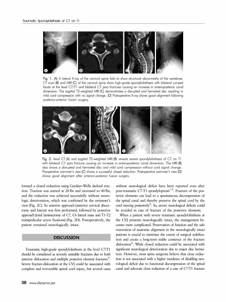
or sensory deficits. A lateral X-ray (Fig. 1A) of the cervical spine failed to show structural abnormality of the vertebrae. How- ever, cervical computed tomography (CT) scan and magnetic resonance imaging (MRI) showed high-grade spondylolisthesis of C7 on T1 with bilateral jumped facets and bilateral C7 pars fractures with an increase in anteroposterior spinal canal dimen- sion (Fig. 1B and C). In addition, the T2-weighted MRI revealed a ruptured, posteriorly herniated disc causing mild cord com- pression with no cord signal change (Fig. 1C). First, we perfor- med a closed reduction using Gardner-Wells skeletal traction. Traction was started at 20 lbs and increased to 60 lbs, but the closed reduction has failed. Therefore, open reduction inclu- ding resection of the fractured facet joint and instrumenta- tion using the Atlas® cable system(Medtronic Sofamor Danek, Mem- phis, USA) were performed posteriorly. In a second operation, a discectomy C7-T1 was performed and a monosegmental fusion was added, using an autologous tricortical iliac autograft and Atlantis® anterior cervical plate system(Medtronic Sofamor Da- nek, Memphis, USA) (Fig. 1D). Post-operatively, the neuro- logical status was not changed.
2. Case 2
A 42-year-old woman presented with severe posterior neck pain after falling 1.5 m down while she was hiking. Neurolo- gical examination showed no neurological deficits. CT (Fig. 2A) and MRI (Fig. 2B) revealed severe spondylolisthesis of C7 on T1 with bilateral C7 pars fracture and increased anteropos- terior canal dimension. Sagittal T2 weighted MRI (Fig. 2B) demonstrated a disrupted and herniated disc causing cervical cord compression with no cord signal change. First, we per-
Traumatic Spondylolisthesis of C7 on T1
38 www.thenerve.net
Fig. 1. (A) A lateral X-ray of the cervical spine fails to show structural abnormality of the vertebrae.CT scan (B) and MRI (C) of the cervical spine show high-grade spondylolisthesis with bilateral jumpedfacets at the level C7/T1 and bilateral C7 pars fractures causing an increase in anteroposterior canaldimension. The sagittal T2-weighted MRI (C) demonstrates a disrupted and herniated disc resulting inmild cord compression with no signal change. (D) Postoperative X-ray shows good alignment followingposterior-anterior fusion surgery.
Fig. 2. Axial CT (A) and sagittal T2-weighted MRI (B) reveals severe spondylolisthesis of C7 on T1 with bilateral C7 pars fracture causing an increase in anteroposterior canal dimension. The MRI (B)also shows a disrupted and herniated disc and mild cord compression without cord signal change.Preoperative swimmer’s view (C) shows a successful closed reduction. Postoperative swimmer’s view (D)shows good alignment after anterior-posterior fusion surgery.
formed a closed reduction using Gardner-Wells skeletal trac- tion. Traction was started at 20 lbs and increased to 40 lbs, and the reduction was achieved successfully without neuro- logic deterioration, which was confirmed by the swimmer’s view (Fig. 2C). So anterior approach (anterior cervical discec- tomy and fusion) was first performed, followed by posterior approach (total laminectomy of C7, C6 lateral mass and T1-T2 transpedicular screw fixations) (Fig. 2D). Postoperatively, the patient remained neurologically intact.
DISCUSSION
Traumatic high-grade spondylolisthesis at the level C7/T1 should be considered as severely unstable fractures due to both anterior dislocation and multiple posterior element fractures1). Severe fracture-dislocation at the CVJ could be associated with complete and irreversible spinal cord injury, but several cases
without neurological deficit have been reported even after post-traumatic C7-T1 spondyloptosis1-3). Fracture of the pos- terior elements can lead to a spontaneous decompression of the spinal canal and thereby preserve the spinal cord by the cord moving posteriorly2). So, severe neurological deficits could be avoided in case of fracture of the posterior elements.
When a patient with severe traumatic spondylolisthesis at the CVJ presents neurologically intact, the management be- comes more complicated. Preservation of function and the safe restoration of anatomic alignment in the neurologically intact patients is crucial to minimize the extent of surgical stabiliza- tion and create a long-term stable construct of the fracture dislocation2). While closed reduction could be associated with significant neurological deterioration due to major disc hernia- tion. However, most spine surgeons believe that close reduc- tion is not associated with a higher incidence of disabling neu- rological deficit due to functional decompression of the spinal canal and advocate close reduction of a case of C7-T1 fracture

Ahn TK et al.
The Nerve 1(1) September 2015 39
dislocation with no neurological deficit1).Although there have been different surgical managements
for traumatic C7-T1 fracture dislocation, combined anterior- posterior instrumentation has been usually chosen as the most stable fixation techniques. An anterior approach has been favo- red as the initial surgical approach to avoid neurological dete- rioration in case of the presence of significant disc herniation and to prevent the risk of additional trauma in case of turning the patient on the operation table for the posterior appro- ach1,2). Posterior open reduction of unreducible dislocations has been considered to be an initial effective surgical manage- ment by allowing direct disengagement of the inferior facet from the superior facet1).
In the present cases, combined anterior-posterior fixation underwent due to the destruction of both anterior and pos- terior elements. Based on success or failure of closed reduction and the presence of significant disc herniation, the initial sur- gical approach was chosen. In the first patient, closed reduction failed and the spinal canal was well maintained. Therefore, pos- terior open reduction and instrumentation was selected as an initial approach. In the second patient, MRI showed a disrup- ted and herniated disc, but the closed reduction was successful without neurological deterioration. So, the anterior approach was first performed, followed by posterior fusion.
CONCLUSION
Cervicothoracic juctional area should be examined carefully in acute traumatic injury to confirm hidden lesions. High-grade traumatic C7-T1 spondylolisthesis without neurological deficits is very rare. The management of a neurologically intact patient
remains challenging due to its potential instability. However, a closed reduction can be recommended to restore anatomic alignment in neurologically intact patients because a posterior element fracture generally leads to a spontaneous posterior de- compression of the spinal canal. Additionally, combined ante- rior-posterior approaches can provide good surgical outcomes, and the initial surgery is chosen based on success or failure of the closed reduction and the presence of the significant disc herniation.
ACKNOWLEDGMENT
This work was supported by the Korea Healthcare Techno- logy Research & Development Project, Ministry for Health & Welfare Affairs (#H11C3245 and #114C3270)
This research was supported by a grant of the Korea Health Technology R&D Project through the Korea Health Industry Development Institute (KHIDI), funded by the Ministry of Health & Welfare, Republic of Korea (HI14)
REFERENCES
1. Acikbas C, Gurkanlar D: Post-traumatic C7-T1 spondyloptosis in a patient without neurological deficit: a case report. Turk Neu- rosurg 20:257-260, 2010
2. Dahdaleh NS, Dlouhy BJ, Greenlee JDW, Smoker WRK, Hitchon PW: An algorithm for the management of posttraumatic cervical spondyloptosis. J Clin Neurosci Off J Neurosurg Soc Australas 20:951-957, 2013
3. Tumialán LM, Dadashev V, Laborde DV, Gupta SK: Management of traumatic cervical spondyloptosis in a neurologically intact patient: case report. Spine 34:E703-E708, 2009


![C7 graham session c7 mon1530_v6[2]](https://static.fdocuments.in/doc/165x107/58ed94c51a28ab717c8b475d/c7-graham-session-c7-mon1530v62.jpg)