HIA framework to investigate additional cancer risk from Ionizing … · HIA framework to...
32
1 HIA framework to investigate additional cancer risk from Ionizing Radiation in Medical Imaging. Top-Down case study Prepared for the project Risk Assessment from Policy to Impact Dimension (RAPID) 2009-2012 EU (DG-SANCO) Grant agreement N o 20081105 Via Project coordinating partner: National Council of Research Institute of Clinical Physiology (IFC-CNR) Unit of Environmental Epidemiology Via Moruzzi, 1 - 56127 Pisa, Italy http://www.ifc.cnr.it By Nunzia Linzalone Elisa Bustaffa and Liliana Cori IFC clicical cardiology group Fabrizio Bianchi Institute of Clinical Physiology - CNR Unit of Environmental Epidemiology May 10 th 2010
Transcript of HIA framework to investigate additional cancer risk from Ionizing … · HIA framework to...
1
HIA framework to investigate additional cancer risk from
Ionizing Radiation in Medical Imaging.
Top-Down case study
Prepared for the project
Risk Assessment from Policy to Impact Dimension (RAPID) 2009-2012
EU (DG-SANCO) Grant agreement No 20081105
Via
Project coordinating partner: National Council of Research
Institute of Clinical Physiology (IFC-CNR) Unit of Environmental Epidemiology
Via Moruzzi, 1 - 56127 Pisa, Italy http://www.ifc.cnr.it
By
Nunzia Linzalone
Elisa Bustaffa and
Liliana Cori IFC clicical cardiology group
Fabrizio Bianchi
Institute of Clinical Physiology - CNR Unit of Environmental Epidemiology
May 10th 2010

2
Contents
Abstract ............................................................................................................................................................. 3
Introduction ....................................................................................................................................................... 4
The model .......................................................................................................................................................... 7
Policy description .............................................................................................................................................. 7
Determinants of health ..................................................................................................................................... 8
Risk factors ...................................................................................................................................................... 11
Health outcomes ............................................................................................................................................. 13
Pilot application ............................................................................................................................................... 14
Conclusion ....................................................................................................................................................... 19
References ....................................................................................................................................................... 21
3
Abstract
Computed tomography (CT) was introduced into medical imaging in the 1970s and has grown exponentially particularly in cardiovascular clinical test for a wide variety of cardiovascular conditions. Cardiovascular CT use has recently been tempered by a string of high-impact publications raising concern about the increase in radiation exposure to the population from medical procedures and the potential cancer risk. This report highlights the framework in which integrated assessment of the effect of current cardiovascular imaging procedures on cancer risk is carried out. The analysis is based on the emerging results of a research project called SUIT-Heart (Stop Useless Ionizing Testing in Heart Disease) conducted by the Institute of Clinical Physiology of National Council of Research (IFC-CNR) in Pisa, Italy and funded by the Istituto Toscano Tumori (ITT). Finally it is outlined the chain of elements contributing to cancer incidence attributed to cardiovascular CT use. It is therefore represented an approach that helps the integration of health impact assessment for decision-making in the sector of ionizing radiation protection policies.
Keywords
IONIZING RADIATION MEDICAL IMAGING COMPUTED TOMOGRAPHY CARDIOVASCULAR IMAGING CANCER RISK RISK ASSESSMENT RADIATION PROTECTION POLICY TUSCANY

4
Introduction
Focusing on single risk factors is not always satisfactory, as strategies and policies developed to
address one risk might increase other risks. To develop effective precautionary policies, policy-
makers and stakeholders need evidences based on an integrated risk assessment. To match this
need RAPID project have included a case study to develop and test a framework and
methodology for a “full chain” health impact assessment. Italian partner selected a topic on
useless imaging testing in medical practice, tackling one of main policy focus at local and
international level as well. Different policies, focused to the issue of awareness in diagnostic use
of ionizing radiation, correspond to changes in individual exposure to cumulative dose and
account for the variation of attributable long term cancer risks.
The intermediate level of determinants of health has been particularly posed under study as
determinants are modified by policies and in turn they modify exposure to risk factors.
Health Impact Assessment (HIA) framework
Since 2000, the need for a new approach integrating the direct and the indirect effects assessed
during health impact assessment has been recognized. In the development of health impact
assessment, two broad approaches are usually acknowledged: the biomedical approach and the
social determinants of health approach (Morgan, 2003). WHO initially promoted the first
approach in the 1980s and defined it as environmental health impact assessment (WHO Regional
Office for Europe, 1987). Environmental health impact assessment is based on the biomedical
model of health, illustrated in direct effects such as mortality and morbidity. The second
approach of health impact assessment evolved from public health considerations and is based
on the interrelationships between the population and the environment, including socioeconomic
determinants of health and institutional factors. This approach allows the indirect effects of
projects and policies on health to be estimated.
More generally health impact assessment provides a structured framework to map the full range
of health effects of any proposal and action, whether these are negative or positive (WHO
Regional Office for Europe, 2002): “Health impact assessment is a combination of procedures,
methods and tools by which a policy, programme or project may be judged as to its potential
effects on the health of a population, and the distribution of those effects within the population”
(WHO European Centre for Health Policy, 1999). An integrated assessment, incorporating health
impact assessment, allows policy development to ensure that health effects are not overlooked.
In the present case study the health impact assessment is done looking at the interrelationships
between the population and the environment, including socioeconomic determinants of health
and institutional factors. This document describes the “full chain” assessment of cancer risk from
Ionizing Radiation in Medical Imaging with a top–down approach. The analysis goes from
present regional regulation and interrelated policies, to the determinants of health, the risk
factors and the attributable health outcomes. Available evidence and knowledge are used to
highlight the full web of connections that lead health effects of policies to individual and
population level.

5
Aim and relevance of the topic
Aim of this study is to develop a model which include either the risk management and the
quantitative assessment of risks, into a framework for the assessment of the full range of health
impacts (figure 1). The overall HIA process is aimed to supply qualitative knowledge for better
decision making in the sector of diagnostic use of computed tomography for cardiovascular
disease, by describing the full scheme of impacts proceeding from current regulatory advice in
clinical practice (top) to a selected health outcome (down).
The challenge on research findings transfer from a Clinical Research Centre (performing the
empirical research on this topic) to policy decisions is discussed, as well.
The topic selection for the case study has been based on considering that inappropriate use of
cardiological ionizing imaging testing, fueled by radiological unawareness, is a significant source
of useless radiation exposure of patients (creating a risk without a commensurate benefit).
Medical imaging is the largest controllable source of radiation exposure in the population of
industrialized countries. One out of two examinations is completely or partially inappropriate
(i.e. risk outweighs benefit) and cardiologists are often unaware of the radiological dose of the
examination they prescribe or practice (Gibbons et al., 2007; Gibbons et al., 2008; Picano et al.
2007). This avoidable exposure is associated with increased, significant cancer risk at both the
individual and population levels and can be minimized through a knowledge-based intervention
targeted to increasing radiological awareness of prescribers and practitioners.
Responding to rising concerns in the radiology community and among the public, the Food and
Drug Administration (FDA) announced a new initiative (US FDA, 2010) in February 2010 aimed at
reducing unnecessary radiation exposure due to medical imaging in US.
In Europe existing guidelines (European Commission, 2001) need duly be reinforced to achieve a
more justified and optimized use of Computed Tomography in each country (Picano, 2004).
In Tuscany, Italy, under the Regional Health Plan 2008-2010 (Regione Toscana, 2008) and in
accordance with the specific objectives on Clinical Risk Management, the Tuscany Region is
committed to promoting initiatives for the prevention and protection of the community, in
particular to the proper use of ionizing radiation (IR) and radioactive substances, which have
enabled major developments in modern medicine with technological innovations such as
tomography (CT), digital angiography and, more recently, Positron Emission Tomography (PET).
The Institute of Clinical Physiology of CNR, in Pisa, has activated in 2008 a collaborative project
with the Istituto Toscano Tumori aiming at primary prevention of cancer through reduction of
inappropriate ionizing testing (SUIT-HEART). Referring to this research issue, IFC researchers and
medical staff represent a multidisciplinary team of experts and a network of practitioners to
undergo testing and validation of case studies. In particular, disclosing how different approaches
of diagnostic practice, responsible of balancing risks and benefits, are modified from background
communication on and knowledge of risks, would lead to strengthen measures for an
appropriate use of IR.
Exposure to ionizing radiation in medical practice
Over the last 30 years imaging techniques have become indispensable as an aid in diagnosis,
prognosis, monitoring of disease and the implementation of interventional procedures (both
diagnostic and therapeutic). Among medical imaging techniques radiological and nuclear
6
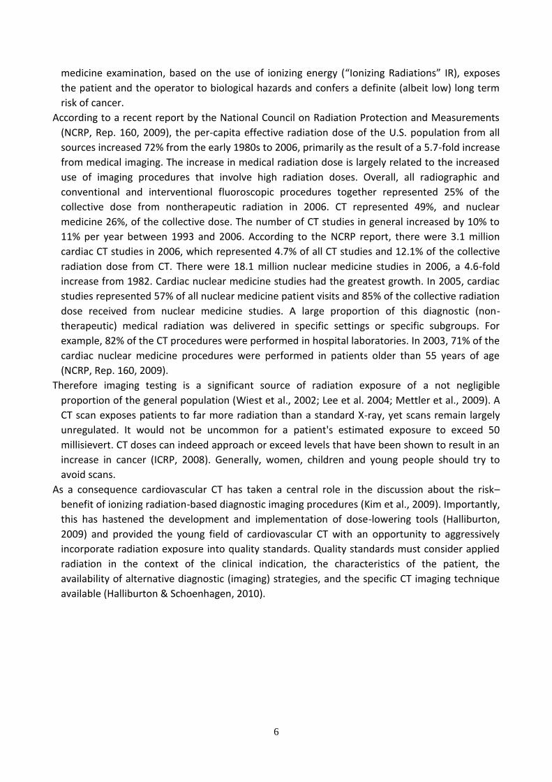
medicine examination, based on the use of ionizing energy (“Ionizing Radiations” IR), exposes
the patient and the operator to biological hazards and confers a definite (albeit low) long term
risk of cancer.
According to a recent report by the National Council on Radiation Protection and Measurements
(NCRP, Rep. 160, 2009), the per-capita effective radiation dose of the U.S. population from all
sources increased 72% from the early 1980s to 2006, primarily as the result of a 5.7-fold increase
from medical imaging. The increase in medical radiation dose is largely related to the increased
use of imaging procedures that involve high radiation doses. Overall, all radiographic and
conventional and interventional fluoroscopic procedures together represented 25% of the
collective dose from nontherapeutic radiation in 2006. CT represented 49%, and nuclear
medicine 26%, of the collective dose. The number of CT studies in general increased by 10% to
11% per year between 1993 and 2006. According to the NCRP report, there were 3.1 million
cardiac CT studies in 2006, which represented 4.7% of all CT studies and 12.1% of the collective
radiation dose from CT. There were 18.1 million nuclear medicine studies in 2006, a 4.6-fold
increase from 1982. Cardiac nuclear medicine studies had the greatest growth. In 2005, cardiac
studies represented 57% of all nuclear medicine patient visits and 85% of the collective radiation
dose received from nuclear medicine studies. A large proportion of this diagnostic (non-
therapeutic) medical radiation was delivered in specific settings or specific subgroups. For
example, 82% of the CT procedures were performed in hospital laboratories. In 2003, 71% of the
cardiac nuclear medicine procedures were performed in patients older than 55 years of age
(NCRP, Rep. 160, 2009).
Therefore imaging testing is a significant source of radiation exposure of a not negligible
proportion of the general population (Wiest et al., 2002; Lee et al. 2004; Mettler et al., 2009). A
CT scan exposes patients to far more radiation than a standard X-ray, yet scans remain largely
unregulated. It would not be uncommon for a patient's estimated exposure to exceed 50
millisievert. CT doses can indeed approach or exceed levels that have been shown to result in an
increase in cancer (ICRP, 2008). Generally, women, children and young people should try to
avoid scans.
As a consequence cardiovascular CT has taken a central role in the discussion about the risk–
benefit of ionizing radiation-based diagnostic imaging procedures (Kim et al., 2009). Importantly,
this has hastened the development and implementation of dose-lowering tools (Halliburton,
2009) and provided the young field of cardiovascular CT with an opportunity to aggressively
incorporate radiation exposure into quality standards. Quality standards must consider applied
radiation in the context of the clinical indication, the characteristics of the patient, the
availability of alternative diagnostic (imaging) strategies, and the specific CT imaging technique
available (Halliburton & Schoenhagen, 2010).
7
The model
A top-down risk assessment model (figure 1) is herein proposed to answer the following issue:
How do current or prospective policies, focused to the issue of awareness in diagnostic use of
ionizing radiation, correspond to changes in individual exposure to cumulative dose?
How do they account for the associated attributable long term cancer risk?
Is currently estimated individual risk comprehensive of cumulative exposures?
With reference to this issue the Tuscany Regional Health Plan 2008-2010 (Regione Toscana, 2008),
presides over initiatives to make it more stringent reduction of ionizing radiations in medical
practice. This policy acts in accordance with local initiatives conveying towards industrials,
physicians and patients awareness with the aim of modifying proximal cultural and socio-
economic health determinants (not usually included in view of cumulative life exposure dose) as
to cause a substantial change in current exposure models to IR. Main risk factors could be
lowered from disseminating at collective and community level a novel understanding of real
risks associated with effective dose of ionizing radiations.
Saving use of radiation has a great potential to reduce incidence case of cancer mainly at
population level. While policy addressed to screen high risk subjects reduce greatly the
individual risk, a policy action modifying exposure model reduce greatly the potential for major
populations at risk (it is the case for categories of male, age over 55, children). As to the balance
risk-benefit, the gain is calculated over a long term perspective including the overall cancers,
attributed to medical imaging.
The following generic methodological steps, validated in a pilot study by IFC-CNR (Regione Emilia
Romagna, 2010), are used to test and validate the model for the assessment of the health
burden of current regulation on ionizing radiation in medical practice:
Selection from literature of relevant knowledge and evidences on medical imaging and
health effects. They constitute a starting rationale of the study context;
Expert advice to validate core factors in model building;
Testing of hypothesis on a real context by a pilot application;
Consensus-building on the developed model;
Results, tools and concrete recommendations summed in a written report. This last has the
function to vehicles information into policy and regulations.
Last two steps completion including the prevented revision by the Rapid whole group.
In essence this framework is an aspiration for a strategy change in the way that the regional health
service provide care as to manage the use of medical imaging optimally, in order to reduce the
likelihood of effective dose of radiation for patients.
Policy description
Protecting the patient from ionizing radiation is regulated by Legislative Decree 187/00, issued to
implement the European directive 97/43 (D. Lgs. 187/00). The Tuscan Regional Council is
committed to monitor the dose to the population and medical workers due to medical
examinations, firstly to allow the information to the patients of the received dose in each
examination and that accumulated during their clinic history. Among aims there is also to
8
promote either a widespread communication campaign for Tuscan citizens and a training
pathway for prescribers on the risks posed by the medical exposure to ionizing radiation. The
objectives of the Regional Health Plan are also targeted to propose guidelines and
recommendations for the implementation of Legislative Decree 187/2000, to be achieved
through the definition of protocols and best practices addressed to regional structures
concerned. Funding is allocated by the Tuscan Region for 3 years to develop a project, by the
Italian Association of Medical Physics, entitled "Communication of patient dose". The project
plans to make automatic the process of measuring the dose and its recording in an electronic
health card, in each Tuscany health-care accredited centers. Funding is also allocated to the
development of the project entitled SUIT-HEART (Stop Useless Ionizing Testing in Heart Disease)
- primary prevention of cancer through reduction of inappropriate ionizing testing, to be carried
out by IFC-CNR and Tuscany Cancer Institute, over 3 years. The overarching aim of the SUIT-
HEART study is to promote a better appreciation of radiation risks in the cardiology community
and in patients, as now unanimously recommended by official documents of the American
College of Radiology 2007, International Atomic Energy Agency 2008 and American Heart
Association 2009.
Consequences of a comprehensive understanding of radioprotection issues inside medical practice
and of the use of advanced health care ionizing radiation technologies, influences different
current policy targets such as: training and information for specialists and practitioners, correct
information for users, implications of informed consent, political intervention to reduce the dose
in the higher risk practices.
Determinants of health
Determinants of health include the range of personal, social, biological, economic and
environmental factors which determine the presence and distribution of risk factors in the
populations. Interaction between a large variety of these factors and different exposure or
baseline health condition, affect the presentation of the outcome. Prescription practice in
medical setting is a major determinant of the cumulative individual and collective dose, having
an effect on the medical practice. However medical practice is also determined from multiple
factors ranging from current regulation, technological updating of devices, type of “license” for
medical device use, continuing training for physicians, physician attitude and perception toward
risks and health equity, patient awareness and informed consent protocols, working
environment regulation and available information on risk-benefit (Figure 2).
Classification of exposed population by gender and young age classes as well as previous exposure
story, also interact determining variations of attributable risks. Moreover there are often
medical conditions, chronic, serious or even fatal, acting as co-determinants of the final outcome
of radiation exposure (Figure 2).
Medical practice and cancer risk
Based on growing clinical experience, guidelines describing appropriate indications for
cardiovascular CT have been established, weighing procedural risk, pre-test probability, and
expected benefit (Hendel et al., 2006). Procedural risk is defined by the need for vascular access,
9
amount of injected contrast media and level of radiation exposure, and depends on patient
specific criteria, including age and sex (Einstein et al., 2007). It is important to note that a
significant reduction in radiation dose for CT imaging of a particular indication (e.g., coronary
CTA with 1 mSv) could shift the risk-benefit ratio and subsequently have an impact on
appropriateness criteria. However, the relatively noninvasive nature of a test alone does not
establish its usefulness for screening, in particular because false positives in patients with low
pre-test probability can be associated with untoward outcome (Nissen, 2008).
If CT is determined to be the most appropriate test, it is important to tailor the imaging protocol to
the clinical question. The CT imaging protocol should also be tailored to patient characteristics.
In addition, X-ray parameters including tube voltage and tube current should be adjusted
according to patient size (Halliburton 2009). A 30% decrease in tube current results in a 30%
reduction in X-ray exposure (DeMarco, 2007).
Although tailoring the cardiovascular CT imaging protocol to the clinical indication and the patient
is critical for the optimization of both image quality and dose, the rapid development of scanner
hardware and software as well as manufacturer differences in scanner design have largely
prevented standardization of protocols. This is reflected in recent studies (Hausleiter, 2009)
demonstrating large variations in coronary CTA protocols, resulting in a wide range of radiation
doses at different centers as well as recent, highly-publicized egregious errors in
noncardiovascular CT imaging that have resulted in dramatic patient overexposure. Clearly, a
coordinated effort is needed to standardize and regulate radiation exposure during
cardiovascular CT, including regular monitoring of the radiation burden (Abbara et al., 2009),
formation of imaging groups with collective experience of various imaging modalities
(multimodality imaging). In such groups, dedicated protocols are designed in collaboration by
radiologists, cardiologists, physicists, and technologists. Based on individualized review of the
clinical indication, the patient is directed toward the most appropriate diagnostic test or
strategy. Choosing the right test for one individual gives possibilities to reduce the burden of
cumulative dose, using radiation as the last option. Adopting an approach based on
appropriateness can lead to spare radiation use at the level of population, increasing the overall
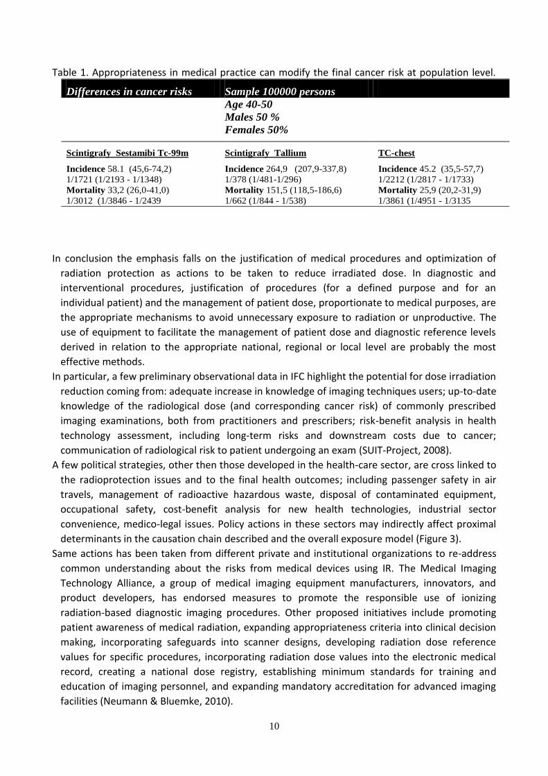
health benefits (figure 3). Differences in estimated cancer risks have a noticeable weights
translating inappropriate clinical decision-making from individual health care to these of a sub-
group of population (table 1).
10
Table 1. Appropriateness in medical practice can modify the final cancer risk at population level.
Differences in cancer risks Sample 100000 persons
Age 40-50
Males 50 %
Females 50%
Scintigrafy Sestamibi Tc-99m Scintigrafy Tallium TC-chest
Incidence 58.1 (45,6-74,2)
1/1721 (1/2193 - 1/1348)
Mortality 33,2 (26,0-41,0)
1/3012 (1/3846 - 1/2439
Incidence 264,9 (207,9-337,8)
1/378 (1/481-1/296)
Mortality 151,5 (118,5-186,6)
1/662 (1/844 - 1/538)
Incidence 45.2 (35,5-57,7)
1/2212 (1/2817 - 1/1733)
Mortality 25,9 (20,2-31,9)
1/3861 (1/4951 - 1/3135
In conclusion the emphasis falls on the justification of medical procedures and optimization of
radiation protection as actions to be taken to reduce irradiated dose. In diagnostic and
interventional procedures, justification of procedures (for a defined purpose and for an
individual patient) and the management of patient dose, proportionate to medical purposes, are
the appropriate mechanisms to avoid unnecessary exposure to radiation or unproductive. The
use of equipment to facilitate the management of patient dose and diagnostic reference levels
derived in relation to the appropriate national, regional or local level are probably the most
effective methods.
In particular, a few preliminary observational data in IFC highlight the potential for dose irradiation
reduction coming from: adequate increase in knowledge of imaging techniques users; up-to-date
knowledge of the radiological dose (and corresponding cancer risk) of commonly prescribed
imaging examinations, both from practitioners and prescribers; risk-benefit analysis in health
technology assessment, including long-term risks and downstream costs due to cancer;
communication of radiological risk to patient undergoing an exam (SUIT-Project, 2008).
A few political strategies, other then those developed in the health-care sector, are cross linked to
the radioprotection issues and to the final health outcomes; including passenger safety in air
travels, management of radioactive hazardous waste, disposal of contaminated equipment,
occupational safety, cost-benefit analysis for new health technologies, industrial sector
convenience, medico-legal issues. Policy actions in these sectors may indirectly affect proximal
determinants in the causation chain described and the overall exposure model (Figure 3).
Same actions has been taken from different private and institutional organizations to re-address
common understanding about the risks from medical devices using IR. The Medical Imaging
Technology Alliance, a group of medical imaging equipment manufacturers, innovators, and
product developers, has endorsed measures to promote the responsible use of ionizing
radiation-based diagnostic imaging procedures. Other proposed initiatives include promoting
patient awareness of medical radiation, expanding appropriateness criteria into clinical decision
making, incorporating safeguards into scanner designs, developing radiation dose reference
values for specific procedures, incorporating radiation dose values into the electronic medical
record, creating a national dose registry, establishing minimum standards for training and
education of imaging personnel, and expanding mandatory accreditation for advanced imaging
facilities (Neumann & Bluemke, 2010).
11
Risk factors
A great deal of data on cancer risk from ionizing radiation technology are at the present available
(BEIR VII, 2006; President’s Cancer Panel 2010). Existing estimation of years of life lost for
attributable cancer poses an alarm for public health policies reinforcement towards an adequate
use of modern health imaging technologies.
Main risk factors, related to induced cancers from IR are: working in the health care sector-staff of
the interventional cardiology, the sharp rise in the number of diagnosis with IR (number of
prescriptions can be used in policy as risk factor indicator), the health care strategies (i.e.
extensive replacement of the classic technologies for diagnosis with new technological devices),
disease in the range of cardio vascular pathologies, patient stratification by clinical risks, other
social factors (i.e. patient expectation to be assisted, underestimation of risks by practitioners
and physicians).
Risk factors (especially environmental), act in different settings as showed in table 2. The radiation
sources may be both natural and artificial. The medical sources of radiation have accounted for
about one fifth that of natural radiations (that first come from radon and after from cosmic rays
and terrestrial radiation) in 1987, close to half in 1993, 100% in 1997, and now 150% that of
natural radiation in most affluent countries. The medical sources of radiation in industrialized
countries are therefore now greater than natural sources. Among the exposures associated with
sources artificially formed, the main contribution to population exposure is related to the use of
radioactive substances in medicine for diagnostic and therapeutic. Medical imaging is the largest
controllable source of radiation exposure in the population of industrialized countries – totalling
around 150 chest x-rays per head per year. Of these exposures, two-thirds come from
cardiovascular testing (cardio-CT, nuclear cardiology and interventional cardiology) (Bedetti,
2008). Interventional cardiologists are today the most exposed among health professionals
(Venneri et al. 2009).
The present report does not describe natural contamination sources, as they are not relevant to
the policy implementation for reduction of exposure from diagnostic use of ionizing radiations.
Other man-made sources of exposure are activities which involve the possession, use, handling
of radioactive materials, products and equipment containing these materials in general,
including treatment, storage and possible disposal of waste. Numerous artificial sources are
listed in current regulation, between those we focus on the use of radiation in medical practice
and exposure coming from computed tomography for cardiovascular testing. The principal risk
factor addressed is related to this specific exposure.
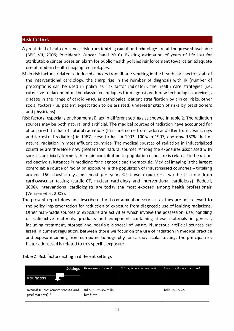
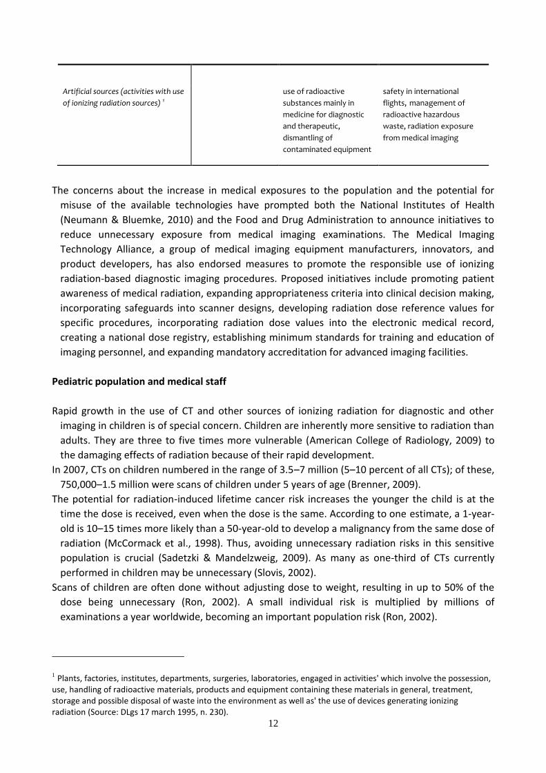
Table 2. Risk factors acting in different settings
Settings
Risk factors
Home environment Workplace environment Community environment
Natural sources (environmental and
food matrices) a)
fallout, DMOS, milk,
beef, etc. fallout, DMOS
12
The concerns about the increase in medical exposures to the population and the potential for
misuse of the available technologies have prompted both the National Institutes of Health
(Neumann & Bluemke, 2010) and the Food and Drug Administration to announce initiatives to
reduce unnecessary exposure from medical imaging examinations. The Medical Imaging
Technology Alliance, a group of medical imaging equipment manufacturers, innovators, and
product developers, has also endorsed measures to promote the responsible use of ionizing
radiation-based diagnostic imaging procedures. Proposed initiatives include promoting patient
awareness of medical radiation, expanding appropriateness criteria into clinical decision making,
incorporating safeguards into scanner designs, developing radiation dose reference values for
specific procedures, incorporating radiation dose values into the electronic medical record,
creating a national dose registry, establishing minimum standards for training and education of
imaging personnel, and expanding mandatory accreditation for advanced imaging facilities.
Pediatric population and medical staff
Rapid growth in the use of CT and other sources of ionizing radiation for diagnostic and other
imaging in children is of special concern. Children are inherently more sensitive to radiation than
adults. They are three to five times more vulnerable (American College of Radiology, 2009) to
the damaging effects of radiation because of their rapid development.
In 2007, CTs on children numbered in the range of 3.5–7 million (5–10 percent of all CTs); of these,
750,000–1.5 million were scans of children under 5 years of age (Brenner, 2009).
The potential for radiation-induced lifetime cancer risk increases the younger the child is at the
time the dose is received, even when the dose is the same. According to one estimate, a 1-year-
old is 10–15 times more likely than a 50-year-old to develop a malignancy from the same dose of
radiation (McCormack et al., 1998). Thus, avoiding unnecessary radiation risks in this sensitive
population is crucial (Sadetzki & Mandelzweig, 2009). As many as one-third of CTs currently
performed in children may be unnecessary (Slovis, 2002).
Scans of children are often done without adjusting dose to weight, resulting in up to 50% of the
dose being unnecessary (Ron, 2002). A small individual risk is multiplied by millions of
examinations a year worldwide, becoming an important population risk (Ron, 2002).
1 Plants, factories, institutes, departments, surgeries, laboratories, engaged in activities' which involve the possession,
use, handling of radioactive materials, products and equipment containing these materials in general, treatment, storage and possible disposal of waste into the environment as well as' the use of devices generating ionizing radiation (Source: DLgs 17 march 1995, n. 230).
Artificial sources (activities with use
of ionizing radiation sources) 1
use of radioactive
substances mainly in
medicine for diagnostic
and therapeutic,
dismantling of
contaminated equipment
safety in international
flights, management of
radioactive hazardous
waste, radiation exposure
from medical imaging
13
The Society for Pediatric Radiology and the National Cancer Institute (NCI) collaborated to develop
and circulate a pamphlet (National Cancer Institute, 2008) for health care providers on pediatric
CT and radiation risks.
Protecting radiation technologists and other medical staff from excessive radiation exposure has
been a concern for many years, with dose limits and lifelong monitoring procedures established
in most countries (Rehani, 2009). Only one-half percent of medical workers reach or exceed this
dose limit (Rehani, 2009). The dose limit recommended by the International Commission on
Radiological Protection (ICRP) and adopted by all but a few countries is 20 mSv annually, or 100
mSv over 5 years (Valentin editor, 2007).
Health outcomes
Ionizing radiation is known to cause harm. High radiation doses tend to kill cells, while low doses
(i.e., 100 mSv) tend to damage or alter the genetic code (DNA) of irradiated cells. The
biological effects of ionizing radiation are divided into two categories: deterministic and
stochastic effects. Deterministic effects, such as erythema or cataract, have a threshold dose
below which the biological response is not observed (BEIR V, 1990; UNSCEAR Report, 2001; Hall,
2000). Some cardiological interventional procedures with long screening times and multiple
image acquisition (e.g. percutaneous coronary intervention, radio-frequency ablation, etc) may
give rise to deterministic effects in both staff and patients (ICRP, Publication 59, 1991; Vano et
al., 2005; Vano et al., 2008). A stochastic effect is a probabilistic event and there is no known
threshold dose. The likelihood of inducing the effect, but not the severity, increases in relation to
dose and may differ among individuals. In fact, the effect of low doses of radiation - less than 50
mSv - do not cause an immediate problem to any body organ, but spread out over long periods
of time after exposure. Damage are at DNA level and is considered to be the main initiating
event by which radiation damage to cells results in development of cancer and hereditary
disease in the future children of exposed parents (Hall, 2000; BEIR VII, 2006). In fact, ionizing
radiation exposure produces long-term health effect through, both directly or indirectly (free
radical interaction), damage to cellular DNA, producing oxidized bases, bulky DNA adducts, and
DNA strands breaks.
The cell has repair mechanisms against damage induced by radiation as well as by chemical
carcinogens. Consequently, biological effects of low dose radiation on living cells may result in
three outcomes: (1) injured or damaged cells repair themselves, resulting in no residual damage;
(2) cells die; or (3) cells incorrectly repair themselves resulting in a biological change.
However, the effects of low-level exposure remain uncertain (Brenner et al., 2003). The
associations between radiation exposure and the development of cancer are mostly based on
populations exposed to relatively high levels of ionizing radiation (e.g., Japanese atomic bomb
survivors).
A linear, no-threshold dose response relationship is used by the IRCP in order to describe the
relationship between radiation dose and the occurrence of cancer (NCRP, Rep. 136, 2001). This
dose-response model suggests that any increase in dose, no matter how small, results in an
incremental increase in risk. Genetic effects are the result of a mutation produced in the
reproductive cells of an exposed individual that are passed on to their offspring. These effects
may show up as birth defects or other conditions in the future children of the exposed individual
14
and succeeding generation. Indeed, studies with laboratory animals have provided a large body
of data on radiation-induced genetic effects (Dubrova, 2003; Foffa et al., 2009)
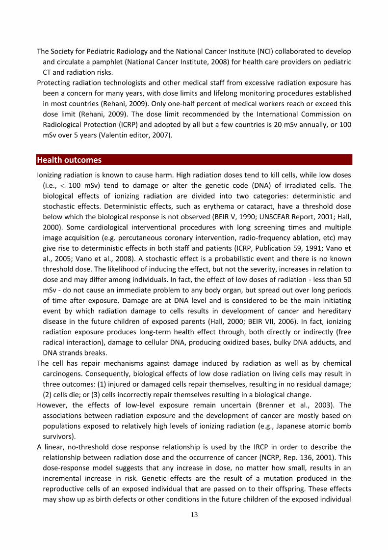
Different health outcomes from radiologic exposure in cardiovascular imaging testing are shown
below.
Table 3. Biological effects of ionizing radiation. (Redrawn and modified from Foffa et al., 2009).
Deterministic effects Stochastic effects
Dose Medium-High Low
Occurrence time Short Long
Threshold dose Yes No
Cell biology Cell Death DNA damage
Clinical effects Skin lesions, erythema, ulcers, epilation,
cataracts, permanent sterility
Cancer, genetic effects
Attributable total cancer risk (fatal cancer + non-fatal cancer) is herein chosen as health outcome.
It is statistically calculated combining evidence of dose estimates and cancer risk estimates.
Pilot application
Two operational phases have been performed with the aim of testing the full chain model (figure
1) referred to the radioprotection policy in Tuscany. An extensive review of present evidence
and an experts consultation, have allowed the identification of factors responsible for modifying
exposures to ionizing radiation in the wide context, and specifically in the healthcare
environment. Inner bibliographic resources, stored at the Institute of Clinical Physiology (located
at the National Research Council, Pisa, Italy), as well as real data gained from the Institute clinical
activity, have been arranged to perform the risks estimates, focusing on the attributable long-
term cancer risk. The two phases are briefly described.
Phase 1. Literature review and experts consultation
The work undertaken in Rapid Italian case study, in particular the causal chain building, was
strongly informed by the completion of state-of-the-art reviews of key areas of research,
including determinants of health, risk factors, health effects. Table 4 summarizes the results of
these reviews based on papers published on scientific journals with high impact factor and
scientific association guidelines and recommendations, focused to medical imaging and/or
computed tomography use. Selected references are primary study or most updated reviews,
published from 2003 to 2010. They totally sum to n. 69.
Reviews have been completed early in the project allowing to refine a checklist for the experts
consultation (table 5). It includes factors that have been pointed out to determine the final
health effects.
Table 4. Summary of results of state-of-the-art reviews used to inform the Rapid case study
method.

15
Chain level Issue Bibliographical Entries
Policy Policy on Radioprotection 2008-2010 Regione Toscana, 2008
National Guidelines ICRP, 2007, 2008; ECRP, 2008;
Health determinants Present medical practice Hall & Brenner, 2008; Brenner & Hall, 2007; Huda & Vance, 2007; Picano et al., 2007; Bedetti et al., 2008; Winslow et al.,2008; ICRP, 2007, 2008; Sodickson, 2009; Griffey & Sodickson, 2009; Fazel et al., 2009; Foffa et al., 2009; Tsapaki et al., 2010; Halliburton & Schoenhagen, 2010;
Technological updating Einstein et al., 2007; Gaztanaga & Garcia, 2009;
Training strategies Jacob et al., 2004; Lee et al., 2004; Correia et al., 2005; Arslanoğlu et al., 2007; Brenner & Hall, 2007; Bruner et al., 2009; Tsapaki et al., 2010
Patient/operator awareness Correia et al., 2005; Brenner & Hall, 2007; Karsli et al., 2009; Bruner et al., 2009;
Workplace environment Vañó, 2003; Delichas et al., 2003; Andreassi, 2004; Venneri et al., 2009; Ciraj-Bjelac et al., 2010;
Community environment Gerber & Gibbons, 2010
Commercial/Economic reasons Picano, 2004; Hall & Brenner, 2008; Street et al., 2009; Hausleiter et al., 2009; Halliburton & Schoenhagen, 2010;
Risk factors
Pediatric population Age Brenner et al., 2001; Brenner & Hall, 2007; Harrison & Day, 2008; Thomas & Wang, 2008; Berrington de Gonzalez et al., 2009; BEIR VII; Gerber & Gibbons, 2010;
Gender Berrington de Gonzalez et al., 2009; Harrison & Day, 2008; BEIR VII;
Diagnosis Huda & Vance, 2007;
Adult population Age Brenner et al., 2001; Brenner & Hall, 2007; Berrington de Gonzalez et al., 2009; Harrison & Day, 2008; BEIR VII;
Gender Berrington de Gonzalez et al., 2009; Harrison & Day, 2008; BEIR VII; Gerber & Gibbons, 2010;
Diagnosis
Health outcomes Fatal cancer Picano, 2004; Picano et al., 2007; Einstein et al., 2007; Brenner & Hall, 2007; BEIR VII; Mettler et al., 2008; Little et al., 2009; Sodickson, 2009; Berrington de Gonzalez et al., 2009; Little et al., 2010; Tsapaki et al. 2010, Halliburton & Schoenhagen, 2010;
Non-fatal cancer Yoshinaga et al., 2004;
Skin and eye lesions (medical staff) Renaud, 1992; McKetty, 1996; Vañó et al., 1998; Finkelstein, 1998; Yoshinaga et al., 2004; Andreassi, 2004; Vano et al., 2005; Vano et al., 2008; Foffa et al., 2009; Ciraj-Bjelac et al., 2010
Teratogenic effects Andreassi et al., 2005,2006a, 2006b; Foffa et al., 2009; Ait-Ali et al., 2010
The checklist is meant to provide a relatively simple approach that can highlight important
information about the potential effect and relevance of listed factors, on final health impacts. In
this desktop exercise, an expert decide whether a factor has, or has not an effect (or it should be
considered uncertain) and assesses the importance of each factor by attributing a score (0 "low",

16
1 "intermediate" and 2 "a lot"). The total score, by factor, helps guide a decision about pursuing
a focused policy action on highly relevant factors.
Table 5. Checklist used for the experts consultation.
Chain level no uncertain yes
Policy Commercial/Financial
Other health care policy
Health determinants Present medical practice
Technological updating
Training strategies
Patient/operator awareness
Workplace environment
Community environment
Other
Risk factors
Pediatric population Age
Gender
Diagnosis
Other
Adult population Age
Gender
Diagnosis
Other
Inner resources from IFC-CNR and collaborative consultants from University and Health care
departments have been selected for the consultation. The multidisciplinary team was constituted
as follow:
Cardiologist (Senior Researcher);
Radiologist (Technical Consultant);
Hemodynamist (Research Director);
Pulmonologist (Researcher);
Nuclear physicist (Principal Investigator);
Geneticist (Researcher);
GP (Generic Physician);
Manager and Scientific Coordinator, Physical co-worker (Sanitary department).
Phase 2. Cancer risk estimates and diagnostic imaging exposure
The risk of cancer associated with diagnostic imaging has been quantified using a computational
software (RADIORISK 1.3, Paterni et al., 2010) based on three main sub-components of
exposure: natural, diagnostic, professional. The result is the amount of cumulative risk either for
the patient or a subpopulation. The simulated risk is secondarily associated with current
indications of appropriateness to inform the physician about the proper clinical decision to be
taken. The software basic function is to estimate the cancer risk based on the personal history of
exposure to ionizing radiation. Current guidelines, dose references and accepted evidence in
BEIRVII are used to calculate risk (BOX 1).

17
Advanced software interfaces display either information on exposure levels by source (mSv unit),
and annual and cumulative exposure by observational period, referred to RX equivalent.
Main results about risks are displayed as well and expressed as incidence cases and mortality
attributed to the different sources of exposure. Medical imaging use extra risk on 100 person (or
on a defined population) is than available, with a calculated range of uncertainty.
Uncertainties in estimated risks
The effective dose, expressed in units of millisieverts (mSv), is the dose quantity most commonly
used to relate exposures from low doses of ionizing radiation to the probability of detrimental
health effects. The effective dose represents the amount of whole body irradiation that yields a
biological risk equivalent to the irradiation of only a portion of the body (as with cardiovascular
CT). Although the effective dose quantity is thought to be the best quantity available for linking
radiation dose and health risk, it must be recognized that the effective dose is associated with a
level of uncertainty on the order of 40% when it is used to quantify dose for medical exposures
(Martin, 2007).
Further, the effective dose is not intended to express absolute patient-specific risk (i.e., risk to
specific persons of known age and sex) but rather risk to the general population. These
limitations of the effective dose underlie the recommendation to use a different metric, the
dose-length product, reported by the CT scanner in units of mGy X cm, to characterize the
amount of radiation from a single CT examination in the patient report (Abbara et al., 2009; Raff
et al., 2009; Hendel et al., 2009) and in research studies.
The calculation of numerical risk from the effective dose estimates is further limited. Cancer risk
from the relatively low doses of ionizing radiation used during medical imaging is linearly
extrapolated from the radiation risk data of atomic bomb survivors in Japan after World War II.
The validity of this approach relies largely on the linear nonthreshold theory, which assumes a
linear relationship between dose and cancer risk even at the smallest doses. However, the linear
BOX 1 Guidelines: Gerber TC et al: Ionizing Radiation in Cardiac Imaging. A Science Advisory from the American Heart Association
Committee on Cardiac Imaging of the Council on Clinical Cardiology and Committee on Cardiovascular Imaging and Intervention of the Council on Cardiovascular Radiology and Intervention. Circulation 2009;119:1056-1065
The Royal College of Radiologists (RCR): Making the best use of clinical radiology services (MBUR), 2007, 6th edition
Budoff MJ, Achenbach S, et al: Assessment of Coronary Artery Disease by Cardiac Computed Tomography. A Scientific Statement from the American Heart Association Committee on Cardiovascular Imaging and Intervention, Council on Cardiovascular Radiology and Intervention, and Committee on Cardiac Imaging, Council on Clinical Cardiology. Circulation 2006;114:1761-1791
Dose Reference: Reference European guideline (2001) Guidelines of Italian Minister of Health Peer reviewed journal Government Agency From each exam data file (if available)
Cancer Risk Estimation - BEIR VII,2006: The estimation is based on 100000 studies, including 87000 Hiroshima and 407000 nuclear workers 2 to 3 confidence intervals of attributable risks estimate X-rays and gamma-rays are a proven carcinogen (WHO’s International Agency of Research of Cancer) Epidemiological evidence up to now above 50 mSv Re-affirm Linear No-Threshold hypothesis
18
no-threshold theory is controversial and the subject of debate (Strzelczyk et al., 2007; Tubiana et
al., 2009; Little et al., 2009). Therefore, estimations of risk from low doses of radiation delivered
during medical imaging examinations must be interpreted with regard to the imprecision of the
calculation.
Further, any potential risk of future stochastic events must be balanced with the risk of forgoing a
medically necessary examination (Gerber et al., 2009).
Usual conventional analysis of risks in health technology assessment consider only acute direct
costs. At present long term risks and downstream costs due to cancer are not included in risk-
benefit analysis. As to radioprotection goal both individual risk calculation (per exam per patient)
and the radiological cumulative risk (lifetime) are necessary to be performed.
However a debate is ongoing regarding the true incremental risk to subjects exposed to doses
currently administered in cardiovascular procedures fails to take into account the uncertainty of
the dose-response relationship in this lower range, as well as tissue-specific reparative
responses, also manifest at lower levels of exposure (Warren et al., 2010). The leap from
radiation exposure to the risk of stochastic effects such as cancer is controversial, particularly for
individual patients, because of known uncertainties in dose estimates and risk models
(Halliburton & Schoenhagen, 2010).
Results Estimated risks for individuals or different sub-populations depend on the exposure model
upstream in the chain of causation. Different approaches in medical practice are already known
to modify individual and population exposure, as to reduce useless radiation and cumulative life
burden of radiation (Figure 3). In particular the case study stresses the relevance of multi-
factorial proximal determinants of health that, acting upon distal risk factors, can lead changes in
the exposure model as well (Table 6). Therefore the wide range of health determinants and risk
factors contributing to the overall exposure have been linked in a causal pathway from policy to
the outcome (Figure 4). Such overall representations of factors which interact each other, has
been duly supported from a few research sub-actions, performed in the IFC tertiary care referral
centre, which have shown that:
radiological awareness is very low even among practitioners and prescribers of exams with
very high radiation exposure (Correia et al., 2005) with at least 40% of stress imaging testing
being inappropriate (Picano et al., 2007).
long-term risks should be included not only in the assessment of diagnostic appropriateness
but also in cost-benefit analysis, with potential to completely overturn on current
approaches to cost-benefit assessment (Bedetti et al., 2008).
an alternative proposal of radiological informed consent form addresses the problem of the
current limitations (Bedetti & Loré, 2007), being consistent with the requirements of
transparency, clarity and legal sustainability proposed by the International Atomic Energy
Agency in 2008.
dose has to be translated into risk and dose reduction has to be translated into number of
spared risks, calculating the cumulative risk for patients (Bedetti et al., 2008) and for
invasive cardiologists, as well (Venneri et al., 2009).
Table 6. Ranking of health determinants and risk factors from experts consultation.

19
Risk factors Health determinants
Very relevant Age - paediatric population Training strategies
Relevant Technological updating
Moderate Gender - paediatric population Present medical practice
Age - adult population Patient/operator awareness
Slight effect Gender - adult population Workplace environment
Diagnosis - adult population
Diagnosis - adult population
Not relevant Familiarity Commercial/Financial policy
Other health care policy
Community environment
Conclusion
In view of an informed policy action a more inclusive web of causation has been delineated,
merging together available information on risk factors with less usual information on indirect
factors, inducing modification in medical practice. The adoption of an integrated approach, in
which applied doses in medical imaging are considered together with other sources of exposure
and modifiable individual behaviors, can be translated in a remarkable health and socio-
economic benefit. The present case study takes a step in this direction allowing decision makers
to identify an optimal strategy.
Recommendations from the 2007 White paper of the American College of Radiology, suggest
“education of all stakeholders in the principles of radiation safety, the appropriate utilization of
imaging to minimize any associated radiation risk, the standardization of radiation dose data for
its ultimate use in benchmark good practice”. This strategy of improving appropriateness
through knowledge-based intervention may be a very cost-effective policy for primary
prevention of cancer in industrialized countries, since 10% of all cancers may be attributable to
the medical radiation. Better knowledge of risks will help to avoid small individual risks
translating into substantial population risks.
Radiologically speaking the doctors (on average) do not know what they do: on one hand this
leads to a colossal amount of waste and risk in our health system, and on the other hand it offers
a unique opportunity to spare an enormous amount of resources (the useless examination and
its linked direct and downstream costs). Reducing inappropriate testing will improve the quality
of health care, shorten waiting lists inflated by useless examinations, and – most importantly -
reduce long-term oncogenesis due to ionizing radiation. This situation is unavoidable in a system
that pays according to volume and not appropriateness. The change in prescription pattern and
reimbursement policy by necessity should by necessarily follow a systematic recognition of
current practice in the “best” (high-tech, technology-oriented, research-driven) institutions.
The 2002 statement of the International Atomic Energy Agency (International Atomic Energy
Agency, 2002) recognized that “Health professionals involved in the process of diagnosis and
treatment are the critical link. Training them properly and ensuring intensive information

20
exchange among them are, therefore, probably the most cost-effective ways of achieving
patients’ safety”.
The SUIT-Heart project was promoted by IFC as an opportunity to reshape current clinical
cardiological practice, with a paradigm shift based on expanding physician knowledge. Inside the
SUIT-HEART research context the application of the delineated RAPID model is a wisdom way for
enlarging the base of knowledge as to achieve the goal of reducing estimated cancer risk due to
radiological examinations, stemming from the general policy on radioprotection.
A shift in the health concept, from a thigh view (limited to the consideration of outcome
depending from the exposure to physical hazards) to a broader one (including the indirect
actions of socioeconomic factors on the final presentation of the outcomes) is expected to build
a more inclusive knowledge background and disseminate it to the stakeholders, physicians and
practitioner involved.
The development of a set of indicator to highlight results from this basic “cultural intervention”
would address the question of quantifying the effects induced on final outcomes in
consideration of the modification of proximal determinants of health.
As to this specific investigation, an evaluation of post interventional actions would in future allow
baseline comparison of diagnostic practice and highlights a different overall balance among risk
and benefit; acting on proximal factors leading to maximizing health benefits and reduce the
expected negative effects. Hence, the best practice on the use of computed tomography in
cardiovascular disease could be comprehensively integrated with the considerations of
socioeconomic determinants of health and institutional factors.
Main outcomes of such further investigation will keep the policy decisions informed reporting the
overall results, research strategy, motivation for choices, subject involved in the consultations,
and a minimum set of recommendation.
It is, therefore, proposed that future quantification of the overall effect upon final outcomes has
to be approached by the model herein described, and characterized by the key features below
described:
Literature consultation
Experts rating
Risk estimates comparison
Overall consideration for best practice
Reinforcement of communication on risks.

21
References
Abbara S, Arbab-Zadeh A, Callister TQ et al. SCCT guidelines for performance of coronary
computed tomographic angiography: a report of the Society of Cardiovascular Computed
Tomography guidelines committee. J Cardiovasc Comput Tomogr. 2009;3:190–204.
Ait-Ali L, Andreassi MG, Foffa I, Spadoni I, Vano E, Picano E. Cumulative patient effective dose and
acute radiation-induced chromosomal DNA damage in children with congenital heart disease.
Heart. 2010 Feb;96(4):269-74.
American College of Radiology. Alliance for Radiation Safety in Pediatric Imaging and imaging
manufacturers agree to collaborate to standardize methods to measure, report pediatric dose
from CT scans . Available at
http://www.acr.org/MainMenuCategories/media_room/FeaturedCategories/PressReleases/Archi
ve/AllianceVendorstoStandardizeDoseReporting.aspx.
Andreassi MG. The biological effects of diagnostic cardiac imaging on chronically exposed
physicians: the importance of being non-ionizing. Cardiovascular Ultrasound. 2004 Nov 22;2:25.
Andreassi MG, Cioppa A, Botto N et al. Somatic DNA damage in interventional cardiologists: a
casecontrol study. FASEB J. 2005;19:998-9.
Andreassi MG, Stigliano I, Cioppa A et al. Chronic low dose radiation exposure indices
chromosomal abnormalities in originally genetically identical twins. Int J Cardiol. 2006a;118:130-1.
Andreassi MG, Ait-Ali L, Botto N et al. Cardiac catheterization and long-term chromosomal damage
in children with congenital heart disease. Eur Heart J. 2006b;27:2703-8.
Arslanoğlu A, Bilgin S, Kubal Z, Ceyhan MN, Ilhan MN, Maral I. Doctors' and intern doctors'
knowledge about patients' ionizing radiation exposure doses during common radiological
examinations. Diagnostic and Interventional Radiology. 2007 Jun;13(2):53-5.
Bedetti G, Botto N, Andreassi MG, Traino C, Vano E, Picano E. Cumulative patient effective dose in
cardiology. British Journal of Radiology. 2008 Sep;81(969):699-705.
Bedetti G, Loré C. Radiological informed consent in cardiovascular imaging: towards the medico
legal perfect storm? Cardiovasc Ultrasound. 2007;5:35.
Berrington de González A, Mahesh M, Kim KP, Bhargavan M, Lewis R, Mettler F, Land C. Projected
cancer risks from computed tomographic scans performed in the United States in 2007. Archives
of Internal Medicine. 2009 Dec 14;169(22):2071-7.

22
Brenner D, Elliston C, Hall E, Berdon W. Estimated risks of radiation-induced fatal cancer from
pediatric CT. AJR American Journal of Roentgenology. 2001 Feb;176(2):289-96.
Brenner DJ, Doll R, Goodhead DT, Hall EJ, Land CE, Little JB, Lubin JH, Preston DL, Preston RJ,
Puskin JS, Ron E, Sachs RK, Samet JM, Setlow RB, Zaider M. Cancer risks attributable to low doses
of ionizing radiation: assessing what we really know. Proceedings of the National Academy of
Sciences of the United States of America. 2003 Nov 25;100(24):13761-6.
Brenner DJ, Hall EJ. Computed tomography-an increasing source of radiation exposure. New
England Journal of Medicine. 2007 Nov 29;357(22):2277-84.
Brenner DJ. Should we be concerned about the rapid increase in CT usage? Presented at the
President’s Cancer Panel meeting. 2009 Jan 27; Phoenix, AZ.
Bruner A, Sutker W, Maxwell G. Minimizing patient exposure to ionizing radiation from computed
tomography scans. Proceedings (Baylor University. Medical Centre). 2009 Apr;22(2):119-23.
Ciraj-Bjelac O, Rehani MM, Sim KH, Liew HB, Vano E, Kleiman NJ. Risk for radiation induced
cataract for staff in interventional cardiology: Is there reason for concern? Catheterization and
Cardiovascular Interventions. 2010 Jun 14.
Correia MJ, Hellies A, Andreassi MG, Ghelarducci B, Picano E. Lack of radiological awareness
among physicians working in a tertiary-care cardiological centre. International Journal of
Cardiology. 2005 Sep 1;103(3):307-11.
Decreto Legislativo del Governo 17 marzo 1995 n° 230. "Attuazione delle direttive
89/618/Euratom, 90/641/Euratom, 92/3/Euratom e 96/29/Euratom in materia di radiazioni
ionizzanti.".
Decreto Legislativo 26 maggio 2000, n. 187. "Attuazione della direttiva 97/43/Euratom in materia
di protezione sanitaria delle persone contro i pericoli delle radiazioni ionizzanti connesse ad
esposizioni mediche" pubblicato nella Gazzetta Ufficiale n. 157 del 7 luglio 2000 - Supplemento
Ordinario n. 105.
Delichas M, Psarrakos K, Molyvda-Athanassopoulou E, Giannoglou G, Sioundas A, Hatziioannou K,
Papanastassiou E. Radiation exposure to cardiologists performing interventional cardiology
procedures. European Journal of Radiology. 2003 Dec;48(3):268-73.
DeMarco JJ, Cagnon CH, Cody DD et al. Estimating radiation doses from multidetector CT using
Monte Carlo simulations: effects of different size voxelized patient models on magnitudes of organ
and effect dose. Phys Med Biol. 2007;52:2583–97.
23
Dubrova YE. Long-term genetic effects of radiation exposure. Mutation Research. 2003 Nov;544(2-
3):433-9.
European Commission Radiation Protection 118. Referral guidelines for imaging. Luxembourg:
Office for Official Publications of the European Communities 2001.
Einstein AJ, Henzlova MJ, Rajagopalan S. Estimating risk of cancer associated with radiation
exposure from 64-slice computed tomography coronary angiography. Journal of American Medical
Association. 2007 Jul 18;298(3):317-23.
Fazel R, Krumholz HM, Wang Y, Ross JS, Chen J, Ting HH, Shah ND, Nasir K, Einstein AJ, Nallamothu
BK. Exposure to low-dose ionizing radiation from medical imaging procedures. National English
Journal of Medicine. 2009 Aug;361(9):849-57.
Finkelstein MM. Is brain cancer an occupational disease of cardiologists? Canadian Journal of
Cardiology. 1998 Nov;14(11):1385-8.
Foffa I, Cresci M, Andreassi MG. Health risk and biological effects of cardiac ionizing imaging: from
epidemiology to genes. International Journal of Environmental Research and Public Health. 2009
Jun;6(6):1882-93.
Gaztanaga J, Garcia MJ. New noninvasive imaging technologies in coronary artery disease. Current
Cardiology Reports. 2009 Jul;11(4):252-7.
Gerber TC, Carr JJ, Arai AE, et al. Ionizing radiation in cardiac imaging. A science advisory from the
American Heart Association Committee on Cardiac Imaging of the Council on Clinical Cardiology
and Committee on Cardiovascular Imaging and Intervention of the Council on Cardiovascular
Radiology and Intervention. Circulation. 2009;119:1056–65.
Gerber TC, Gibbons RJ. Weighing the risks and benefits of cardiac imaging with ionizing radiation.
JACC Cardiovascular Imaging. 2010 May;3(5):528-35.
Gibbons RJ, Miller TD, Hodge D, Urban L, Araoz PA, Pellikka P, McCully RB. Application of
appropriateness criteria to stress single-photon emission computed tomography sestamibi studies
and stress echocardiograms in an academic medical center. Journal of the American College of
Cardiology: Cardiovascular Imaging. 2008 Apr 1;51(13):1283-9.
Gibbons RJ. Leading the elephant out of the corner: the future of health care: presidential address
at the American Heart Association 2006 scientific sessions. Circulation. 2007 Apr 24;115(16):2221-
30.
Griffey RT, Sodickson A. Cumulative radiation exposure and cancer risk estimates in emergency
department patients undergoing repeat or multiple CT. AJR American Journal of Roentgenology.
2009 Apr;192(4):887-92.

24
Hall EJ. Radiobiology for the Radiologist. 5th edition. Lippincott Williams & Wilkins, Philadelphia,
2000. Available at:
http://books.google.it/books?hl=it&lr=&id=6HhjwRyqBzgC&oi=fnd&pg=PP11&dq=Radiobiology+f
or+the+radiologist&ots=0slFCBjnF3&sig=bDwd0JPT7E-9uBW9wn4l6e56VE#v=onepage&q&f=false.
Hall EJ, Brenner DJ. Cancer risks from diagnostic radiology. The British Journal of Radiology. 2008
May;81(965):362-78.
Halliburton S. Options for reducing patient radiation dose with cardiovascular computed
tomography. Int J Cardiovasc Imaging. 2009;25:153–64.
Halliburton SS, Schoenhagen P. Cardiovascular imaging with computed tomography: responsible
steps to balancing diagnostic yield and radiation exposure. Journal of the American College of
Cardiology: Cardiovascular Imaging. 2010 May;3(5):536-40.
Harrison J, Day P. Radiation doses and risks from internal emitters. Journal of Radiological
Protection. 2008 Jun;28(2):137-59.
Hausleiter Jörg, Meyer T, Hermann F, Hadamitzky M, Krebs M, Gerber TC, McCollough C, Martinoff
S, Kastrati A, Schömig A, Achenbach S. Estimated radiation dose associated with cardiac CT
angiography. Journal of the American College of Cardiology: Cardiovascular Imaging. 2009 Feb
4;301(5):500-7.
Health Risks from Exposure to Low Levels of Ionizing Radiation. BEIR V. Health effects of exposure
to low levels of ionizing radiation. 1990. Available at:
http://www.nap.edu/openbook.php?isbn=0309039959.
Health Risks from Exposure to Low Levels of Ionizing Radiation. BEIR VII—phase 2. 2006,
Committee to assess health risks from exposure to low levels of ionizing radiation. National
ResearchCouncil.
Hendel RC, Patel MR, Kramer CM, et al. ACCF/ACR/SCCT/SCMR/ ASNC/NASCI/SCAI/SIR 2006
Appropriateness criteria for cardiac computed tomography and cardiac magnetic resonance
imaging: a report of the American College of Cardiology Foundation Quality Strategic Directions
Committee Appropriateness Criteria Working Group, American College of Radiology, Society of
Cardiovascular Computed Tomography, Society for Cardiovascular Magnetic Resonance, American
Society of Nuclear Cardiology, North American Society for Cardiac Imaging, Society for
Cardiovascular Angiography and Interventions, and the Society of Interventional Radiology. J Am
Coll Cardiol. 2006 48:1475–97.
Hendel RC, Berman DS, Di Carli MF, et al. ACCF/ASNC/ACR/AHA/ASE/SCCT/SCMR/SNM 2009
appropriate use criteria for cardiac radionuclide imaging: A report of the American college of
Cardiology Foundation Appropriate Use Criteria Task Force, the American Society of Nuclear
Cardiology, the American College of Radiology, the American Heart Association, the American

25
Society of Echocardiography, the Society of Cardiovascular Computed Tomography, the Society for
Cardiovascular Magnetic Resonance, and the Society of Nuclear Medicine. J Am Coll Cardiol.
2009;53:2201–9.
Huda W, Vance A. Patient radiation doses from adult and pediatric CT. American Journal
Roentgenology. 2007 Feb;188(2):540-6.
International Atomic Energy Agency, International Action Plan on the Radiological Protection of
patients, IAEA, Wien 2002.
International Commission on Radiological Protection (ICRP). The biological basis for dose limitation
in the skin. ICRP Publication 59. Annals ICRP. 1991:22.
International Commission on Radiological Protection (ICRP). The 2007 Recommendations of the
International Commission on Radiological Protection. ICRP Publication 103. Annals ICRP.
2007;37:1-332.
International Commission on Radiological Protection (ICRP). Radiological protection in medicine.
Publication 105. Annals ICRP. Volume 37 Issue 6, oct 2008.
Jacob K, Vivian G, Steel JR. X-ray dose training: are we exposed to enough? Clinical Radiology. 2004
Oct;59(10):928-34; discussion 926-7.
Karsli T, Kalra MK, Self JL, Rosenfeld JA, Butler S, Simoneaux S. What physicians think about the
need for informed consent for communicating the risk of cancer from low-dose radiation. Pediatric
Radiology. 2009 Sep;39(9):917-25.
Kim KP, Einstein AJ, Berrington de González A. Coronary artery calcification screening: estimated
radiation dose and cancer risk. Arch Intern Med. 2009;169:1188 –94.
Lee CI, Haims AH, Monico EP, Brink JA, Forman HP. Diagnostic CT scans: assessment of patient,
physician, and radiologist awareness of radiation dose and possible risks. Radiology. 2004
May;231(2):393-8.
Little MP, Wakeford R, Tawn EJ, Bouffler SD, Berrington de Gonzalez A. Risks associated with low
doses and low dose rates of ionizing radiation: why linearity may be (almost) the best we can do.
Radiology. 2009 Apr;251(1):6-12.
Little MP, Tawn EJ, Tzoulaki I, Wakeford R, Hildebrant G, Paris F, Tapio S, Elliott P. Review and
meta-analysis of epidemiological associations between low/moderate doses of ionizing radiation
and circulatory disease risks, and their possible mechanism. Radiation and Environmental
Biophysics. 2010 May;49(2):139-53.
Martin CJ. Effective dose: how should it be applied to medical exposures? Br J Radiol.
2007;80:639–47.

26
McKetty MH. Study of radiation doses to personnel in a cardiac catheterization laboratory. Health
Physics. 1996 Apr;70(4):563-7.
Mettler FA, Thomadsen BR, Bhargavan M, Gilley DB, Gray JE, Lipoti JA, McCrohan J, Yoshizumi TT,
Mahesh M. Medical radiation exposure in the U.S. in 2006: preliminary results. Health Physics.
2008 Nov;95(5):502-7.
Mettler FA, Bhargavan M, Faulkner K, et al. Radiologic and nuclear medicine studies in the United
States and worldwide: frequency, radiation dose, and comparison with other radiation sources—
1950 –2007. Radiology. 2009;253:520 –31.(NEL TESTO CITI METTLER 2007, E QUESTO è 2009; è
UN ERRORE DEL TESTO? QUALE VOCE BIBLIOGRAFICA VA INSERITA?)
National Cancer Institute. Radiation risks and pediatric computed tomography (CT): a guide for
health care providers [Internet]. Bethesda (MD): National Institutes of Health; 2008. Available
from: http://www.nci.nih.gov/cancertopics/causes/radiation-risks-pediatric-CT.
National Council on Radiation Protection and Measurements: Evaluation of the Linear-
Nonthreshold Dose-Response Model for Ionizing Radiation. NCRP, Bethesda 2001. Report No.136.
National Council on Radiation Protection and Measurements. Ionizing radiation exposure of the
population of the United States. National Council on Radiation Protection report no. 160.
Bethesda, Md: National Council on Radiation Protection and Measurements, 2009.
Neumann RD, Bluemke DA. Tracking radiation exposure from diagnostic imaging devices at the
NIH. J Am Coll Radiol. 2010;7:87–9.
Nissen SE. Limitations of computed tomography coronary angiography. J Am Coll Cardiol. 2008;
52:2145–7.
Paterni M, Carpeggiani C, Ripoli A, Caramella D, Lazzeri M, Traino C, Picano E. RadioRisk Software
to calculate and communicate radiological risk to patients and doctors. In: SUIT - Stop Useless
Imaging Testing. Project of the Institute of Clinical Physiology and of the Tuscany Region to search
new strategies to reduce inappropriate use of Imaging Testing, 2010.
Picano E. Sustainability of medical imaging. BMJ. 2004; 328:578-80.
Picano E, Pasanisi E, Brown J, Marwick TH. A gatekeeper for the gatekeeper: inappropriate
referrals to stress echocardiography. Am Heart J. 2007;154:285-90.
President’s Cancer Panel. Reducing Environmental Cancer Risk, What We Can Do Now. 2008–2009
Annual Report, U.S. DEPARTMENT OF HEALTH AND HUMAN SERVICES.
27
Raff GL, Abidov A, Achenbach S, et al. SCCT guidelines for the interpretation and reporting of
coronary computed tomographic angiography. J Cardiovasc Comput Tomogr. 2009; 3:122–36.
Regione Emilia Romagna. La Valutazione di Impatto sulla salute. Un nuovo strumento a supporto
delle decisioni. Bologna, Ottobre 2010.
Regione Toscana. Piano Sanitario Regionale 2008-2010. 30.7.2008 Bollettino Ufficiale della
Regione Toscana N. 25.
Rehani MM. Smart protection: a “smart” card that contains patients’ information including
radiation dose data would help protect them from radiation effects. IAEA Bulletin. 2009 May;
50‑2:1-3.
Renaud L. A 5-y follow-up of the radiation exposure to in-room personnel during cardiac
catheterization. Health Physics. 1992 Jan;62(1):10-5.
Ron E. Ionising radiation and cancer risk: evidence from epidemiology. Pediatr Radiol.
2002;32:232-7.
Sadetzki S, Mandelzweig L. Childhood exposure to external ionizing radiation and solid cancer risk.
Br J Cancer. 2009;100:1021-5.
Slovis TL. The ALARA (as low as reasonably achievable) concept in pediatric CT intelligent dose
reduction. Multidisciplinary conference organized by the Society of Pediatric Radiology, August 18-
19, 2001. Ped Radiol. 2002 Apr;32:217-317.
Sodickson A, Baeyens PF, Andriole KP, Prevedello LM, Nawfel RD, Hanson R, Khorasani R.
Recurrent CT, cumulative radiation exposure, and associated radiation-induced cancer risks from
CT of adults. Radiology. 2009 Apr;251(1):175-84.
Street M, Brady Z, Van Every B, Thomson KR. Radiation exposure and the justification of computed
tomography scanning in an Australian hospital emergency department. International Medicine
Journal. 2009 Nov;39(11):713-9.
Strzelczyk J, Damilakis J, Marx MV, Macura KJ. Facts and controversies about radiation exposure,
part 2: low level exposures and cancer risk. J Am Coll Radiol. 2007;4:32–9.
Thomas KE, Wang B. Age-specific effective doses for pediatric MSCT examinations at a large
children's hospital using DLP conversion coefficients: a simple estimation method. Pediatric
Radiology. 2008 Jun;38(6):645-56.
Tsapaki V, Rehani M, Saini S. Radiation safety in abdominal computed tomography. Seminars in
ultrasound CT and MRI. 2010 Feb;31(1):29-38.

28
Tubiana M, Feinendegen LR, Yang C, Kaminski JM. The linear no-threshold relationship is
inconsistent with radiation biologic and experimental data. Radiology. 2009;251:13–22.
UNSCEAR Report. Hereditary Effects of Radiation. United Nations Scientific Committee on the
Effects of Atomic Radiation. Report to the General Assembly, with Scientific Annex. UNSCEAR
Report, 2001.
U.S. Food and Drug Administration. FDA unveils initiative to reduce unnecessary radiation
exposure from medical imaging [Internet]. News release 2010 Feb 9. Rockville (MD): FDA.
Available from:
http://www.fda.gov/NewsEvents/Newsroom/PressAnnouncements/ucm200085.htm.
Valentin J, editor. The 2007 recommendations of the International Commission on Radiological
Protection: ICRP Publication 103. Ann ICRP. 2007 Mar;37:2-4.
Vañó E, González L, Beneytez F, Moreno F. Lens injuries induced by occupational exposure in non-
optimized interventional radiology laboratories. British Journal of Radiology. 1998 Jul;71(847):728-
33.
Vañó E. Radiation exposure to cardiologists: how it could be reduced. Heart. 2003
Oct;89(10):1123-4.
Vañó E, Gonzalez L, Guibelalde E, Aviles P, Fernandez JM, Prieto C, Galvan C. Evaluation of risk of
deterministic effects in fluoroscopically guided procedures. Radiation Protection. 2005;117(1-
3):190-4.
Vañó E, Gonzalez L, Fernández JM, Haskal ZJ. Eye lens exposure to radiation in interventional
suites: caution is warranted. Radiology. 2008 Sep;248(3):945-53.
Venneri L, Rossi F, Botto N, Andreassi MG, Salcone N, Emad A, Lazzeri M, Gori C, Vano E, Picano E.
Cancer risk from professional exposure in staff working in cardiac catheterization laboratory:
insights from the National Research Council's Biological Effects of Ionizing Radiation VII Report.
American Heart Journal. 2009 Jan;157(1):118-24.
Warren K, Laskey MD, Ludwig E, Feinendegen MD, Ronald D, Neumann, MD, Vasken Dilsizian MD.
Low-Level Ionizing Radiation From Noninvasive Cardiac Imaging: Can We Extrapolate Estimated
Risks From Epidemiologic Data to the Clinical Setting? J AmColl Cardiol Img. 2010;3(5):517–24.
WHO Regional Office for Europe. Air quality guidelines for Europe. Copenhagen, WHO Regional
Publications, 1987; European Series,No. 23.
WHO European Centre for Health Policy. Health impact assessment: main concepts and suggested
approach. Gothenburg consensus paper. World Health Organization European Centre for Health
Policy, Brussels, Belgium, 1999.

29
WHO Regional Office for Europe. Prisons, drugs and society, a consensus statement on principles,
policies and practices, WHO Regional Office for Europe, 2002, Geneva.
Wiest PW, Locken JA, Heintz PH, Mettler FA Jr. CT scanning: a major source of radiation exposure.
Seminars in Ultrasound CT MR. 2002 Oct;23(5):402-10.
Winslow JE, Hinshaw JW, Hughes MJ, Williams RC, Bozeman WP. Quantitative assessment of
diagnostic radiation doses in adult blunt trauma patients. Annals of Emergency Medicine. 2008
Aug;52(2):93-97.
Yoshinaga S, Mabuchi K, Sigurdson AJ, Doody MM, Ron E. Cancer risks among radiologists and
radiologic technologists: review of epidemiologic studies. Radiology. 2004 Nov;233(2):313-21.
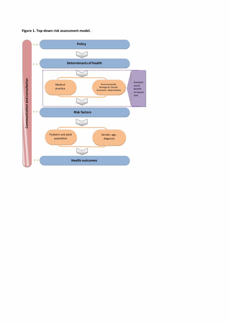
Figure 1. Top-down risk assessment model.
Policy
Determinants of health
Risk factors
Health outcomes
Medical
practice
Environmental, Biological, Clinical,
Economic determinants
Co
mm
un
ica
tio
n a
nd
co
nsu
lta
tio
n
Greatestsocial benefit at lowestcost
Pediatric and adult
population
Gender, age,
diagnosis

31
Figure 2. Top-down risk assessment model fed with current knowledge.
Tuscany Policy on Radiprotection 2008-2010
Proximal health determinants (physical environment)
Cumulative individual dose, working tasks, technologies
availability, co-morbidity,…
Fatal and non fatal cancer, other non cancer outcomes
Current guideline,
technological updating,
continuing training, patient
and operator awareness,
etc…….
Workplace environment
and community
environment
Co
mm
un
icat
ion
an
d c
on
sult
atio
n
Gre
ates
t so
cial
ben
efit
at
low
est
cost
Pediatric and
adult population Gender, age,
diagnosis

32
Figure 3. Decision-making in Clinical Practice
Figure 4. The RAPID framework applied to regional radioprotection policy and health.