HHS Public Access 1,* Meenakshi A. Chellaiah Jing Zou Mark … · Writing the article -- -- ... in...
13
In vitro BMP2 stimulation of osteoblast citrate production in concert with mineralized bone nodule formation Leslie C. Costello 1,* , Meenakshi A. Chellaiah 1 , Jing Zou 1 , Mark A. Reynolds 2 , and Renty B. Franklin 1 1 Department of Oncology and Diagnostic Sciences, Dental School, University of Maryland, Baltimore, USA 2 Department of Periodontics, Dental School, University of Maryland, Baltimore, USA Abstract Background—That citrate is a major indispensible component of bone in humans and in all osteovertebrates has been known for about seventy-five years. Yet, its role and importance in the structure and function of bone and bone formation have remained unknown. However, recent studies have identified that citrate is a major and essential component of the apatite/collagen structure of bone; and that the biomechanical properties of bone (e.g., stability, strength, resistance to fracture) depend on the appropriate incorporation of citrate in the structure of bone. The osteoblasts have recently been identified as citrate-producing cells that provide the citrate that is incorporated in the apatite/collagen structure during osteogenesis. Little is known regarding the factors and mechanisms involved in the regulation of citrate that is incorporated along with mineralization during the process of bone formation. Because of the importance of BMP2 in the initiation of osteogenesis and the development of the osteoblasts, it is essential to determine its This is an Open Access article distributed under the terms of Creative Commons Attribution License (http://creativecommons.org/ licenses/by/3.0). This permits unrestricted use, distribution, and reproduction in any medium, provided the original work is properly cited. * Correspondence: [email protected]. Competing interests The authors declare that they have no competing interests. Authors’ contributions Authors’ contributions LCC MAC JZ RBF MAR Research concept and design ✓ ✓ -- ✓ ✓ Collection and/or assembly of data ✓ ✓ ✓ ✓ -- Data analysis and interpretation ✓ ✓ ✓ ✓ -- Writing the article ✓ -- -- ✓ ✓ Critical revision of the article ✓ ✓ -- ✓ -- Final approval of article ✓ -- -- ✓ -- Statistical analysis ✓ -- ✓ ✓ -- HHS Public Access Author manuscript J Regen Med Tissue Eng. Author manuscript; available in PMC 2015 December 01. Published in final edited form as: J Regen Med Tissue Eng. 2015 ; 4: . doi:10.7243/2050-1218-4-2. Author Manuscript Author Manuscript Author Manuscript Author Manuscript
Transcript of HHS Public Access 1,* Meenakshi A. Chellaiah Jing Zou Mark … · Writing the article -- -- ... in...

In vitro BMP2 stimulation of osteoblast citrate production in concert with mineralized bone nodule formation
Leslie C. Costello1,*, Meenakshi A. Chellaiah1, Jing Zou1, Mark A. Reynolds2, and Renty B. Franklin1
1Department of Oncology and Diagnostic Sciences, Dental School, University of Maryland, Baltimore, USA
2Department of Periodontics, Dental School, University of Maryland, Baltimore, USA
Abstract
Background—That citrate is a major indispensible component of bone in humans and in all
osteovertebrates has been known for about seventy-five years. Yet, its role and importance in the
structure and function of bone and bone formation have remained unknown. However, recent
studies have identified that citrate is a major and essential component of the apatite/collagen
structure of bone; and that the biomechanical properties of bone (e.g., stability, strength, resistance
to fracture) depend on the appropriate incorporation of citrate in the structure of bone. The
osteoblasts have recently been identified as citrate-producing cells that provide the citrate that is
incorporated in the apatite/collagen structure during osteogenesis. Little is known regarding the
factors and mechanisms involved in the regulation of citrate that is incorporated along with
mineralization during the process of bone formation. Because of the importance of BMP2 in the
initiation of osteogenesis and the development of the osteoblasts, it is essential to determine its
This is an Open Access article distributed under the terms of Creative Commons Attribution License (http://creativecommons.org/licenses/by/3.0). This permits unrestricted use, distribution, and reproduction in any medium, provided the original work is properly cited.*Correspondence: [email protected].
Competing interestsThe authors declare that they have no competing interests.
Authors’ contributions
Authors’ contributions LCC MAC JZ RBF MAR
Research concept and design ✓ ✓ -- ✓ ✓
Collection and/or assembly of data ✓ ✓ ✓ ✓ --
Data analysis and interpretation ✓ ✓ ✓ ✓ --
Writing the article ✓ -- -- ✓ ✓
Critical revision of the article ✓ ✓ -- ✓ --
Final approval of article ✓ -- -- ✓ --
Statistical analysis ✓ -- ✓ ✓ --
HHS Public AccessAuthor manuscriptJ Regen Med Tissue Eng. Author manuscript; available in PMC 2015 December 01.
Published in final edited form as:J Regen Med Tissue Eng. 2015 ; 4: . doi:10.7243/2050-1218-4-2.
Author M
anuscriptA
uthor Manuscript
Author M
anuscriptA
uthor Manuscript

possible implication in the development of the citrate-producing capability of the osteoblasts (i.e.,
“citration”) during the formation of mineralized bone nodules.
Methods—The goal of this study was to determine if BMP2 promotes the development of
citrate-producing osteoblasts for increased citrate incorporation in the formation of mineralized
bone nodules. The study employed MC3T3 mesenchyme stem cell osteogenic differentiation in
the presence and absence of BMP2.
Results—The results showed that BMP2 treatment increased the osteogenic development of
mineralized bone nodules. In addition, BMP2 increased osteoblast citrate production and
incorporation in the mineralized bone nodule. This was accompanied by increased ZIP1
transporter, which is an essential genetic/metabolic event for citrate-producing cells.
Conclusions—The results demonstrate, for the first time, that BMP2 facilitates the osteoblast
“citration” process in concert with mineralization during bone formation; and provide
confirmation of the important role of osteoblasts as specialized citrate-producing cells in the
process of bone formation. However, it is essential to determine if these in vitro effects will occur
in vivo in BMP2-implant induction of bone formation. “Citration” is essential for osteoinductive
bone to represent the chemical, structural, and biomechanical properties of “normal” bone.
Keywords
BMP2; citrate production; ZIP1 transporter; mineralized bone formation; osteoblasts; apatite/collagen complex; osteogenesis; citration and mineralization; mesenchyme stem cells
Introduction
High levels of citrate constitute a major component of bone (and teeth) in humans and in all
osteovertebrates. It comprises ~1.6 % of the bone content; ~5% of the organic component of
bone; and ~80% of the total body citrate resides in bone. The fact that all osteovertebrates
exhibit this high bone citrate composition (cartilage does not contain the high citrate levels)
is evidence that citrate is an indispensible essential component of bone. Although this has
been known since 1941, the implications of citrate in bone have received little attention or
recognitions over the past~35 years. Consequently, progress and advances in the
identification and elucidation of citrate relationships in normal bone and in bone disorders
remain largely unknown. However, recent studies [1–4] have identified that citrate is an
important component of the apatite/collagen structure of bone; and it is essential to achieve
optimal manifestation of the important biomechanical properties of bone (such as stability,
strength, resistance to fracture).
This important role of citrate now brings attention to the necessity for increased research
into citrate implications in normal bone formation and in bone disorders. One of the
unresolved fundamental issues has been the identification of the source of citrate for
incorporation into bone. We recently identified [5,6] that the osteoblasts are specialized
citrate-producing cells, which provide the citrate incorporation (i.e., “citration”), along with
mineralization, during bone formation. The osteoblast metabolic and functional capability
occurs during osteogenic differentiation of the mesenchyme stem cells. This is a new
Costello et al. Page 2
J Regen Med Tissue Eng. Author manuscript; available in PMC 2015 December 01.
Author M
anuscriptA
uthor Manuscript
Author M
anuscriptA
uthor Manuscript

understanding of the role of osteoblasts and also the events of osteogenesis and bone
formation.
Now it becomes essential to identify the factors and events that regulate osteoblast citrate
production and incorporation into bone. BMP2 is important for initiating and optimizing
early osteogenic events leading to bone formation, including mineralized bone nodule
formation during mesenchyme stem cells differentiation [7–9]. Therefore, we initiated
studies to determine if BMP2 also stimulated osteoblast citrate production and “citration” of
mineralized bone nodules in osteogenic differentiation of MC3T3 cells.
Methods
Cell culture
Mouse mesenchyme stem cells (MC3T3) were obtain from American Type Culture
Collection (ATCC, Manassas, VA). The mesenchyme cells were grown and maintained in
mesenchymal cell growth medium which consisted α-MEM supplemented with L-glutamine
and containing 30 mg/ml gentamicin, 15 ug/ml amphotericin and 10% fetal bovine serum.
Cells were cultured at 37°C in the presence of 5% CO2 until ~75% confluent. The
mesenchyme cells were induced to differentiate to the osteoblast linage by culturing the cells
in growth medium containing ascorbic acid (50 mg/ml) and B-glycerophosphate (10 mM).
For BMP2 treatment, cells were maintained in osteogenic medium containing 100 ng/ml
BMP2 with media changes every two days. Control cells were maintained in the osteogenic
medium without BMP2.
Alizarin red staining
Mineralization was assessed by staining with Alizarin Red for calcium deposits. Briefly, the
medium was aspirated from the wells and the osteoblast cells and bone nodules fixed by
incubation in iced cold 70% ethanol for 1 hour. The ethanol was removed and the wells
rinsed twice with water. The water was then removed and enough Alizarin Red Solution (40
mM) added to cover the wells. After 30 min the Alizarin Red solution was removed and the
wells washed four times with water. Mineralization was documented using an inverted
microscope with a Qicam Fas1394 digital camera.
Citrate and calcium assay
For the determination of citrate and calcium production, the medium was aspirated from the
wells, and replaced by a volume of PBS. The PBS was removed by aspiration, followed by
the addition of lysing buffer. The content of the well was scraped and mixed in the lysing
buffer. The lysate was sonicated and deproteinized by addition of 7% trichroloacetic acid
and centrifugation at 300g for 5 min. The citrate concentration of the lysate extract was
measured by the fluoroenzymatic method previously described [10]. Calcium concentration
in the lysate extract was assayed using the Calcium Colorimetric Assay kit from BioVision,
Inc.
Costello et al. Page 3
J Regen Med Tissue Eng. Author manuscript; available in PMC 2015 December 01.
Author M
anuscriptA
uthor Manuscript
Author M
anuscriptA
uthor Manuscript

Western blot analysis
Osteoblast cells were lysed using RIPA buffer (Upstate Biotech). The protein concentration
of the lysates was determined using the BioRad protein assay based on the method of
Bradford [11]. Proteins were separated by SDS-PAGE and transferred to nitrocellulose
membrane. The membranes were blocked by incubating in PBS containing 5% non-fat milk
and 0.1% tween 20 for 2 hours at room temperature. ZIP1 was detected by incubating the
membranes overnight with ZIP1 chicken polyclonal antibody as described previously [12].
ZIP1 bound antibody was detected by incubating the membranes with hydrogen peroxide
labeled goat anti-chicken secondary antibody and enhanced chemiluminescence detection
reagents. The membranes were stripped and re-probed using anti β-actin antibody.
Experimental and statistical analyses
The experiment was repeated so as to establish reproducibility of the results obtained. The
results presented below are representative of the repeated experiments. In each experiment,
the control and experimental groups were prepared in triplicate. T-test determination of the
mean±sem values was employed to determine statistical significance.
Results
We first determined the effects of BMP2 on mineralized bone nodule formation resulting
from osteogenic differentiation of MC3T3 cells. Alizarin Red staining reveals the apparent
BMP2-induced increase in mineralized nodule formation (Figure 1A). This is corroborated
by the quantitative determination of calcium which shows a 100% increase in calcium
incorporated into the BMP2 treated cultures (Figure 1B). These results are consistent with
the well-established role of BMP2 in enhancing the osteogenic differentiation of
mesenchyme stem cells resulting in osteoblast mineralization during bone formation. The
determination of citrate (Figure 1B) shows that BMP2 markedly increased (+238%) the
level of citrate production and incorporation compared to the control. Most importantly, the
citrate/calcium ratio was significantly increased (+75%) by BMP2 treatment. This reveals
that in addition to its affect on increasing the differentiation of osteoblasts resulting in
mineralized bone nodules, BMP2 additionally increases osteoblast citrate production and
incorporation during mineralized bone formation (i.e., the “citration” of bone). Prior to
differentiation, the undifferentiated MSCs exhibit very little citrate production (~5 nmols/mg
protein) and calcium incorporation (~360 nmols/mg protein) compared to the respective
concentrations following differentiation (see Figure 1B for values of the control and BMP2
treated differentiated cultures).
Since the upregulation of ZIP1 expression is required for optimal osteogenic differentiation
of human mesenchyme stem cells leading to mineralizing and citrate producing osteoblasts
[6,13], we determined if this relationship was evident in the BMP2 effects. Figure 1C shows
the marked increase in ZIP1 transporter abundance in response to BMP2 treatment. Along
with this, we obtained an increase in zinc levels (umols/mg protein) from 0.20±0.06
(control) to 0.29±0.03 for BMP2 treatment; an increase of 45%. However, the difference
was not significant due to the small sample size. Nevertheless, the upregulation during
Costello et al. Page 4
J Regen Med Tissue Eng. Author manuscript; available in PMC 2015 December 01.
Author M
anuscriptA
uthor Manuscript
Author M
anuscriptA
uthor Manuscript

differentiation of the MC3T3 cells is consistent with the observations obtained with human
mesenchyme stem cell differentiation.
Discussion
The effects of BMP2 in this study must be discussed in relation to the new and more
appropriate understanding of the implications of citrate in the composition and structure of
normal bone formation. Until recently, descriptions of the apatite/collagen structure of bone
focused on the mineralization of collagen; with no inclusion of citrate as a major component
of the apatite/collagen complex. Recent studies have identified that citrate is incorporated in
the structure of the apatite/collagen complex; and that it is essential for bone to exhibit its
important biomechanical properties [1–4]. Figure 2 provides a representation of the apatite/
collagen structure of bone; in which citrate is incorporated in the apatite nanoncrystal
(mineral) component, and in the apatite/collagen complex [4]. Heretofore, this essential
incorporation of citrate in the structure of bone had not been appropriately represented in the
contemporary view of the structure of bone.
This leads to the new understanding that the process of bone formation must include the
incorporation of citrate (which we refer to as “citration”) along with mineralization in the
apatite nanocrystal/collagen complex. This also implies that the source of the citrate during
bone formation must be identified. Our recent studies with human mesenchyme cells and
primary osteoblasts [5,6] identified the osteoblasts as specialized “citrate-producing” cells.
Under appropriate conditions, osteogenic mesenchyme stem cell differentiation results in
osteoblasts with citrate-producing as well as mineralizing capabilities. In this study, the
osteogenic differentiation of the MC3T3 cells (in the absence of added BMP2) resulting in
osteoblast citrate production along with formation of mineralized bone nodules corroborates
and extends our earlier studies. In addition, the concurrent BMP2-induced increase in citrate
production and increased mineralized bone nodules provides additional evidence that
osteoblast citrate production and “citration” are coordinated events in bone formation.
Therefore, Figure 3 provides the new and more appropriate representation of osteogenesis
during bone formation. In the absence of citration, the bone will not exhibit its important
structural and biomechanical properties; and is not representative of “normal” bone.
That BMP2 is an important factor in inducing the differentiation of stem cells or precursor
cells into osteoblastic lineages is well established [7–9,14,15]; although the downstream
factors associated with the development of the functional osteoblasts and the early formation
of mineralized bone nodules during the bone formation process remain largely unknown. In
this study, the BMP2-induced increase in osteoblast mineralized bone nodule formation
(Figure 1) is representative of the reported BMP2 induction of osteoblast differentiation and
mineralization. However, the importance of BMP2 is additionally revealed by its specific
induction of osteoblast citrate production (i.e., increased citration), in addition to increasing
mesenchyme stem cell differentiation and development of the osteoblasts.
The mechanisms by which BMP2 facilitates osteoblast citrate production requires an
understanding of the unique cellular metabolic relationships involved in “net citrate
production”; which is a specialized functional metabolic activity that does not typically exist
Costello et al. Page 5
J Regen Med Tissue Eng. Author manuscript; available in PMC 2015 December 01.
Author M
anuscriptA
uthor Manuscript
Author M
anuscriptA
uthor Manuscript

in mammalian cells (Figure 4). The cellular metabolic pathway of net citrate production has
been identified by our studies with prostate epithelial cells, which are specialized citrate-
producing cells [16]. This functional metabolic relationship now applies to osteoblasts. It
requires genetic/metabolic changes in the typical mammalian cell citraterelated intermediary
metabolism; such as represented by the osteogenic differentiation transformation of
mesenchymal cells to citrate-producing osteoblasts [5,6,17] (Figure 4). The most important
initiating metabolic event is the increased cellular/mitochondrial concentration of zinc,
which results in the specific inhibition of m-aconitase [18]. This is essential so that the
citrate that is synthesized in the mitochondria is not converted to isocitrate and oxidized via
the Krebs cycle; but instead, the citrate is accumulated for its transport out of the
mitochondria and secreted into the extracellular environment. The increase in zinc uptake is
achieved by the upregulation of ZIP1 zinc uptake transporter (Slc39A1).
Therefore, it is especially significant that the BMP2-induced increased citrate production is
accompanied by increased ZIP1 transporter abundance (Figure 1C), which increases zinc
accumulation; and is the major genetic/metabolic event for the osteoblasts to achieve net
citrate production. However, other genetic/metabolic alterations as represented in Figure 4
are also required for osteoblast citrate production; and some of these might be induced by
BMP2. Tang et al., [13] reported that optimal osteogenic differentiation of human MSC to
mineralizing osteoblasts requires an accompanying increase in ZIP1 expression; and that the
increased ZIP1 expression leads to altered expression of other genes that are implicated in
the osteogenic process. This included the upregulation of mAAT, which is another genetic/
metabolic alteration required for net citrate production (Figure 4). Thus, these BMP2-
induced effects are consistent with the other events required for the osteogenic
differentiation of human mesenchyme cells [7–9,14,15]. It is now important to elucidate the
mechanism, factors, and signaling pathway(s) associated with BMP2 regulation of
osteoblast citrate production.
It must also be recognized that the incorporation of calcium and the incorporation of citrate
in the formation of the bone nodules result from different sources and processes (Figure 5).
The calcium is derived by the osteoblast uptake and transport from the calcium component
of the extracellular medium. In contrast, the extracellular medium is devoid of citrate.
Therefore, osteoblast de novo synthesis of citrate provides the citrate incorporated into the
mineralized bone nodules. So, it becomes apparent that BMP2 stimulates osteoblast de novo
citrate production. Furthermore, the results demonstrate that an exogenous extracellular
source of citrate, unlike calcium, is not required for citrate incorporation during bone
formation. The long-held prevailing view of plasma citrate homeostasis and bone turnover
has represented that citrate, along with calcium, is transported into bone during bone
formation. Based on our studies [5,6], that view is no longer tenable; and this impacts the
understanding of hormonal/humoral actions implicated in the homeostatic regulation of
citrate, calcium, bone and plasma.
The important implications of citrate should be considered in relation to the application of
regenerative medicine and implant technology for osteoinductive treatment of bone
disorders and repair of bone defects. The optimal bone formation from such treatments
should be the generation of new bone that exhibits the chemical, structural, and
Costello et al. Page 6
J Regen Med Tissue Eng. Author manuscript; available in PMC 2015 December 01.
Author M
anuscriptA
uthor Manuscript
Author M
anuscriptA
uthor Manuscript

biomechanical properties of the “normal” bone. Presently, no information or reported studies
exist in which the presence or status of citrate has been determined or considered in implant-
induced bone formation. The results of this study now implicate BMP2 in facilitating
osteoblast citration in addition to facilitating osteoblast differentiation. Since BMP2 is
osteoinductive in vivo, it is important to determine if BMP2 implants result in “citrated”
mineralized bone formation, so as to exhibit the composition and biomechanical properties
as exists in normal bone formation.
Most importantly, the results of this study, along with recent reports [1–6], provide
compelling evidence of the essential and indispensible requirements of citrate incorporation
to achieve normal bone formation. As such, the absence of recognition and consideration of
the role of citrate in bone over the past forty years should be remedied by renewed interest
and research into the implications of citrate in bone. This is essential to achieve the
appropriate and accurate understanding of the process and factors involved in normal bone
formation; the implications in the development of bone disorders; and the translation for the
treatment of bone disorders and defects.
Acknowledgement
These studies were supported by NIH grant AR064808.
References
1. Hu YY, Rawal A, Schmidt-Rohr K. Strongly bound citrate stabilizes the apatite nanocrystals in bone. Proc Natl Acad Sci U S A. 2010; 107:22425–22429. [PubMed: 21127269]
2. Hu Y-Y, Liu XP, Ma X, Rawal A, Prozorov T, Akinc M, Mallapragada SK, Schmidt-Rohr K. Biomimetic self-assembling copolymer_hydroxyapatite nanocomposites with the nanocrystal size controlled by citrate. Chem. Mater. 2011; 23:2481–2490.
3. Davies E, Muller KH, Wong WC, Pickard CJ, Reid DG, Skepper JN, Duer MJ. Citrate bridges between mineral platelets in bone. Proc Natl Acad Sci U S A. 2014; 111:E1354–E1363. [PubMed: 24706850]
4. Costello LC, Chellaiah M, Zou J, Franklin RB, Reynolds MA. The status of citrate in the hydroxyapatite/collagen complex of bone; Its role in bone formation. J Regen Med Tissue Eng. 2014; 3:4. [PubMed: 25745562]
5. Costello LC, Franklin RB, Reynolds MA, Chellaiah M. The Important Role of Osteoblasts and Citrate Production in Bone Formation: “Osteoblast Citration” as a New Concept for an Old Relationship. Open Bone J. 2012; 4
6. Franklin RB, Chellaiah M, Zou J, Reynolds MA, Costello LC. Evidence that Osteoblasts are Specialized Citrate-producing Cells that Provide the Citrate for Incorporation into the Structure of Bone. Open Bone J. 2014; 6:1–7. [PubMed: 25745519]
7. Yamaguchi A, Ishizuya T, Kintou N, Wada Y, Katagiri T, Wozney JM, Rosen V, Yoshiki S. Effects of BMP-2, BMP-4, and BMP-6 on osteoblastic differentiation of bone marrow-derived stromal cell lines, ST2 and MC3T3-G2/PA6. Biochem Biophys Res Commun. 1996; 220:366–371. [PubMed: 8645311]
8. Chatakun P, Nunez-Toldra R, Diaz Lopez EJ, Gil-Recio C, Martinez-Sarra E, Hernandez-Alfaro F, Ferres-Padro E, Giner-Tarrida L, Atari M. The effect of five proteins on stem cells used for osteoblast differentiation and proliferation: a current review of the literature. Cell Mol Life Sci. 2014; 71:113–142. [PubMed: 23568025]
9. Rosen V. BMP2 signaling in bone development and repair. Cytokine Growth Factor Rev. 2009; 20:475–480. [PubMed: 19892583]
Costello et al. Page 7
J Regen Med Tissue Eng. Author manuscript; available in PMC 2015 December 01.
Author M
anuscriptA
uthor Manuscript
Author M
anuscriptA
uthor Manuscript

10. Costello LC, O’Neill JJ. A simplified and sensitive method for citrate determination in biological samples. J Appl Physiol. 1969; 27:120–122. [PubMed: 4389164]
11. Bradford MM. A rapid and sensitive method for the quantitation of microgram quantities of protein utilizing the principle of protein-dye binding. Anal Biochem. 1976; 72:248–254. [PubMed: 942051]
12. Franklin RB, Ma J, Zou J, Guan Z, Kukoyi BI, Feng P, Costello LC. Human ZIP1 is a major zinc uptake transporter for the accumulation of zinc in prostate cells. J Inorg Biochem. 2003; 96:435–442. [PubMed: 12888280]
13. Tang Z, Sahu SN, Khadeer MA, Bai G, Franklin RB, Gupta A. Overexpression of the ZIP1 zinc transporter induces an osteogenic phenotype in mesenchymal stem cells. Bone. 2006; 38:181–198. [PubMed: 16203195]
14. Zachos TA, Shields KM, Bertone AL. Gene-mediated osteogenic differentiation of stem cells by bone morphogenetic proteins-2 or -6. J Orthop Res. 2006; 24:1279–1291. [PubMed: 16649180]
15. Knippenberg M, Helder MN, Zandieh Doulabi B, Wuisman PI, Klein-Nulend J. Osteogenesis versus chondrogenesis by BMP-2 and BMP-7 in adipose stem cells. Biochem Biophys Res Commun. 2006; 342:902–908. [PubMed: 16500625]
16. Costello LC, Franklin RB. The clinical relevance of the metabolism of prostate cancer; zinc and tumor suppression: connecting the dots. Mol Cancer. 2006; 5:17. [PubMed: 16700911]
17. Costello LC, Franklin RB. A review of the important central role of altered citrate metabolism during the process of stem cell differentiation. J Regen Med Tissue Eng. 2013; 2
18. Costello LC, Liu Y, Franklin RB, Kennedy MC. Zinc inhibition of mitochondrial aconitase and its importance in citrate metabolism of prostate epithelial cells. J Biol Chem. 1997; 272:28875–28881. [PubMed: 9360955]
Costello et al. Page 8
J Regen Med Tissue Eng. Author manuscript; available in PMC 2015 December 01.
Author M
anuscriptA
uthor Manuscript
Author M
anuscriptA
uthor Manuscript
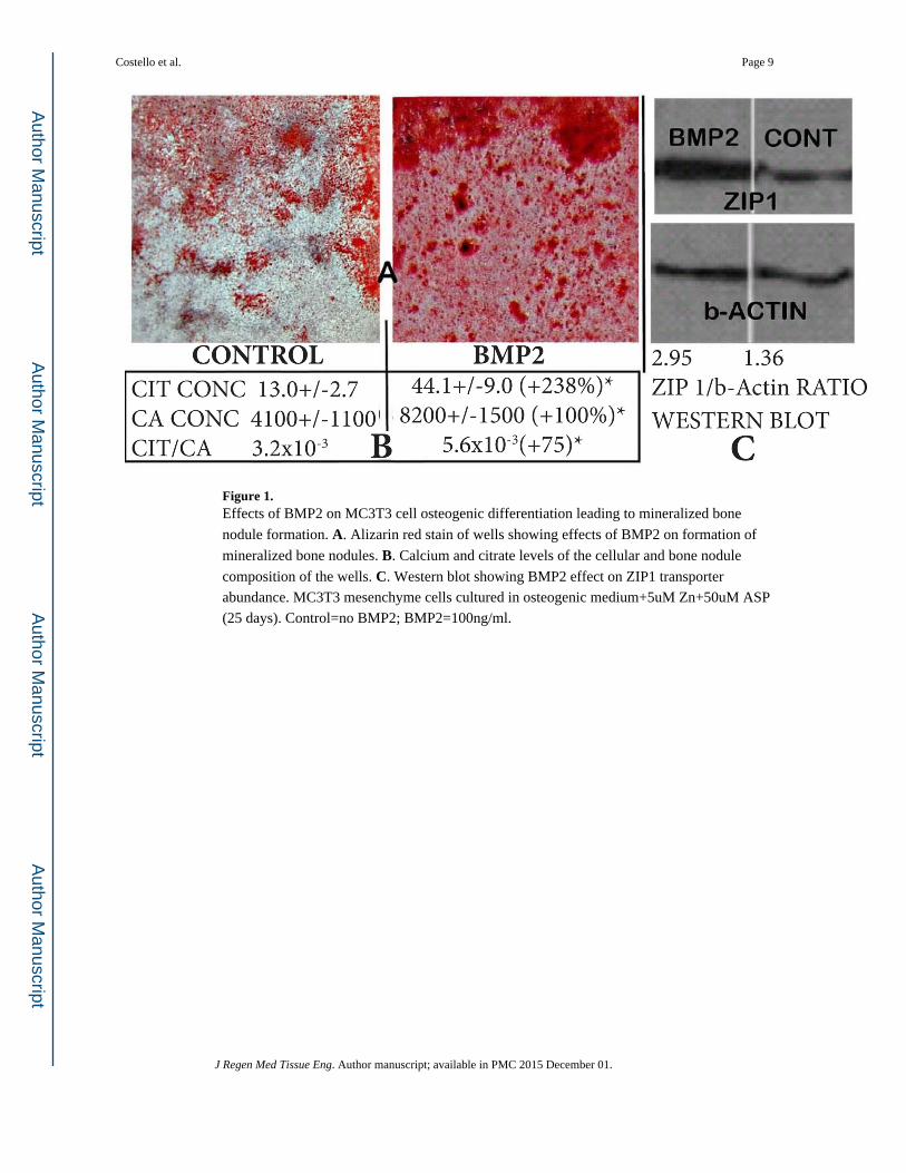
Figure 1. Effects of BMP2 on MC3T3 cell osteogenic differentiation leading to mineralized bone
nodule formation. A. Alizarin red stain of wells showing effects of BMP2 on formation of
mineralized bone nodules. B. Calcium and citrate levels of the cellular and bone nodule
composition of the wells. C. Western blot showing BMP2 effect on ZIP1 transporter
abundance. MC3T3 mesenchyme cells cultured in osteogenic medium+5uM Zn+50uM ASP
(25 days). Control=no BMP2; BMP2=100ng/ml.
Costello et al. Page 9
J Regen Med Tissue Eng. Author manuscript; available in PMC 2015 December 01.
Author M
anuscriptA
uthor Manuscript
Author M
anuscriptA
uthor Manuscript
Figure 2. The status of citrate in the apatite/collagen component of bone. A. The apatite component.
B. The apatite/collagen complex. M: Mineral; CI: Citrate; CO: Collagen.
Costello et al. Page 10
J Regen Med Tissue Eng. Author manuscript; available in PMC 2015 December 01.
Author M
anuscriptA
uthor Manuscript
Author M
anuscriptA
uthor Manuscript

Figure 3. Normal bone formation showing osteogenic differentiation and the role of OSB citrate
production and “citration” in concert with the formation of the apatite/collagen complex.
Also shows the ZIP1 transporter upregulation and zinc uptake requirement for
differentiation to OSB citrate-producing cells.
Costello et al. Page 11
J Regen Med Tissue Eng. Author manuscript; available in PMC 2015 December 01.
Author M
anuscriptA
uthor Manuscript
Author M
anuscriptA
uthor Manuscript

Figure 4. Important genetic/metabolic alterations (in blue) for the differentiation of mesenchyme stem
cells to citrate-producing osteoblasts. ASP-Tr: Aspartate transporter; ZIP1: Zinc uptake
transporter; CTP: Citrate transport protein; AAT: Aspartate aminotransferase; ACON:
Aconitase.
Costello et al. Page 12
J Regen Med Tissue Eng. Author manuscript; available in PMC 2015 December 01.
Author M
anuscriptA
uthor Manuscript
Author M
anuscriptA
uthor Manuscript

Figure 5. Osteoblast de novo citrate production; and calcium transport from plasma for incorporation
into the a patite/collagen structure in the process of bone formation.
Costello et al. Page 13
J Regen Med Tissue Eng. Author manuscript; available in PMC 2015 December 01.
Author M
anuscriptA
uthor Manuscript
Author M
anuscriptA
uthor Manuscript