Heterotopic ossification in the interosseous membrane of ... · Heterotopic ossification in the...
3
Case Report International Journal of Anatomical Variations (2013) 6: 71–73 eISSN 1308-4038 Heterotopic ossification in the interosseous membrane of right forearm bones Introduction Interosseous membrane is a broad thin collagenous sheet whose fibers slant distomedially between the radial and ulnar interosseous borders. The distal part of the membrane is attached to the posterior division of the radial border. The membrane gives attachment to muscles. Its fibers help in transmission of forces which act proximally from the hand to the radius, hence to the ulna and humerus. The posterior relations near the carpus are the anterior interosseous artery and posterior interosseous nerve [1]. Heterotopic ossification is the abnormal formation of true bone within extra skeletal soft tissues such as muscles, fascial planes, tendons, other mesenchymal soft tissues [2]. It is seen in 2% of all forearm injuries leading to ossification of interosseous membrane and causes significant functional impairment [3]. Case Report During routine osteology demonstration classes for the 1st year MBBS students in the Department of Anatomy, BLDEU’s Shri B. M. Patil Medical College, Bijapur, we observed an abnormal piece of bone at the site of distal part of interosseous membrane of right radius and ulna. This piece of bone was the heterotopic ossification in the interosseous membrane hence resulting in fusion near the distal ends of right radius and ulna (Figure 1). The anterior surface was rough and posterior surface was smooth as shown in Figures 2 and 3. The inferior radio-ulnar joint was normal. The measurements were taken using spreading caliper (Table 1). Sulabha H. DESHPANDE Shankreppa D. DESAI Vijayendra N. KULKARNI Ravi S. BULAGOUDA Department of Anatomy BLDEU’S Shri. B. M. Patil Medical College, Bijapur, Karnataka, INDIA. Dr. Sulabha H. Deshpande Assistant Professor Department of Anatomy BLDEU’S Shri B. M. Patil Medical College Bijapur, Karnataka, INDIA. +91 8352-262770 [email protected] Received March 27th, 2012; accepted October 21st, 2012 Abstract Interosseous membrane is a broad thin collagenous sheet whose fibers slant distomedially between the radial and ulnar interosseous borders. Its distal part is attached to the posterior division of the radial border. Its fibers help in transmission of forces that act proximally from the hand to the radius, hence to the ulna and humerus. The posterior relations near the carpus are the anterior interosseous artery and posterior interosseous nerve. Heterotopic ossification resulting in radio-ulnar cross union occurs in 2% of all forearm injuries. During the routine undergraduate demonstration classes in the department of Anatomy at BLDEU’s Shri B. M. Patil Medical College, Bijapur, we found an unusual piece of bone at the site of distal one-third of interosseous membrane of right radius and ulna, which indicates the heterotopic ossification resulting in the fusion near the distal ends of radius and ulna. The ossified part measured 1.27 cm x 2.1 cm x 1.63 cm, using spreading calipers. Factors resulting in heterotopic ossification include open fractures, hematoma formation, infection, callus formation. Complications include loss of forearm supination and pronation, which impairs the function of entire upper limb. Also one would expect compression over the anterior interosseous artery and posterior interosseous nerve. It is of importance for the orthopedists while dealing with compound fractures and their management such as internal fixation. © Int J Anat Var (IJAV). 2013; 6: 71–73. Key words [heterotopic ossification] [interosseous membrane] [forearm bones] Published online April 21st, 2013 © http://www.ijav.org Table 1. Measurements of the case. Length of ulna 26.77 cm Length of radius 21.07 cm Length of ossified part 3.77 cm Thickness of ossified part 1.27 cm – upper end 2.10 cm – midregion 1.63 cm – lower end Discussion Heterotopic ossification leading to cross union between forearm bones was first described by Gross in 1864. Although it is more common with fractures of proximal and middle third, the distal forearm is not entirely spared of this troublesome complication [4]. Reidel in 1883, Dejerne and Ceillier in 1918
Transcript of Heterotopic ossification in the interosseous membrane of ... · Heterotopic ossification in the...
Case Report
International Journal of Anatomical Variations (2013) 6: 71–73eISSN 1308-4038
Heterotopic ossification in the interosseous membrane of right forearm bones
IntroductionInterosseous membrane is a broad thin collagenous sheet whose fibers slant distomedially between the radial and ulnar interosseous borders. The distal part of the membrane is attached to the posterior division of the radial border. The membrane gives attachment to muscles. Its fibers help in transmission of forces which act proximally from the hand to the radius, hence to the ulna and humerus. The posterior relations near the carpus are the anterior interosseous artery and posterior interosseous nerve [1]. Heterotopic ossification is the abnormal formation of true bone within extra skeletal soft tissues such as muscles, fascial planes, tendons, other mesenchymal soft tissues [2]. It is seen in 2% of all forearm injuries leading to ossification of interosseous membrane and causes significant functional impairment [3].
Case ReportDuring routine osteology demonstration classes for the 1st year MBBS students in the Department of Anatomy, BLDEU’s Shri B. M. Patil Medical College, Bijapur, we observed an abnormal piece of bone at the site of distal part of interosseous membrane of right radius and ulna. This piece of bone was the heterotopic ossification in the interosseous membrane hence resulting in fusion near the distal ends of right radius and ulna (Figure 1). The anterior surface was rough and posterior
surface was smooth as shown in Figures 2 and 3. The inferior radio-ulnar joint was normal.The measurements were taken using spreading caliper (Table 1).
Sulabha H. DESHPANDE
Shankreppa D. DESAIVijayendra N. KULKARNIRavi S. BULAGOUDA
Department of Anatomy BLDEU’S Shri. B. M. Patil Medical College, Bijapur, Karnataka, INDIA.
Dr. Sulabha H. Deshpande Assistant Professor Department of Anatomy BLDEU’S Shri B. M. Patil Medical College Bijapur, Karnataka, INDIA. +91 8352-262770 [email protected]
Received March 27th, 2012; accepted October 21st, 2012
AbstractInterosseous membrane is a broad thin collagenous sheet whose fibers slant distomedially between the radial and ulnar interosseous borders. Its distal part is attached to the posterior division of the radial border. Its fibers help in transmission of forces that act proximally from the hand to the radius, hence to the ulna and humerus. The posterior relations near the carpus are the anterior interosseous artery and posterior interosseous nerve. Heterotopic ossification resulting in radio-ulnar cross union occurs in 2% of all forearm injuries. During the routine undergraduate demonstration classes in the department of Anatomy at BLDEU’s Shri B. M. Patil Medical College, Bijapur, we found an unusual piece of bone at the site of distal one-third of interosseous membrane of right radius and ulna, which indicates the heterotopic ossification resulting in the fusion near the distal ends of radius and ulna. The ossified part measured 1.27 cm x 2.1 cm x 1.63 cm, using spreading calipers. Factors resulting in heterotopic ossification include open fractures, hematoma formation, infection, callus formation. Complications include loss of forearm supination and pronation, which impairs the function of entire upper limb. Also one would expect compression over the anterior interosseous artery and posterior interosseous nerve. It is of importance for the orthopedists while dealing with compound fractures and their management such as internal fixation.
© Int J Anat Var (IJAV). 2013; 6: 71–73.
Key words [heterotopic ossification] [interosseous membrane] [forearm bones]
Published online April 21st, 2013 © http://www.ijav.org
Table 1. Measurements of the case.
Length of ulna 26.77 cmLength of radius 21.07 cmLength of ossified part 3.77 cmThickness of ossified part 1.27 cm – upper end
2.10 cm – midregion
1.63 cm – lower end
DiscussionHeterotopic ossification leading to cross union between forearm bones was first described by Gross in 1864. Although it is more common with fractures of proximal and middle third, the distal forearm is not entirely spared of this troublesome complication [4]. Reidel in 1883, Dejerne and Ceillier in 1918
72 Deshpande et al.
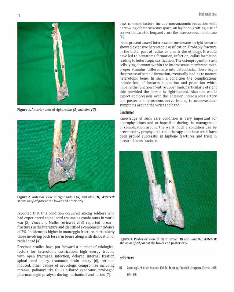
Figure 1. Anterior view of right radius (R) and ulna (U).
UR
Figure 2. Anterior view of right radius (R) and ulna (U). Asterisk shows ossified part at the lower end anteriorly.
U
*
R
Figure 3. Posterior view of right radius (R) and ulna (U). Asterisk shows ossified part at the lower end posteriorly.
U
*
R
reported that this condition occurred among soldiers who had experienced spinal cord trauma as combatants in world war [5]. Vince and Muller reviewed 2381 reported forearm fractures in the literature and identified a combined incidence of 2%. Incidence is higher in monteggia fracture, particularly those involving both forearm bones along with dislocation of radial head [4].Previous studies have put forward a number of etiological factors for heterotopic ossification: high energy trauma with open fractures, infection, delayed internal fixation, spinal cord injury, traumatic brain injury [6], retinoid induced, other causes of neurologic compromise including tetanus, poliomyelitis, Guillain-Barré syndrome, prolonged pharmacologic paralysis during mechanical ventilation [7].
Less common factors include non-anatomic reduction with narrowing of interosseous space, on lay bone-grafting, use of screws that are too long and cross the interosseous membrane [4].In the present case of interosseous membrane in right forearm showed extensive heterotopic ossification. Probably fracture in the distal part of radius or ulna is the etiology. It would have led to hematoma formation, infection, callus formation leading to heterotopic ossification. The osteoprogenitor stem cells lying dormant within the interosseous membrane, with proper stimulus, differentiate into osteoblasts. These begin the process of osteoid formation, eventually leading to mature heterotopic bone. In such a condition the complications include loss of forearm supination and pronation which impairs the function of entire upper limb, particularly of right side provided the person is right-handed. Also one would expect compression over the anterior interosseous artery and posterior interosseous nerve leading to neurovascular symptoms around the wrist and hand.
ConclusionKnowledge of such rare condition is very important for neurophysicians and orthopedists during the management of complication around the wrist. Such a condition can be prevented by prophylactic radiotherapy and these trials have been proved successful in hipbone fractures and tried in forearm bones fracture.
References
[1] Standring S, ed. Gray’s Anatomy. 40th Ed., Edinburg, Churchill Livingstone, Elsevier. 2008;
839–840.

73Heterotopic ossification in forearm
[2] Moore DS, Cho G. Heterotopic Ossification Imaging. http://emedicine.medscape.com/article/390416 (accessed February 2013)
[3] McAuliffe JA. Heterotopic ossification in the forearm. J Am Soc Surg Hand. 2005; 5: 30–41.
[4] Browner BD, Levine AM, Jupiter JB, Trafton PG, Krettek C. Skeletal trauma: basic science, management and reconstruction. 4th Ed., Saunders. 2008; 1487.
[5] Shehab D, Elgazzar AH, Collier BD. Heterotopic ossification. J Nucl Med. 2002; 43: 346–353.
[6] Lee DY, Cho TJ, Choi IH, Chung CY, Yoo WJ, Kim JH, Park YK. Clinical and radiological manifestations of osteogenesis imperfecta type V. J Korean Med Sci. 2006; 21: 709–714.
[7] Dodd HJ, Dootson G, Sarkani I. Ossification in the interosseous membrane of forearm after etretinate treatment for psoriasis. Br J Dermatol. 1988; 119: 105–106.