HETEROGENEITY OF C ERB B FAMILY MEMBERS EXPRESSION IS RELATED TO CELL MORPHOLOGY AND IMMUNOPROFILE...
7
_____________________________ A.M. Cîmpean et al 33 ORIGINAL ARTICLE INTRODUCTION Targeted therapies represent the “gold standard” of various malignancies treatment intensely tested in experimental models and clinical trials [1-3], especially for those cancers with a high incidence and agressiveness as breast, renal or lung cancers [4-6]. Actual modern trends in clinical practice are focus on the discovery and testing of various thyrosine-kinase receptors family. Between them, epidermal growth factor receptor (EGFR, ErbB-1, HER1 in humans) and human epidermal growth factor receptor 2 (HER2, ErbB-1) are already therapeutic targets approved in clinical practice.Their amplification or overexpression is rarely encountered in normal tissues but is present in the development and progression of different types of malignant tumors (mamary, colonic, gastric), being considered a poor prognostic factor for metastasis and due to this fact an agressiveness factor. HER2 is already accepted by FDA and used as a therapeutic target in case of metastatic breast cancer [7]. In gastric cancer, HER2 overexpression is widely accepted but the interpretation and the evaluation of its expression are still controversial and due to this aspect, the transtuzumab based therapy is not approved for clinical application [8]. Another receptor that is recognized as a therapeutic target and as a prognostic factor in malignant tumors is EGFR or HER1. Its identification as an oncogene has led to the development of a targeted therapy based on gefitinib and erlotinib that are applied in lung cancer [9, 10] and also cetuximab that is used in colon cancer [11]. In pituitary adenomas, EGFR and HER2 expressions are sporadically studied and, for most of the cases literature data reporting results that derive from in vitro or in vivo experimental models. Except prolactinomas, there is a totally lack of correlations between the expression of HER2, EGFR, pituitary adenomas morphologic type and hormone profile. Moreover, the data regarding the potential role of HER2 and EGFR in the development and progression of pituitary adenomas was sporadically studied and the findings were not integrated into clinical or therapeutic context, neither in a potential molecular classification of pituitary adenomas which is more and more frequently discuss nowadays. MATERIAL AND METHODS A retrospective study has been designed by randomly choosing and including a total number of 80 HETEROGENEITY OF C ERB B FAMILY MEMBERS EXPRESSION IS RELATED TO CELL MORPHOLOGY AND IMMUNOPROFILE IN PITUITARY ADENOMAS Anca Maria Cîmpean 1 *, Eugen Melnic 2 , Ana Corlan 3 , Amalia Raluca Ceaușu 1 , Marius Raica 1 ABSTRACT Purpose. Epidermal growth factor receptor (EGFR, HER1) and human epidermal receptor 2 (HER2) assessement in pituitary adenomas related to hormone profile. Design and methods. For 60 retrospective cases of pituitary adenomas, we established the histopathologic diagnosis by using morphological stains, followed by case selection for immunoprofile and EGFR and HER 2 assessement. Results. More than one third of the studied pituitary adenomas (33,33%) were positive for HER2, with membranar pattern in basophilic cells and with predominantly cytoplasmic, granular pattern for acidophils cells. HER2 immuno-expression characterized PRL secreting adenomas (p=0.005) and associations between FSH-LH (p< 0.001) TSH-FSH (p=0,024) and TSH-LH (p=0.028). In situ hybridization confirmed HER2 gene amplification in 33,34% out of all positive cases for HER2 by immunohistochemistry. EGFR positivity was found significantly for GH-prolactin (p=0.000) and prolactin-ACTH (p=0.045) co-expressing pituitary adenomas, peritumoral macrophages and folliculostellate cells. Conclusions. Differential HER2 and EGFR expression related to hormone profile heterogeneity can define different subclasses of pituitary adenomas and could explain clinical, prognostic and therapeutic heterogeneity which are observed in clinical practice. Our results support re-classification of pituitary adenomas based on molecular approach which should include markers with well certified prognostic and therapeutic impact. Key Words: HER2, EGFR,pituitary adenomas, targeted therapy 1 Department of Microscopic Morphology/Histology, Angiogenesis Research Center, „Victor Babeș” University of Medicine and Pharmacy Timișoara, Romania, 2 Department of Pathology, Nicolae Testemițanu University of Medicine and Pharmacy Chișinău, Moldova, 3 Department of Endocrinology, Vasile Goldiș University, Arad, Romania * Department of Microscopic Morphology/Histology, Angiogenesis Research Center, Piata Eftimie Murgu 2, 300041 Timisoara, Romania. E-mail: [email protected]
-
Upload
anca-maria-cimpean -
Category
Health & Medicine
-
view
25 -
download
0
Transcript of HETEROGENEITY OF C ERB B FAMILY MEMBERS EXPRESSION IS RELATED TO CELL MORPHOLOGY AND IMMUNOPROFILE...
_____________________________
A.M. Cîmpean et al 33
ORIGINAL ARTICLE
INTRODUCTION
Targeted therapies represent the “gold standard” of various malignancies treatment intensely tested in experimental models and clinical trials [1-3], especially for those cancers with a high incidence and agressiveness as breast, renal or lung cancers [4-6]. Actual modern trends in clinical practice are focus on the discovery and testing of various thyrosine-kinase receptors family. Between them, epidermal growth factor receptor (EGFR, ErbB-1, HER1 in humans) and human epidermal growth factor receptor 2 (HER2, ErbB-1) are already therapeutic targets approved in clinical practice.Their amplification or overexpression is rarely encountered in normal tissues but is present in the development and progression of different types of malignant tumors (mamary, colonic, gastric), being considered a poor prognostic factor for metastasis and due to this fact an agressiveness factor. HER2 is already accepted by FDA and used as a therapeutic target in case of metastatic breast cancer [7]. In gastric cancer, HER2 overexpression is widely accepted but the interpretation and the evaluation of its expression are still controversial and due to this aspect, the transtuzumab based therapy is not approved for clinical application [8]. Another
receptor that is recognized as a therapeutic target and as a prognostic factor in malignant tumors is EGFR or HER1. Its identification as an oncogene has led to the development of a targeted therapy based on gefitinib and erlotinib that are applied in lung cancer [9, 10] and also cetuximab that is used in colon cancer [11].
In pituitary adenomas, EGFR and HER2 expressions are sporadically studied and, for most of the cases literature data reporting results that derive from in vitro or in vivo experimental models. Except prolactinomas, there is a totally lack of correlations between the expression of HER2, EGFR, pituitary adenomas morphologic type and hormone profile. Moreover, the data regarding the potential role of HER2 and EGFR in the development and progression of pituitary adenomas was sporadically studied and the findings were not integrated into clinical or therapeutic context, neither in a potential molecular classification of pituitary adenomas which is more and more frequently discuss nowadays.
MATERIAL AND METHODS
A retrospective study has been designed by randomly choosing and including a total number of 80
HETEROGENEITY OF C ERB B FAMILY MEMBERS EXPRESSION IS RELATED TO CELL MORPHOLOGY AND IMMUNOPROFILE
IN PITUITARY ADENOMASAnca Maria Cîmpean1*, Eugen Melnic2, Ana Corlan3, Amalia Raluca Ceaușu1,
Marius Raica1
ABSTRACTPurpose. Epidermal growth factor receptor (EGFR, HER1) and human epidermal receptor 2 (HER2) assessement in pituitary adenomas related to hormone profile. Design and methods. For 60 retrospective cases of pituitary adenomas, we established the histopathologic diagnosis by using morphological stains, followed by case selection for immunoprofile and EGFR and HER 2 assessement. Results. More than one third of the studied pituitary adenomas (33,33%) were positive for HER2, with membranar pattern in basophilic cells and with predominantly cytoplasmic, granular pattern for acidophils cells. HER2 immuno-expression characterized PRL secreting adenomas (p=0.005) and associations between FSH-LH (p< 0.001) TSH-FSH (p=0,024) and TSH-LH (p=0.028). In situ hybridization confirmed HER2 gene amplification in 33,34% out of all positive cases for HER2 by immunohistochemistry. EGFR positivity was found significantly for GH-prolactin (p=0.000) and prolactin-ACTH (p=0.045) co-expressing pituitary adenomas, peritumoral macrophages and folliculostellate cells. Conclusions. Differential HER2 and EGFR expression related to hormone profile heterogeneity can define different subclasses of pituitary adenomas and could explain clinical, prognostic and therapeutic heterogeneity which are observed in clinical practice. Our results support re-classification of pituitary adenomas based on molecular approach which should include markers with well certified prognostic and therapeutic impact. Key Words: HER2, EGFR,pituitary adenomas, targeted therapy
1 Department of Microscopic Morphology/Histology, Angiogenesis Research Center, „Victor Babeș” University of Medicine and Pharmacy Timișoara, Romania, 2 Department of Pathology, Nicolae Testemițanu University of Medicine and Pharmacy Chișinău, Moldova,
3 Department of Endocrinology, Vasile Goldiș University, Arad, Romania* Department of Microscopic Morphology/Histology, Angiogenesis Research Center, Piata Eftimie Murgu 2, 300041 Timisoara, Romania.
E-mail: [email protected]

_____________________________34 Research and Clinical Medicine, 2016, Vol. 1, Nr. 1
cases of pituitary adenomas which were collected by open surgery or by transsphenoidal approach and processed in a similar manner following routine standard protocols for paraffin embedding. A histopathologic re-evaluation was performed by three independent experienced pathologist and a final number of 60 cases were considered proper for the future immunohistochemical and molecular methods. We focused first on the identification of hormone immunoprofile for growth hormone (GH), prolactin (PRL), adrenocorticotropic hormone (ACTH), thyroid stimulating hormone (TSH), folliculo-stimulating hormone (FSH) and luteinizing hormone (LH). There were performed immunohistochemical procedures to highlight all six pituitary hormones such as growth hormone (GH, polyclonal rabbit anti-human, dilution 1 : 300), prolactin (PRL, polyclonal rabbit anti-human, dilution 1 : 250), adrenocorticotropin (ACTH, monoclonal mouse anti-human, clone 02A3, dilution 1 : 50), thyroid stimulating hormone (TSH, monoclonal mouse anti-human, clone 0042, dilution 1 : 50), luteinizing hormone (LH, monoclonal mouse anti-human, clone C93, dilution 1 : 50), and follicle stimulating hormone (FSH, monoclonal mouse anti-human, clone C10, dilution 1 : 50). All primary antibodies were supplied by Dako Cytomation, Carpinteria, USA.
HER2 protein was first quantify by immunohistochemistry using fully automated assay Bond Oracle HER2 IHC System, interpretation being performed according with the protocol provided in the specification sheet of the assay. Because of the lack of data regarding HER2 expression in pituitary adenomas, we consider that HER2 immunohistochemistry is needed to be validated by in situ hybridisation performed with SPoT-Light® CISH Polymer Detection Kit (Invitrogen, USA). For epidermal growth factor receptor (EGFR), a mouse anti human monoclonal antibody clone EGFR.25 has been used together with Bond Refine Detection System DAB, also from Leica Microsystems.
Microscopic evaluation was made by two independent observers using Nikon Eclipse E600 microscope (Nikon Corporation Japan). The images were captured and processed using Lucia G system. Statistical analysis was made using the SPSS system, version 17. The statistic methods included correlation tests such as: Pearson, Kendall and Spearman. The correlatios were considered statistically significant if the p value was less than 0.05.
RESULTS
Our evaluation included both immunohistochemical expression and gene amplification of HER2 in pituitary adenomas. The reaction presented two patterns, membraneous (fig. 1a) and cytoplasmic (fig.1b), but the membranous one predominated. The restricted membraneous expression of HER2 was observed in the basophilic cells from pituitary adenomas. In
case of the acidophilic cells from pituitary adenomas, the immunohistochemical expression of HER2 was predominantly cytoplasmatic and presented a granular pattern .
For most of the cases, the membraneous reaction presented a discontinuous pattern, with dotted-like contour (fig.1c), located along the circumference of the cell membrane. The cytoplasmic expression was homogenous, with a granular pattern. Regarding the distribution, positive cells presented a focal layout being organized as small groups or islands of positive tumor cells.
After the quantification of HER2 immunohistochemical expression, we assessed HER2 presence with histopathologic types and growth patterns of pituitary adenomas.
From the total number of HER2 positive cases, a percentage of 40 % were mixed pituitary adenomas. The chromophobe areas were present in all mixed pituitary adenomas being most frequently associated with acidophilic type areas (in 62,5 % of cases) and basophilic type areas (37,5 % of cases). The chromophobe areas had a greater intensity of protein HER2 expression compared to the acidophilic or basophilic areas.
Regarding the growth pattern, papillary type was frequently encountered, being observed in 45 % of HER2 positive cases. HER2 positive papillary adenomas were heterogeneously stained. About half of them (55,5 %) were papillary type adenomas with basophilic cells, 33,3 % were papillary adenomas with acidophilic cells, the rest (11, 2 %) being chromophobe.
The compact (solid) growth pattern was also present in 45 % HER2 positive cases. The distribution of cell types was as follows : 33, 3 % were constituted of acidophilic cells, 44, 4 % were of mixed type with cromophobe cells that were associated with acidophilic or basophilic cells and 22,3 % were composed of basophilic cells. The rest of 10 % of HER2 positive pituitary adenomas presented a trabecular pattern with basophilic (50 %) and chromophobe cells (50 %).
There was no statistically significant correlation registered between HER2 expression and the GH secreting pituitary adenomas (p=0,162). On the other hand, the PRL type hormone profile presented a statistically significant correlation with HER2 immunohistochemical expression. For the other types of hormones no statistically significant correlations were registered with HER2, the values for each of these associations being represented as follows : TSH-HER2 (p=0,232), ACTH-HER2 (p=0,727), FSH-HER2 (p=0,949) and LH-HER2 (p=0,364).
The study of HER2 expression in pituitary adenomas continued with the evaluation of the correlation between the pituitary hormones in order to determine whether in case of HER2 positive pituitary adenomas, hormone associations differ compared to those obtained for pituitary adenomas secreting one hormone only.

_____________________________
A.M. Cîmpean et al 35
Thus, in HER2 positive pituitary adenomas, GH-PRL co-expression was registered, and it had a statistically significant value (p=0,005). Also, a partial Kendall type correlation (p=0,036) and Spearman correlation (p=0, 032) was registered for PRL-LH co-expression.
For TSH-FSH and TSH-LH, in the cases that were HER2 positive, the co-expression correlation was statistically significant for both cases, having a p=0,028 and a p=0,024 respectively. Another statistically significant correlation was registered between FSH-LH (p=0,000) for HER2 positive adenomas.
Half of the HER2 positive pituitary adenomas (50%) got a +2 score when microscopically interpreting the immunohistochemical reaction, 45 % were clasified as +1 score and only 5 % presented an intense HER2 expression which was noted +3. Due to these reasons, in order to respect the evaluation protocol for HER2 applied in mamary tumors, all +2 positive cases were reevaluated in order to detect the genetic amplification by means chromogenic in situ hybridization (CISH).
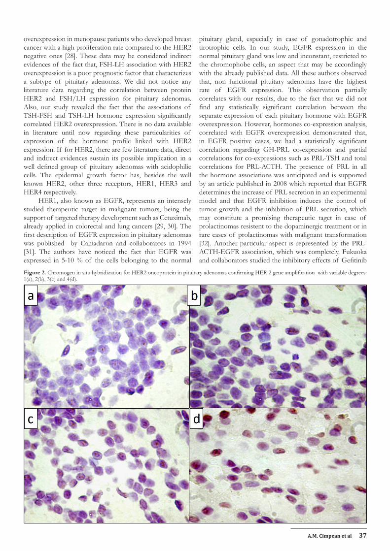
The genic amplification confirmed the immunohistochemical expression of HER2 protein in 33,34 % of cases noted as + 2 by immunohistochemsitry. Cases with genic amplification were extremely heterogenous, on one hand in that which considers the number of tumor cells and, on the other hand regarding the amplification score evidenced by the nuclear presence of three or more dot-like distinct signals or clusters of positive signals(fig.2 a-d)
Analysis of EGFR expression in pituitary adenomas has revealed a percentage of 48,33 % of positive cases for this marker. Three patterns of expression have been observed : the classical one, restricted to the cell membrane (M, fig.1d) with a continuous aspect and a homogenous intensity, a membraneous (continuous or discontinuous) plus cytoplasmatic one (with a granular aspect) (MC, fig. 1e) and cytoplasmic only (C, fig. 1f) with a granular or diffuse aspect. About 51,72 % of cases presented a +2 or +3 score, the rest being noted as +1. The solid growth pattern was observed in 45 % of cases, , while the papillary pattern was observed in 35 % of EGFR positive cases. The rest were, in variable proportions, trabecular type pituitary adenomas, alveolar type, fusiform type and with a mixed growth pattern.
As we have done before for HER2, the study continued with the identification of the correlations between the hormone profile and EGFR expression. Unlike HER2 quantification in which a statistically significant correlation has been demostrated between HER2 expression and PRL secretion, in case of EGFR there was no co-expression demostrated for none of the evaluated pituitary hormones that were quantified separately in comparison with EGFR expression. In order to identify the most frequent hormone associations in the EGFR positive groups the co-expressions of the pituitary hormones were quantified.
In case of GH it has been demonstrated that the
most frequent co-expression was established with PRL, supported by the statistically significant correlation for GH-PRL secreting pituitary adenomas and EGFR (p=0,000). Partial correlation has been registered for PRL-TSH secretion (Kendall=0,053, Spearman=0,047) and a complete correlation for PRL-ACTH (Pearson=0,045, Kendall=0,008 and Spearman=0,009).
EGFR expression was not restricted to tumor epithelial cells only in pituitary adenomas. Other two cell types were frequently observed as being EGFR positive in the present study. Some pituitary adenomas were characterized by a massive acumulation of EGFR positive macrophages situated at the tumor periphery. EGFR expression in pituitary adenomas did not correlate with the expression of other growth factors such as VEGF but neither did it correlate with the overexpression of protein HER2. EGFR negative pituitary adenomas contain cells with a stellate morphology, having cytoplasmic processes inserted amongst tumor cells (fig.1f, inset). Due to the morphology and the pattern of distribution of these EGFR positive cells, we interpreted them as being EGFR positive folicular stellate cells.
DISCUSSIONS
HER1 and HER2 are recognized as being important therapeutic targets in several malignancies as breast [12, 13], lung [14, 15] or gastric cancer [16, 17]. For such tyrosin-kinase receptors tageted therapies were developed, the most known being trastuzumab, approved and used on a large scale in metastatic breast cancer [18], but also targeted anti EGFR therapies [19].
Studies regarding two receptors in pituitary adenomas are scattered. In the past 10 years less than 10 articles focused on the implication of oncoprotein HER2/neu in pituitary adenomas were published, the majority being of an experimental type (http://www.ncbi.nlm.nih.gov/pubmed/?term=c+erbB2%2C+pituitary+adenomas).
Except a single article about EGFR expression and the expression of oncoprotein HER2/neu is described in detail as an expression found in lactotrophic and corticotrophic cells [20], nowadays the prognostic role of these factors in the evolution and progression of pituitary adenomas has not been certified. Unlike what has been described in the previously mentioned article, we did nor observe the expression of protein HER2/neu in the normal pituitary tissue. On the other hand, the percentage of pituitary adenomas that were positive for HER2/neu oncoprotein was similar to the one encountered in case of the metastatic breast cancer, about one third of the analysed cases respectively [21]. Pituitary adenomas had a membraneous expression pattern and a differentiated cytoplasmic one depending on the cell types from pituitary adenomas structure. In basophilic cells the expression of oncoprotein HER2/neu was restricted to the membrane, in a similar manner to that observed and quantified in tumor cells from mamary neoplasms

_____________________________36 Research and Clinical Medicine, 2016, Vol. 1, Nr. 1
[22], while in acidophilic cells the expression of HER2/neu was predominantly granular cytoplasmatic, with or without membraneous intensification, in a similar manner to that described in the majority of gastric cancer that are positive for this marker [23]. The cytoplasmic expression of HER2/neu in gastric cancer was significantly correlated from a statistical point of view with the age and the expression of HER3, a well known receptor that presents the affinity to highly heterodimerize with HER2 and, through this action, being a poor prognostic factor in mamary cancer by determining Transtuzumab therapy resistance [24]. In pituitary adenomas with acidophilic cells the cytoplasmic expression was predominant, an aspect that may suggest a strong hetherodymerization of HER2 with HER3 that may constitute a poor prognostic factor in pituitary adenomas with acidophilic cells also. Unlike other studied regarding the expression of protein HER2 in pituitary adenomas, the present study observed its overexpression, also with a cytoplasmic pattern in chromophobe cells pituitary adenomas, an aspect that was not previously mentioned. Without depending on the solid or the papillary type, HER2 was positive in about 66 % of cases with acidophilic cells. In pure pituitary adenomas, HER2 overexpression was correlated with PRL secretion but not with GH secretion. This correlation sustains even more the implication of HER2 with HER3 hetherodymerization in pituitary adenomas that secrete PRL, an aspect based on the fact that there are previously published data that demonstrated the poor prognostic role of HER2/HER3 association as a factor of high agressiveness in pituitary adenomas that secrete PRL and suggested the fact that the targeted inhibition
of this overexpression may be a useful alternative therapy in case of prolactinomas with an unfavourable evolution and resistant to conventional therapy [25]. In order to support this hypothesis the data published by Zhao and Ren may be taken into consideration; they have demonstrated that Neuregulin secretion (a ligand for HER3) acts as a regulator of PRL synthesis possibly by means of a paracrine/juxtacrine mechanism [26]. On the other hand, in case of pituitary adenomas with a double hormone profile, the simultaneous expression of GH-PRL was significantly correlated from a statistical point of view with HER2 expression. These data are accordingly to the previously published ones [27], which demonstrated the fact that, more than half of the GH-PRL pituitary adenomas cases were positive for HER2 oncoprotein and from these, half presented the overexpression of protein p53, partially ovelapped with a high proliferation index. A particular aspect of our study is represented by types TSH-LH and TSH-FSH hormone associations which were significantly correlated with protein HER2 overexpression. This association was not reported before in literature and may be studied as a prognostic factor. Another pair of hormones that expressed themselves in tandem in the HER2 positive pituitary adenomas was the one constituted by FSH and LH, their co-expression having a complete correlation with the overexpression of protein HER2 in pituitary adenomas. This correlation confirmed the high expression of HER2 in chromophobe cells from the studied pituitary adenomas. The high serum levels of FSH and LH were incriminated as poor prognostic factors associated with (or possible inductors) HER2
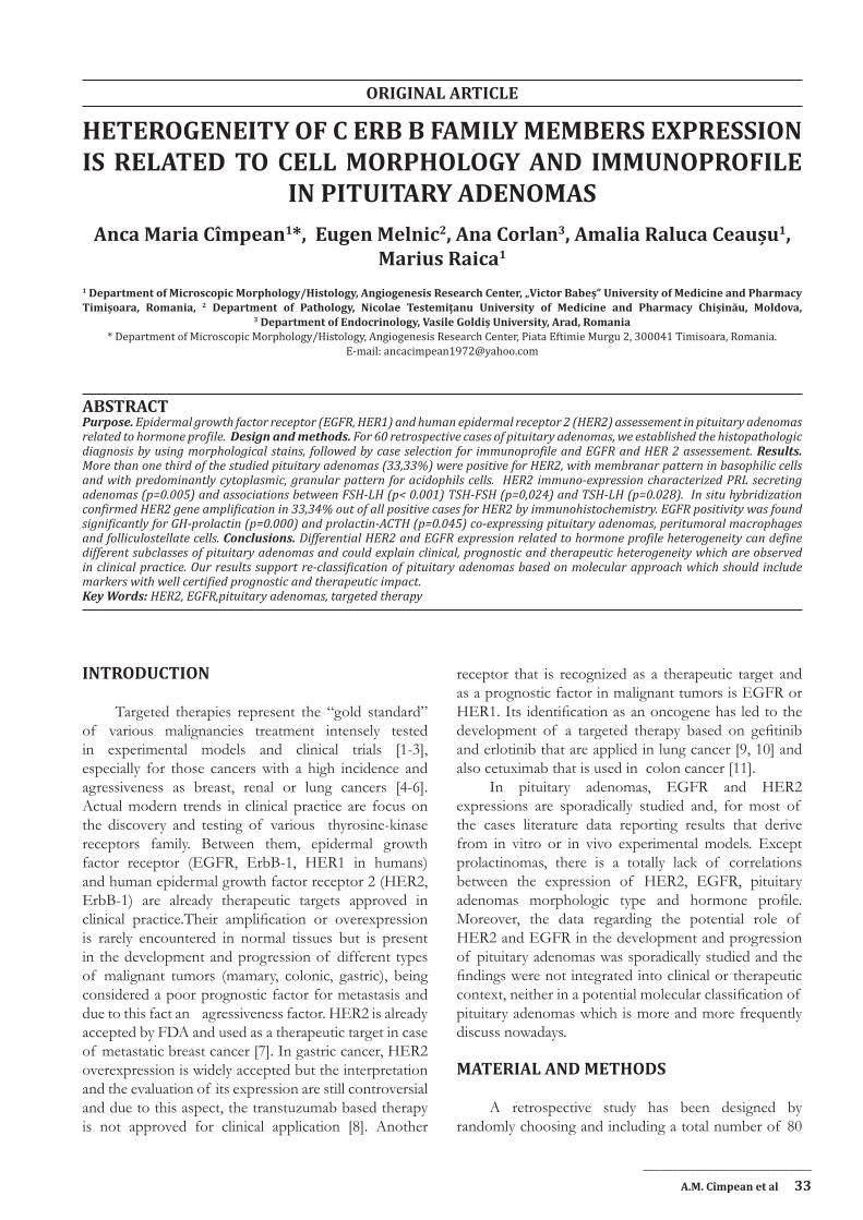
Figure 1. HER2 (a, b, c) and EGFR (d, e, f) immunohistochemical expression in pituitary adenomas. Both members have different and heterogeneous expression patterns membranar restricted (a, d) combined membranar and cytoplasmic (b, e) and cytoplasmic restricted with granular pattern (c, f). Note that EGFR expression was also present in folliculostellate cells in those case where tumor cells were EGFR negative (f, inset).
_____________________________
A.M. Cîmpean et al 37
overexpression in menopause patients who developed breast cancer with a high proliferation rate compared to the HER2 negative ones [28]. These data may be considered indirect evidences of the fact that, FSH-LH association with HER2 overexpression is a poor prognostic factor that characterizes a subtype of pituitary adenomas. We did not notice any literature data regarding the correlation between protein HER2 and FSH/LH expression for pituitary adenomas. Also, our study revealed the fact that the associations of TSH-FSH and TSH-LH hormone expression significantly correlated HER2 overexpression. There is no data available in literature until now regarding these particularities of expression of the hormone profile linked with HER2 expression. If for HER2, there are few literature data, direct and indirect evidences sustain its possible implication in a well defined group of pituitary adenomas with acidophilic cells. The epidermal growth factor has, besides the well known HER2, other three receptors, HER1, HER3 and HER4 respectively.
HER1, also known as EGFR, represents an intensely studied therapeutic target in malignant tumors, being the support of targeted therapy development such as Cetuximab, already applied in colorectal and lung cancers [29, 30]. The first description of EGFR expression in pituitary adenomas was published by Cahiadarun and collaborators in 1994 [31]. The authors have noticed the fact that EGFR was expressed in 5-10 % of the cells belonging to the normal
pituitary gland, especially in case of gonadotrophic and tirotrophic cells. In our study, EGFR expression in the normal pituitary gland was low and inconstant, restricted to the chromophobe cells, an aspect that may be accordingly with the already published data. All these authors observed that, non functional pituitary adenomas have the highest rate of EGFR expression. This observation partially correlates with our results, due to the fact that we did not find any statistically significant correlation between the separate expression of each pituitary hormone with EGFR overexpression. However, hormones co-expression analysis, correlated with EGFR overexpression demonstrated that, in EGFR positive cases, we had a statistically significant correlation regarding GH-PRL co-expression and partial correlations for co-expressions such as PRL-TSH and total correlations for PRL-ACTH. The presence of PRL in all the hormone associations was anticipated and is supported by an article published in 2008 which reported that EGFR determines the increase of PRL secretion in an experimental model and that EGFR inhibition induces the control of tumor growth and the inhibition of PRL secretion, which may constitute a promising therapeutic taget in case of prolactinomas resistent to the dopaminergic treatment or in rare cases of prolactinomas with malignant transformation [32]. Another particular aspect is represented by the PRL-ACTH-EGFR association, which was completely. Fukuoka and collaborators studied the inhibitory effects of Gefitinib
Figure 2. Chromogen in situ hybridization for HER2 oncoprotein in pituitary adenomas confirming HER 2 gene amplification with variable degrees: 1(a), 2(b), 3(c) and 4(d).

_____________________________38 Research and Clinical Medicine, 2016, Vol. 1, Nr. 1
on ACTH secretory isolated cells from pituitary adenomas that secrete ACTH of human and canine origin and demonstrated that EGFR inhibition determines the decrease of tumor dimensions, corticosteroids level as well as the reduction of the omental adipose mass [33]. Due to all these results, the authors consider that the therapy based on Gefitinib may be a welcomed alternative that may reduce surgical interventions carried out for these types of pituitary tumors, of course with an increase in the patient’s comfort. Other studies regarding EGFR expression in pituitary adenomas support our data. Kontogeorgeous and collaborators demonstrated the EGFR overexpression in over 60 % of corticotrophic pituitary adenomas and only in 20 % of the somatotrophic and latotrophic pituitary adenomas [34]. These results sustain the statistically significant correlation obtained for the association of GH-PRL and of PRL-ACTH with EGFR in our study. The decrease of the protein p27/Kip1 in the cases of ACTH secreting pituiray adenomas, correlated with the inhibition of the same type of protein by EGFR expression, suggests the fact that EGFR is implicated in the tumorigenesis of ACTH secreting pituitary adenomas by means of a p27/Kip1 inhibition mechanism [35].
GH-PRL secreting adenomas are characterized by HER2 and EGFR overexpression this finding suggesting a possible existence of different molecular subclasses with a specific phenotype. Foliculo/stelate cells are important components of pituitary adenomas because of their ability to secrete several growth factors with prognostic or therapeutic role. Presence of distinct pituitary adenomas subclasses is also sustain on the one hand by association between FSH/LH and HER2 overexpression and, on the other hand by EGFR association with ACTH secreting pituitary adenomas with or without PRL secretion.
Pituitary adenomas overexpressing growth factors could be targets for new therapies based on humanized monoclonal antibodies against such factors. In this moment, this kind of therapy is not usually applied in pituitary adenomas treatment. Based on different growth factors expression, a reclassification of pituitary adenomas is required. Together with hormone profile, this new classification should include growth factors which can define and identify new molecular classes of pituitary adenomas with different clinical and therapeutic behaviour influencing recurrences and prognosis. Presented findings together with literature data, sugessted that prolactin secreting adenomas represent a special group with particular evolution and prognosis most probably given by interaction between prolactin and growth factors with still unknown involvement in pathogenesis.
ACKNOWLEDGEMENTS
The authors declare that they have no conflict of interests. We are grateful to „Victor Babes” University
of Medicine and Pharmacy which supported us in the development of our work and also, to Professor Mihail Coculescu for his valuable experience in the field.
REFERENCES
1. Korsmeyer R. Critical questions in development of targeted nanoparticle therapeutics. Regen Biomater. 2016;3:143-147.2. Han Li C, Chen Y. Small and Long Non-Coding RNAs: Novel Targets in Perspective Cancer Therapy. Curr Genomics. 2015;16:319-326.3. Pectasides E. Genomic Alterations and Targeted Therapy in Gastric and Esophageal Adenocarcinoma. Clin Ther. 2016 ; pii: S0149-2918(16)30153-9.4. Wisinski KB, Tevaarwerk AJ, Burkard ME, et al. Phase 1 study of an AKT-inhibitor (MK-2206) combined with lapatinib in adult solid tumors followed by dose-expansion in advanced HER2+ breast cancer. Clin Cancer Res. 2016 ; pii: clincanres.2365.2015. 5. Langrand-Escure J, Vallard A, Rivoirard R, et al. Safety assessment of molecular targeted therapies in association with radiotherapy in metastaticrenal cell carcinoma: a real-life report. Anticancer Drugs. 2016;27:427-432. 6. Berardi R, Rinaldi S, Santoni M, et al. Prognostic models to predict survival in patients with advanced non-small cell lung cancer treated with first-line chemo- or targeted therapy. Oncotarget. 2016;doi: 10.18632/oncotarget.8309.7. Michiels S, Pugliano L, Marguet S, et al. Progression-free survival as surrogate endpoint for overall survival in clinical trials of HER2-targeted agents in HER2-positive metastatic breast cancer. Ann Oncol. 2016; Mar 8. pii: mdw132. 8. Kataoka H, Mori Y, Shimura T, et al. A phase II prospective study of the trastuzumab combined with 5-weekly S-1 and CDDP therapy forHER2-positive advanced gastric cancer. Cancer Chemother Pharmacol. 2016;77:957-62.9. Carbonnaux M, Souquet PJ, Meert AP, et al. Inequalities in lung cancer: a world of EGFR. Eur Respir J. 2016;47:1502-9.10. Martínez E, Martínez M, Rico M, et al. Feasibility, tolerability, and efficacy of the concurrent addition of erlotinib to thoracic radiotherapy in locally advanced unresectable non-small-cell lung cancer: a Phase II trial. Onco Targets Ther. 2016;9:1057-1066. 11. Yan Y, Grothey A. Molecular profiling in the treatment of colorectal cancer: focus on regorafenib. Onco Targets Ther. 2015;8:2949-2957. 12. Davis NM, Sokolosky M, Stadelman K, et al. Deregulation of the EGFR/PI3K/PTEN/Akt/mTORC1 pathway in breast cancer: possibilities for therapeutic intervention. Oncotarget. 2014;5:4603-4650. 13. Ieni A, Barresi V, Caltabiano R, et al. Discordance rate of HER2 status in primary breast carcinomas versus synchronous axillary lymph node metastases: a multicenter retrospective investigation. OncoTargets Ther. 2014 ;7:1267-1272. 14. Heymach JV, Lockwood SJ, Herbst RS, et al. EGFR biomarkers predict benefit from vandetanib in combination with docetaxel in a randomized phase III study of second line treatment for patients with advanced non-small cell lung cancer. Ann Oncol. 2014;25:1941-1948. 15. Cretella D, Saccani F, Quaini F, et al. Trastuzumab emtansine is active on HER-2 overexpressing NSCLC cell lines and overcomes gefitinib resistance. Mol Cancer. 2014;13:143. 16. Qi W, Li X, Zhang Y, Yao R, et al. Overexpression of Her-upregulates FoxM1 in gastric cancer. Int J Mol Med. 2014;33:1531-1538.

_____________________________
A.M. Cîmpean et al 39
17. Nielsen TO, Friis-Hansen L, Poulsen SS, et al. Expression of the EGF family in gastric cancer: downregulation of HER4 and its activating ligand NRG4. PLoS One. 2014;9:e94606. 18. Callahan R. Ado-Trastuzumab Emtansine in Metastatic HER2-Positive Breast Cancer. J Adv Pract Oncol. 2014;5:134-139. 19. Kuiper JL, Smit EF. Challenges in the Management of EGFR -Mutated NonSmall Cell Lung Cancer Patients with Acquired Resistance to Tyrosine Kinase Inhibitors. Oncology. 2014;87:83-94. 20. Cooper O, Vlotides G, Fukuoka H, et al. Expression and function of ErbB receptors and ligands in the pituitary. Endocr Relat Cancer. 2011;18: R197-211. 21. Pinto AC, Ades F, de Azambuja E, et al. Trastuzumab for patients with HER2 positive breast cancer: delivery, duration and combination therapies. Breast. 2013; 22:S152-5. 22. Martin V, Cappuzzo F, Mazzucchelli L, et al. HER2 in solid tumors: more than 10 years under the microscope; where are we now? Future Oncol. 2014 ;10:146986. 23. Jácome AA, Wohnrath DR, Scapulatempo Neto C, et al. Prognostic value of epidermal growth factor receptors in gastric cancer: a survival analysis by Weibull model incorporating long-term survivors. Gastric Cancer. 2014;17:76-86. 24. Green AR, Barros FF, Abdel-Fatah TM, et al. HER2/HER3 heterodimers and p21 expression are capable of predicting adjuvant trastuzumab response in HER2+ breast cancer. Breast Cancer Res Treat. 2014;145:33-44. 25. Vlotides G, Cooper O, Chen YH, et al. Heregulin regulates prolactinoma gene expression. Cancer Res. 2009;69:4209-4216. 26. Zhao W, Ren SG. Neuregulin-1 (Nrg1) is mainly expressed in rat pituitary gonadotroph cells and possibly regulates prolactin (PRL) secretion in a juxtacrine manner. J Neuroendocrinol. 2011;23:1252-1262.
27. Botelho CH, Magalhães AV, Mello PA, et al. Expression of p53, Ki-67 and c-erb B2 in growth hormone-and/or prolactin-secreting pituitary adenomas. Arq Neuropsiquiatr. 2006;64:60-66. 28. Zhou J, Chen Y, Huang Y, et al. Serum follicle-stimulating hormone level is associated with human epidermal growth factor receptor type and Ki67 expression in post-menopausal females with breast cancer. Oncol Lett. 2013;6:1128-1132. 29. Ciombor KK, Berlin J. Targeting metastatic colorectal cancer - present and emerging treatment options. Pharmgenomics Pers Med. 2014;7:137-144. 30. Komaki R, Paulus R, Blumenschein GR Jr, et al. EGFR expression and survival in patients given cetuximab and chemoradiation for stage III non-small cell lung cancer: A secondary analysis of RTOG 0324. Radiother Oncol. 2014;112:30-36. 31. Chaidarun SS, Eggo MC, Sheppard MC, et al. Expression of epidermal growth factor (EGF), its receptor, and related oncoprotein (erbB-2) in human pituitary tumors and response to EGF in vitro. Endocrinology. 1994;135:2012-2021. 32. Vlotides G, Siegel E, Donangelo I, et al. Rat prolactinoma cell growth regulation by epidermal growth factor receptor ligands. Cancer Res. 2008;68:6377-6386. 33. Fukuoka H, Cooper O, Ben-Shlomo A, et al. EGFR as a therapeutic target for human, canine, and mouse ACTH-secreting pituitary adenomas. J Clin Invest. 2011;121:4712-4721. 34. Kontogeorgos G, Stefaneanu L, Kovacs K, et al. Localization of Epidermal Growth Factor (EGF) and Epidermal Growth Factor Receptor (EGFr) in Human Pituitary Adenomas and Nontumorous Pituitaries: An Immunocytochemical Study. Endocr Pathol. 1996;7:63-70. 35. Theodoropoulou M, Arzberger T, Gruebler Y, et al. Expression of epidermal growth factor receptor in neoplastic pituitary cells: evidence for a role in corticotropinoma cells. J Endocrinol. 2004;183:385-394.