herg Encodes a K+ Current Highly Conserved in Tumors of...
9
[CANCER RESEARCH 58. 815-822. February 15. 19981 herg Encodes a K+ Current Highly Conserved in Tumors of Different Histogenesis: A Selective Advantage for Cancer Cells?1 Laura Bianchi, Barbara Wible, Annarosa Arcangeli, Maurizio Tagliatatela, Ferdinando Morra, Pasqualina Castaido, Olivia Crociani, Barbara Rosati, Laura Faravelli, Massimo Olivotto, and Enzo Wanke2 Dipartimento di Fisiologìae Biochimica Generali. Università degli Studi di Milano, Milan, Italy {B. R., L. F., E. W.], Rammelkamp Center for Education and Research, MelroHealth Campus. Case Western Resen-e University: Cleveland. Ohio 44109-1998 IL B., B. W.]. Istituto di Patologia Generale. Università degli Studi di Firenze, Florence. Italy ¡A.A., O. C., M. O.}: and Dipartimento di Neuroscienze, Sezione di Farmacologia. Università degli Studi di Napoli Federico II, Naples, Italy ¡M.T., F. M., P. C.l ABSTRACT The human ether-a-go-go-related gene (herg) encodes a K+ current (7HERG)that plays a fundamental role in heart excitability by regulating the action potential repolarization </Kr>:mutations of this gene are re sponsible for the chromosome 7-linked long QT syndrome (LQT2). In this report, we show that in a variety (n = 17) of tumor cell lines of different species (human and murine) and distinct histogenesis (neuroblastoma, rhabdomyosarcoma, adenocarcinoma, lung microcytoma, pituitary tu mors, insulinoma ß-cells,and monoblastic leukemia), a novel K ' inward- rectifier current </,K), which is biophysically and pharmacologically sim ilar to /nim.- can be recorded with the patch-clamp technique. Northern blot experiments with a human herg cDNA probe revealed that both in human and murine clones the very high expression of herg transcripts can be quantified in at least three clearly identifiable bands, suggesting an alternative splicing of HERG niRN A. Moreover, we cloned a cDNA en coding for /,„from the SH-SY5Y human neuroblastoma. The sequence of this cDNA result was practically identical to that already reported for herg, indicating a high conservation of this gene in tumors. Consistently, the expression of this clone in Xenopus oocytes showed that the encoded K ' channel had substantially all of the biophysical and pharmacological properties of the native /(R described for tumor cells. In addition, in the tumor clones studied, /,K governs the resting potential, whereas it could not be detected either by the patch clamp or the Northern blot techniques in cells obtained from primary cell cultures of parental tissues (sensory neurons and myotubes), whose resting potential is controlled by the classical k ' anomalous rectifier current. This current substitution had a profound impact on the resting potential, which was markedly depolar ized in tumors as compared with normal cells. These results suggest that /iR is normally only expressed during the early stages of cell differentia tion frozen by neoplastic transformation, playing an important patho- physiological role in the regulatory mechanisms of neoplastic cell survival. In fact, because of its biophysical features, /, K, besides keeping the resting potential within the depolarized values required for unlimited tumor growth, could also appear suitable to afford a selective advantage in an ischemie environment. INTRODUCTION It has long been known that cancer cells have remarkably lower resting potentials (VREST)than normal cells from the same tissues, and it has been proposed that this is an essential prerogative for cells destined to unlimited growth (reviewed in Ref. l). Although an obvious explanation of this feature might be ascribed to type and density of K+ channels, this topic has been scarcely explored with Received 6/24/97; accepted 12/16/97. The costs of publication of this article were defrayed in part by the payment of page charges. This article must therefore be hereby marked advertisement in accordance with 18 U.S.C. Section 1734 solely to indicate this fact. ' This work was supported by grants from the Associazione Italiana per la Ricerca sul Cancro, Consiglio Nazionale delle Ricerche (finalized project. Applicazioni Cllniche per la Ricerca Oncologica). Ministero dell'Università e della Ricerca Scientifica e Tecno logica, Associazione Italiana contro le Leucemie, and NIH Grant HL36930. L. B. is a PhD student from Dipartimento di Scienze Fisiologiche, Università di Firenze. L. F. and B. R. are PhD students from Dipartimento di Fisiologia e Biochimica Generali, Università di Milano. 2 To whom requests for reprints should be addressed, at University of Milan, Dipar timento di Fisiologia e Biochimica Generali, Via Celoria 26-20133 Milano. Italy. Phone: 39-2-70644609; Fax: 39-2-70632884; E-mail: [email protected]. up-to-date electrophysiological and molecular biology techniques. This scientific gap is particularly amazing in view of the huge knowl edge available about K+ channels and their role in the control of mitogenesis (2, 3). In this field, special attention has been devoted recently to the members of an evolutionarily conserved multigene family of voltage-activated K+ channels that contribute to the signal ing capacity of excitable as well as nonexcitable cells (4). The prototype of one such family is the Drosophila eag* locus (5), which encodes a K+ channel protein, the heterologous expression of which in Xenopus oocytes produces a voltage-dependent outward K+ current (6). A number of eag-related genes have now been cloned, including: elk (Drosophila), m-eag (mouse), and herg (human-eci^-related gene), which was first isolated from a human hippocampus cDNA library and mapped to chromosome 7 (4). The first clue that the herg gene product may play a role in the regulation of human tissue excitability was provided by Curran et al. (7), who found that herg mutations were related to an inherited ventricular arrhythmia (LQT2). Heterologous expression of the herg product (8) showed that it encodes for a K+ current (/HERC;)-tne major subunit of /Kr channels, which contribute to the repolarization of cardiac action potential (9). This current is specifically blocked by class III antiarrhythmic drugs, such as sotalol and its analogue E4031, as well as by WAY 123,398 and dofetilide (9, 10). Evidence for the expression of HERG-like currents in tissues other than those of the heart emerged from the discovery that a current having the biophysical and pharmacological properties of HERG (/IR) was constitutively and highly expressed in neuroblastoma cell lines across various species, from mouse to human, and shared the depend ence of the conductance on [K+]0 and, like the /HKR(; expressed in Xenopus oocytes, was blocked by E4031, WAY-123,398, Cs+, Ba2+, and La3+ (11-14). The voltage-dependent gating of the neuroblas toma /|R was characterized by intrinsic inactivation and activation curves that together make the channel operate within the central region of VR[;STof neuroblastoma cells (about -40 mV). These gating properties are linked to the mechanisms controlling the cell cycle in such a way that the /,R activation curves vary widely in the cells of unsynchronized populations, whereas the synchronization of cells in the G, phase of the cell cycle greatly reduces this variability (12). We here report that /IR is not only present in a number of different neuroblastoma cell lines but also in tumors of different histogenesis. On the contrary, in normal cells derived from tissues with the same histogenesis as the corresponding tumor cell lines, /,R was undetect- able, and VRESTwas governed by different channels displaying bio physical properties similar to the classical inward rectifier current (IRK). Interestingly, the cloning of a herg cDNA from SH-SY5Y human neuroblastoma cells and its expression in Xenopus oocytes revealed that herg and the corresponding K * current are maintained substantially unmodified after the neoplastic transformation, provid ing a new experimental explanation for the depolarized VRHST,which characterizes cancer cells. ' The abbreviations used are: eag, ether-a-go-go; DRG, dorsal root ganglion. 815 Research. on February 17, 2019. © 1998 American Association for Cancer cancerres.aacrjournals.org Downloaded from
Transcript of herg Encodes a K+ Current Highly Conserved in Tumors of...
[CANCER RESEARCH 58. 815-822. February 15. 19981
herg Encodes a K+ Current Highly Conserved in Tumors of Different Histogenesis:
A Selective Advantage for Cancer Cells?1
Laura Bianchi, Barbara Wible, Annarosa Arcangeli, Maurizio Tagliatatela, Ferdinando Morra, Pasqualina Castaido,Olivia Crociani, Barbara Rosati, Laura Faravelli, Massimo Olivotto, and Enzo Wanke2
Dipartimento di Fisiologìae Biochimica Generali. Università degli Studi di Milano, Milan, Italy {B. R., L. F., E. W.], Rammelkamp Center for Education and Research,MelroHealth Campus. Case Western Resen-e University: Cleveland. Ohio 44109-1998 IL B., B. W.]. Istituto di Patologia Generale. Università degli Studi di Firenze, Florence.
Italy ¡A.A., O. C., M. O.}: and Dipartimento di Neuroscienze, Sezione di Farmacologia. Università degli Studi di Napoli Federico II, Naples, Italy ¡M.T., F. M., P. C.l
ABSTRACT
The human ether-a-go-go-related gene (herg) encodes a K+ current
(7HERG)that plays a fundamental role in heart excitability by regulatingthe action potential repolarization </Kr>:mutations of this gene are responsible for the chromosome 7-linked long QT syndrome (LQT2). In thisreport, we show that in a variety (n = 17) of tumor cell lines of different
species (human and murine) and distinct histogenesis (neuroblastoma,rhabdomyosarcoma, adenocarcinoma, lung microcytoma, pituitary tumors, insulinoma ß-cells,and monoblastic leukemia), a novel K ' inward-
rectifier current </,K), which is biophysically and pharmacologically similar to /nim.- can be recorded with the patch-clamp technique. Northern
blot experiments with a human herg cDNA probe revealed that both inhuman and murine clones the very high expression of herg transcripts canbe quantified in at least three clearly identifiable bands, suggesting analternative splicing of HERG niRN A. Moreover, we cloned a cDNA encoding for /,„from the SH-SY5Y human neuroblastoma. The sequence of
this cDNA result was practically identical to that already reported forherg, indicating a high conservation of this gene in tumors. Consistently,the expression of this clone in Xenopus oocytes showed that the encodedK ' channel had substantially all of the biophysical and pharmacological
properties of the native /(R described for tumor cells. In addition, in thetumor clones studied, /,K governs the resting potential, whereas it couldnot be detected either by the patch clamp or the Northern blot techniquesin cells obtained from primary cell cultures of parental tissues (sensoryneurons and myotubes), whose resting potential is controlled by theclassical k ' anomalous rectifier current. This current substitution had a
profound impact on the resting potential, which was markedly depolarized in tumors as compared with normal cells. These results suggest that/iR is normally only expressed during the early stages of cell differentiation frozen by neoplastic transformation, playing an important patho-
physiological role in the regulatory mechanisms of neoplastic cell survival.In fact, because of its biophysical features, /, K, besides keeping the restingpotential within the depolarized values required for unlimited tumorgrowth, could also appear suitable to afford a selective advantage in anischemie environment.
INTRODUCTION
It has long been known that cancer cells have remarkably lowerresting potentials (VREST)than normal cells from the same tissues, andit has been proposed that this is an essential prerogative for cellsdestined to unlimited growth (reviewed in Ref. l). Although anobvious explanation of this feature might be ascribed to type anddensity of K+ channels, this topic has been scarcely explored with
Received 6/24/97; accepted 12/16/97.The costs of publication of this article were defrayed in part by the payment of page
charges. This article must therefore be hereby marked advertisement in accordance with18 U.S.C. Section 1734 solely to indicate this fact.
' This work was supported by grants from the Associazione Italiana per la Ricerca sul
Cancro, Consiglio Nazionale delle Ricerche (finalized project. Applicazioni Cllniche perla Ricerca Oncologica). Ministero dell'Università e della Ricerca Scientifica e Tecno
logica, Associazione Italiana contro le Leucemie, and NIH Grant HL36930. L. B. is a PhDstudent from Dipartimento di Scienze Fisiologiche, Università di Firenze. L. F. and B. R.are PhD students from Dipartimento di Fisiologia e Biochimica Generali, Università diMilano.
2 To whom requests for reprints should be addressed, at University of Milan, Dipar
timento di Fisiologia e Biochimica Generali, Via Celoria 26-20133 Milano. Italy. Phone:39-2-70644609; Fax: 39-2-70632884; E-mail: [email protected].
up-to-date electrophysiological and molecular biology techniques.
This scientific gap is particularly amazing in view of the huge knowledge available about K+ channels and their role in the control of
mitogenesis (2, 3). In this field, special attention has been devotedrecently to the members of an evolutionarily conserved multigenefamily of voltage-activated K+ channels that contribute to the signal
ing capacity of excitable as well as nonexcitable cells (4). Theprototype of one such family is the Drosophila eag* locus (5), whichencodes a K+ channel protein, the heterologous expression of whichin Xenopus oocytes produces a voltage-dependent outward K+ current
(6). A number of eag-related genes have now been cloned, including:elk (Drosophila), m-eag (mouse), and herg (human-eci^-related gene),
which was first isolated from a human hippocampus cDNA libraryand mapped to chromosome 7 (4).
The first clue that the herg gene product may play a role in theregulation of human tissue excitability was provided by Curran et al.(7), who found that herg mutations were related to an inheritedventricular arrhythmia (LQT2). Heterologous expression of the hergproduct (8) showed that it encodes for a K+ current (/HERC;)-tne major
subunit of /Kr channels, which contribute to the repolarization ofcardiac action potential (9). This current is specifically blocked byclass III antiarrhythmic drugs, such as sotalol and its analogue E4031,as well as by WAY 123,398 and dofetilide (9, 10).
Evidence for the expression of HERG-like currents in tissues other
than those of the heart emerged from the discovery that a currenthaving the biophysical and pharmacological properties of HERG (/IR)was constitutively and highly expressed in neuroblastoma cell linesacross various species, from mouse to human, and shared the dependence of the conductance on [K+]0 and, like the /HKR(; expressed inXenopus oocytes, was blocked by E4031, WAY-123,398, Cs+, Ba2+,and La3+ (11-14). The voltage-dependent gating of the neuroblas
toma /|R was characterized by intrinsic inactivation and activationcurves that together make the channel operate within the centralregion of VR[;STof neuroblastoma cells (about -40 mV). These gating
properties are linked to the mechanisms controlling the cell cycle insuch a way that the /,R activation curves vary widely in the cells ofunsynchronized populations, whereas the synchronization of cells inthe G, phase of the cell cycle greatly reduces this variability (12).
We here report that /IR is not only present in a number of differentneuroblastoma cell lines but also in tumors of different histogenesis.On the contrary, in normal cells derived from tissues with the samehistogenesis as the corresponding tumor cell lines, /,R was undetect-
able, and VRESTwas governed by different channels displaying biophysical properties similar to the classical inward rectifier current(IRK). Interestingly, the cloning of a herg cDNA from SH-SY5Y
human neuroblastoma cells and its expression in Xenopus oocytesrevealed that herg and the corresponding K * current are maintained
substantially unmodified after the neoplastic transformation, providing a new experimental explanation for the depolarized VRHST,whichcharacterizes cancer cells.
' The abbreviations used are: eag, ether-a-go-go; DRG, dorsal root ganglion.
815
Research. on February 17, 2019. © 1998 American Association for Cancercancerres.aacrjournals.org Downloaded from
HERO K* CHANNELS AND CLONING OF HEKC IN TUMOR CELLS
MATERIALS AND METHODS
Cell Culture. The human rhabdomyosarcoma TE671. the mouse neuroblastoma N18TG2 (provided by Prof. G. Augusti-Tocco, Department of Cel
lular Biology, University of Rome, Rome, Italy), the DRG X neuroblastoma
hybrid cell line Fl I. and the human neuroblastoma SHSY5Y (Prof. G. Tarone,Department of Genetics, Biology and Medical Chemistry. University of Turin.Turin. Italy) were cultured in DMEM containing 4.5 g/1 of glucose and 10%PCS (5% for the SHSY5Y). The mouse/rat neuroblastoma-glioma NG108-15(provided by Prof. Augusti-Tocco) was cultured in DMEM-HAT medium
containing 4.5 g/1 of glucose and 10% of FCS. The rat pheochromocytomaPC12 and small cell lung cancer cells NCI-N592, GLC8, and H69 (kindly
provided by Prof. F dementi. Department of Pharmacology. University ofMilan. Milan, Italy); the pituitary cell lines GH,, GH4. and MMQ (kindlyprovided by Dr. I. S. Login. University of Virginia. School of Medicine,Charlottesville. VA); the human mammary gland adenocarcinoma cells SK-BR-3 and the monoblastic leukemia line FLG29.1 (kindly provided by P.
Bernabei. Hematology Unit. Florence. Italy) were all cultured in RPMI 1640containing 10% horse serum and 5% FCS. The human myotubes were culturedin MEM containing 15% FCS. epidermal growth factor (Life Technologies.Inc.) and insulin (Sigma Chemical Co.). The neonatal rat DRG cells wereprepared as described (15). The anterior pituitary cells were dissected from150-g Wistar rats; dissociated with trypsin (25%). collagenase A (25%). andDNase (2.5%); and incubated for 30 min at 37°C.The resulting suspension wasplated onto 35-mm dishes at a density of 3 x 10' cells/ml. The plating medium
was DMEM containing 4.5 g/1 of glucose and 10% FCS. All of the cells wereincubated at 37°C in a humidified atmosphere with 5% CO2 (10% for
SHSY5Y andFll).Molecular Biology. Total RNA from subconfluent cultures of SH-SY5Y
cells was isolated using RNA STAT-60 isolation kit (Tel-Test B. Inc.).
Overlapping partial fragments of herg cDNA were isolated by means ofreverse transcription-PCR using the following oligo pairs (the nucleotide
numbers refer to the HERG sequence: accession number U04270): fragment 1.forward primer S'-GATCGGGCCCCTCAGGATGCCGGTGCGGAGGG-GCCAC-3' (part of the untranslated region, APAI restriction site, and nucle-
otides I to 21 of the translated region; the starting codon is underlined) andreverse primer 5'-GCCCTTGAGGTCGACAAAGTTGAGGGTG-3' (nucleo-
tides 1008 to 1035); fragment 2. forward primer in which a SALI restriction sitewas introduced (C instead of G. underlined) S'-CTCAACTTTGTCGACCT-CAAGGGCGAC-3' (nucleotides 1013 to 1035) and reverse primer 5'-ACT-CAGGGAAGCCCTTCAGC-3' (nucleotides 2148 to 2167); and fragment 3,forward primer S'-TCCAGCGGCTGTACTCGGGC-S' (nucleotides 1988 to2007) and reverse primer S'-GGACCAGAAGTGGTCGGAGAACTC-S' (nucleotides 2539 to 2562); fragment 4, forward primer 5'-GGCTCCATC-GAGATCGAGATCCTGCGGGGC-3' (nucleotides 2353 to 2376) and reverseprimer 5 ' -GATCGAATTCCTAACTGCCCGGGTCCGAGCC-3 '.
PCR was performed as follows: 35 cycles (95°C,30 s; 55°C,30 s: and 72°C,30 s), followed by a final extension for 10 min at 72°C.The bands of the
expected molecular weight were gel purified, subcloned into PCRII (Invitro-gen. San Diego. CA). and sequenced. The full-length herg cDNA was assem
bled from the overlapping fragments in a modified pSP64 vector in which twonew restriction sites were introduced (EcoRV and Nru\) 3' to the polyfA)* tail.
For expression in Xenopus oocytes, the construct was linearized using Nrul andcRNA prepared with the mMESSAGEmMACHINE in vitro transcription kit(Ambion. Austin. TX) with SP6 polymerase. The cRNA was precipitated andresuspended in 0.1 M KCI. The integrity of the cRNA was confirmed, andconcentration was estimated on a formaldehyde/agarose gel by comparisonwith an RNA ladder (Life Technologies, Inc., Gaithersburg, MD).
Northern Blot Analysis. Northern blotting was performed using theNorthernMax kit (Ambion). Five to 20 jj.g of total RNA from each of the tumorcell lines and rat brain and heart (Ambion) were electrophoresed on a 1%agarose-formaldehyde gel. Ethidium bromide (10 /¿g/ml)was added to the
samples prior to electrophoresis to allow visualization of the total RNA beforeblotting. The transfer to BrightStar-Plus membranes (Ambion) was carried outusing the manufacturer's protocol. The blot was probed with the entire herg or
a fragment (444 bp) encompassing nucleotides 1691-2135 (accession number
U04270), which was obtained by means of PCR amplification from the hergplasmid. Both probes were labeled with 12P (DecaPrime kit; Ambion) by
means of random priming. Hybridization was overnight at 42°Cin the solution
provided in the NorthernMax kit at a probe concentration of 2 X IO6 cpm/ml.
The membrane was washed with the buffers supplied with the kit: twice for 10min at room temperature in wash buffer I and twice for 20 min at 50°Cin wash
buffer 2. The transcripts were visualized by autoradiography using KodakBiomax MS film and an intensifying screen for 16-52 h. Higher stringencyconditions (hybridization at 65°Cand washing at 65°C)gave similar results.
Patch Clamp Recordings. The currents were recorded at room temperature using an amplifier Axopatch 1-D (Axon Instruments, Inc., Foster City,CA). The whole-cell configuration of the patch-clamp technique was adoptedusing pipettes, the resistances of which were in the range of 5-10 MÃŽÃŽ.Pipette
resistance, cell capacitance, and series resistance errors were carefully compensated for by introducing a compensation of about 80-90% before each
voltage clamp protocol run. The cells were perfused with an extracellularsolution delivered into the chamber where the cells were plated by means ofhypodermic needles inserted into a capillary with a small hole. The standardextracellular solution contained 130 mM NaCI, 2 mM KCI, 2 HIMCaCl2, 2 mMMgCU. 10 mM HEPES-NaOH. and 5 mM glucose 5, pH 7.4. When necessary.
the NaCI was replaced by KCI 5 and 40 mM. The standard pipette solution at[Ca2+]¡= 10~7 M (pCa 7) contained 120 mM K+-aspartate, 10 mM NaCI, 2 mM
MgCU, 4 mM CaCU, 10 mM EGTA-KOH, and 10 mM HEPES-KOH. Thejunction potential (~-5 mV) was not corrected.
Oocyte Handling and Voltage-Clamp Recordings. The Xenopus wereanesthetized by immersion in 0.2% tricaine for 15-30 min. The ovarian lobes
were digested using 2 mg/ml type IA collagenase (Sigma Chemical Co.) in aCa2+-free solution to remove follicle cells. Stage IV and V oocytes wereinjected with herg cRNA (0.1-0.5 /ig/^1; 40 nl) and then cultured in Earth's
solution (88 mM NaCI 88, 1 mM KCI, 0.4 mM CaCU. 0.33 mM Ca(NO,)2, 1 mMMgSO4, 2.4 mM NaHCO,, and 10 mM HEPES. pH 7.4). The oocyte measurements were made 2-5 days after the injection at room temperature usingstandard two microelectrode voltage-clamp techniques. Glass microelectrodes
were filled with 3 M KCI. and their tips were broken to obtain tip resistancesof 3-6 MÌÌ.The oocytes were voltage clamped using a commercially available
amplifier (Warner Instrument Corp.). The standard bath solution contained 120mM NaOH, 2 mM KOH, 1 mM CaCU, 1 mM MgCU, 10 mM HEPES, and 122mM methanesulfonic acid. When necessary, the NaOH was replaced by 5 and40 mM KOH. During the acquisition and data analysis of both the current- andvoltage-clamp recordings, the pClamp (Axon Instruments, Foster City, CA)
and Origin (Microcal, Inc.. Northampton. MA) software were routinely usedon a 486DX2 PC.
RESULTS
Expression of herg Transcripts and /,,< Currents in VariousNeuroblastoma Cell Lines. Using Northern blot analysis and patchclamp recordings, we studied the expression of herg transcripts andthe corresponding K+ currents in the following neuroblastoma cell
lines: SH-SY5Y and SK-NBE (human); N18T42 and 41A3 (murine);Fil (rat DRG-mouse N18TG2 neuroblastoma hybrid); andNG108-15 (mouse-rat hybrid neuroblastoma-glioma), as shown in
Fig. 1/4, bottom.The Northern blot of RNA from SH-SY5Y cells showed three
bands of 4.4, 2.9, and 2.4 kb, which hybridized to the herg probe; thelargest band (4.4 kb) was the most intense of the three. The same threebands were also shown by the other human line, SK-NBE (Fig. IB,
bottom), although the second band (2.9 kb) was much more prevalentthan the others. The existence of multiple transcripts suggests thatalternative splicing of herg mRNA may occur in human neuroblasto-
mas, with quantitative differences from one cell line to another; thealternative splicing of herg mRNA has been suggested for humanheart mRNA with two bands of 4.4 and 4.1 kb visible on Northernblots (7). The patch clamp recordings obtained using the protocolshown in Fig. 1C revealed no substantial differences in the inwardcurrents of the SH-SY5Y and SK-N-BE cells (Fig. 1, A and B, top).The mouse and mouse-rat hybrid lines (panels C-F) expressed the
same /,R profile as the human lines but showed only a single band of4 kb in the Northern blot. This means that either the fragment of
816
Research. on February 17, 2019. © 1998 American Association for Cancercancerres.aacrjournals.org Downloaded from

HERG K* CHANNELS AND CLONING OF HERC IN TUMOR CELLS
Human NB Mouse NB
rDRG/ mNB ml rHYBRID NB-GLIOMA
SH-SY5Y SK-N-BE N18TG2 D 41A3 F-ll NG108-15 Q Protocol|10mV
1-80 mV
-120 mV
H RatBrain Heart
Human Non-Nervous Tumours
TE671
/ -I
SK-BR-3 M NCI-N592 FLG29
SH-SY5Y
SK-N-BE
N18TO2
X 41A3
V FilNG108-15
• TE671O SK-BR-3
D N1H-N592
FLO29
0.0
-80 -60 -40 0 mV
Fig. 1. Expression of herg mRNA and the corresponding HERG currents in various tumor cell lines. Each panel shows the /,R currents (upper} and Northern blots (lower) of eachcell type. The currents were elicited in a solution containing 40 IHMKC1 according to the protocol shown in G: the membrane potential was held at various potentials from -80 to-10 for 20 s (preconditioning step) and then stepped up to the test potential ( - 120 mV) for 200 ms. The Northern blot analyses were performed on total RNA. The blots were hybridizedusing a 12P-labeled probe encoding the whole sequence or only the region between S3 and S6. The arrows on the left indicate the molecular weights of the bands observed in SY5Y
cells (4.4, 2.9, and 2.4 kb). H. Northern blots of rat brain and heart mRNA. In O. the experimental points (symbols) were obtained from the currents shown in the various insets (A-fand 7-/V) by plotting their normalized peak amplitude (///max) against the preconditioning potential. The points were then best fitted to Boltzmann equations and gave the followingvalues of Vl/2 and slope (k; mV) for each of the cell types: SH-SY5Y, -33.1 and 6.7; SK-N-BE, -32 and 6.1; NI8TG2, -36.5 and 7.1; 41A3. -38.5 and 5.2; Fl 1, -41 and 4.2;NG108, 15, -43, and 5.1; TE67I, -33.3 and 4.74; SK-BR-3, -38.7 and 3.8; NCI-N592, -56.1 and 4.9; FLG29, -23.4 and 4.2. The scale burs in A-F and I-N were 25 ms for timeand the following for each cell type (pA): SH-SY5Y. 100; SK-N-BE. 75; NI8TG2, 200; 41 A3, 150; FI 1, 200; NG108, 300; TE671, 500; SK-BR-3, 500; NCI-N592. 1000; and FLG29,
100.
human herg cDNA used as a probe is not capable of detecting thealternatively spliced transcripts revealed by SH-SY5Y and SK-N-BE RNA or the mouse and/or rat herg transcripts are not
alternatively spliced. Unlike those of human tissues, Northern blotsof rat RNAs from normal tissues known to express erg. brain andheart, also show only a single band (Fig. 1/f). Here the transcriptwas of about 4.3 kb, which is closer in size to the largest transcriptin human tumor cells than to the mRNA detected in mouse/ratneuroblastomas.
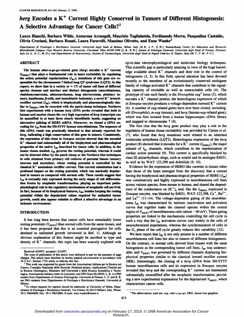
Although we did not carry out strictly quantitative comparisonsamong various cell types, we checked the mRNA load for eachsample from the intensity of the 28S and 18S mRNA bands asrevealed by ethidium bromide prior to blotting. We considered thisprocedure the most suitable to have a roughly comparative estimateof herg expression in cells of completely different histogenesis, forwhich it was hard to choose a priori a gene expressed at the samelevel, herg expression (Fig. 2A) and mRNA loading (Fig. 20) areshown for some of the cell lines reported in Fig. 1 and for normaltissues.
herg Expression and /IK in Nonnervous Tumors of DistinctHistogenesis. The high level of herg expression in all of the testedneuroblastoma cell lines prompted us to explore whether herg and/or/1Rwere also detectable in nonnervous tumors of varying histogenesis.As shown in Fig. 1, I-N, the tumor cells from human tissues with a
completely different histogenesis, such as TE671 rhabdomyosarcoma,SK-BR3 mammary adenocarcinoma, NCI-N592 lung microcytoma,
and FLG29 monoblastic leukemia (16), all had transcripts that weredetectable using the herg probe. The number and relative abundanceof herg transcripts in all four cell lines was similar to those of thehuman neuroblastomas except in the case of FLG29, the RNA ofwhich had a unique profile that was characterized by two bands (4.0and 2.2 kb), of which the smallest was the most prevalent. In all ofthese nonneuronal tumors, as well as in other clones, including ratpheochromocytoma PC 12; small cell lung cancer GLC8 and H69cells; the GH,, GH4, and MMQ pituitary tumors; and RIN and INS-1pancreatic ß-celltumors (data not shown), 7,R was expressed atcomparable levels and with the same typical voltage-dependent acti
vation curves as the neuroblastomas. Interestingly, the deactivation
817
Research. on February 17, 2019. © 1998 American Association for Cancercancerres.aacrjournals.org Downloaded from
HERO K' CHANNELS AND CLONING OF HERG IN TUMOR CELLS
— 00
£ Z
o gI/lre
2 S
Fig. 2. Northern blot and corresponding mRNA loading for normal and tumor cells. A.Northern hlol with hrrx probe (see "Materials and Methods") of 5 /ig of total mRNA
extracted from the tissues indicated in the figure. R. the ethidium bromide stained gel priorto blotting. Arrows. 28S and I8S RNAs.
lime constant was faster in the FLG29 cells (Fig. 1A/,top). Because thesmallest transcript in this cell line was the most predominant, thefaster deactivation of the resulting /,R is consistent with recent results(17) showing that herg transcripts lacking most of the NH2 terminushave faster deactivation kinetics. Fig. 10 shows the currents obtainedfrom tumor cells after they were normalized and plotted against thepreconditioning potential to obtain the corresponding voltage-depen
dent activation curves of /m. The experimental data were fitted usinga Boltzmann equation, which revealed a marked variability in voltagegating among the different cell lines. The most right-shifted curve was
for the FLG29 clone, consistent with the taster deactivation referred toabove. A similar effect on voltage-dependent gating has been observed in F-ll cells treated with La3+ (13). Importantly, the currents
shown in Fig. 1 were all pharmacologically identified as HERGcurrents; all could be blocked by the antiarrhythmics agents E4031and WAY 123-398. as well as by the inorganic cations Cs + and Ba2 +
in the same way as ar|d /ÃŽR(data not shown: Ref. 13). Ourresults show that similar /IR currents are expressed at comparablelevels in tumors of completely different histogenesis, from neuroblastoma to monoblastic leukemia. These cell lines ostensibly have littlein common except the phenotypic characteristics of malignancy, i.e..anaplasia and invasiveness.
Cloning of a herg cDNA from SH-SY5Y Human Neuroblastoma Cells. A herg cDNA was cloned from SH-SY5Y cells usingreverse transcriptase-PCR. for which eight oligonucleotide primers
were synthesized on the basis of the herg sequence available in theGenBank database (accession number U04270). From the total RNAof SH-SY5Y human neuroblastoma cells, four overlapping cDNAfragments were obtained, sequenced, and assembled into a full-length
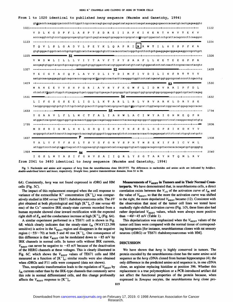
herg coding sequence. The latter resulted to be in some cDNA totallyidentical to the sequence isolated from human hippocampus byWarmke and Ganetzky (4), whereas in one cDNA clone, we observeda variation of five nucleotides (in boldface and double-underlined
letters) as reported in Fig. 3.This change implies a single amino acid substitution, i.e., an argi
nine instead of a histidine at position 396 (indicated by the box). Thissubstitution occurs just before the putative S l segment. It is not yetclear whether these differences are due to mutations introduced duringthe PCR or represent a true herg polymorphism. On the whole, thesedata suggest that the herg in neuroblastoma cells is substantiallysimilar to that originally described in the cDNA of human hippocam
pus, indicating that this gene is not affected by the transformationprocess.
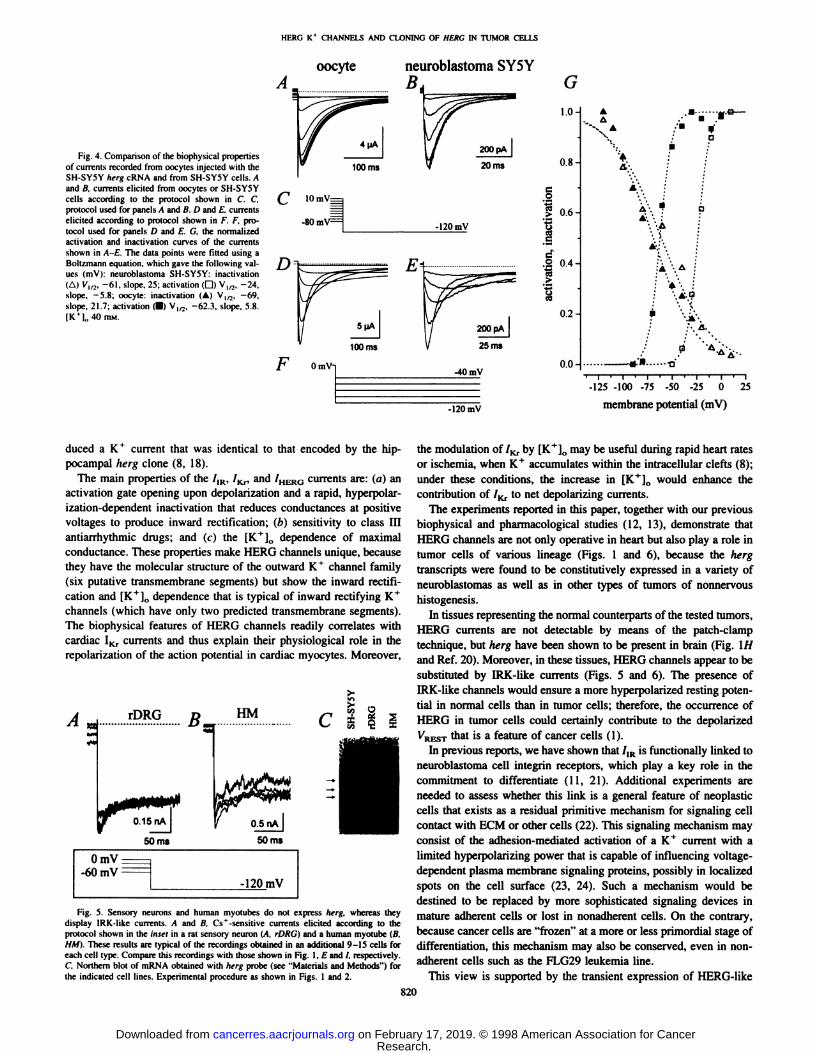
Comparison of Native 7,R with Currents Expressed by theSH-SY5Y herg Clone in Xenopus Oocytes. To compare the native7IRcurrents with those generated by the SH-SY5Y herg clone (Fig. 3),
we used the heterologous expression in Xenopus oocytes.Fig. 4 shows the inward SH-SY5Y HERG currents recorded in
Xenopus oocytes compared with the SH-SY5Y /IR current; no com
parison of the outward currents is shown because their recording inneuroblastoma cells is complicated by an overlapping delayed rectifying K+ current that is activated in the same voltage range as HERG.
To study the activation properties, we used the protocol described inFig. 4C. The tail currents elicited at -120 mV (Fig. 4A for oocytes
and Fig. 4B for SH-SY5Y cells) were normalized to obtain the
activation curves illustrated in Fig. 4G (closed and open squares forthe oocytes and SH-SY5Y cells, respectively). The inactivation curves(Fig. 4G, closed and open triangles for the oocytes and SH-SY5Y
cells, respectively) were obtained by plotting the normalized peakchord conductance (g^^ = /p^I^M ~ £|J) aga'nst tne membrane
potential VM(Fig. 4D and E). The experimental points were fitted toBoltzmann equations, which gave the Vl/2 and slope values indicatedin the figure legend. The shape of the activation curves was identicalin the oocytes and SH-SY5Y cells, although the curve was shifted 38
mV to the left in the former. This shift should be not regarded as apeculiarity of the mutated cDNA indicated in Fig. 3 because it wasalso observed with the unmutated clones (data not shown).
Three possible explanations may account for this shift: (a) differences in the internal and/or external surface charge of the plasmamembrane that produce changes in the surface potential: (/>) uniqueposttranslational modifications of the herg gene product in the twocell types; or (c) the association of HERG with different subunits inneuroblastoma cells and oocytes.
Activation curves of the herg expressed currents reported by San-
guinetti et al. (8), Trudeau et al. (18), and Schönherrand Heinemann(19) (V1/2 = -69 mV versus -15, 6, and -19.5, respectively) are
shifted to the right as compared with our data obtained from oocytecurrents. This discrepancy is explained by the much shorter preconditioning prepulse used in these studies. In fact, we have evidence4
that shorter prepulses than those shown in Fig. 4C (10 s) and adifferent choice of the Vh (-80 mV versus 0 mV in our experiments)
led to a rightward shift of the activation curve, because the channelsrequire this amount of time to attain a true steady-state at each voltage.
In this light, we consider it a pure chance that the activation curvesreported by the cited authors fall close the our SH-SY5Y clone.
Taken together, the properties of the neuroblastoma /,R currentwere largely reproduced by the currents resulting from the SH-SY5Y
herg clone.Neoplastic Transformation Is Accompanied by the Replace
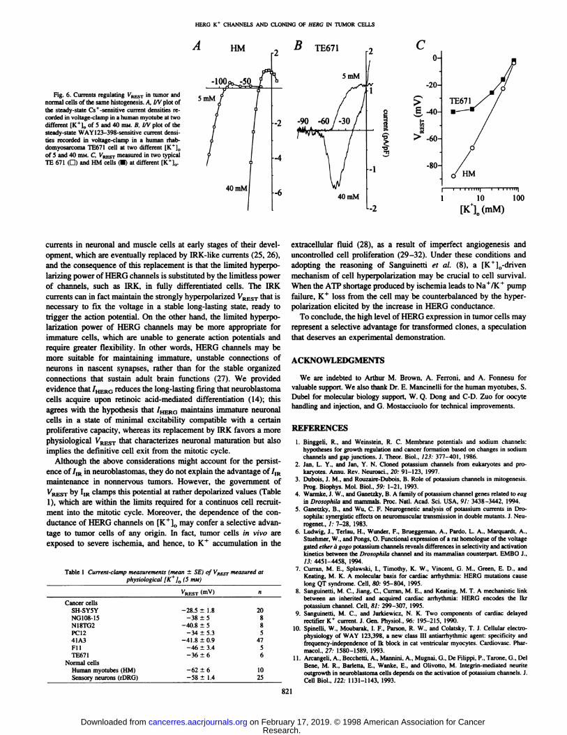
ment of IRK Currents with ¡IKCurrents: Effects on VKESTatDifferent [K+]0. As a first insight into the oncological relevance of
the recurrent expression of I,R in tumor cells, we conducted a comparative study of the inward K+ currents in two pairs of histogenet-
ically identical normal and tumor cells, i.e., rDRG versus Fllneuroblastoma, and human myotube (HM) versus TE671 rhabdomy-
osarcoma. Interestingly, each of these comparisons revealed that,instead of the /,R recorded in the tumor lines (Fig. l, E and /),thenormal cells possessed a K ' current with the biophysical and pharmacological features (Cs+ sensitive and E-4031 insensitive; data not
shown) of the classical inward rectifier IRK (Fig. 5 A and B and Fig.
4 B. Rosati. A. Arcangeli. M. Olivotto, and E. Wanke, manuscript in preparation.
818
Research. on February 17, 2019. © 1998 American Association for Cancercancerres.aacrjournals.org Downloaded from
HERG K* CHANNELS AND CLONING OF HERG IN TUMOR CELLS
From 1 to 1020 identical to published herg sequence (Warmke and Ganetzky, 1994)
gtfigacctcaagggcgaccccttcttggcttcgcccaccagtgaccgtgagatoatagcacctaagataaaggagcgaacccacaatgtcactgagaaggtc
1021 + + + + + + + + + +—
VDLKGDPFLASPTSDREIIAPKIKERTHNVTEKV
acccaggtcctgtccctgggcgccgacgtgctgcctgagtacaagctgcaggcaccgcgcatccgccgctggaccatcctgcattacagccccttcaaggcc
1123 + + + + + + —-
TQVLSLGADVLPEYKLCAPRI \R \ RWTILHYSPFKA
gtgtgggactggctcatcctgctgctggtcatctacacggctgtcttcacaccctactcggctgccttcctgctgaaggagacggaagaaggcccgcctgct
1225 ^—^—^^^^-^ SI ^^^^^^^^^^^^^— + + * + +
VWDWLILLLVIYTAVFTPYSAAFLLKETEEGPPA
accgagtgtggctacgcotgccagccgctggctgtggtggacctcatcgtggacatcatgttcattgtggacatcctcatcaacttccgoaccacctacgtc
1327 + + + S2 ————————^-^ +
TECGYACQPLAVVDLIVDIMFIVDILINFRTTYV
aatgccaacgaggaggtggtcagccaccccggccgcattgccgtccactacttcaagggctggttcctcatcgacatggtggccgccatccccttcgacctg
1429 - +
NAN EVVSH
4-
GR AVHYFKGWFL DM AA DL
ctcatctttggctctggctctgaggagctgatcgggctgctgaagactgcgcggctgctgcggctggtgcgcgtggcgcggaagctggatcgctactcagag
1531
1633
1735
1837
S4 —
T A R L L
+—
D RLIFGSGSEELIGLLKTARLLRLVRVARKLDRYSE
tacggcgcggccgtgctgttcttgctcatgtgcacctttgcgctcatcgcgcactggctggcctgcatctggtacgccatcggcaacatggagcagccacac
•S5
IAHYGAAVLFLLMCTFALIAHWLACIWYAIGNMEQPH
atggactcacgcatcggctggctgcacaacctgggcgaccagataggcaaaccctacaacagcagcggcctgggcggcccctccatcaaggacaagtatgtg
----- +--------- +--------- +--------- +--------- +--------- +--------- +--------- +--------- +--------- +----—
MDSRIGWLHNLGDQIGKPYNSSGLGGPSIKDKYV
acggcgctctacttcaccttcagcagcctcaccagtgtgggcttcggcaacgtctctcccaacaccaactcagagaagatcttctccatctgcgtcatgctc
1122
1224
1326
1428
1530
1632
1734
1836
1938
TALYFTFSSLTSVGFGNVSPNTNSEKIFSICVML
attggctccctcatgtafigctagcatcttcggcaacgtgtcggccatcatccagcggctgtactcgggcacagcccgctaccacacacagatgctgcgggtg
1939 ——^^^^^—^^^^^^ §g ^—^^^^ + -l- + + + 2040
IGSLMYASIFGNVSAIIQRLYSGTARYHTQMLRV
from 2041 to 3480 identicalto herg sequence (Warmkeand Ganetzky, 1994)
Fig. 3. Nucleotide and amino acid sequence of herg from the neuroblastoma clone SH-SY5Y. The differences in nucleotides and amino acids are indicated by boldface,double-underlined letters and boxes, respectively. Straight lines, putative transmembrane domains, from SI to S6.
6A). Consistently, herg was not found expressed in rDRG and HMcells (Fig. 5Q.
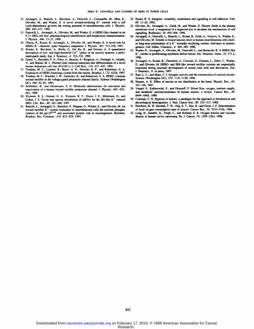
The impact of this replacement emerged when the cell response tovariance of the extracellular K+ concentration ([K+]0) was compar
atively studied in HM versus TE671 rhabdomyosarcoma cells. The I/Vplot obtained at both physiological and high [K+]0 (5 mM versus 40mM) of the Cs+-sensitive IRK steady-state currents recorded from a
human myotube showed clear inward rectification with the expectedright shift of £Kand the conductance increase at high [K+]0 (Fig. 6A).
A similar experiment performed in a TE671 cell is shown in Fig.6ß,which clearly indicates that the steady-state 7,R (WAY 123.398
sensitive) is active in the VRESTregion and disappears in the negativeregion (—557—70)at both 5 and 40 mM [K^]0. One consequence of
this difference is that VRESTcan be modulated down to -85 mV by
IRK channels in normal cells. In tumor cells without IRK currents,VRESTcan never be negative to -45 mV because of the deactivation
of the HERG channels at these voltages. This is clearly illustrated inFig. 6C, which shows the VRESTvalues of TE671 cells and HMmeasured as a function of [K+]0; similar results were also obtained
when rDRGs and Fl 1 cells were compared (data not shown).Thus, neoplastic transformation leads to the regulation of VRESTby
/iR currents rather than by the IRK-type channels that commonly serve
this role in normal differentiated cells, and this change profoundlyaffects the VRESTresponse to [K+]0.
Measurements of VRKSTin Tumors and in Their Normal Counterparts. We have demonstrated that, in neuroblastoma cells, a directcorrelation exists between the V¡/2of the activation curve of /,R andthe value of VREST,so that the more the activation curve was shiftedto the right, the more depolarized VRESTbecame (12). Consistent withthe observation that most of the tumor cell lines we tested havemarkedly right-shifted activation curves (Fig. 10), these lines also had
rather depolarized VRESTvalues, which were always more positivethan -40/-45 mV (Table 1).
This depolarization was emphasized when the VRHSTvalues of thetumor cell lines were compared with the normal tissue of corresponding histogenesis [for instance, neuroblastomas clones with rat sensoryneurons (rDRG) or TE671 rhabdomyosarcomas with HM].
DISCUSSION
We have shown that herg is highly conserved in tumors. Theprotein encoded by the neuroblastoma clone has the same amino acidsequence as the herg cDNA cloned from human hippocampus (4); theonly difference in the predicted amino acid sequence is, in one cDNAclone, an arginine replacing histidine at position 396. Whether thisreplacement is a true polymorphism or a PCR-introduced artifact did
not affect the functional properties of the protein because, whenexpressed in Xenopus oocytes, the neuroblastoma herg clone pro-
819
Research. on February 17, 2019. © 1998 American Association for Cancercancerres.aacrjournals.org Downloaded from
HERO K* CHANNELS AND CLONING OF HERC IN TUMOR CELLS
Fig. 4. Comparison of the biophysical propertiesof currents recorded from oocytes injected with theSH-SY5Y herg cRNA and from SH-SY5Y cells. Aand ß.currents elicited from oocytes or SH-SY5Ycells according to Ihe protocol shown in C. C.protocol used for panels A and B. I) and E, currentselicited according to protocol shown in F. F, protocol used for panels /) and E. C. the normalizedactivation and inaclivation curves of the currentsshown in A-E. The data points were filled using aBolt/mann equation, which gave Ihe following values (mV): neuroblastoma SH-SY5Y: inactivalion(A) Vm, -61, slope, 25; activation (D) V1/2, -24,slope, -5.8; oocyte: inactivation (A) V1/2, —69,slope, 21.7; activation (•)V,,,, -62.3, slope, 5.8.|K'],,40 mm.
oocyte neuroblastoma SY5Y
10mV=
-80 m\ -120mV
100 ms
OmV-i-40 mV
G1.0-
0.8-
II 0.6
oesc
c.2 0.4
0.2-
0.0-
AA
A-,
A'-
A .
: '*'• i: ':*•<?
•'»'•
-120 mV
-125 -100 -75 -50 -25 O
membrane potential (mV)
25
duced a K+ current that was identical to that encoded by the hip- the modulation of/Kr by [K+]„may be useful during rapid heart rates
pocampal herg clone (8, 18).The main properties of the /]R, /Kr, and /HEROcurrents are: (a) an
activation gate opening upon depolarization and a rapid, hyperpolar-ization-dependent inactivation that reduces conductances at positive
voltages to produce inward rectification; (b) sensitivity to class IIIantiarrhythmic drugs; and (c) the [K ' ]„dependence of maximal
conductance. These properties make HERO channels unique, becausethey have the molecular structure of the outward K+ channel family
(six putative transmembrane segments) but show the inward rectification and [K+]() dependence that is typical of inward rectifying K +
channels (which have only two predicted transmembrane segments).The biophysical features of HERO channels readily correlates withcardiac IKr currents and thus explain their physiological role in therepolarization of the action potential in cardiac myocytes. Moreover,
rDRG HM
0.15 nA
50ms
C
t
OmV—AfimV-120
mV
Fig. 5. Sensory neurons and human myolubes do noi express herg, whereas theydisplay IRK-like currents. A and B. Cs +-sensitive currents elicited according lo the
protocol shown in the inset in a ral sensory neuron (A. rDRG} and a human myotube (B,HM}. These results are typical of Ihe recordings obtained in an additional 9-15 cells for
each cell type. Compare this recordings with those shown in Fig. l, E and /. respectively.C Northern blot of mRNA obtained with herg probe (see "Materials and Methods") for
the indicated cell lines. Experimental procedure as shown in Figs. 1 and 2.
or ischemia, when K *"accumulates within the intracellular clefts (8);
under these conditions, the increase in [K+]0 would enhance the
contribution of /Kr to net depolarizing currents.The experiments reported in this paper, together with our previous
biophysical and pharmacological studies (12, 13), demonstrate thatHERO channels are not only operative in heart but also play a role intumor cells of various lineage (Figs. 1 and 6), because the hergtranscripts were found to be constitutively expressed in a variety ofneuroblastomas as well as in other types of tumors of nonnervoushistogenesis.
In tissues representing the normal counterparts of the tested tumors,HERG currents are not detectable by means of the patch-clamp
technique, but herg have been shown to be present in brain (Fig. \Hand Ref. 20). Moreover, in these tissues, HERG channels appear to besubstituted by IRK-like currents (Figs. 5 and 6). The presence ofIRK-like channels would ensure a more hyperpolarized resting poten
tial in normal cells than in tumor cells; therefore, the occurrence ofHERG in tumor cells could certainly contribute to the depolarizedVRESTthat is a feature of cancer cells (1).
In previous reports, we have shown that /IR is functionally linked toneuroblastoma cell integrin receptors, which play a key role in thecommitment to differentiate (11, 21). Additional experiments areneeded to assess whether this link is a general feature of neoplasticcells that exists as a residual primitive mechanism for signaling cellcontact with ECM or other cells (22). This signaling mechanism mayconsist of the adhesion-mediated activation of a K+ current with a
limited hyperpolarizing power that is capable of influencing voltage-
dependent plasma membrane signaling proteins, possibly in localizedspots on the cell surface (23, 24). Such a mechanism would bedestined to be replaced by more sophisticated signaling devices inmature adherent cells or lost in nonadherent cells. On the contrary,because cancer cells are "frozen" at a more or less primordial stage of
differentiation, this mechanism may also be conserved, even in non-
adherent cells such as the FLG29 leukemia line.This view is supported by the transient expression of HERG-like
820
Research. on February 17, 2019. © 1998 American Association for Cancercancerres.aacrjournals.org Downloaded from
HERO K+ CHANNELS AND CLONING OF HEKC IN TUMOR CELLS
B
Fig. 6. Currents regulating V/RESTin tumor andnormal cells of the same histogenesis. A, l/V plot ofthe steady-state Cs+-sensitive current densities recorded in voltage-clamp in a human myotube at twodifferent [K+]0 of 5 and 40 HIM.B, l/V plot of thesteady-state WAY123-398-sensitive current densities recorded in voltage-clamp in a human rhab-domyosarcoma TE671 cell at two different IK*],,
of 5 and 40 HIM.C, VRESTmeasured in two typicalTE 671 (G) and HM cells (•)at different [K+]„.
Co-
-20-
¿-40-
> -60-
-80-
TE671
1 10 100
[Kl(mM)
currents in neuronal and muscle cells at early stages of their development, which are eventually replaced by IRK-like currents (25, 26),and the consequence of this replacement is that the limited hyperpo-
larizing power of HERG channels is substituted by the limitless powerof channels, such as IRK, in fully differentiated cells. The IRKcurrents can in fact maintain the strongly hyperpolarized VRESTthat isnecessary to fix the voltage in a stable long-lasting state, ready to
trigger the action potential. On the other hand, the limited hyperpolarization power of HERG channels may be more appropriate forimmature cells, which are unable to generate action potentials andrequire greater flexibility. In other words, HERG channels may bemore suitable for maintaining immature, unstable connections ofneurons in nascent synapses, rather than for the stable organizedconnections that sustain adult brain functions (27). We providedevidence that /HERGreduces the long-lasting firing that neuroblastomacells acquire upon retinoic acid-mediated differentiation (14); this
agrees with the hypothesis that /HERG maintains immature neuronalcells in a state of minimal excitability compatible with a certainproliferative capacity, whereas its replacement by IRK favors a morephysiological VRESTthat characterizes neuronal maturation but alsoimplies the definitive cell exit from the mitotic cycle.
Although the above considerations might account for the persistence of 7IRin neuroblastomas, they do not explain the advantage of 7IRmaintenance in nonnervous tumors. However, the government ofVRESTby IIR clamps this potential at rather depolarized values (Table1), which are within the limits required for a continuos cell recruitment into the mitotic cycle. Moreover, the dependence of the conductance of HERG channels on [K+]0 may confer a selective advan
tage to tumor cells of any origin. In fact, tumor cells in vivo areexposed to severe ischemia, and hence, to K+ accumulation in the
Table 1 Current-clamp measurements (mean ±SE) of VKESTmeasured at
physiological [K /0 f5 mid)
CancercellsSH-SY5YNG108-15N18TG2PC1241A3FllTE671Normal
cellsHumanmyotubes (HM)
Sensory neurons (rDRG)VREST
(">V)-28.5
±1.8-38±5-40.8±5-34±5.3-41.8±0.9-46±3.4-36
±6-62
±6-58±1.4n2088Î47561025
extracellular fluid (28), as a result of imperfect angiogenesis anduncontrolled cell proliferation (29-32). Under these conditions andadopting the reasoning of Sanguinetti et al. (8), a [K+](,-driven
mechanism of cell hyperpolarization may be crucial to cell survival.When the ATP shortage produced by ischemia leads to Na+/K * pumpfailure, K+ loss from the cell may be counterbalanced by the hyper-
polarization elicited by the increase in HERO conductance.To conclude, the high level of HERG expression in tumor cells may
represent a selective advantage for transformed clones, a speculationthat deserves an experimental demonstration.
ACKNOWLEDGMENTS
We are indebted to Arthur M. Brown, A. Ferroni, and A. Fonnesu forvaluable support. We also thank Dr. E. Mancinelli for the human myotubes, S.Dubel for molecular biology support, W. Q. Dong and C-D. Zuo for oocyte
handling and injection, and G. Mostacciuolo for technical improvements.
REFERENCES
1. Binggeli, R., and Weinstein, R. C. Membrane potentials and sodium channels:hypotheses for growth regulation and cancer formation based on changes in sodiumchannels and gap junctions. J. Theor. Biol., 123: 377-401. 1986.
2. Jan, L. Y.. and Jan, Y. N. Cloned potassium channels from eukaryotes and pro-karyotes. Annu. Rev. Neurosci., 20: 91-123, 1997.
3. Dubois, J. M., and Rouzaire-Dubois, B. Role of potassium channels in mitogenesis.Prog. Biophys. Mol. Biol., 59: 1-21, 1993.
4. Warmke, J. W., and Ganetzky, B. A family of potassium channel genes related to eagin Drosophila and mammals. Proc. Nati. Acad. Sci. USA, 91: 3438-3442, 1994.
5. Ganetzky, B., and Wu, C. F. Neurogenetic analysis of potassium currents in Drosophila: synergislic effects on neuromuscular transmission in double mutants. J. Neu-rogenet., /: 7-28, 1983.
6. Ludwig, J.. Terlau, H., Wunder, F., Brueggeman. A-, Pardo, L. A., Marquardt. A..Stuehmer, W., and Pongs, O. Functional expression of a rat homologue of the voltagegated ether à gogo potassium channels reveals differences in selectivity and activationkinetics between the Drosophilu channel and its mammalian counterpart. EMBO J.,13: 4451-4458, 1994.
7. Curran, M. E., Splawski. I.. Timothy, K. W., Vincent, G. M., Green, E. D.. andKeating, M. K. A molecular basis for cardiac arrhythmia: HERG mutations causelong QT syndrome. Cell, 80: 95-804, 1995.
8. Sanguinetti, M. C., Jiang, C., Curran. M. E., and Keating. M. T. A mechanistic linkbetween an inherited and acquired cardiac arrhythmia: HERG encodes the Ikrpotassium channel. Cell, 81: 299-307. 1995.
9. Sanguinetti. M. C., and Jurkiewicz, N. K. Two components of cardiac delayedrectifier K" current. J. Gen. Physiol., 96: 195-215. 1990.
10. Spinelli. W., Moubarak, I. F.. Parson, R. W., and Colatsky, T. J. Cellular electro-physiology of WAY 123,398, a new class III antiarrhythmic agent: specificity andfrequency-independence of Ik block in cat ventricular myocytes. Cardiovasc. Phar-macol.. 27: 1580-1589, 1993.
11. Arcangeli, A., Becchetti, A., Mannini. A., Mugnai, G., De Filippi, P., Tarone, G.. DelBene, M. R., Barletta, E., Wanke, E., and Olivotto, M. Integrin-mediated neuriteoutgrowth in neuroblastoma cells depends on the activation of potassium channels. J.Cell Biol., /22: 1131-1143, 1993.
821
Research. on February 17, 2019. © 1998 American Association for Cancercancerres.aacrjournals.org Downloaded from
HERO K* CHANNELS AND CLONING OF HERC IN TUMOR CELLS
12. Arcangeli, A., Bianchi. L.. Becchetti, A., Faravelli. L., Coronnello, M.. Mini. E.,Olivotto. M., and Wanke. E. A novel inward-rectifying K* current with a cell
cycle-dependence governs the resting potential of neuroblastoma cells. J. Physiol..489: 455-471, 1995.
13. Faravelli, L.. Arcangeli, A.. Olivotto. M., and Wanke. E. A HERG-like channel in ratF-11 DRG cell line: pharmacological identification and biophysical characterisation.J. Physiol., 496: 13-23, 1996.
14. Chiesa. N., Rosati. B., Arcangeli. A.. Olivotto. M., and Wanke, E. A novel role forHERG K' channels: spike frequency adaptation. J. Physiol.. SOI: 313-318. 1997.
15. Wanke, E.. Becchetti. A.. Biella. G.. Del Bo. R., and Ferroni, A. A quantitativedescription of low- and high-threshold Ca:* spikes in rat sensory neurons: a perfo
rated-patch study. Eur. J. Neurosa., 4: 723-732, 1992.16. Gattei. V., Bemabei. P. A., Pinto, A.. Bezzini, A., Ringressi, A., Formigli, A.. Attadia.
V.. and Brandi. M. L. Phornol ester induced osieoclasl-like differentiation of a novelhuman leukaemia cell line (FLG29.1I. J. Cell Biol.. 116: 437-447, 1992.
17. Trudeau, M. C.. London, B., Beyer. A. K.. Newton. K. P.. and Robertson, G. A.Expression of HERG homologs cloned from the mouse. Biophys. J.. 72: A224, 1997.
18. Trudeau, M. C.. Warmke. J. W., Ganetzky, B., and Robertson. G. A. HERG. a humaninward rectifier in the voltage-gated potassium channel family. Science (WashingtonDC), 269: 92-95. 1995.
19. Schönherr. R.. and Hcinemann. S. H. Molecular determinants for activation andinactivation of a human inward rectifier potassium channel. J. Physiol.. 493: 653-
642, 1996.20. Wymore. R. S.. Gintant. G. A.. Wymore, R. T., Dixon, J. E.. Mckinnon. D., and
Cohen, I. S. Tissue and species distribution of mRNA for the IKr-like K ^ channel
ERG. Circ. Res.. 80: 261-268. 1997.
21. Bianchi. L.. Arcangeli. A.. Bartolini, P.. Mugnai. G.. Wanke. E., and Olivotto. M. Aninw ard rectifier K " current modulates in neuroblasloma cells the tyrosine phospho-rylation of the ppl25'AK and associated protein: role in neuritogenesis. Biochem.
Biophys. Res. Commun., 210: 823-829, 1995.
22. Hynes. R. O. Integrins: versatility, modulation and signalling in cell adhesion. Cell.69: 11-25, 1992.
23. Olivolto, M., Arcangeli, A., Carla, M., and Wanke. E. Electric fields at the plasmamembrane level: a reappraisal of a neglected key to decipher the mechanisms of cellsignalling. BioEssays. 18: 495-504, 1996.
24. Arcangeli, A., Faravelli. L., Bianchi, L., Rosati. B.. Grilli, A., Vescovi, A., Wanke, E.,and Olivotto. M. Soluble or bound Lumini elicit in human neuroblastoma cells short-or long-term potentiation of a K+ inwardly rectifying current: relevance to neurito
genesis. Cell Adhes. Commun.. 4: 369-385. 1996.25. Wanke, E.. Arcangeli, A.. Olivotto. M.. Faravelli. L., and Bonecchi. R. A HERG-like
K* current in proliferating myoblasts before fusion. Soc. Neurosa. Abstr., 22: 571.2,
1996.26. Arcangeli, A., Rosati. B.. Cherubini. A.. Crociani, O., Fontana, L., Ziller. C., Wanke,
E., and Olivotto. M. HERG- and IRK-like inward rectifier currents are sequentially
expressed during neuronal development of neural crest cells and derivatives. Eur.J. Neurosci., 9: in press. 1997.
27. Katz, L. C.. and Shatz, C. J. Synaptic activity and the construction of cortical circuits.Science (Washington DC). 274: 1133-1138, 1996.
28. Hansen, A. K. Effect of anoxia on ion distribution in the brain. Physiol. Rev., 65:101-148, 1987.
29. Vaupel. P., Kallinowski. F.. and Okunieff, P. Blood flow, oxygen, nutrient supply,and metabolic microenvironment of human tumors: a review. Cancer Res., 49:6449-6465. 1989.
30. Coleman. C. N. Hypoxia in tumors: a paradigm for the approach to biochemical andphysiological heterogeneity. J. Nati. Cancer Inst., 80: 310-317, 1988.
31. Dewhirst, M. W. Secomb, T. W., Ong, E. T.. Hsu. R.. and Gross, J. F. Determinationof loca! oxygen consumption rates in tumors. Cancer Res., 54: 3333-3336, 1994.
32. Lyng, H.. Sundfor, K.. Trope. C.. and Rofstad. E. K. Oxygen tension and vasculardensity in human cervix carcinoma. Br. J. Cancer. 74: 1559-1563, 1996.
822
Research. on February 17, 2019. © 1998 American Association for Cancercancerres.aacrjournals.org Downloaded from

1998;58:815-822. Cancer Res Laura Bianchi, Barbara Wible, Annarosa Arcangeli, et al.
Cells?Different Histogenesis: A Selective Advantage for Cancer Current Highly Conserved in Tumors of+ Encodes a Kherg
Updated version
http://cancerres.aacrjournals.org/content/58/4/815
Access the most recent version of this article at:
E-mail alerts related to this article or journal.Sign up to receive free email-alerts
Subscriptions
Reprints and
To order reprints of this article or to subscribe to the journal, contact the AACR Publications
Permissions
Rightslink site. Click on "Request Permissions" which will take you to the Copyright Clearance Center's (CCC)
.http://cancerres.aacrjournals.org/content/58/4/815To request permission to re-use all or part of this article, use this link
Research. on February 17, 2019. © 1998 American Association for Cancercancerres.aacrjournals.org Downloaded from