Brain Dopamine Transporter in Spontaneously Hypertensive Rats
1
Hepatotoxic effects of fenofibrate in spontaneously hypertensive rats expressing human
C-reactive protein
Vojtěch Škop,1 Jaroslava Trnovská,1 Olena Oliyarnyk,1 Irena Marková,1 Hana Malínská,1
Ludmila Kazdová,1 Václav Zídek,2 Vladimír Landa,2 Petr Mlejnek,2 Miroslava Šimáková,2
Miroslav Kůdela3, Michal Pravenec2, Jan Šilhavý2
1Center for Experimental Medicine, Institute for Clinical and Experimental Medicine, Prague,
Czech Republic
2Institute of Physiology, Czech Academy of Sciences, Prague, Czech Republic
3Department of Forensic Medicine, Masaryk Hospital in Ústí nad Labem, Czech Republic
Correspondence Address:
Jan Šilhavý
Institute of Physiology, Czech Academy of Sciences
Vídeňská 1083
14220 Prague 4
Czech Republic
Phone: (420)241062153; Fax: (420)241062488; Email: [email protected]
Short title: Hepatotoxic effects of fenofibrate in SHR-CRP rats
2
SUMMARY
Dyslipidemia and inflammation play an important role in the pathogenesis of cardiovascular
and liver disease. Fenofibrate has a well-known efficacy to reduce cholesterol and
triglycerides. Combination with statins can ameliorate hypolipidemic and anti-inflammatory
effects of fibrates. In the current study, we tested the anti-inflammatory and metabolic effects
of fenofibrate alone and in combination with rosuvastatin in a model of inflammation and
metabolic syndrome, using spontaneously hypertensive rats expressing the human C-reactive
protein transgene (SHR-CRP transgenic rats). SHR-CRP rats treated with fenofibrate alone
(100 mg/kg body weight) or in combination with rosuvastatin (20 mg/kg body weight) versus
SHR-CRP untreated controls showed increased levels of proinflammatory marker IL6,
increased concentrations of ALT, AST and ALP, increased oxidative stress in the liver and
necrotic changes of the liver. In addition, SHR-CRP rats treated with fenofibrate, or with
fenofibrate combined with rosuvastatin versus untreated controls, exhibited increased serum
triglycerides and reduced HDL cholesterol, as well as reduced hepatic triglyceride, cholesterol
and glycogen concentrations. These findings suggest that in the presence of high levels of
human CRP, fenofibrate can induce liver damage even in combination with rosuvastatin.
Accordingly, these results caution against the possible hepatotoxic effects of fenofibrate in
patients with high levels of CRP.
KEY WORDS: fenofibrate; rosuvastatin; C-reactive protein; transgenic; spontaneously
hypertensive rat; inflammation; hepatotoxic
3
INTRODUCTION
Dyslipidemia and inflammation play an important role in the pathogenesis of cardiovascular
and liver disease. Fenofibrate is a lipid-lowering drug whose effects are mediated by its
interactions with peroxisome proliferator-activated receptor alpha (PPARα), which regulates
expression of genes that code for enzymes involved in fatty acid oxidation. Fenofibrate also
increases lipoprotein lipolysis and reduces liver triglyceride production. In addition to its
hypolipidemic effects, fenofibrate can reduce inflammation as evidenced by decreased levels
of CRP and IL6 in both experimental studies and clinical trials (Hao et al. 2011; Coban et al.
2005; Noureldein et al. 2015; Min et al. 2012; Sun et al. 2015; Yesilbursa et al. 2005). On the
other hand, the anti-inflammatory effects of fenofibrate remain controversial because in some
studies no effects of fenofibrate on CRP reduction have been observed (Mulvey et al. 2012).
Fenofibrate is recommended for therapy of hypertriglyceridemia and hypercholesterolemia
often in combination with statins for improving its efficiency to reduce inflammation and
atherogenic dyslipidemia.
Major controversy exists as to whether increased CRP contributes to the inflammation and
pathogenesis of metabolic and cardiovascular disturbances or whether it is just a secondary
response to inflammatory disease processes (Scirica and Morrow 2006). To test whether
increased levels of human CRP per se can promote disturbances in glucose and lipid
metabolism characteristic of metabolic syndrome, we transgenically expressed human CRP in
spontaneously hypertensive rats (SHR) and demonstrated a causal role of CRP-induced
inflammation in the pathogenesis of several components of metabolic syndrome in this animal
model (Pravenec et al. 2011). Recently, we studied the anti-inflammatory effects of
4
rosuvastatin in SHR-CRP transgenic rats and in non-transgenic SHR controls, as a result of
which we found that rosuvastatin treatment decreased circulating levels of inflammatory
response markers IL6 and TNFα without decreasing circulating levels of human CRP.
Rosuvastatin also reduced cardiac and renal inflammation and oxidative tissue damage
(Šilhavý et al. 2014; Šilhavý et al. 2015). In the current study, we tested the anti-inflammatory
and metabolic effects of fenofibrate alone and in combination with rosuvastatin in SHR-CRP
transgenic rats.
METHODS
Animals.
Transgenic SHR (hereafter referred to as SHR-CRP) were derived by microinjections of SHR
ova with a construct containing cDNA for human CRP under control of the apoE promoter in
order to drive expression of the CRP transgene in the liver, where CRP is normally produced
(Pravenec et al. 2011). To investigate the effects of fenofibrate (Lipanthyl, Recipharm
Fontaine, Dijon, France) alone and in combination with rosuvastatin (Rosucard, Zentiva,
Prague, Czech Republic) on inflammation caused by human CRP, we randomized 7-month-
old transgenic SHR-CRP into 3 groups treated with placebo, fenofibrate alone (100 mg/kg
body weight), and with fenofibrate in combination with rosuvastatin (20 mg/kg body weight)
for 4 weeks. In each group, we studied 7 males. The rats were housed in an air-conditioned
animal facility and allowed free access to their chow and water. At the end of the
experiments, rats were decapitated in their postprandial state and tissues were collected for
analysis. No experiments on live animals were performed. All experiments were performed in

5
agreement with the Animal Protection Law of the Czech Republic and were approved by the
Ethics Committee of the Institute of Physiology, Czech Academy of Sciences, Prague.
Basal- and insulin-stimulated glycogen synthesis and glucose oxidation in skeletal muscle.
For measurement of insulin-stimulated incorporation of glucose into glycogen and its
oxidation into CO2, diaphragmatic muscles were incubated for 2 hours in 95% O2 + 5% CO2
in Krebs-Ringer bicarbonate buffer, pH 7.4, containing 0.1 μCi/ml of 14C-U glucose, 5
mmol/L of unlabeled glucose, and 2.5 mg/ml of bovine serum albumin (Sigma, Fraction V,
Czech Republic), with or without 250 μU/ml insulin. After the incubation, glycogen was
extracted from the tissue, counted for radioactivity, and insulin-stimulated incorporation of
glucose into glycogen was determined. To determine glucose oxidation, 0.2 ml of 1M
hyamine hydroxide was injected into the central compartment of the incubation vial and 0.5
ml of 1M H2SO4 was added to the main compartment to liberate CO2. The vials were
incubated for another 45 min and the hyamine hydroxide was then quantitatively transferred
to the scintillation vial containing 10 ml of toluene-based scintillation fluid for counting of
radioactivity.
Glucose utilization in isolated epididymal adipose tissue. Glucose utilization in adipose
tissue was determined ex vivo by measuring the incorporation of radioactive glucose into
adipose tissue lipids. Distal parts of epididymal adipose tissue were rapidly dissected and
incubated for 2 hours in Krebs-Ringer bicarbonate buffer with 5 mmol/L glucose, 0.1 Ci
14C-U glucose/mL (UVVR, Prague, Czech Republic) and 2% bovine serum albumin with a
gaseous phase of 95% O2 and 5% CO2 in the presence (250 U/mL) or absence of insulin in
6
incubation media. All incubations were performed at 37°C in sealed vials in a shaking water
bath. Estimation of 14C-glucose incorporation into neutral lipids was performed. Briefly,
adipose tissue was removed from the incubation medium, rinsed in saline, and immediately
put into chloroform. The pieces of tissue were dissolved using a Teflon pestle homogenizer,
methanol was added (chloroform:methanol 2:1), and lipids were extracted at 4°C overnight.
The remaining tissue was removed, KH2PO4 was added, and a clear extract was taken for
further analysis. An aliquot was evaporated, reconstituted in scintillation liquid, and the
radioactivity measured by scintillation counting.
Tissue triglyceride and cholesterol measurements. In order to determine triglycerides and
cholesterol in the liver, heart and gastrocnemius muscle, tissues were powdered under liquid
N2 and extracted for 16 hours in chloroform:methanol, after which 2% KH2PO4 was added
and the solution centrifuged. The organic phase was removed and evaporated under N2. The
resulting pellet was dissolved in isopropyl alcohol, and triglyceride and cholesterol content
was determined by enzymatic assay (Erba-Lachema, Brno, Czech Republic).
Liver glycogen determination. Liver tissues were hydrolyzed by boiling in 30% KOH
containing oyster glycogen. After digestion, 96% ethanol was added and glycogen was
allowed to precipitate at 4°C overnight. The precipitate was centrifuged, washed with ethanol,
and dissolved in H2O. Aliquots were used to measure glycogen using the glucose oxidase
assay (Erba-Lachema, Brno, Czech Republic).
Biochemical analyses. Rat serum CRP and human serum CRP were measured using ELISA
7
kits (Alpha Diagnostics International, San Antonio, U.S.A.). Blood glucose levels were
measured by the glucose oxidase assay (Erba-Lachema, Brno, Czech Republic) using tail vein
blood drawn into 5% trichloracetic acid and promptly centrifuged. NEFA levels were
determined using an acyl-CoA oxidase-based colorimetric kit (Roche Diagnostics GmbH,
Mannheim, Germany). Serum triglyceride and cholesterol concentrations were measured
using standard enzymatic methods (Erba-Lachema, Brno, Czech Republic). Serum insulin
concentrations were determined using a rat insulin ELISA kit (Mercodia, Uppsala, Sweden).
Serum IL6 was measured using rat ELISA kits (BioSource International, Inc., Camarillo,
U.S.A.). Rat MCP-1 was measured by ELISA kits (Instant ELISA eBioscience, Austria).
ALT, AST, and ALP enzyme activities were determined spectrophotometrically by routine
clinical biochemistry methods using a Roche kit. HDL-cholesterol was measured after double
precipitation with dextran and MgCl2 by spectrophotometric methods using commercially
available kits (Roche Diagnostics GmbH, Mannheim, Germany).
Parameters of oxidative stress. The activity of superoxide dismutase (SOD) was analyzed
using the reaction of blocking nitrotetrazolium blue reduction and nitroformazan formation.
Catalase (CAT) activity measurement was based on the ability of H2O2 to produce with
ammonium molybdate a color complex detected spectrophotometrically. The activity of
seleno-dependent glutathione peroxidase (GSH-Px) was monitored by oxidation of
glutathione using Ellman’s reagent (0.01М solution of 5,5'-dythiobis-2 nitrobenzoic acid).
The level of reduced glutathione (GSH) was determined in the reaction of SH-groups using
Ellman’s reagent. The levels of reduced (GSH) and oxidized (GSSG) forms of glutathione
were determined using a high-performance liquid chromatography method with fluorescent
8
detection according to the HPLC diagnostic kit (Chromsystems, Munich, Germany).
Glutathione reductase (GR) and glutathione S-transferase (GST) activities were measured
using Cayman Chemicals assay kits (Michigan, USA). Lipoperoxidation products were
assessed by the levels of thiobarbituric acid reactive substances (TBARS) determined by
assaying reactions with thiobarbituric acid. The levels of conjugated dienes were analyzed by
extraction in media (heptane:isopropanol = 2:1) and measured spectrophotometrically in a
heptane layer.
Expression of the Scd1 gene in the liver. Reverse transcription and quantitative real-time
PCR analyses were performed using the TaqMan RNA-to-CTTM 1-Step Kit and the TaqMan
Gene Expression Assay (Life Technologies, Applied Biosystems, USA) and carried out in a
Vii ATM 7 Real-Time PCR System (Applied Biosystems, USA). Relative expression of the
Scd1 gene was determined after normalization against Pgk1 (phosphoglycerate kinase 1) as an
internal reference and calculated using the 2-Ct method.
Histology
Pieces of tissue were fixated in 4% formaldehyde for at least 48 hours. Afterwards they were
processed using standard techniques into paraffin blocks by dehydration through a series of
graded ethanol baths to displace the water and infiltrate with wax. Once infiltrated, the tissues
were embedded into wax blocks, which were then sectioned into slices up to 3 mm thick and
dyed routinely using hematoxylin and eosin staining.
Statistical analysis. Results are expressed as means ± S.E.M. One-way ANOVA was used to

9
search for significant differences between the 3 groups. The Holm-Sidak method was used for
all pairwise multiple comparison procedures with an overall significance level defined as
P<0.05.
RESULTS
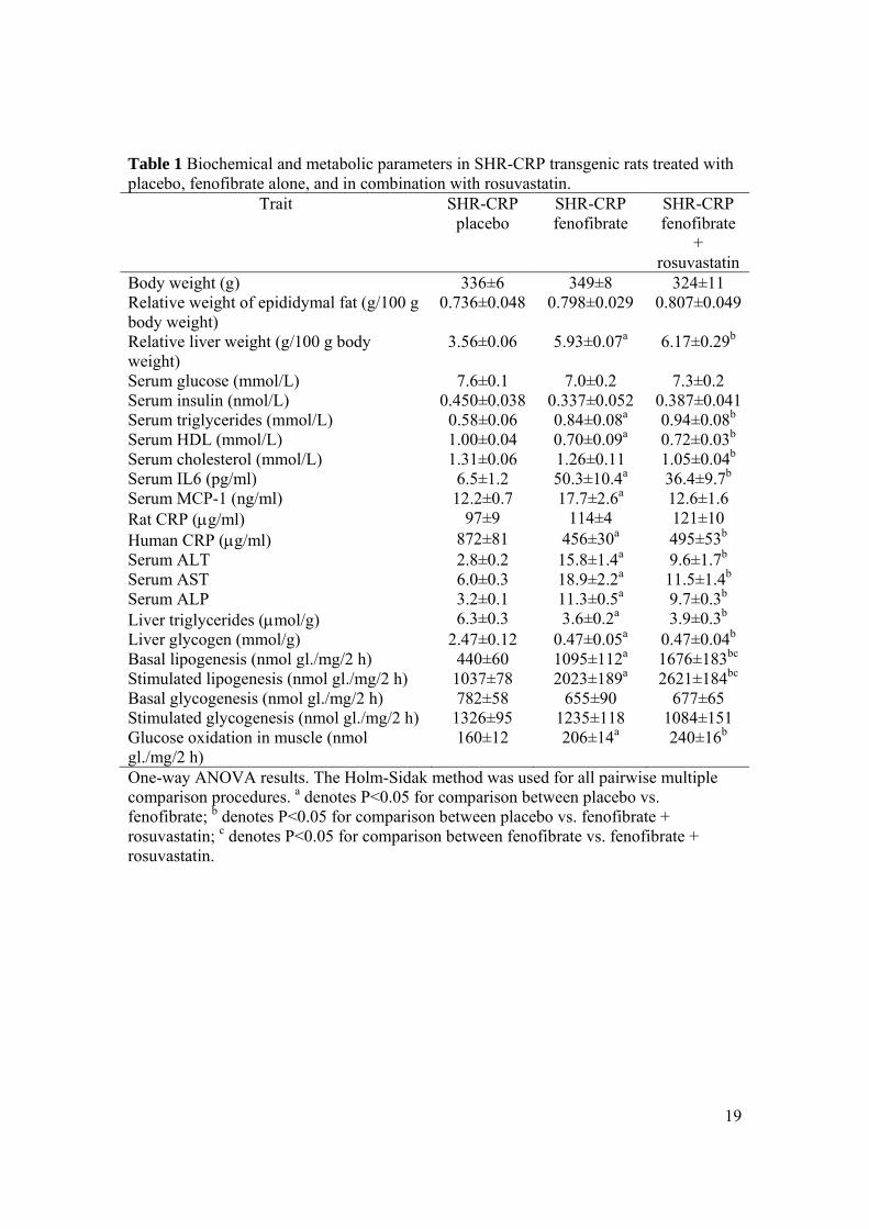
Effects of fenofibrate alone, and in combination with rosuvastatin, on serum levels of
human CRP and rat CRP and on inflammation induced by human CRP. Table 1 shows the
serum levels of human CRP and endogenous rat CRP in the different experimental groups. As
shown, serum levels of human CRP were significantly reduced in SHR-CRP transgenic rats
treated with fenofibrate alone and in combination with rosuvastatin when compared to
untreated SHR-CRP controls. On the other hand, endogenous rat CRP levels were not affected
by the treatment. Table 1 shows the effects of treatment on serum levels of IL6. Treatment
with both fenofibrate alone and in combination with rosuvastatin induced significant increases
in serum levels of IL6 when compared to untreated SHR-CRP controls. In addition, SHR-
CRP rats treated with fenofibrate exhibited significantly higher levels of MCP-1 (Monocyte
Chemoattractant Protein-1), one of the key chemokines that regulate migration and infiltration
of monocytes/macrophages (Table 1).
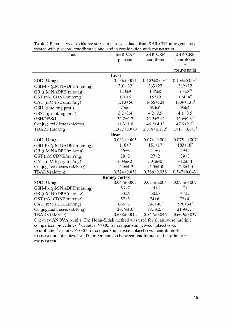
Effects of fenofibrate alone or in combination with rosuvastatin on oxidative stress-related
parameters. We assessed the effects of fenofibrate alone and in combination with rosuvastatin
on human CRP-induced oxidative stress in tissues by measuring activities of antioxidative
enzymes, glutathione, levels of lipoperoxidation products, conjugated dienes and TBARS
10
(Table 2). In the transgenic SHR-CRP strain expressing human CRP, both fenofibrate alone
and in combination with rosuvastatin induced a significant increase in conjugated dienes,
TBARS, and reduced GSH/GSSG ratios in the liver. On the other hand, oxidative stress did
not increase in the heart and kidney cortex after treatment with fenofibrate alone and in
combination with rosuvastatin. Increased oxidative stress in the liver was associated with
reduced SOD, whereas CAT increased (Table 2).
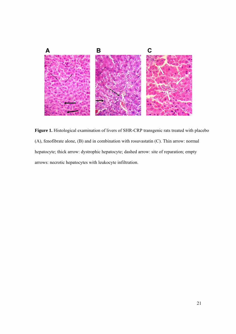
Effects of fenofibrate alone and in combination with rosuvastatin on target organ damage.
Table 1 shows significantly increased serum levels of ALT, AST, and ALP in SHR-CRP rats
treated with fenofibrate alone and in combination with rosuvastatin when compared to
untreated SHR-CRP controls. SHR-CRP rats treated with fenofibrate alone and in
combination with rosuvastatin exhibited similar changes in liver tissue (Figures 1B and 1C),
which ranged from dystrophy, presented by hydropic changes of hepatocytes with granulation
and vacuolization of the cytoplasm, to signs of centrilobular reparation, to focal necrosis of
hepatocytes with leukocyte infiltration. In contrast, microscopic examination of the livers of
untreated SHR-CRP rats revealed only mild dystrophic changes with centrilobular reparation
(Figure 1A). In kidneys, chronic changes including diffuse hyaline deposition in glomeruli
were found, however, no inflammatory or necrotic changes were present (data not shown).
Metabolic effects of fenofibrate alone and in combination with rosuvastatin. As shown in
Table 1, treatment of SHR-CRP rats with fenofibrate alone and in combination with
rosuvastatin when compared to untreated controls was associated with increased serum
triglyceride and reduced HDL cholesterol levels, with reduced hepatic triglyceride and

11
glycogen concentrations but increased liver weight, increased basal and insulin stimulated
incorporation of glucose in epididymal fat triglycerides, and higher glucose oxidation in
skeletal muscle.
Expression of Scd1 (stearoyl-CoA desaturase) in the liver. Treatment with fenofibrate alone
and in combination with rosuvastatin was associated with a more than 100-fold increase in
hepatic expression of the Scd1 gene (Figure 2).
DISCUSSION
In the current study, we found that treatment of SHR-CRP rats with fenofibrate alone and in
combination with rosuvastatin induced liver damage, including increased serum levels of
ALT, AST, and ALP, oxidative stress (increased levels of lipoperoxidation products, reduced
GSH/GSSG ratio, and reduced activity of SOD) and dystrophic, inflammatory, and necrotic
changes revealed by histology. In addition, the treatment was associated with increased
systemic inflammation, as evidenced by high levels of serum IL6 and MCP-1 in SHR-CRP
rats treated with fenofibrate alone and in combination with rosuvastatin. On the other hand,
treatment with fenofibrate in combination with rosuvastatin was associated with significant
reduction of transgenic human CRP, whereas treatment with rosuvastatin alone had no effects
on transgenic human CRP (Šilhavý et al. 2014). Since the human CRP transgene is under the
control of the ApoE (Apolipoprotein E) promoter, it is possible that fenofibrate can reduce the
expression of the transgene by interacting with the ApoE promoter, similar to its negative
effects on apolipoprotein A-I expression (Vu-Dac et al. 1994). The lack of beneficial effects
12
of reduced human CRP levels might be explained by the fact that this decrease was not
sufficient to ameliorate the adverse effects of transgenic human CRP. Mild, transiently
increased levels of serum aminotransferases were reported in up to 20% of patients treated
with fenofibrate (Kobayashi et al. 2009; Geng et al. 2013; Guo et al. 2012). However,
increased levels of aminotransferases in patients treated with fenofibrate are usually
asymptomatic and transient. On the other hand, there have also been multiple reports that
patients treated with fenofibrate develop liver injury (Livertox 2015).
The treatment with fenofibrate alone and in combination with rosuvastatin was associated
with a more than 100-fold increase in hepatic Scd1 expression. Scd1 gene codes for stearoyl-
CoA desaturase that catalyzes production of monounsaturated fatty acids, which serve as
substrates for the synthesis of triglycerides, cholesteryl esters, and phospholipids. Scd1 also
plays an important role in inflammation (Liu et al 2011). Its expression is under the control of
transcription factors SREBP and LXR, which are regulated by PPARα (Hebbachi et al. 2008).
It can be therefore expected that the effect of fenofibrate on ScdI is mediated by PPARα
activation. It has been demonstrated in a rat model of non-alcoholic steatohepatitis (NASH)
(rats fed a methionine and choline-deficient diet) that pharmacological inhibition of Scd1 is
associated with reduced hepatic steatosis, lower ALT and AST levels, and amelioration of
hepatocellular degeneration and inflammation (Kurikawa et al. 2013). These findings are in
agreement with the results of the current study, where increased expression of Scd1 was
associated with hepatotoxic effects. On the other hand, more than a 200-fold increase in
hepatic Scd1 expression was observed in Wistar rats fed a standard diet and treated with
fenofibrate, while no increase in Scd1 expression was observed in untreated controls

13
(Yamazaki et al. 2012). No liver damage was reported in Wistar rats treated with fenofibrate,
which suggests that fenofibrate alone is not hepatotoxic in Wistar rats and that its adverse
effects on the liver are likely to be caused in combination with additional harmful factors,
such as high fat and high fructose diets and inflammation. Thus, it is possible that in our study
the combined adverse effects of high levels of human CRP and fenofibrate resulted in severe
hepatotoxicity.
SHR-CRP rats treated with fenofibrate alone and in combination with rosuvastatin showed
significantly increased oxidative stress in the liver, with significantly higher levels of
conjugated dienes and TBARS and lower GSH/GSSG ratios compared to untreated controls.
On the other hand, no significant increase of lipoperoxidation products was observed in the
heart and kidney cortex after treatment. In our previous studies (Šilhavý et al. 2014), SHR-
CRP rats treated with rosuvastatin alone exhibited significantly reduced levels of both
conjugated dienes and TBARS and lower levels of serum IL6 when compared to untreated
rats. Despite these observations, combined treatment with fenofibrate and rosuvastatin in
SHR-CRP rats resulted in similar adverse effects to treatment with fenofibrate alone.
Although the mechanisms of the hepatotoxic effects of fenofibrate are not known, we suggest
that they are, at least in part, caused by tissue damage due to oxidative stress. Fenofibrate,
through the activation of PPARα, causes the proliferation of peroxisomes and an increased
number of peroxisomal enzymes, including acyl-CoA oxidase (ACO). This results in massive
generation of H2O2 (Nemali et al. 1989; Yeldandi et al. 2000; Peters et al. 2005). The
increased quantity of H2O2 is not sufficiently eliminated by catalase, even though its activity
is also increased by fenofibrate. Increased H2O2 can react with cellular components and cause
14
oxidative stress and, consequently, cell and tissue damage.
The treatment of SHR-CRP rats with fenofibrate was associated with reduced concentrations
of liver triglycerides, cholesterol, and glycogen along with increased serum triglyceride and
reduced HDL cholesterol levels when compared to untreated SHR-CRP controls.
Inflammation is associated with increased plasma triglyceride levels due to increased lipolysis
in adipose tissue and suppressed fatty acid uptake and oxidation in skeletal muscle, shifting its
metabolism from fatty acids to glucose as a preferred fuel substrate (Khovidhunkit et al.
2004). In our previous studies in SHR-CRP transgenic rats treated with rosuvastatin alone
versus SHR-CRP untreated controls, we observed significantly reduced serum triglycerides.
In contrast, cholesterol levels were unchanged and hepatic triglycerides and oxidative stress
were significantly reduced (Šilhavý et al. 2014). These beneficial effects of rosuvastatin were
not observed in the current study, where treatment of SHR-CRP rats with combination of
fenofibrate and rosuvastatin was associated with increased serum triglycerides and liver
oxidative stress.
In conclusion, the results of the current study strongly suggest that, in the presence of high
levels of human CRP, fenofibrate can induce liver damage even in combination with
rosuvastatin, which has been previously shown to protect against the adverse effects of human
CRP in this animal model. Accordingly, these findings caution against the possible
hepatotoxic effects of fenofibrate in patients with high levels of CRP.
Acknowledgments
This work was supported grant NT/14325-3/2013 from the Ministry of Health of the Czech
15
Republic.
References
COBAN E, OZDOGAN M, YAZICIOGLU G, SARI R: The effect of fenofibrate on the
levels of high sensitivity C-reactive protein in dyslipidaemic hypertensive patients, Int J Clin
Pract 59: 415-458, 2005.
GENG Q, REN J, CHEN H, LIANG W: Adverse events following statin-fenofibrate therapy
versus statin alone: a meta-analysis of randomized controlled trials. Clin Exp Pharmacol
Physiol 40: 219-226. 2013.
GUO J, MENG F, MA N, LI C, DING Z, WANG H, HOU R, QIN Y: Meta-analysis of safety
of the coadministration of statin with fenofibrate in patients with combined hyperlipidemia.
Am J Cardiol 110: 1296-1301, 2012.
HAO Y, ZHANG H, YANG X, WANG L, GU D: Effects of fibrates on C-reactive protein
concentrations: a meta-analysis of randomized controlled trials, Clin Chem Lab Med 50: 391-
397, 2011.
HEBBACHI AM, KNIGHT BL, WIGGINS D, PATEL DD, GIBBONS GF: Peroxisome
proliferator-activated receptor alpha deficiency abolishes the response of lipogenic gene
expression to re-feeding: restoration of the normal response by activation of liver X receptor
alpha. J Biol Chem 283: 4866-4876, 2008.
KHOVIDHUNKIT W, KIM MS, MEMON RA, SHIGENAGA JK, MOSER AH,
FEINGOLD KR, GRUNDFELD C: Effects of infection and inflammation on lipid and
lipoprotein metabolism: mechanisms and consequences to the host. J Lipid Res 45: 1169-
1196, 2004.
16
KOBAYASHI A, SUZUKI Y, KUNO H, SUGAI S, SAKAKIBARA H, SHIMOI K: Effects
of fenofibrate on plasma and hepatic transaminase activities and hepatic transaminase gene
expression in rats. J Toxicol Sci 34: 377-387, 2009.
KURIKAWA N, TAKAGI T, WAKIMOTO S, UTO Y, TERASHIMA H, KONO K,
OGATA T, OHSUMI J: A novel inhibitor of stearoyl-CoA desaturase-1 attenuates hepatic
lipid accumulation, liver injury and inflammation in model of nonalcoholic steatohepatitis.
Biol Pharm Bull 36: 259-267, 2013.
LIVERTOX – National Institute for Diabetes and Digestive and Kidney Diseases [Internet]:
available from http://livertox.nih.gov/Fenofibrate.htm [Accessed 24 September 2015]
LIU X, STRABLE MS, NTAMBI JM: Stearoyl CoA desaturase 1: role in cellular
inflammation and stress. Adv Nutr 2: 15-22, 2011.
MIN YJ, CHOI YH, HYEON CW, CHO JH, KIM KJ, KWON JE, KIM EY, LEE WS, LEE
KJ, KIM SW, KIM TH, KIM CJ: Fenofibrate reduces C-reactive protein levels in
hypertriglyceridemic patients with high risks for cardiovascular diseases. Korean Circ J 42:
741-746, 2012.
MULVEY CK, FERGUSON JF, TABITA-MARTINEZ J, KONG S, SHAH RY, PATEL PN,
MASTER SR, USMAN MH, PROPERT KJ, SHAH R, MEHTA NN, REILLY MP:
Peroxisome proliferator-activated receptor-α agonism with fenofibrate does not suppress
inflammatory responses to evoked endotoxemia. J Am Heart Assoc 1: e002923, 2012.
NEMALI MR, REDDY MK, USUDA N, REDDY PG, COMEAU LD, RAO MS, REDDY
JK: Differential induction and regulation of peroxisomal enzymes: predictive value of
peroxisome proliferation in identifying certain nonmutagenic carcinogens. Toxicol Appl
Pharmacol 9: 772-87, 1989.

17
NOURELDEIN MH, ABD EL-RAZEK RS, EL-HEFNAWY MH, EL-MESALLAMY HO:
Fenofibrate reduces inflammation in obese patients with or without type 2 diabetes mellitus
via sirtuin 1/fetuin A axis. Diab Res Clin Pract 109: 513-520, 2015.
PETERS JM, CHEUNG C, GONZALEZ FJ: Peroxisome proliferator-activated receptor-alpha
and liver cancer: where do we stand? J Mol Med (Berl) 83:774-785, 2005.
PRAVENEC M, KAJIYA T, ZÍDEK V, LANDA V, MLEJNEK P, SIMÁKOVÁ M,
SILHAVÝ J, MALÍNSKÁ H, OLIYARNYK O, KAZDOVÁ L, FAN J, WANG J, KURTZ
TW: Effects of human C-reactive protein on pathogenesis of features of the metabolic
syndrome. Hypertension 57: 731-737, 2011.
SCIRICA BM, MORROW DA.: Is C-reactive protein an innocent bystander or
proatherogenic culprit? The verdict is still out. Circulation 113: 2128-2134, 2006.
SUN B, XIE Y, JIANG J, WANG Y, XU X, ZHAO C, HUANG F: Pleiotropic effects of
fenofibrate therapy on rats with hypertriglycemia. Lipids Health Dis 14: 27, 2015.
ŠILHAVÝ J, ZÍDEK V, LANDA V, ŠIMÁKOVÁ M, MLEJNEK P, ŠKOP V,
OLIYARNYK O, KAZDOVÁ L, MANCINI M, SAAR K, SCHULZ H, HÜBNER N,
KURTZ TW, PRAVENEC M: Rosuvastatin can block pro-inflammatory actions of transgenic
human C-reactive protein without reducing its circulating levels. Cardiovasc Ther 32: 59-65,
2014.
ŠILHAVÝ J, ZÍDEK V, LANDA V, ŠIMÁKOVÁ M, MLEJNEK P, OLIYARNYK O,
MALÍNSKÁ H, KAZDOVÁ L, MANCINI M, PRAVENEC M: Rosuvastatin ameliorates
inflammation, renal fat accumulation, and kidney injury in transgenic spontaneously
hypertensive rats expressing human C-reactive protein. Physiol Res 64: 295-301, 2015.
VU-DAC N, SCHOONJANS K, LAINE B, FRUCHART JC, AUWERX J, STAELS B:
18
Negative regulation of the human apolipoprotein A-I promoter by fibrates can be attenuated
by the interaction of the peroxisome proliferator-activated receptor with its response element.
J Biol Chem 269: 31012-31018, 1994.
YAMAZAKI T, OKADA H, SAKAMOTO T, SUNAGA K, TSUDA T, MITSUMOTO A,
KUDO N, KAWASHIMA Y: Differential induction of stearoyl-CoA desaturase 1 and 2 genes
by fibrates in the liver of rats. Biol Pharm Bull 35: 116-120, 2012.
YELDANDI AV, RAO MS, REDDY JK: Hydrogen peroxide generation in peroxisome
proliferator-induced oncogenesis. Mutat Res 448: 159-177, 2000.
YESILBURSA D, SERDAR A, SALTAN Y, SERDAR Z, HEPER Y, GUCLU S, CORDAN
J: The effect of fenofibrate on serum paraoxonase activity and inflammatory markers in
patients with combined hyperlipidemia. Kardiol Pol 62: 526-530. 2005.
19
Table 1 Biochemical and metabolic parameters in SHR-CRP transgenic rats treated with placebo, fenofibrate alone, and in combination with rosuvastatin.
Trait SHR-CRP placebo
SHR-CRP fenofibrate
SHR-CRP fenofibrate
+ rosuvastatin
Body weight (g) 336±6 349±8 324±11Relative weight of epididymal fat (g/100 g body weight)
0.736±0.048 0.798±0.029 0.807±0.049
Relative liver weight (g/100 g body weight)
3.56±0.06 5.93±0.07a 6.17±0.29b
Serum glucose (mmol/L) 7.6±0.1 7.0±0.2 7.3±0.2 Serum insulin (nmol/L) 0.450±0.038 0.337±0.052 0.387±0.041Serum triglycerides (mmol/L) 0.58±0.06 0.84±0.08a 0.94±0.08b
Serum HDL (mmol/L) 1.00±0.04 0.70±0.09a 0.72±0.03b
Serum cholesterol (mmol/L) 1.31±0.06 1.26±0.11 1.05±0.04b
Serum IL6 (pg/ml) 6.5±1.2 50.3±10.4a 36.4±9.7b
Serum MCP-1 (ng/ml) 12.2±0.7 17.7±2.6a 12.6±1.6 Rat CRP (g/ml) 97±9 114±4 121±10 Human CRP (g/ml) 872±81 456±30a 495±53b
Serum ALT 2.8±0.2 15.8±1.4a 9.6±1.7b
Serum AST 6.0±0.3 18.9±2.2a 11.5±1.4b
Serum ALP 3.2±0.1 11.3±0.5a 9.7±0.3b
Liver triglycerides (mol/g) 6.3±0.3 3.6±0.2a 3.9±0.3b
Liver glycogen (mmol/g) 2.47±0.12 0.47±0.05a 0.47±0.04b
Basal lipogenesis (nmol gl./mg/2 h) 440±60 1095±112a 1676±183bc
Stimulated lipogenesis (nmol gl./mg/2 h) 1037±78 2023±189a 2621±184bc
Basal glycogenesis (nmol gl./mg/2 h) 782±58 655±90 677±65 Stimulated glycogenesis (nmol gl./mg/2 h) 1326±95 1235±118 1084±151 Glucose oxidation in muscle (nmol gl./mg/2 h)
160±12 206±14a 240±16b
One-way ANOVA results. The Holm-Sidak method was used for all pairwise multiple comparison procedures. a denotes P<0.05 for comparison between placebo vs. fenofibrate; b denotes P<0.05 for comparison between placebo vs. fenofibrate + rosuvastatin; c denotes P<0.05 for comparison between fenofibrate vs. fenofibrate + rosuvastatin.
20
Table 2 Parameters of oxidative stress in tissues isolated from SHR-CRP transgenic rats treated with placebo, fenofibrate alone, and in combination with rosuvastatin.
Trait SHR-CRP placebo
SHR-CRP fenofibrate
SHR-CRP fenofibrate
+ rosuvastatin
Liver SOD (U/mg) 0.136±0.011 0.103±0.004a 0.104±0.003b
GSH-Px (M NADPH/min/mg) 301±32 263±22 269±12 GR (M NADPH/min/mg) 123±9 132±8 168±8bc
GST (nM CDNB/min/mg) 130±6 157±9 174±8c
CAT (mM H2O2/min/mg) 1283±56 1666±124 1839±136b
GSH (mol/mg prot.) 75±5 59±5a 59±2b
GSSG (mol/mg prot.) 3.2±0.4 4.2±0.5 4.1±0.5 GSH/GSSG 26.2±2.7 15.3±2.4a 15.6±1.9b
Conjugated dienes (nM/mg) 31.3±1.8 45.3±4.1a 47.9±2.2b
TBARS (nM/mg) 1.332±0.070 2.018±0.132a 1.911±0.147b
Heart SOD (U/mg) 0.063±0.005 0.074±0.004 0.075±0.007 GSH-Px (M NADPH/min/mg) 118±7 151±17 183±18b
GR (M NADPH/min/mg) 46±3 41±5 49±4 GST (nM CDNB/min/mg) 28±2 27±2 29±3 CAT (mM H2O2/min/mg) 603±32 591±30 612±44 Conjugated dienes (nM/mg) 15.6±1.3 14.5±1.0 12.8±1.5 TBARS (nM/mg) 0.724±0.071 0.766±0.056 0.547±0.043c
Kidney cortex SOD (U/mg) 0.067±0.007 0.074±0.004 0.075±0.007 GSH-Px (M NADPH/min/mg) 63±7 44±4 47±4 GR (M NADPH/min/mg) 53±4 50±5 47±2 GST (nM CDNB/min/mg) 57±3 74±6a 72±4b
CAT (mM H2O2/min/mg) 646±31 796±40a 576±34c
Conjugated dienes (nM/mg) 20.7±1.0 19.1±2.1 21.9±2.1 TBARS (nM/mg) 0.638±0.042 0.547±0.046 0.669±0.037 One-way ANOVA results. The Holm-Sidak method was used for all pairwise multiple comparison procedures. a denotes P<0.05 for comparison between placebo vs. fenofibrate; b denotes P<0.05 for comparison between placebo vs. fenofibrate + rosuvastatin; c denotes P<0.05 for comparison between fenofibrate vs. fenofibrate + rosuvastatin.
21
Figure 1. Histological examination of livers of SHR-CRP transgenic rats treated with placebo
(A), fenofibrate alone, (B) and in combination with rosuvastatin (C). Thin arrow: normal
hepatocyte; thick arrow: dystrophic hepatocyte; dashed arrow: site of reparation; empty
arrows: necrotic hepatocytes with leukocyte infiltration.

22
Figure 2. Hepatic expression of the Scd1 gene in SHR-CRP transgenic rats treated with
fenofibrate alone, fenofibrate in combination with rosuvastatin, and with placebo. * denotes
P<0.0001.