Hepatoprotective effect of the root extract of Decalepis hamiltonii against carbon...
7
Hepatoprotective effect of the root extract of Decalepis hamiltonii against carbon tetrachloride-induced oxidative stress in rats Anup Srivastava a, * , T. Shivanandappa b a Department of Pathology, Center for Free Radical Biology, University of Alabama at Birmingham, 901, 19th St. S., Rm #347, Birmingham, AL 35294, USA b Department of Food Protectants and Infestation Control, Central Food Technological Research Institute, Mysore-570020, Karnataka, India article info Article history: Received 4 February 2009 Received in revised form 27 April 2009 Accepted 5 May 2009 Keywords: Decalepis hamiltonii Hepatoprotective Antioxidant enzymes Lipid peroxidation, protein carbonyls abstract Decalepis hamiltonii, a climbing shrub, grows in the forests of peninsular India and is consumed for its health promoting properties. The hepatoprotective activity of the aqueous extract of the roots of D. ham- iltonii with known antioxidant constituents was studied against carbon tetrachloride (CCl 4 )-induced oxidative stress and liver injury in rats. Pretreatment of rats with aqueous extract of the roots of D. ham- iltonii, single (50, 100 and 200 mg/kg b.w.) and multiple doses (50 and 100 mg/kg b.w. for 7 days) signif- icantly prevented the CCl 4 (1 ml/kg b.w.) induced hepatic damage as indicated by the serum marker enzymes (AST, ALT, ALP, and LDH). Parallel to these changes, the root extract also prevented CCl 4 -induced oxidative stress in the rat liver by inhibiting lipid peroxidation and protein carbonylation, and restoring the levels of antioxidant enzymes (SOD, CAT, GPx, GR, and GST) and glutathione. The biochemical changes were consistent with histopathological observations suggesting marked hepatoprotective effect of the root extract in a dose dependent manner. Protective effect of the aqueous extract of the roots of D. ham- iltonii against CCl 4 -induced acute hepatotoxicity could be attributed to the antioxidant constituents. Ó 2009 Elsevier Ltd. All rights reserved. 1. Introduction Liver damage is a widespread pathology which in most cases in- volves oxidative stress and is characterised by a progressive evolu- tion from steatosis to chronic hepatitis, fibrosis, cirrhosis, and hepatocellular carcinoma. Various xenobiotics are known to cause hepatotoxicity one among them is carbon tetrachloride (CCl 4 ) (Kodavanti, Joshi, Young, Meydrech, & Mehendale, 1989). Reductive dehalogenation of CCl 4 by the P450 enzyme system to the highly reactive trichloromethyl radical initiates the process of lipid perox- idation which is considered to be the most important mechanism in the pathogenesis of liver damage induced by CCl 4 (Demirdag et al., 2004). Trichloromethyl radical can even react with sulfhydryl groups of glutathione (GSH) and protein thiols. In addition, CCl 4 also alters the antioxidant profile of the liver including the antioxidant enzymes like superoxide dismutase (SOD), catalase (CAT), glutathi- one peroxidase (GPx), glutathione reductase (GR), and glutathione transferase (GST) (Sheweita, El-Gabar, & Bastawy, 2001). Steroids, vaccines, and antiviral drugs, have been used as ther- apies for liver pathologies, have potential adverse side-effects, especially if administered chronically or sub-chronically. There- fore, herbal products and traditional medicines with better effec- tiveness and safe profiles are needed as a substitute for chemical therapeutics. As oxidative stress plays a central role in liver pathol- ogies and their progression, the use of antioxidants have been pro- posed as therapeutic agents, as well as drug co-adjuvants, to counteract liver damage. A number of studies have shown that the plant extracts having antioxidant activity protect against CCl 4 hepatotoxicity by inhibiting lipid peroxidation and enhancing anti- oxidant enzyme activity (Shahjahan, Sabitha, Jainu, & Devi, 2004; Sheweita et al., 2001). Decalepis hamiltonii (Wight and Arn.) (family: Asclepiadaceae), a climbing shrub, grows in the forests of peninsular India. Its tuber- ous roots are consumed as pickles and juice for its health promot- ing properties in southern India. The roots are also used in folk medicine and ayurvedic (the ancient Indian traditional system of medicine) preparations as general vitaliser and blood purifier (Nayar, Shetty, Mary, & Yoganarshimhan, 1978). We have earlier shown that the roots of D. hamiltonii possess potent antioxidant properties, which could be associated with their health benefits (Srivastava, Shereen, Harish, & Shivanandappa, 2006). Our recent work has shown that the aqueous extract of the roots of D. hamil- tonii is a cocktail of antioxidants (activity guided purification) namely, 4-hydroxyisophthalic acid, ellagic acid, 14-aminotetra- decanoic acid, 4-(1-hydroxy-1-methylethyl)-1-methyl-1, 2-cyclo- hexane diol, 2-hydroxymethyl-3-methoxybenzaldehyde, 2,4,8 trihydroxybicyclo [3.2.1]octan-3-one; out of these five are novel antioxidants (Srivastava, Harish, & Shivanandappa, 2006; 0308-8146/$ - see front matter Ó 2009 Elsevier Ltd. All rights reserved. doi:10.1016/j.foodchem.2009.05.014 * Corresponding author. Tel.: +1 205 975 9576; fax: +1 205 975 7447. E-mail address: [email protected] (A. Srivastava). Food Chemistry 118 (2010) 411–417 Contents lists available at ScienceDirect Food Chemistry journal homepage: www.elsevier.com/locate/foodchem
-
Upload
anup-srivastava -
Category
Documents
-
view
217 -
download
1
Transcript of Hepatoprotective effect of the root extract of Decalepis hamiltonii against carbon...
Food Chemistry 118 (2010) 411–417
Contents lists available at ScienceDirect
Food Chemistry
journal homepage: www.elsevier .com/locate / foodchem
Hepatoprotective effect of the root extract of Decalepis hamiltonii againstcarbon tetrachloride-induced oxidative stress in rats
Anup Srivastava a,*, T. Shivanandappa b
a Department of Pathology, Center for Free Radical Biology, University of Alabama at Birmingham, 901, 19th St. S., Rm #347, Birmingham, AL 35294, USAb Department of Food Protectants and Infestation Control, Central Food Technological Research Institute, Mysore-570020, Karnataka, India
a r t i c l e i n f o
Article history:Received 4 February 2009Received in revised form 27 April 2009Accepted 5 May 2009
Keywords:Decalepis hamiltoniiHepatoprotectiveAntioxidant enzymesLipid peroxidation, protein carbonyls
0308-8146/$ - see front matter � 2009 Elsevier Ltd. Adoi:10.1016/j.foodchem.2009.05.014
* Corresponding author. Tel.: +1 205 975 9576; faxE-mail address: [email protected] (A. Srivastava).
a b s t r a c t
Decalepis hamiltonii, a climbing shrub, grows in the forests of peninsular India and is consumed for itshealth promoting properties. The hepatoprotective activity of the aqueous extract of the roots of D. ham-iltonii with known antioxidant constituents was studied against carbon tetrachloride (CCl4)-inducedoxidative stress and liver injury in rats. Pretreatment of rats with aqueous extract of the roots of D. ham-iltonii, single (50, 100 and 200 mg/kg b.w.) and multiple doses (50 and 100 mg/kg b.w. for 7 days) signif-icantly prevented the CCl4 (1 ml/kg b.w.) induced hepatic damage as indicated by the serum markerenzymes (AST, ALT, ALP, and LDH). Parallel to these changes, the root extract also prevented CCl4-inducedoxidative stress in the rat liver by inhibiting lipid peroxidation and protein carbonylation, and restoringthe levels of antioxidant enzymes (SOD, CAT, GPx, GR, and GST) and glutathione. The biochemical changeswere consistent with histopathological observations suggesting marked hepatoprotective effect of theroot extract in a dose dependent manner. Protective effect of the aqueous extract of the roots of D. ham-iltonii against CCl4-induced acute hepatotoxicity could be attributed to the antioxidant constituents.
� 2009 Elsevier Ltd. All rights reserved.
1. Introduction
Liver damage is a widespread pathology which in most cases in-volves oxidative stress and is characterised by a progressive evolu-tion from steatosis to chronic hepatitis, fibrosis, cirrhosis, andhepatocellular carcinoma. Various xenobiotics are known to causehepatotoxicity one among them is carbon tetrachloride (CCl4)(Kodavanti, Joshi, Young, Meydrech, & Mehendale, 1989). Reductivedehalogenation of CCl4 by the P450 enzyme system to the highlyreactive trichloromethyl radical initiates the process of lipid perox-idation which is considered to be the most important mechanism inthe pathogenesis of liver damage induced by CCl4 (Demirdag et al.,2004). Trichloromethyl radical can even react with sulfhydrylgroups of glutathione (GSH) and protein thiols. In addition, CCl4 alsoalters the antioxidant profile of the liver including the antioxidantenzymes like superoxide dismutase (SOD), catalase (CAT), glutathi-one peroxidase (GPx), glutathione reductase (GR), and glutathionetransferase (GST) (Sheweita, El-Gabar, & Bastawy, 2001).
Steroids, vaccines, and antiviral drugs, have been used as ther-apies for liver pathologies, have potential adverse side-effects,especially if administered chronically or sub-chronically. There-fore, herbal products and traditional medicines with better effec-
ll rights reserved.
: +1 205 975 7447.
tiveness and safe profiles are needed as a substitute for chemicaltherapeutics. As oxidative stress plays a central role in liver pathol-ogies and their progression, the use of antioxidants have been pro-posed as therapeutic agents, as well as drug co-adjuvants, tocounteract liver damage. A number of studies have shown thatthe plant extracts having antioxidant activity protect against CCl4
hepatotoxicity by inhibiting lipid peroxidation and enhancing anti-oxidant enzyme activity (Shahjahan, Sabitha, Jainu, & Devi, 2004;Sheweita et al., 2001).
Decalepis hamiltonii (Wight and Arn.) (family: Asclepiadaceae), aclimbing shrub, grows in the forests of peninsular India. Its tuber-ous roots are consumed as pickles and juice for its health promot-ing properties in southern India. The roots are also used in folkmedicine and ayurvedic (the ancient Indian traditional system ofmedicine) preparations as general vitaliser and blood purifier(Nayar, Shetty, Mary, & Yoganarshimhan, 1978). We have earliershown that the roots of D. hamiltonii possess potent antioxidantproperties, which could be associated with their health benefits(Srivastava, Shereen, Harish, & Shivanandappa, 2006). Our recentwork has shown that the aqueous extract of the roots of D. hamil-tonii is a cocktail of antioxidants (activity guided purification)namely, 4-hydroxyisophthalic acid, ellagic acid, 14-aminotetra-decanoic acid, 4-(1-hydroxy-1-methylethyl)-1-methyl-1, 2-cyclo-hexane diol, 2-hydroxymethyl-3-methoxybenzaldehyde, 2,4,8trihydroxybicyclo [3.2.1]octan-3-one; out of these five are novelantioxidants (Srivastava, Harish, & Shivanandappa, 2006;
412 A. Srivastava, T. Shivanandappa / Food Chemistry 118 (2010) 411–417
Srivastava, Rao, & Shivanandappa, 2007). We have also reportedthat the methanolic extract contains several antioxidant com-pounds (Harish, Divakar, Srivastava, & Shivanandappa, 2005). Thepresent study investigates the hepatoprotective potential of D.hamiltonii aqueous extract pretreatment against CCl4-induced livertoxicity in rats.
2. Materials and methods
2.1. Chemicals
Nicotinamide adenine dinucleotide phosphate reduced(NADPH), 1-chloro-2,4-dinitrobenzene (CDNB), thiobarbituric acid(TBA), glutathione (GSH), oxidised glutathione (GSSG), glutathionereductase (GR), cumene hydroperoxide (CHP), pryogallol, bovineserum albumin (BSA), 2,4-dinitrophenyl hydrazine (DNPH), tetra-ethoxypropane were purchased from Sigma Chemical Co. (St. Louis,MO, USA). Trichloroacetic acid (TCA), hydrogen peroxide (H2O2),5,50dithiobis(2-nitrobenzoic acid) (DTNB) and other chemicalswere purchased from Sisco Research Laboratories, Mumbai, India.All the chemicals used were of highest purity grade available.
2.2. Preparation of the root powder and extraction
Roots of D. hamiltonii were washed with water, followed bycrushing with a roller to separate the inner woody core from theouter fleshy layer. The fleshy portions were pooled, dried at 40 �Cin a hot air oven and fine powdered. The powder was used forextraction.
The aqueous extract was prepared by homogenising the rootpowder in warm water (50 �C) and allowed to stand for 24 h, fil-tered with Whatman paper No. 1 and the filtrate was lyophilisedand weighed (17% of root powder). To quantify/characterise the ex-tract composition total polyphenolic content was measured. Theaqueous extract had a total polyphenolic content of 13.8 mg/g ex-tract. Aqueous extract of D. hamiltonii (DHA) was chosen for thisstudy as it shows highest antioxidant activity among the differentsolvent extracts (Srivastava et al., 2006).
2.3. Animals and treatments
Sixty day old adult male Wistar rats (180–200 g) were dividedinto groups of eight each. Appropriate guidelines of the local ani-mal ethics committee were followed for the animal experiments.In a 90 days dietary study on rats it was established that the rootextract of D. hamiltonii is safe to the mammalian system at thehighest dose used in this study. Based on the preliminary experi-ments the hepatoprotective dose of the aqueous extract of D. ham-iltonii was decided. In single dose pretreatment (oral) experiment,administration of aqueous extract of the roots of D. hamiltonii at 50,100 and 200 mg/kg b.w. was followed, after 1 h, by oral adminis-tration of CCl4 (1/2 LD50-1 ml/kg b.w.). In multiple dose pretreat-ment experiment, aqueous extract of the roots of D. hamiltoniiwas administered for seven consecutive days at 50 and 100 mg/kgb.w. followed by a single oral dose of CCl4 (1 ml/kg b.w.) on the7th day. Animals were sacrificed by anesthesia 16 h after CCl4
administration, the liver perfused with saline were dissected outand processed immediately for biochemical assays.
2.4. Experimental groupings
Single dose: Group I – control; Group II – CCl4 (sunflower oil wasused as the vehicle); Group III – D. hamiltonii aqueous extract(DHA) 50 mg/kg b.w. + CCl4; Group IV – DHA 100 mg/kgb.w. + CCl4; Group V – DHA 200 mg/kg b.w. + CCl4; Group VI –DHA 200 mg/kg b.w.
Multiple dose: Group I – Control; Group II – CCl4; Group III –DHA 50 mg/kg b.w. + CCl4; Group IV – DHA 100 mg/kg b.w. + CCl4;Group V – DHA 100 mg/kg b.w.
2.5. Serum enzymes
Blood samples were collected in tubes, allowed to clot and theserum was collected by centrifugation at 2000g for 10 min andstored at 4 �C for biochemical analysis. Serum transaminases (ala-nine transaminase (ALT) and aspartate transaminase (AST)) weredetermined by the method of Reitman and Frankel (Reitman &Frankel, 1957). The reaction mixture containing the substrates(L-alanine (200 mM) or L-aspartate (200 mM) with a-ketoglutar-ate) and enzyme in phosphate buffer (0.1 M, pH 7.4) was incubatedfor 30 min and 60 min for ALT and AST, respectively. After incuba-tion, DNPH (1 mM) was added and kept for another 30 min at roomtemperature. The colour was developed by the addition of NaOH(0.4 M) and read at 505 nm in a spectrophotometer. Lactate dehy-drogenase (LDH) activity was assayed by the method of Kornberg(1955). The reaction mixture consisted of NADH (0.02 M), sodiumpyruvate (0.01 M) in sodium phosphate buffer (0.1 M, pH 7.4).The change in the absorbance was recorded at 340 nm at 30 sinterval for 3 min. Alkaline phosphatase (ALP) activity was assayedby the method of Walter and Schutt (1974) with p-nitrophenylphosphate (1.25 mM) as the substrate. The enzyme activity wascalculated using the extinction coefficient, 1.85 � 10�3 M�1 cm�1
for p-nitrophenol.
2.6. Lipid peroxidation
Lipid peroxidation (LPO) in the tissue homogenate was mea-sured by estimating the formation of thiobarbituric acid reactivesubstances (TBARS) (Ohkawa, Ohishi, & Yagi, 1979). Tissue homog-enate (10% w/v in 50 mM phosphate buffer, pH 7.4) was boiled inTCA (10%) and TBA (0.34%) for 15 min, cooled and centrifuged.Absorbance of the supernatant was read at 535 nm. TBARS was cal-culated using tetraethoxypropane as the standard.
2.7. Antioxidant enzymes
Liver tissue was homogenised (10% w/v) in ice-cold 50 mMphosphate buffer (pH 7.4), centrifuged at 10,000g for 20 min at4 �C and the supernatant was used to assay the enzyme activi-ties. Superoxide dismutase (SOD) activity was measured usingpyrogallol (2 mM) autoxidation in Tris buffer (Marklund &Marklund, 1974). Catalase (CAT) activity was measured usingH2O2 (3%) as the substrate in phosphate buffer (Aebi, 1974).Glutathione peroxidase (GPx) activity was measured by the indi-rect assay method using glutathione reductase. Cumene hydro-peroxide (1 mM) and glutathione (0.25 mM) were used assubstrates and oxidation of NADPH by glutathione reductase(0.25 U) in tris buffer (0.05 M, pH 7.4) was monitored at340 nm (Mannervik, 1985). Glutathione reductase (GR) activitywas estimated using oxidised glutathione (0.5 mM) and NADPH(2 mM) in potassium phosphate buffer (0.1 M, pH 7.4) (Calberg& Mannervik, 1985). Glutathione-S-transferase (GST) activitywas assayed by the method of Warholm, Guthenberg, Bahr,and Mannervik (1985) in phosphate buffer (0.1 M, pH 7.6) con-taining glutathione (0.5 mM) and CDNB (0.5 mM) and changein the absorbance at 344 nm was monitored in a UV–visiblespectrophotometer.
2.8. Glutathione
A 10% (w/v) liver homogenate was prepared in 5% (w/v)trichloroacetic acid, centrifuged at 2000g for 10 min and the
A. Srivastava, T. Shivanandappa / Food Chemistry 118 (2010) 411–417 413
glutathione (GSH) content in the deproteinised supernatant wasestimated by Ellman’s reagent with a standard curve(Ellman, 1959).
2.9. Protein carbonyls
Liver homogenate (10% w/v) was prepared in 20 mM Tris–HClbuffer, pH 7.4 with 0.14 M NaCl, centrifuged at 10,000g for10 min at 4 �C. Supernatant (1.0 ml) was precipitated with anequal volume of 20% TCA and centrifuged. The pellet was resus-pended in 1.0 ml of DNPH (10 mM in 2 M HCl) and allowed tostand at room temperature for 60 min with occasional vortexing.0.5 ml of 20% TCA was added to the reaction mixture and centri-fuged, the pellet obtained was washed three times with acetoneand 1.0 ml of 2% of SDS (in 20 mM Tris–HCl, 0.1 M NaCl, pH 7.4)was added to solublise the pellet. The absorbance of the solutionwas read at 360 nm and the carbonyl content was calculatedusing a molar extinction coefficient of 22,000 M�1 cm�1 (Levineet al., 1990).
Protein content was estimated by the method of Lowry,Rosenbrough, Farr, and Randall (1951) with bovine serum albuminas the standard.
2.10. Histopathological examination
Pieces of liver from the left lobe were fixed in Bouin’s fluid for24 h and processed for paraffin embedding. Sections (6 lm thick)were stained with hematoxylin and eosin and imaged with Olym-pus photomicroscope.
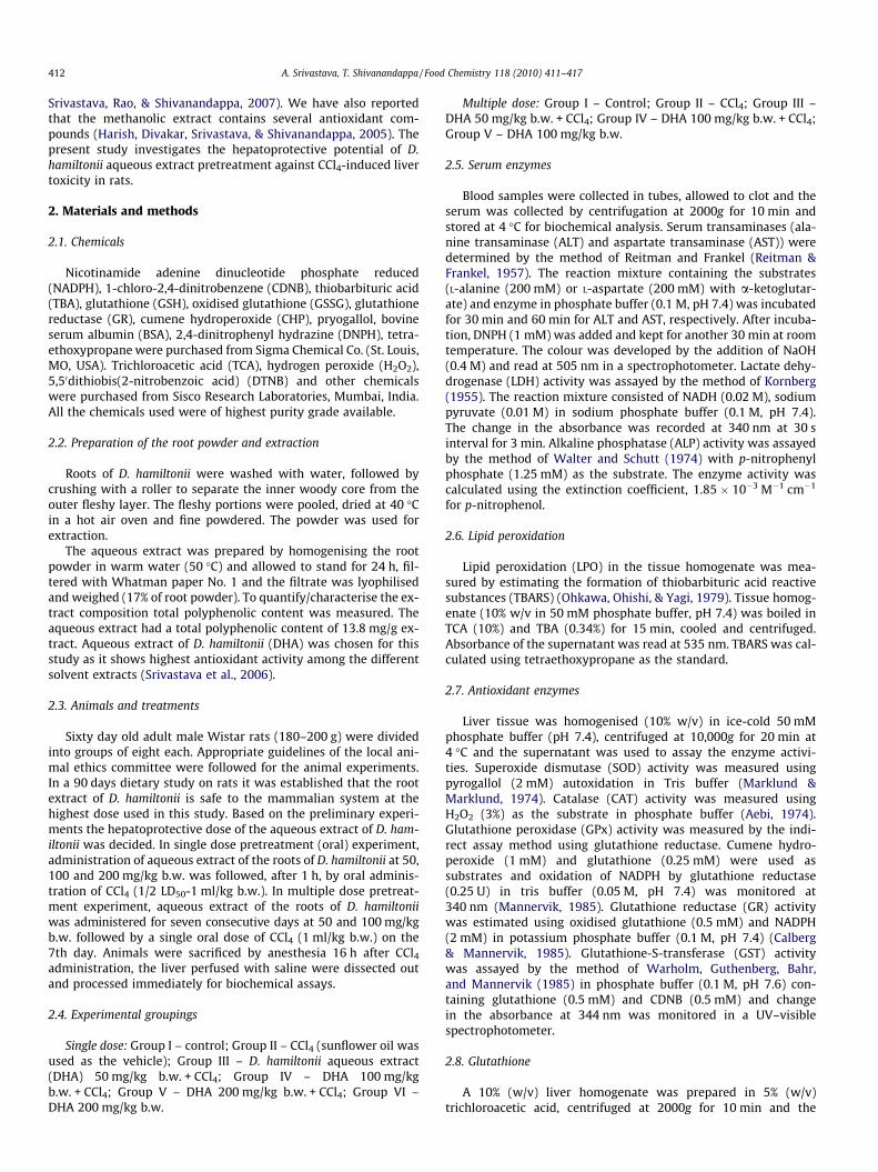
Fig. 1. Protective effect of the aqueous extract of the roots of D. hamiltonii (pretreatmenCCl4; Group III – DHA 50 mg/kg b.w. + CCl4; Group IV – DHA 100 mg/kg b.w. + CCl4; Grdehydrogenase; ALT, alanine aminotransferase; AST, aspartate aminotransferase; ALP, aalphabets differ significantly at p < 0.05 level (DMRT).
2.11. Statistics
Data were expressed as mean ± SE (n = 8) and significant differ-ence between the groups was statistically analysed by Duncan’smultiple range test (Statistica Software, 1999). A difference wasconsidered significant at p < 0.05.
3. Results
The food consumption and body weights of the DHA treatedrats were comparable to the control group.
3.1. Serum enzymes
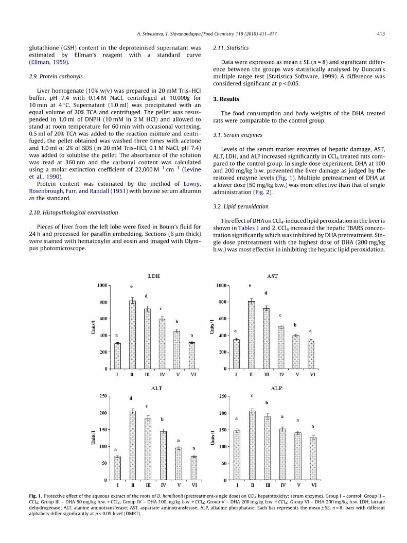
Levels of the serum marker enzymes of hepatic damage, AST,ALT, LDH, and ALP increased significantly in CCl4 treated rats com-pared to the control group. In single dose experiment, DHA at 100and 200 mg/kg b.w. prevented the liver damage as judged by therestored enzyme levels (Fig. 1). Multiple pretreatment of DHA ata lower dose (50 mg/kg b.w.) was more effective than that of singleadministration (Fig. 2).
3.2. Lipid peroxidation
The effect of DHA on CCl4-induced lipid peroxidation in the liver isshown in Tables 1 and 2. CCl4 increased the hepatic TBARS concen-tration significantly which was inhibited by DHA pretreatment. Sin-gle dose pretreatment with the highest dose of DHA (200 mg/kgb.w.) was most effective in inhibiting the hepatic lipid peroxidation.
t-single dose) on CCl4 hepatotoxicity: serum enzymes. Group I – control; Group II –oup V – DHA 200 mg/kg b.w. + CCl4; Group VI – DHA 200 mg/kg b.w. LDH, lactatelkaline phosphatase. Each bar represents the mean ± SE, n = 8; bars with different
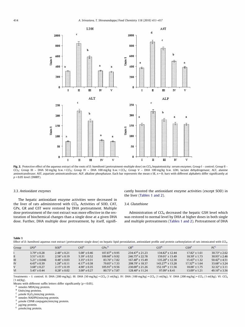
Fig. 2. Protective effect of the aqueous extract of the roots of D. hamiltonii (pretreatment-multiple dose) on CCl4 hepatotoxicity: serum enzymes. Group I – control; Group II –CCl4; Group III – DHA 50 mg/kg b.w. + CCl4; Group IV – DHA 100 mg/kg b.w. + CCl4; Group V – DHA 100 mg/kg b.w. LDH, lactate dehydrogenase; ALT, alanineaminotransferase; AST, aspartate aminotransferase; ALP, alkaline phosphatase. Each bar represents the mean ± SE, n = 8; bars with different alphabets differ significantly atp < 0.05 level (DMRT).
414 A. Srivastava, T. Shivanandappa / Food Chemistry 118 (2010) 411–417
3.3. Antioxidant enzymes
The hepatic antioxidant enzyme activities were decreased inthe liver of rats administered with CCl4. Activities of SOD, CAT,GPx, GR and GST were restored by DHA pretreatment. Multipledose pretreatment of the root extract was more effective in the res-toration of biochemical changes than a single dose at a given DHAdose. Further, DHA multiple dose pretreatment, by itself, signifi-
Table 1Effect of D. hamiltonii aqueous root extract (pretreatment-single dose) on hepatic lipid per
Group LPOA SODB CATC GPx.D
I 3.79a ± 0.28 2.48a ± 0.21 5.08c ± 0.46 107.87b ± 9.95II 3.51a ± 0.31 2.58a ± 0.19 5.39c ± 0.52 109.66b ± 9.92III 5.21c ± 0.046 0.48c ± 0.03 3.35a ± 0.31 83.76a ± 7.82IV 4.43b ± 0.39 1.28b ± 0.11 4.17b ± 0.38 79.83a ± 7.33V 3.68a ± 0.27 2.15a ± 0.19 4.90c ± 0.35 105.91b ± 9.56VI 5.45c ± 0.44 0.28c ± 0.02 3.00a ± 0.27 80.73a ± 7.87
Treatments – I: control; II: DHA (200 mg/kg); III: DHA (50 mg/kg) + CCl4 (1 ml/kg); IV:(1 ml/kg).Means with different suffix letters differ significantly (p < 0.05).
A nmoles MDA/mg protein.B Units/mg proteins.C lmole H2O2/min/mg protein.D nmoles NADPH/min/mg protein.E lmole CDNB conjugate/min/mg protein.F lg/mg protein.G lmole/mg protein.
cantly boosted the antioxidant enzyme activities (except SOD) inthe liver (Tables 1 and 2).
3.4. Glutathione
Administration of CCl4 decreased the hepatic GSH level whichwas restored to normal level by DHA at higher doses in both singleand multiple pretreatments (Tables 1 and 2). Pretreatment of DHA
oxidation, antioxidant profile and protein carbonylation of rats intoxicated with CCl4.
GRD GSTE GSHF PCG
234.47d ± 21.23 134.82b ± 12.44 17.82c ± 1.61 30.73a ± 2.62246.75d ± 22.78 159.01c ± 13.49 18.50c ± 1.73 30.93a ± 2.46167.48b ± 15.49 135.28b ± 12.38 15.43b ± 1.32 50.42b ± 4.51208.79c ± 18.37 143.27bc ± 13.28 17.32bc ± 1.64 33.68a ± 3.24230.00d ± 21.26 152.18bc ± 11.56 18.06c ± 1.75 32.32a ± 3.11128.40a ± 11.24 97.09a ± 8.41 13.09a ± 1.21 49.16b ± 3.56
DHA (100 mg/kg) + CCl4 (1 ml/kg); V: DHA (200 mg/kg) + CCl4 (1 ml/kg); VI: CCl4
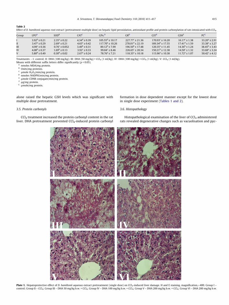
Table 2Effect of D. hamiltonii aqueous root extract (pretreatment-multiple dose) on hepatic lipid peroxidation, antioxidant profile and protein carbonylation of rats intoxicated with CCl4.
Group LPOA SODB CATC GPx.D GRD GSTE GSHF PCG
I 3.92b ± 0.21 2.35a ± 0.22 4.34d ± 0.39 105.55d ± 10.17 227.77c ± 21.36 170.93c ± 16.20 16.17c ± 1.38 33.20a ± 2.91II 3.47a ± 0.29 2.69a ± 0.21 4.65e ± 0.42 117.70e ± 10.26 278.01d ± 22.19 189.34d ± 17.55 17.41d ± 1.59 33.38a ± 3.27III 4.99c ± 0.36 0.76c ± 0.052 3.49b ± 0.31 89.12b ± 7.99 196.50b ± 17.48 120.35a ± 11.45 14.36b ± 1.24 38.45b ± 3.43IV 4.08b ± 0.37 1.69b ± 0.13 3.92c ± 0.33 99.84c ± 8.46 226.65c ± 20.34 150.21b ± 12.38 14.50c ± 1.32 33.68a ± 3.24V 5.80d ± 0.49 0.30d ± 0.02 2.67a ± 0.24 78.76a ± 7.21 118.35a ± 10.18 115.96a ± 10.38 11.72a ± 1.07 50.42c ± 4.12
Treatments – I: control; II: DHA (100 mg/kg); III: DHA (50 mg/kg) + CCl4 (1 ml/kg); IV: DHA (100 mg/kg) + CCl4 (1 ml/kg); V: CCl4 (1 ml/kg).Means with different suffix letters differ significantly (p < 0.05).
A nmoles MDA/mg protein.B Units/mg proteins.C lmole H2O2/min/mg protein.D nmoles NADPH/min/mg protein.E lmole CDNB conjugate/min/mg protein.F lg/mg protein.G lmole/mg protein.
A. Srivastava, T. Shivanandappa / Food Chemistry 118 (2010) 411–417 415
alone raised the hepatic GSH levels which was significant withmultiple dose pretreatment.
3.5. Protein carbonyls
CCl4 treatment increased the protein carbonyl content in the ratliver. DHA pretreatment prevented CCl4-induced protein carbonyl
Plate 1. Hepatoprotective effect of D. hamiltonii aqueous extract pretreatment (single docontrol; Group II – CCl4; Group III – DHA 50 mg/kg b.w. + CCl4; Group IV – DHA 100 mg/kg
formation in dose dependent manner except for the lowest dosein single dose experiment (Tables 1 and 2).
3.6. Histopathology
Histopathological examination of the liver of CCl4 administeredrats revealed degenerative changes such as vacuolisation and pyc-
se) on CCl4-induced liver damage. H and E staining, magnification,�400. Group I –b.w. + CCl4; Group V – DHA 200 mg/kg b.w. + CCl4; Group VI – DHA 200 mg/kg b.w.
416 A. Srivastava, T. Shivanandappa / Food Chemistry 118 (2010) 411–417
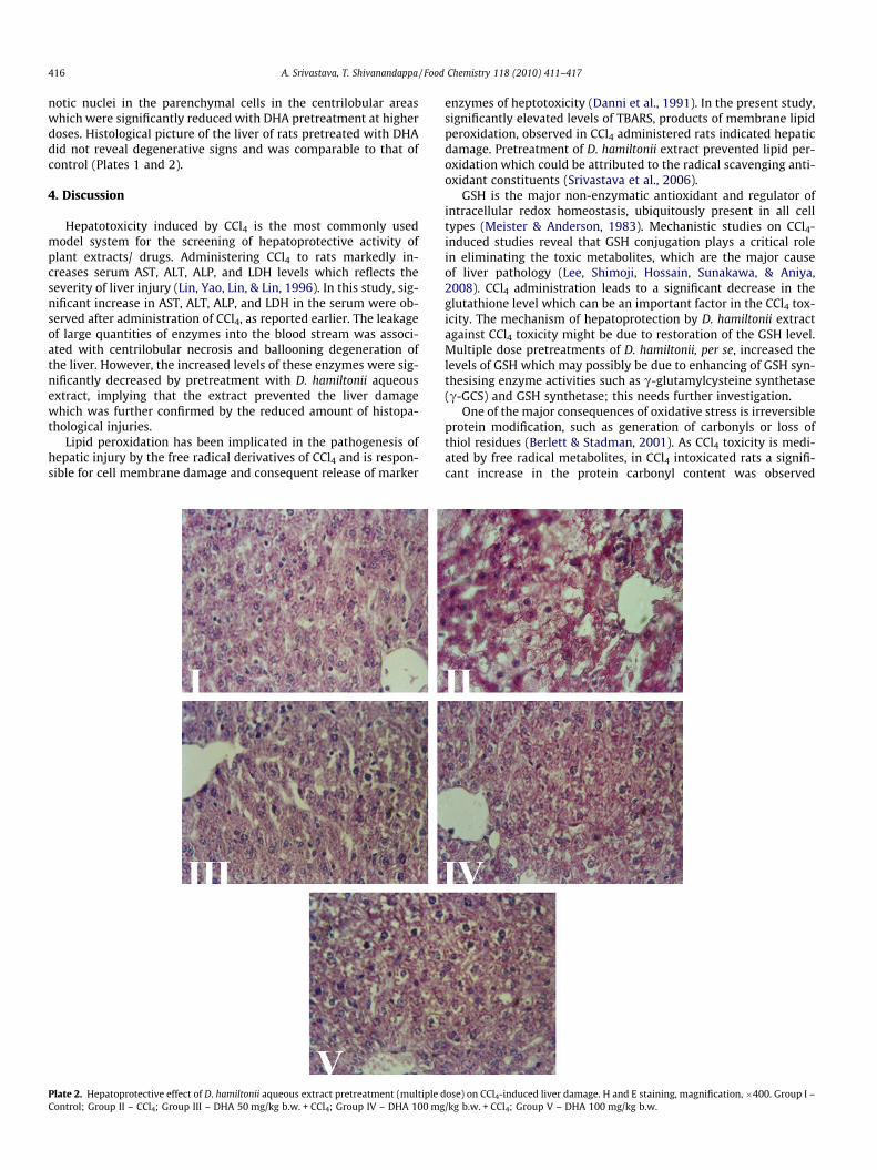
notic nuclei in the parenchymal cells in the centrilobular areaswhich were significantly reduced with DHA pretreatment at higherdoses. Histological picture of the liver of rats pretreated with DHAdid not reveal degenerative signs and was comparable to that ofcontrol (Plates 1 and 2).
4. Discussion
Hepatotoxicity induced by CCl4 is the most commonly usedmodel system for the screening of hepatoprotective activity ofplant extracts/ drugs. Administering CCl4 to rats markedly in-creases serum AST, ALT, ALP, and LDH levels which reflects theseverity of liver injury (Lin, Yao, Lin, & Lin, 1996). In this study, sig-nificant increase in AST, ALT, ALP, and LDH in the serum were ob-served after administration of CCl4, as reported earlier. The leakageof large quantities of enzymes into the blood stream was associ-ated with centrilobular necrosis and ballooning degeneration ofthe liver. However, the increased levels of these enzymes were sig-nificantly decreased by pretreatment with D. hamiltonii aqueousextract, implying that the extract prevented the liver damagewhich was further confirmed by the reduced amount of histopa-thological injuries.
Lipid peroxidation has been implicated in the pathogenesis ofhepatic injury by the free radical derivatives of CCl4 and is respon-sible for cell membrane damage and consequent release of marker
Plate 2. Hepatoprotective effect of D. hamiltonii aqueous extract pretreatment (multiple dControl; Group II – CCl4; Group III – DHA 50 mg/kg b.w. + CCl4; Group IV – DHA 100 mg
enzymes of heptotoxicity (Danni et al., 1991). In the present study,significantly elevated levels of TBARS, products of membrane lipidperoxidation, observed in CCl4 administered rats indicated hepaticdamage. Pretreatment of D. hamiltonii extract prevented lipid per-oxidation which could be attributed to the radical scavenging anti-oxidant constituents (Srivastava et al., 2006).
GSH is the major non-enzymatic antioxidant and regulator ofintracellular redox homeostasis, ubiquitously present in all celltypes (Meister & Anderson, 1983). Mechanistic studies on CCl4-induced studies reveal that GSH conjugation plays a critical rolein eliminating the toxic metabolites, which are the major causeof liver pathology (Lee, Shimoji, Hossain, Sunakawa, & Aniya,2008). CCl4 administration leads to a significant decrease in theglutathione level which can be an important factor in the CCl4 tox-icity. The mechanism of hepatoprotection by D. hamiltonii extractagainst CCl4 toxicity might be due to restoration of the GSH level.Multiple dose pretreatments of D. hamiltonii, per se, increased thelevels of GSH which may possibly be due to enhancing of GSH syn-thesising enzyme activities such as c-glutamylcysteine synthetase(c-GCS) and GSH synthetase; this needs further investigation.
One of the major consequences of oxidative stress is irreversibleprotein modification, such as generation of carbonyls or loss ofthiol residues (Berlett & Stadman, 2001). As CCl4 toxicity is medi-ated by free radical metabolites, in CCl4 intoxicated rats a signifi-cant increase in the protein carbonyl content was observed
ose) on CCl4-induced liver damage. H and E staining, magnification, �400. Group I –/kg b.w. + CCl4; Group V – DHA 100 mg/kg b.w.
A. Srivastava, T. Shivanandappa / Food Chemistry 118 (2010) 411–417 417
which was restored to normalcy by D. hamiltonii extract pretreat-ment. The antioxidant activity of D. hamiltonii extract and itsGSH status elevating effect could be responsible for thisobservation.
Among the cellular antioxidants, SOD, CAT, GST, GPx, and GRhave received extensive studies. SOD catalyses the dismutation ofsuperoxide anion to H2O2 and O2. Because H2O2 is still harmfulto cells, catalase and GPx further catalyse the decomposition ofH2O2 to water. In the reaction catalysed by GPx, GSH is oxidisedto GSSG, which can then be reduced back to GSH by GR. GSH is alsoa cofactor for GST, a phase II enzyme, primarily involved in thedetoxification of electrophilic xenobiotics via catalysing the forma-tion of GSH-electrophile conjugate (Hayes, Flanagan, & Jowsey,2005). Thus, the coordinate actions of various cellular antioxidantsin mammalian cells are critical for effectively detoxifying free rad-icals. CCl4 administration to rats declined antioxidant capacity ofthe rat liver as evinced in decreased activity of the antioxidant en-zymes, which is in agreement with earlier reports (Shahjahan et al.,2004). D. hamiltonii extract pretreatment prevented the reductionin the antioxidant enzyme activities and consequent oxidativedamage to the liver. In fact, the multiple dose pretreatment of D.hamiltonii extract alone significantly boosted the antioxidant en-zyme activities. Similar studies have shown a positive effect of dif-ferent classes of polyphenols on antioxidant enzyme activitiesin vivo (Molina, Sanchez-Reus, Iglesias, & Benedi, 2003; Scharf,Prustomersky, Knasmuller, Schulte-Hermann, & Huber, 2003).
In conclusion, the D. hamiltonii aqueous extract effectively pre-vented CCl4-induced hepatotoxicity which explains its health pro-moting property. Our previous study with ethanol, an inducer ofoxidative stress, also shows hepatoprotective effect of D. hamiltoniiextract (Srivastava & Shivanandappa, 2006). Even though ethanoland CCl4 have different mechanisms of toxicity D. hamiltonii aque-ous extract could protect the rat liver. The hepatoprotective activ-ity of D. hamiltonii extract could be a result of free radicalscavenging activity or boosting the antioxidant capacity of the li-ver. The bioactive antioxidant principles of the aqueous extractthat have been identified (Srivastava et al., 2006) could be respon-sible for the observed hepatoprotective effect. Further studies withthe individual antioxidant compounds isolated from D. hamiltoniiroot extract on hepatocytes are underway which will enable usto understand the exact mechanism of hepatoprotective actionby D. hamiltonii.
Acknowledgements
This work was done at Central Food Technological ResearchInstitute, Mysore, India. The authors wish to thank the Directorof the institute for his keen interest in this study. The first authoracknowledges Council for Scientific and Industrial Research, NewDelhi for awarding the research fellowship.
References
Aebi, H. (1974). Catalase. In H. U. Bergmeyer (Ed.), Methods of enzymatic analysis. Vol.2 (pp. 674–678). Weinheim: Verlag Chenie.
Berlett, B. S., & Stadman, E. R. (2001). Protein oxidation in aging, disease andoxidative stress. Journal of Biological Chemistry, 272, 20313–20316.
Calberg, I., & Mannervik, B. (1985). Glutathione reductase. In A. Meister (Ed.).Methods in enzymology (Vol. 113, pp. 484–490). FL: Academic Press.
Danni, O., Chiarpotto, E., Aragno, M., Biasi, F., Comoglio, A., Belliardo, F., et al. (1991).Lipid peroxidation and irreversible cell damage: Synergism between carbontetrachloride and 1,2-dibromoethane in isolated rat hepatocytes. Toxicology andApplied Pharmacology, 110, 216–222.
Demirdag, K., Bakcecioglu, I. H., Ozercan, I. H., Ozden, M., Yilmaz, S., & Kalkan, A.(2004). Role of L-carnitine in the prevention of acute liver damage induced bycarbon tetrachloride in rats. Journal of Gastroenterology and Hepatology, 19,333–338.
Ellman, G. L. (1959). Tissue sulfhydryl groups. Archives of Biochemistry andBiophysics, 82, 70–77.
Harish, R., Divakar, S., Srivastava, A., & Shivanandappa, T. (2005). Isolation ofantioxidant compounds from the methanolic extract of the roots of Decalepishamiltonii (Wight and Arn.). Journal of Agricultural and Food Chemistry, 53,7709–7714.
Hayes, J. D., Flanagan, J. U., & Jowsey, I. R. (2005). Glutathione transferases. AnnualReviews in Pharmacology and Toxicology, 45, 51–88.
Kodavanti, P. R., Joshi, U. M., Young, Y. A., Meydrech, E. F., & Mehendale, H. M.(1989). Protection of hepatotoxic and lethal effects of CCl4 by partialhepatectomy. Toxicology and Pathology, 17, 494–505.
Kornberg, A. (1955). Lactic dehydrogenase of muscle. Methods in Enzymology, 1,441–443.
Lee, K. K., Shimoji, M., Hossain, Q. S., Sunakawa, H., & Aniya, Y. (2008). Novelfunction of glutathione transferase in rat liver mitochondrial membrane: Rolefor cytochrome c release from mitochondria. Toxicology and AppliedPharmacology, 232, 109–118.
Levine, R. L., Garland, D., Oliver, C. N., Amici, A., Climent, I., Lenz, A., et al. (1990).Determination of carbonyl content of oxidatively modified proteins. Methods inEnzymology, 186, 464–478.
Lin, S. C., Yao, C. J., Lin, C. C., & Lin, Y. H. (1996). Hepatoprotective activity of Taiwanfolk medicine: Eclipta prostrate Linn. against various hapatotoxins inducedacute hapatotoxicity. Phototherapy Research, 10, 483–490.
Lowry, O. H., Rosenbrough, N. J., Farr, A. L., & Randall, R. J. (1951). Proteinmeasurement with Folin-Phenol reagent. Journal of Biological Chemistry, 193,265–275.
Mannervik, B. (1985). Glutathione peroxidase. In A. Miester (Ed.). Methods inenzymology (Vol. 113, pp. 490–495). FL: Academic Press.
Marklund, S., & Marklund, G. (1974). Involvement of the superoxide anion in theautoxidation of pyrogallol and a convenient assay for superoxide dismutase.European Journal of Biochemistry, 47, 469–474.
Meister, A., & Anderson, M. E. (1983). Glutathione. Annual Reviews in Biochemistry,52, 711–760.
Molina, M. F., Sanchez-Reus, I., Iglesias, I., & Benedi, J. (2003). Quercetin, a flavonoidantioxidant, prevents and protects against ethanol-induced oxidative stress inmouse liver. Biological and Pharmaceutical Bulletin, 26, 1398–1402.
Nayar, R. C., Shetty, J. K. P., Mary, Z., & Yoganarshimhan, S. N. (1978).Pharmacognostical studies on the root of Decalepis hamiltonii Wt. and Arn.and comparison with Hemidesmus indicus (L.) R. Br. Proceedings of IndianAcademy of Sciences, 87, 37–48.
Ohkawa, H., Ohishi, N., & Yagi, K. (1979). Assay for lipid peroxides in animal tissuesby thiobarbituric acid reaction. Analytical Biochemistry, 95, 351–358.
Reitman, S., & Frankel, S. (1957). A colorimetric method for the determination ofserum oxaloacetic and glutamic pyruvic transaminases. American Journal ofClinical Pathology, 28, 56–63.
Scharf, G., Prustomersky, S., Knasmuller, S., Schulte-Hermann, R., & Huber, W. W.(2003). Enhancement of glutathione and g-glutamylcystein synthetase, the ratelimiting enzyme of glutathione synthesis, by chemoprotective plant-derivedfood and beverage components in the human hepatoma cell line Hep G2.Nutrition and Cancer, 45, 74–83.
Shahjahan, M., Sabitha, K. E., Jainu, M., & Devi, C. S. S. (2004). Effect of Solanumtrilobatum against carbon tetrachloride induced hapatic damage in albino rats.Indian Journal of Medical Research, 120, 194–198.
Sheweita, S. A., El-Gabar, M. A., & Bastawy, M. (2001). Carbon tetrachloride-inducedchanges in the activity of phase II drug-metabolizing enzyme in the liver ofmale rats: Role of antioxidants. Toxicology, 165, 217–224.
Srivastava, A., Harish, R., & Shivanandappa, T. (2006). Novel antioxidant compoundsfrom the aqueous extract of the roots of Decalepis hamiltonii (Wight and Arn.)and their inhibitory effect on low-density lipoprotein oxidation. Journal ofAgricultural and Food Chemistry, 54, 790–795.
Srivastava, A., Rao, L. J. M., & Shivanandappa, T. (2007). Isolation of ellagic acid fromthe aqueous extract of the roots of Decalepis hamiltonii: Antioxidant activity andcytoprotective effect. Food Chemistry, 103, 224–233.
Srivastava, A., Shereen, Harish, R., & Shivanandappa, T. (2006). Antioxidant activityof the roots of Decalepis hamiltonii (Wight and Arn.). LWT-Food Science andTechnology, 39, 1059–1065.
Srivastava, A., & Shivanandappa, T. (2006). Hepatoprotective effect of the aqueousextract of the roots of Decalepis hamiltonii against ethanol-induced oxidativestress in rats. Hepatology Research, 35, 267–275.
Walter, K., & Schutt, C. (1974). Acid and alkaline phosphatase in serum (Two pointmethod). In H. U. Bergmeyer (Ed.). Methods of enzymatic analysis (Vol. 2,pp. 856–860). Deerfield Beach, FL: Verlag Chemie.
Warholm, M., Guthenberg, C., Bahr, C. V., & Mannervik, B. (1985). Glutahtionetransferase from human liver. Methods in enzymology (Vol. 113, pp. 499–504).Deerfield Beach, FL: Academic Press.