Hepatocyte Proliferation in Fatty Liver-Hemorrhagic and ...
28
Page 1/28 Salidroside Alleviates Lipid Metabolism Disorder and Inammatory Response, and Promotes Hepatocyte Proliferation in Fatty Liver-Hemorrhagic Syndrome of Laying Hens Zhifu Cui Sichuan Agricultural University - Chengdu Campus Ningning Jin Sichuan Agricultural University - Chengdu Campus Felix Kwame Amevor Sichuan Agricultural University - Chengdu Campus Liang Li Sichuan Agricultural University - Chengdu Campus Gang Shu Sichuan Agricultural University - Chengdu Campus Xiaxia Du Sichuan Agricultural University - Chengdu Campus Xincheng Kang Sichuan Agricultural University - Chengdu Campus Dan Xu Sichuan Agricultural University - Chengdu Campus Zifan Ning Sichuan Agricultural University - Chengdu Campus Xun Deng Sichuan Agricultural University - Chengdu Campus Weizhen Song Sichuan Agricultural University - Chengdu Campus Yaofu Tian Sichuan Agricultural University - Chengdu Campus Qing Zhu Sichuan Agricultural University - Chengdu Campus Yan Wang Sichuan Agricultural University - Chengdu Campus Diyan Li Sichuan Agricultural University - Chengdu Campus
Transcript of Hepatocyte Proliferation in Fatty Liver-Hemorrhagic and ...

Page 1/28
Salidroside Alleviates Lipid Metabolism Disorderand In�ammatory Response, and PromotesHepatocyte Proliferation in Fatty Liver-HemorrhagicSyndrome of Laying HensZhifu Cui
Sichuan Agricultural University - Chengdu CampusNingning Jin
Sichuan Agricultural University - Chengdu CampusFelix Kwame Amevor
Sichuan Agricultural University - Chengdu CampusLiang Li
Sichuan Agricultural University - Chengdu CampusGang Shu
Sichuan Agricultural University - Chengdu CampusXiaxia Du
Sichuan Agricultural University - Chengdu CampusXincheng Kang
Sichuan Agricultural University - Chengdu CampusDan Xu
Sichuan Agricultural University - Chengdu CampusZifan Ning
Sichuan Agricultural University - Chengdu CampusXun Deng
Sichuan Agricultural University - Chengdu CampusWeizhen Song
Sichuan Agricultural University - Chengdu CampusYaofu Tian
Sichuan Agricultural University - Chengdu CampusQing Zhu
Sichuan Agricultural University - Chengdu CampusYan Wang
Sichuan Agricultural University - Chengdu CampusDiyan Li
Sichuan Agricultural University - Chengdu Campus

Page 2/28
Yao Zhang Sichuan Agricultural University - Chengdu Campus
Xiaoqi Wang Agriculture and Animal Husbandry Comprehensive Service Center of Razi County, Tibet Autonomous
Region, P. R. China.Xue Han
Guizhou Institute of Animal Husbandry and Veterinary Medicine, Guizhou Province, P. R. China.Jing Feng
Institute of Animal Husbandry and Veterinary Medicine, College of Agriculture and Animal Husbandry,Tibet Autonomous Region, P. R. China.Xiaoling Zhao ( [email protected] )
Sichuan Agricultural University - Chengdu Campus
Research
Keywords: Layer, Fatty liver hemorrhagic syndrome, Salidroside, In�ammatory response, Hepatocyteproliferation.
Posted Date: September 21st, 2021
DOI: https://doi.org/10.21203/rs.3.rs-885594/v1
License: This work is licensed under a Creative Commons Attribution 4.0 International License. Read Full License
Page 3/28
AbstractBackground: Fatty liver hemorrhagic syndrome (FLHS) is a chronic hepatic disease which occurs whenthere is a disorder in lipid metabolism. This disease is often observed in caged laying hens andcharacterized by a decrease in egg production and dramatic increase of mortality. Salidroside (SDS) is anherbal drug which has shown numerous pharmacological activities, such as protective effects onmitochondrial function, attenuates cell apoptosis and in�ammation, and promotes strong antioxidantdefense system. We aimed to determine the therapeutic effects of SDS on FLHS in laying hens andinvestigate the underlying mechanisms through which SDS operates these functions. We constructedoleic acid (OA)-induced fatty liver model in vitro and high-fat diet-induced FLHS of laying hens in vivo.
Results: Results indicated that SDS inhibited OA-induced lipid accumulation in chicken primaryhepatocytes, increased hepatocyte activity, elevated the mRNA expression of proliferation related genesPCNA, CDK2, and cyclinD1 and increased the protein levels of PCNA and CDK2, as well as decreased thecleavage levels of Caspase-9, Caspase-8, and Caspase-3 and apoptosis in hepatocytes. Moreover, SDSpromoted the phosphorylation levels of PDK1, AKT, and Gsk3-β, while inhabited the PI3K inhibitor.Additionally, we found that high-fat diet-induced FLHS of laying hens in vivo resulted in heavier bodyweight, liver weight, and abdominal fat weight, and severer steatosis in histology, compared with thecontrol group (Con). However, SDS maintained lighter body weight, liver weight, and abdominal fat weightand alleviate hepatic steatosis in Model+SDS group. In addition, high-fat diet-induced FLHS (Model) oflaying hens had higher total cholesterol (TC), triglyceride (TG), alanine transaminase (ALT), and aspartateaminotransferase (AST) levels in serum than Con group, while SDS maintained low TC, TG, ALT, and ASTlevels and high Superoxide dismutase (SOD) activity in Model+SDS group. Moreover, SDS decreased themRNA expression abundances of PPARγ, SCD, and FAS in liver, whereas increased those of PPARα andMTTP, and decreased the mRNA expression of TNF-α, IL-1β, IL-6, and IL-8 in Model+SDS group.
Conclusions: Generally, SDS attenuated OA-induced ROS generation, inhibited lipid accumulation andhepatocyte apoptosis, and promoted hepatocyte proliferation by targeting the pathway PI3K/AKT/Gsk3-βin OA-induced fatty liver model in vitro, and alleviated high-fat diet-induced hepatic steatosis, oxidativestress, and in�ammatory response in laying hens in vivo.
IntroductionIn poultry, liver has strong lipid synthesis ability but poor lipid storage capacity. Excessive storage of lipidsubstances in liver results in a series of pathological occurrences in aging chickens, in which the mostrepresentative disease is fatty liver-hemorrhagic syndrome (FLHS) of laying hens, which seriouslydecreases egg production [1–4]. Further deterioration of fatty liver may lead to steatohepatitis, liver�brosis, and even cirrhosis [5], having sudden death of individuals in the layer �ocks [6, 7].
Studies indicated that high fat diets led to fat transport obstruction in chicken liver, disrupted the dynamicbalance of fat metabolism in hepatocytes, adipocytes, and bloods. Excessive deposition of lipids were

Page 4/28
mainly composed of neutral fat in hepatocytes [8], which is an important pathological feature of non-alcoholic fatty liver disease (NAFLD) [9–11]. Excessive lipid accumulation and estrogen secretiondepressed the mitochondrial function in hepatocytes, and produced harmful reactive oxygen species(ROS) which damaged hepatocytes, and further reduced the activity of antioxidant enzymes in organismsby inhibiting gene transcription, thus causing damage to the antioxidant system of animals [12].
Plant extracts were reported to improve liver steatosis [13–15]. Rhodiola rosea L. radix (RRL) known asplateau ginseng is a rare medicinal herb that grows in cold high-altitude areas. In Ming Dynasty, China(�ve hundred years ago) RRL has used as a tonic to treat diseases, eliminate fatigue and resist cold.Modern pharmacological studies have con�rmed that RRL extract has anti-aging, anti-tumor, anti-hypoxia, anti-fatigue, and anti-oxidation functions [16–21].
Salidroside (SDS) is the main pharmacological component of RRL. It can scavenge free radicals,decrease oxidative stress, enhance immunity, and prevent aging in animals [16–21]. Meanwhile, SDS hasno genetic toxicity or mutagenesis [22]. Mao et al. induced the aging model with D-galactose in mice, andfound that SDS could inhibit the formation of advanced glycation end products in vivo, which increasedthe production of lymphocyte mitosis and interleukin-2 (IL-2), and had an anti-aging effect [23]. Lu et al.studied the effects of SDS on the number of helper T cells and the delayed type hypersensitivity responsein aged rats, and found that SDS stimulated the body's humoral and cellular immune responses [16].Zhang found that SDS inhibited H2O2-induced cell viability loss, reduced cell apoptosis rate, inhibited
mitochondrial membrane potential decline, and increased intracellular Ca2+ concentration, which can beused to treat and prevent neurodegenerative diseases [24]. Guan demonstrated SDS resistedlipopolysaccharide (LPS)-induced acute pneumonia in a mouse model [25]. Li reported that SDSprevented mouse mastitis induced by LPS, reduced the activity of myeloperoxidase and theconcentrations of TNF-α, IL-1β, and IL-6 in breast muscle tissue, inhibited the in�ammatory cellin�ltration, and produced anti-in�ammatory effects [26].
Additionally, SDS protected against LPS-induced liver failure in mice by its antioxidant activity andinhibited hypoxia-inducible factor-1α expression [27], protected against acetaminophen-inducedhepatotoxicity by preventing or mitigating intracellular glutathione depletion and oxidative damage [28],and inhibited chronic hepatitis C virus by inhibiting serine protease activity [29]. Recent studies indicatedthat SDS alleviated hypoxia-induced liver injury and inhibited cell apoptosis via the IRE1α/JNK pathway[30], alleviated carbon tetrachloride-induced liver damage in mice by activating mitochondria to resistoxidative stress [31], inhibited apoptosis and autophagy in a Concanavalin A-induced liver injury model[32], protected oxidative stress induced by diabetes mellitus, effectively reduced blood TC and TG levels[33], alleviated liver steatosis in type 2 diabetic mice [34], and reduced obesity and liver lipid depositioninduced by a high-fat diet by inhibiting oxidative stress and activation of in�ammatory bodies in the liver[35].
Generally, SDS protected liver and treated liver diseases in mammalian studies. Thus, we hypothesis thatSDS may have positive effect on relieving the fatty liver in laying hens. Here, we used oleic acid (OA) [36–

Page 5/28
38], dietary energy, and protein structure recombination [39–42] to establish chicken fatty liver models invitro and in vivo to explore the effects of SDS on lipid metabolism, proliferation, apoptosis, andmitochondrial function of fatty liver in chickens.
Materials And Methods
Purity determination of SDSThe SDS was purchased from Nanjing Herb-Source Biotechnology Co. Ltd (Nanjing, China). Its purity andstructure were examined via High Performance Liquid Chromatography (HPLC) (Agilent Technologies Inc.,California, USA), Nuclear Magnetic Resonance Spectrometer (NMR) (Avance III HD 400, Bruker,Switzerland), and Mass Spectrometry (MS) (Thermo Fisher Scienti�c, San Jose, CA).
AnimalsA total of three hundred 35-week-old Rohman layers were raised at the Animal Breeding Farm, SichuanAgricultural University (Ya’an, China). All animal experiments were approved by the Institutional AnimalCare and Use Committee of Sichuan Agricultural University (Certi�cation No. YCS-B2018102013). Allexperiments were conducted in accordance with the Laboratory Animal Welfare and Ethics guidelines ofSichuan Agricultural University.
Cell culture and treatmentPrimary hepatocytes were isolated from the Rohman layers according to the methods described by [43,44]. The hepatocytes were resuspended in William’s medium E (Sigma, Shanghai, China) and plated into6-well plates (2 × 10⁶ cells/well) after cell counting. The hepatocytes were cultured in a cell cultureincubator at a constant temperature of 37 ℃, 5 % CO2, and 95 % air saturated humidity. After 24 h, thehepatocytes were cultured with 0.6 mM OA for 12, 24, 36 and 48 h and treated with/without SDS. Allhepatocytes were collected for further experiments.
RNA extraction and quantitative real time PCR (qRT-PCR)Total RNA was extracted from all samples according to the instructions of the Trizol reagent (MolecularResearch Center, Cincinnati, OH). The RNA concentration and purity were estimated by determining theA260/A280 absorbance ratio, and the 18 S and 28 S bands in a 1 % agarose gel. Reverse transcriptionand qRT-PCR were performed as described previously [45]. GAPDH and β-actin were used as endogenouscontrols to normalize gene expression using the 2−ΔΔCt method [46]. Gene-speci�c primers designed withsoftware Primer Premier 5.0 according to the coding sequences of target genes are summarized inSupplementary table 1.
Protein extraction and western blot analysisHepatocyte protein was isolated using a protein extraction kit (BestBio Biotech Co. Ltd., Shanghai, China),and the BCA protein assay kit (BestBio) was used to determine the concentration of protein samples.

Page 6/28
Western blotting was performed with the method described by [47] with the following primary antibodies:anti-PPARα (Abcam, Cambridge, UK), anti-PCNA (ABclonal Technology, Wuhan, China), anti-CDK2 (Zen-Bio, Chengdu, Chin), anti-caspase-3 and caspase-8 (Abcam), and caspase-9 (Bioss, Beijing, China), anti-PI3K [Cell Signaling Technology (CST), USA], anti-p-PI3K (CST), anti-AKT and p-AKT (CST), and anti-Gsk3-β and p-Gsk3-β (ABclonal). Goat anti-mouse and goat anti-rabbit secondary antibodies (Zen-Bio) β-Tubulin (Zen-Bio) was used as a reference.
Staining for liver tissue and hepatocyteLiver tissues (µm/g) were �xed with 4 % paraformaldehyde for 24 h and dehydrated with differentconcentrations of alcohol. Thereafter, the tissue mass was embedded in para�n, cut into thin slices andplaced on a slide. After hematoxylin and eosin (HE) staining, the sections were sealed with neutral resin.Liver tissue was sampled as a size of 24 mm×24 mm×2 mm, frozen and then placed on a tissue bearerdripping with an OTC embedding agent. Slices (thickness 10 µm) were obtained with the slicing machine(Leica CM1520, Germany) and a�xed to the anti-slip slide. After staining with oil red O staining solutionfor 15 min, the slices were sealed with a neutral resin.
Hepatocyte Oil red O staining was performed following the manufacturers’ protocol. Brie�y, hepatocyteswere washed with phosphate buffered saline (PBS) and �xed with ORO Fixative for 30 min, and then thestationary �uid was discarded and washed with PBS twice. Thereafter, 60% isopropanol was added andmaintained for 5min, after which the ORO Stain was added and retained for 20 min. After washing 5times, the Mayer Hematoxylin staining solution was added and kept for 2 min. Hepatocytes were washedand ORO Buffer was added and sustained for 1 min. After washing with PBS, the hepatocytes werecovered with distilled water. All sections and hepatocytes were viewed under a microscope (DP80Digital,Olympus, Tokyo, Japan) and ten �elds were randomly selected for statistical analysis.
Cell counting kit-8 (CCK‐8) and 5‐ethynyl‐2‐deoxyuridine(EdU) assayThe CCK-8 Kit (MeilunBio, Dalian, China) was used to test hepatocyte activity and choose the optimumconcentration and treatment time of SDS in the OA-induced fatty liver model of primary hepatocytes. TenµL of CCK-8 reagent was added to each well and incubated in a cell culture incubator for 2 h after beingtreated with SDS for 12, 24, 36, and 48 h. A microplate reader (Varioskan LUX, Thermo Fisher, USA) wasused to determine the optical density (OD) of each sample at 450 nm. The proliferation state ofhepatocytes was determined using a Cell‐Light™ EdU kit (RiboBio, Guangzhou, China) according to themanufacturer’s instructions. A �uorescent microscope was used to calculate the number of EdU-positivecells.
Cell apoptosis and reactive oxygen species (ROS) analysisHepatocyte apoptosis was detected using Flow Cytometry (CytoFLEX, Beckman, USA) and Kaluza 2.1software. Hepatocytes were washed with PBS and the concentration was adjusted to 1×10⁶ cells/mL,and determination was performed as previously described [48]. DCFH-DA (2, 7-Dichloro�uorescin; Sigma)

Page 7/28
was added to hepatocytes at a �nal concentration of 10 µM for 20 min at room temperature andhepatocyte ROS were analyzed using a Shimadzu RF-3501 �uorescence spectrophotometer at anexcitation wavelength of 488 nm and an emission wavelength of 525 nm.
PI3K/AKT/Gsk3-β pathway analysisLY294002 (Houston, Texas, United States) was used as a PI3K inhibitor to explore whether SDS exerts itsbiological function through PI3K/AKT/Gsk3-β. LY294002 was purchased from Selleck Chemicals and pre-incubated with hepatocytes for 2 h.
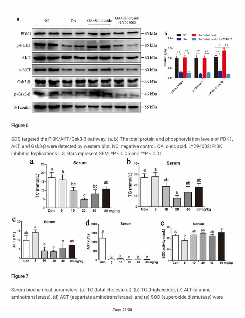
In vivo experiments in laying hensIn Trial 1, we wanted to �nd out the optimal feeding concentration of SDS. Thus, 180 35-week-oldRohman layers were randomly divided into six groups, including the Control (Con) and SDS feedinggroups (5, 10, 20, 40, and 80 mg/kg, respectively). Additive feeding experiment lasted for 4 weeks. Serumbiochemical parameters including total cholesterol (TC), Triglyceride (TG), alanine aminotransferase(ALT), aspartate aminotransferase (AST), and superoxide dismutase (SOD) were measured with enzyme-linked immunosorbent assay (ELISA) kits (Baolai Biotechnology Co., Ltd, Yancheng, China), following themanufacturer’s instructions. Moreover, the expression abundances of lipid metabolism-related genes andin�ammatory factors were detected using qRT-PCR.
In Trial 2, our purpose was to make clear whether SDS can prevent fatty liver in laying birds. Thus, 144 35-week-old Rohman layers were adopted and separated into four groups: Con, Con + SDS, Model, andModel + SDS. Based on Trial 1, optimal feeding dosage of SDS (20 mg/kg) was used to feed the birds for4 weeks in group Con + SDS and Model + SDS. The fatty liver model (Model) of laying hens in vivo wasestablished with the method previously described by [39–42]. The diet composition was summarized inSupplementary table 2.
Statistical analysisThe statistical analyses were carried out using SAS 9.3 software (SAS Institute Inc., Cary, NC, USA), andthe mean of three replicates were evaluated and presented as mean ± standard error (SE). Signi�cancelevel was determined using Duncan’s multiple range tests and displayed as P < 0.05 (*) and P < 0.01 (**).
Results
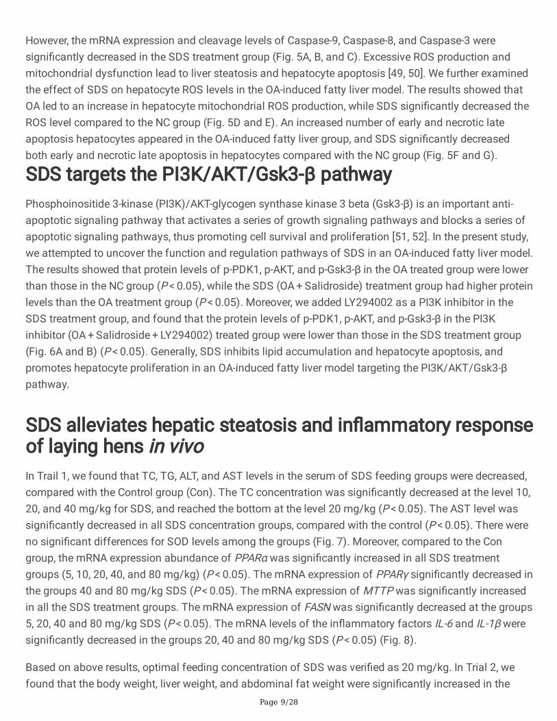
Purity determination of SDSThe purity and structure of SDS (C14H20O7, 300.30, Fig. 1A) were detected using HPLC, NMR, and MS. TheHPLC analysis results showed that the area reached a peak at a run time of 14.196 min, with a peak arearate of 99.81 % (Fig. 1B). NMR analysis results showed that the structure complied with the molecular
Page 8/28
formula of C14H20O7 (Fig. 1C). MS analysis showed that the structure complied with the molecular weightof 300.30 Da (Fig. 1D).
SDS treatment with OA-induced fatty liver model in primaryhepatocytesOA (0.6 mM) was used for inducing fatty liver model in primary hepatocytes. Cells were treated with 0.1,0.2, 0.3, 0.4 mg/mL SDS for 48 h, respectively (Supplementary table 3). According to the OD values of theCCK-8 assay at 12, 24, 36, and 48 h, OA signi�cantly decreased hepatocyte activity compared with thecontrol, while SDS signi�cantly increased hepatocyte activity at the concentration of 0.3 mg/mL at 36 h(Fig. 2).
SDS inhibits OA-induced lipid accumulation in chickenprimary hepatocytesOil red O staining was used to test lipid accumulation and the results showed that numerous lipiddroplets appeared in the OA group. SDS signi�cantly decreased OA-induced lipid accumulation inhepatocytes (Fig. 3A). In addition, the mRNA abundance and protein level of peroxisome proliferator-activated α (PPARα) signi�cantly decreased in the OA group, whereas SDS signi�cantly increased themRNA and protein levels of PPARα (Fig. 3B and C). The mRNA expression of acyl-CoA synthetase long-chain 1 (ACSL1) and microsomal triglyceride transport protein (MTTP) were decreased in the OA group.However, SDS increased their mRNA expression levels in group OA + SDS. The mRNA expressionabundances of peroxisome proliferator-activated γ (PPARγ), fatty acid synthase (FASN), Fatty aciddesaturase 1 (FADS1), and fatty acid desaturase 2 (FADS2) were signi�cantly increased in the OA groupwhile SDS decreased their mRNA expression levels in group OA + SDS (Fig. 3B).
SDS promotes hepatocyte proliferation in OA-induced fattyliver modelWe investigated the effect of SDS on chicken hepatocyte proliferation in an OA-induced fatty liver model.We found that OA resulted in a decrease in the mRNA and protein levels of proliferation related genes[cyclin-dependent kinase 2 (CDK2) and proliferating cell nuclear antigen (PCNA)], and decreased themRNA expression of cyclinD1. SDS signi�cantly increased the mRNA expression of PCNA, CDK2, andcyclinD1, as well as the protein expression level of PCNA and CDK2 (Fig. 4A, B and C). The EdU assayrevealed a decrease in the number of proliferating cells in response to treatment with OA, while the cellproliferation rate signi�cantly increased in the SDS treatment group (Fig. 4D and E).
SDS inhibits hepatocyte apoptosis in OA-induced fatty livermodelWe further investigated whether SDS in�uenced hepatocyte apoptosis in OA-induced fatty liver and foundthat OA signi�cantly increased the mRNA expression of Caspase-8, Caspase-9, and Caspase-3, andprotein expression levels of Caspase-9 and Caspase-3, didn’t affect the protein expression of Caspase-8.
Page 9/28
However, the mRNA expression and cleavage levels of Caspase-9, Caspase-8, and Caspase-3 weresigni�cantly decreased in the SDS treatment group (Fig. 5A, B, and C). Excessive ROS production andmitochondrial dysfunction lead to liver steatosis and hepatocyte apoptosis [49, 50]. We further examinedthe effect of SDS on hepatocyte ROS levels in the OA-induced fatty liver model. The results showed thatOA led to an increase in hepatocyte mitochondrial ROS production, while SDS signi�cantly decreased theROS level compared to the NC group (Fig. 5D and E). An increased number of early and necrotic lateapoptosis hepatocytes appeared in the OA-induced fatty liver group, and SDS signi�cantly decreasedboth early and necrotic late apoptosis in hepatocytes compared with the NC group (Fig. 5F and G).
SDS targets the PI3K/AKT/Gsk3-β pathwayPhosphoinositide 3-kinase (PI3K)/AKT-glycogen synthase kinase 3 beta (Gsk3-β) is an important anti-apoptotic signaling pathway that activates a series of growth signaling pathways and blocks a series ofapoptotic signaling pathways, thus promoting cell survival and proliferation [51, 52]. In the present study,we attempted to uncover the function and regulation pathways of SDS in an OA-induced fatty liver model.The results showed that protein levels of p-PDK1, p-AKT, and p-Gsk3-β in the OA treated group were lowerthan those in the NC group (P < 0.05), while the SDS (OA + Salidroside) treatment group had higher proteinlevels than the OA treatment group (P < 0.05). Moreover, we added LY294002 as a PI3K inhibitor in theSDS treatment group, and found that the protein levels of p-PDK1, p-AKT, and p-Gsk3-β in the PI3Kinhibitor (OA + Salidroside + LY294002) treated group were lower than those in the SDS treatment group(Fig. 6A and B) (P < 0.05). Generally, SDS inhibits lipid accumulation and hepatocyte apoptosis, andpromotes hepatocyte proliferation in an OA-induced fatty liver model targeting the PI3K/AKT/Gsk3-βpathway.
SDS alleviates hepatic steatosis and in�ammatory responseof laying hens in vivoIn Trail 1, we found that TC, TG, ALT, and AST levels in the serum of SDS feeding groups were decreased,compared with the Control group (Con). The TC concentration was signi�cantly decreased at the level 10,20, and 40 mg/kg for SDS, and reached the bottom at the level 20 mg/kg (P < 0.05). The AST level wassigni�cantly decreased in all SDS concentration groups, compared with the control (P < 0.05). There wereno signi�cant differences for SOD levels among the groups (Fig. 7). Moreover, compared to the Congroup, the mRNA expression abundance of PPARα was signi�cantly increased in all SDS treatmentgroups (5, 10, 20, 40, and 80 mg/kg) (P < 0.05). The mRNA expression of PPARγ signi�cantly decreased inthe groups 40 and 80 mg/kg SDS (P < 0.05). The mRNA expression of MTTP was signi�cantly increasedin all the SDS treatment groups. The mRNA expression of FASN was signi�cantly decreased at the groups5, 20, 40 and 80 mg/kg SDS (P < 0.05). The mRNA levels of the in�ammatory factors IL-6 and IL-1β weresigni�cantly decreased in the groups 20, 40 and 80 mg/kg SDS (P < 0.05) (Fig. 8).
Based on above results, optimal feeding concentration of SDS was veri�ed as 20 mg/kg. In Trial 2, wefound that the body weight, liver weight, and abdominal fat weight were signi�cantly increased in the
Page 10/28
Model group, and signi�cantly decreased in the Con + SDS and Model + SDS group, compared with theCon group (Table 1). H & E staining of the liver tissues in the Con group showed that the cytoplasm waslightly stained, and appeared a few small fatty vacuoles. Oil red O staining showed that small lipiddroplets appeared in the liver, which indicated mild steatosis. In the Model group we found a largenumber of fatty vacuoles, the hepatic cord was disorganized the sinuses were atretic, and oil red Ostaining showed that numerous lipid droplets appeared in the liver, which indicated severe steatosis. Ingroup Con + SDS and Model + SDS, we observed that the hepatocytes were arranged neatly and clearly,the nucleus were at the center of the cell (blue) and the cytoplasm of the hepatocytes were evenlydistributed (pink). Oil red O staining showed that both Con + SDS and Model + SDS groups had fewer fatdroplets than the Con group (Fig. 9).
Table 1
Body, liver and abdominal fat weight measurementsWeight (g) Con Con + SDS Model Model + SDS
Body (0 d) 1760 ± 190 1740 ± 190 1730 ± 140 1740 ± 130
Body (28 d) 1770 ± 110b 1650 ± 110c 1920 ± 150a 1700 ± 90c
Liver 50.67 ± 7.52b 33.53 ± 3.62c 58.72 ± 6.88a 26.17 ± 1.82c
Abdominal fat 72.34 ± 15.35b 33.05 ± 12.52c 94.98 ± 16.12a 40.97 ± 13.02c
Data was shown as the mean ± standard error (SE) (n = 9). Means within a line marked without thesame superscripts differed signi�cantly (P < 0.05).
Compared with the Con group, both TC and TG levels in serum were signi�cantly increased in Modelgroup and decreased in Con + SDS and Model + SDS groups (P < 0.05). The ALT and AST levels in groupCon + SDS and Model + SDS were signi�cantly lower than those in Model group (P < 0.05). Moreover, wefound that SOD activity in group Con + SDS was higher than those in group Con and Model (P < 0.05, Fig.10).
Compared with the Con group, the mRNA expression abundances of PPARα and MTTP was signi�cantlyincreased in the group Con + SDS and Model + SDS (P < 0.05). The mRNA expressions abundances ofPPARγ, SCD, and FAS were signi�cantly increased in the Model group while decreased in the Con + SDSand Model + SDS group (P < 0.05). Moreover, the mRNA expressions of TNF-α, IL-1β, IL-6, and IL-8 weresigni�cantly increased in the Model group while decreased in the Con + SDS and Model + SDS groups (P < 0.05) (Fig. 11). Our results suggest that SDS alleviates high-fat diet-induced hepatic steatosis, oxidativestress and the in�ammatory response of laying hens in vivo.
DiscussionFatty liver hemorrhagic syndrome (FLHS) is a chronic hepatic disease caused by a disorder of lipidmetabolism, which usually presents as steatosis, cirrhosis, liver �brosis, and NAFLD [53, 54]. FLHS occurs
Page 11/28
in cage laying hens with high frequency and is characterized by decreased egg production andunexplained death of laying hens. FLHS accounts for 74% of the total mortality of cage laying hens inQueensland, Australia [40]. In addition, FLHS is the most common cause of non-communicable chickendeaths in Northern California [55]. FLHS caused huge losses to the poultry industry.
SDS is a herbal drug which grows at high altitude areas [56] and have numerous pharmacologicaleffects, e.g. protective effects on mitochondrial function [57], anti-apoptotic and anti-in�ammatoryeffects [58], and antioxidant effects [59, 60]. Multiple studies suggested that SDS could reduce the liverlipid accumulation in both the type 2 diabetic [34] and NAFLD mice [35]. In the current study, we foundthat SDS inhibited OA-induced lipid accumulation in primary chicken hepatocytes. Moreover, SDSpromoted hepatocyte proliferation and inhibited its apoptosis in an OA-induced fatty liver model. SDSincreased the hepatocyte activity and the mRNA expression of proliferation related genes PCNA, CDK2,and cyclinD1, and the protein expression levels of PCNA and CDK2. Moreover, SDS decreased thecleavage levels of Caspase-9, Caspase-8, and Caspase-3, and also the hepatocytes apoptosis. Theseresults were consistent with the previous studies which indicated that SDS increased the proteinexpression of cyclin-dependent kinases (CDKs) [61] and Cyclin D1 [62], and suppressed cell apoptosis byinhibiting the pro-apoptotic protein expression of cleaved-Caspase-3/9 [62]. Our study showed that SDSsigni�cantly attenuated OA-induced ROS generation, which was consistent with the �nding that SDSattenuated high-fat diet-induced ROS generation in NAFLD mice [35].
PI3K/AKT/Gsk3-β is a critical anti-apoptotic signaling pathway which activates a series of growthsignaling pathways and blocks apoptotic signaling pathways, thereby promoting cell survival andproliferation [51, 52]. Zhang et al. suggested that SDS protected against 1-methyl-4-phenylpyridine-induced cell apoptosis in part by regulating the PI3K/AKT/Gsk3-β pathway. SDS increased thephosphorylation levels of AKT and Gsk3-β, and inhibited the activation of caspase-3, caspase-6, andcaspase-9 [52]. SDS dose-dependently increased the phosphorylation of the mitochondria-associatedPI3K/AKT/Gsk3-β pathway in hepatocytes [63], and alleviated sepsis induced myocarditis in rats byregulating the PI3K/AKT/Gsk3-β signaling pathway [64]. In the present study, we found that SDSincreased the phosphorylation levels of PDK1, AKT, and Gsk3-β but decreased the PI3K inhibitor. Wedetermined that SDS alleviated lipid accumulation, hepatocyte apoptosis, and promoted hepatocyteproliferation in the OA-induced fatty liver model by targeting the PI3K/AKT/Gsk3-β pathway.
Furthermore, we investigated the effects of SDS on HFD-induced FLHS in laying hens in vivo. Resultsindicated that the layer’s body weight, liver weight, and abdominal fat weight were signi�cantly increasedin the Model group which having severe steatosis, whereas all those traits were decreased and improvedin SDS feeding groups. Additionally, high-fat diet-induced FLHS of laying hens showed the high levels ofTC, TG, ALT, and AST in serum, while SDS improved all those indexes and increased SOD activity of high-fat diet-fed birds. Zheng et al. detected that SDS alleviated hepatic steatosis, oxidative stress, andin�ammatory reactions in the liver of NAFLD mice, characterized by the decreased levels of TC, TG, ALT,and AST in serum, inhibition of hepatic lipid deposition and the gene expression of FAS, and suppressionof the gene expression of TNF-α and IL-1β in the liver [35]. Moreover, Amevor et al. [65] reported there were

Page 12/28
decreased levels of AST and ALT in serum, and no liver steatosis in chickens fed with the diets containingboth dietary quercetin and vitamin E (dietary antioxidants). The increased expression of lipogenesis geneFAS accelerated the ectopic deposition of TG [66]. In the current study, we found that SDS decreased theliver mRNA expression of PPARγ, SCD, and FAS, whereas increased the mRNA expression of PPARα andMTTP in high-fat diet-induced FLHS of laying hens in vivo. In�ammatory cytokine TNF-α, IL-6, IL-8, and IL-1β played important roles in the in�ammatory response [67]. Previous studies reported that SDSalleviated cell injury and lipid accumulation, and inhibited the mRNA expression of IL-1β and IL-6 inhuman NAFLD [68], alleviated LPS-induced injury in humans by decreasing in�ammatory chemokines IL-6 and TNF-α [62], and improved the survival rate of endotoxemia mice by blocking the activation of NF-κBand inhibiting the expression and release of in�ammatory cytokines TNF-α, IL-6, and IL-1β [69]. We foundthat SDS decreased the mRNA expression of TNF-α, IL-1β, IL-6, and IL-8. These results indicate that SDSalleviates HFD-induced hepatic steatosis, oxidative stress, and the in�ammatory response of laying hensin vivo.
ConclusionsOleic acid (OA)-induced fatty liver model and high-fat diet-induced FLHS were successfully built in vitroand in vivo in the present study. SDS attenuated OA-induced ROS generation, inhibited lipid accumulationand hepatocyte apoptosis, and promoted hepatocyte proliferation in the OA-induced fatty liver model invitro by targeting the PI3K/AKT/Gsk3-β pathway. Moreover, SDS alleviated high-fat diet-induced hepaticsteatosis, oxidative stress, and the in�ammatory response of laying hens in vivo.
AbbreviationsACSL1, acyl-CoA synthetase long-chain 1; ALT, alanine transaminase; AST, aspartate aminotransferase;CDK2, cyclin-dependent kinase 2; FADS1, Fatty acid desaturase 1; FASN, fatty acid synthase; FLHS, fattyliver hemorrhagic syndrome; Gsk3-β, glycogen synthase kinase 3 beta; HPLC, High Performance LiquidChromatography; MS, Mass Spectrometry; MTTP, microsomal triglyceride transport protein; NMR, NuclearMagnetic Resonance Spectrometer; OA, oleic acid; PCNA, proliferating cell nuclear antigen; PI3K,Phosphoinositide 3-kinase; PPARα, peroxisome proliferator-activated α; ROS, reactive oxygen species;SDS, salidroside; SOD, superoxide dismutase; TC, total cholesterol; TG, triglyceride.
DeclarationsAuthors’ contributions
ZC wrote the paper draft; FA and XZ corrected the draft; XZ, GS and JF supervised the experimentators;ZC and NJ performed the experiments. ZC, LL, XD, XK, DX, ZN, XD, and WS analyzed the data; ZC, YT, QZ,YW, DL, YZ, XW, and XH generated the images.

Page 13/28
All data were generated in-house, and no paper mill was used. All authors agree to be accountable for allaspects of work ensuring integrity and accuracy.
Funding
The authors thank the Sichuan Science and Technology Program (2020JDRC0104), the Key Research &Development Plan of the Department of Science and Technology of Tibet Autonomous Region(XZ202101ZY0002N), the Local Projects Guided by the Central Government from Razi County, TibetAutonomous Region, and the Projects Funded by the Central Government to Guide Local Scienti�c andTechnological Development from Guizhou province ([2021]4003) for funding this work.
Informed Consent Statement
Not applicable.
Data Availability Statement
All data presented in this study are available on request from the corresponding author.
Ethics approval and consent to participate
All animal experiments were approved by the Institutional Animal Care and Use Committee of SichuanAgricultural University (Certi�cation No. YCS-B2018102013). All experiments were conducted inaccordance with the Laboratory Animal Welfare and Ethics guidelines of Sichuan Agricultural University.
Consent for publication
All the authors read and agree to the content of this paper and its publication.
Con�ict of interest
The authors have declared that no competing interest exists.
References1. Jensen LS, Casey JM, Savage SI, Britton WM. An association of hardness of water with incidence of
fatty liver syndrome in laying hens. Poult Sci. 1976;55(2):719-24.
2. Wolford JH, Polin D. Lipid accumulation and hemorrhage in livers of laying chickens. A study onfatty liver-hemorrhagic syndrome (FLHS). Poult Sci. 1972;51(5):1707-13.
3. Neuschwander-Tetri BA. Fatty liver and the metabolic syndrome. Curr Opin Gastroenterol.2007;23(2):193-8.
4. Squires EJ, Leeson S. Aetiology of fatty liver syndrome in laying hens. Br Vet J. 1988;144(6):602-9.

Page 14/28
5. Koek GH, Liedorp PR, Bast A. The role of oxidative stress in non-alcoholic steatohepatitis. Clin ChimActa. 2011;412(15-16):1297-305.
�. Shini A, Shini S, Bryden WL. Fatty liver haemorrhagic syndrome occurrence in laying hens: impact ofproduction system. Avian Pathol. 2019;48(1):25-34..
7. Butler EJ. Fatty liver diseases in the domestic fowl--a review. Avian Pathol. 1976;5(1):1-14.
�. Egnatchik RA, Leamy AK, Noguchi Y, Shiota M, Young JD. Palmitate-induced activation ofmitochondrial metabolism promotes oxidative stress and apoptosis in H4IIEC3 rat hepatocytes.Metabolism. 2014;63(2):283-95.
9. Wang LJ, Zhang HW, Zhou JY, Liu Y, Yang Y, Chen XL, Zhu CH, Zheng RD, Ling WH, Zhu HL. Betaineattenuates hepatic steatosis by reducing methylation of the MTTP promoter and elevating genomicmethylation in mice fed a high-fat diet. J Nutr Biochem. 2014;25(3):329-36.
10. Koo SH. Nonalcoholic fatty liver disease: molecular mechanisms for the hepatic steatosis. Clin MolHepatol. 2013;19(3):210-5.
11. Reccia I, Kumar J, Akladios C, Virdis F, Pai M, Habib N, Spalding D. Non-alcoholic fatty liver disease: Asign of systemic disease. Metabolism. 2017;72:94-108.
12. Mantena SK, King AL, Andringa KK, Eccleston HB, Bailey SM. Mitochondrial dysfunction andoxidative stress in the pathogenesis of alcohol- and obesity-induced fatty liver diseases. Free RadicBiol Med. 2008;44(7):1259-72.
13. Feng X, Yu W, Li X, Zhou F, Zhang W, Shen Q, Li J, Zhang C, Shen P. Apigenin, a modulator of PPARγ,attenuates HFD-induced NAFLD by regulating hepatocyte lipid metabolism and oxidative stress viaNrf2 activation. Biochem Pharmacol. 2017;136:136-49.
14. Wang X, Xing C, Yang F, Zhou S, Li G, Zhang C, Cao H, Hu G. Abnormal expression of liver autophagyand apoptosis-related mRNA in fatty liver haemorrhagic syndrome and improvement function ofresveratrol in laying hens. Avian Pathol. 2020;49(2):171-8.
15. Huang WC, Chen YL, Liu HC, Wu SJ, Liou CJ. Ginkgolide C reduced oleic acid-induced lipidaccumulation in HepG2 cells. Saudi Pharm J. 2018;26(8):1178-84.
1�. Lu L, Yuan J, Zhang S. Rejuvenating activity of salidroside (SDS): dietary intake of SDS enhancesthe immune response of aged rats. Age (Dordr). 2013;35(3):637-46.
17. Lv C, Huang Y, Liu ZX, Yu D, Bai ZM. Salidroside reduces renal cell carcinoma proliferation byinhibiting JAK2/STAT3 signaling. Cancer Biomark. 2016;17(1):41-7.
1�. Yang ZR, Wang HF, Zuo TC, Guan LL, Dai N. Salidroside alleviates oxidative stress in the liver withnon- alcoholic steatohepatitis in rats. BMC Pharmacol Toxicol. 2016;17:16.
19. Dhar P, Bajpai PK, Tayade AB, Chaurasia OP, Srivastava RB, Singh SB. Chemical composition andantioxidant capacities of phytococktail extracts from trans-Himalayan cold desert. BMC ComplementAltern Med. 2013;13:259.
20. Yuan Y, Wu SJ, Liu X, Zhang LL. Antioxidant effect of salidroside and its protective effect againstfuran-induced hepatocyte damage in mice. Food Funct. 2013;4(5):763-9.

Page 15/28
21. Yu P, Hu C, Meehan EJ, Chen L. X-ray crystal structure and antioxidant activity of salidroside, aphenylethanoid glycoside. Chem Biodivers. 2007;4(3):508-13.
22. Zhu J, Wan X, Zhu Y, Ma X, Zheng Y, Zhang T. Evaluation of salidroside in vitro and in vivogenotoxicity. Drug Chem Toxicol. 2010;33(2):220-6.
23. Mao GX, Deng HB, Yuan LG, Li DD, Li YY, Wang Z. Protective role of salidroside against aging in amouse model induced by D-galactose. Biomed Environ Sci. 2010;23(2):161-6.
24. Zhang L, Yu H, Sun Y, Lin X, Chen B, Tan C, Cao G, Wang Z. Protective effects of salidroside onhydrogen peroxide-induced apoptosis in SH-SY5Y human neuroblastoma cells. Eur J Pharmacol.2007;564(1-3):18-25.
25. Guan S, Xiong Y, Song B, Song Y, Wang D, Chu X, Chen N, Huo M, Deng X, Lu J. Protective effects ofsalidroside from Rhodiola rosea on LPS-induced acute lung injury in mice. ImmunopharmacolImmunotoxicol. 2012;34(4):667-72.
2�. Li D, Fu Y, Wen Z. Salidroside attenuates in�ammatory responses by suppressing nuclear factor-κBand mitogen activated protein kinases activation in lipopolysaccharide-induced mastitis in mice.In�amm Res. 2013;62(1):9-15.
27. Wu YL, Lian LH, Jiang YZ, Nan JX. Hepatoprotective effects of salidroside on fulminant hepaticfailure induced by D-galactosamine and lipopolysaccharide in mice. J Pharm Pharmacol.2009;61(10):1375-82.
2�. Wu YL, Piao DM, Han XH, Nan JX. Protective effects of salidroside against acetaminophen-inducedtoxicity in mice. Biol Pharm Bull. 2008;31(8):1523-9.
29. Zuo G, Li Z, Chen L, Xu X. Activity of compounds from Chinese herbal medicine Rhodiola kirilowii(Regel) Maxim against HCV NS3 serine protease. Antiviral Res. 2007;76(1):86-92.
30. Xiong Y, Wang Y, Xiong Y, Gao W, Teng L. Salidroside alleviated hypoxia-induced liver injury byinhibiting endoplasmic reticulum stress-mediated apoptosis via IRE1α/JNK pathway. BiochemBiophys Res Commun. 2020;529(2):335-40.
31. Lin SY, Dan X, Du XX, Ran CL, Lu X, Ren SJ, Tang ZT, Yin LZ, He CL, Yuan ZX, Fu HL, Zhao XL, Shu G.Protective Effects of Salidroside against Carbon Tetrachloride (CCl4)-Induced Liver Injury by InitiatingMitochondria to Resist Oxidative Stress in Mice. Int J Mol Sci. 2019;20(13):3187.
32. Feng J, Niu P, Chen K, Wu L, Liu T, Xu S, Li J, Li S, Wang W, Lu X, Yu Q, Liu N, Xu L, Wang F, Dai W, XiaY, Fan X, Guo C. Salidroside mediates apoptosis and autophagy inhibition in concanavalin A-inducedliver injury. Exp Ther Med. 2018 Jun;15(6):4599-614.
33. Li F, Tang H, Xiao F, Gong J, Peng Y, Meng X. Protective effect of salidroside from Rhodiolae Radix ondiabetes-induced oxidative stress in mice. Molecules. 2011;16(12):9912-24.
34. Zhang XR, Fu XJ, Zhu DS, Zhang CZ, Hou S, Li M, Yang XH. Salidroside-regulated lipid metabolismwith down-regulation of miR-370 in type 2 diabetic mice. Eur J Pharmacol. 2016;779:46-52.
35. Zheng T, Yang X, Li W, Wang Q, Chen L, Wu D, Bian F, Xing S, Jin S. Salidroside Attenuates High-FatDiet-Induced Nonalcoholic Fatty Liver Disease via AMPK-Dependent TXNIP/NLRP3 Pathway. OxidMed Cell Longev. 2018;2018:8597897.

Page 16/28
3�. Li L, Chu X, Yao Y, Cao J, Li Q, Ma H. (-)-Hydroxycitric Acid Alleviates Oleic Acid-Induced Steatosis,Oxidative Stress, and In�ammation in Primary Chicken Hepatocytes by Regulating AMP-ActivatedProtein Kinase-Mediated Reactive Oxygen Species Levels. J Agric Food Chem. 2020;68(40):11229-41.
37. Espe M, Xie S, Araujo P. development of a fatty liver model. Aquacult Nutr. 2019, 25.
3�. Weijler AM, Schmidinger B, Kapiotis S, Laggner H, Hermann M. Oleic acid induces the novelapolipoprotein O and reduces mitochondrial membrane potential in chicken and human hepatomacells. Biochimie. 2018;147:136-42.
39. Zhuang Y, Xing C, Cao H, Zhang C, Luo J, Guo X, Hu G. Insulin resistance and metabonomicsanalysis of fatty liver haemorrhagic syndrome in laying hens induced by a high-energy low-proteindiet. Sci Rep. 2019;9(1):10141.
40. Rozenboim I, Mahato J, Cohen NA, Tirosh O. Low protein and high-energy diet: a possible naturalcause of fatty liver hemorrhagic syndrome in caged White Leghorn laying hens. Poult Sci.2016;95(3):612-21.
41. Maurice DV, Jensen LS. In�uence of diet composition on hepatic lipid accumulation andhemorrhages in caged layers. Poult Sci. 1978;57(4):989-97.
42. Yang F, Ruan J, Wang T, Luo J, Cao H, Song Y, Huang J, Hu G. Improving effect of dietary soybeanphospholipids supplement on hepatic and serum indexes relevant to fatty liver hemorrhagicsyndrome in laying hens. Anim Sci J. 2017;88(11):1860-9.
43. Zhou XD, Dong XF, Tong JM, Xu P, Wang ZM. High levels of vitamin E affect retinol binding proteinbut not CYP26A1 in liver and hepatocytes from laying hens. Poult Sci. 2012;91(5):1135-41.
44. Qi XL, Wang J, Yue HY, Wu SG, Zhang YN, Ni HM, Guo Y, Zhang HJ, Qi GH. Trans10, cis12-conjugatedlinoleic acid exhibits a stronger antioxidant capacity than cis9, trans11-conjugated linoleic acid inprimary cultures of laying hen hepatocytes. Poult Sci. 2018;97(12):4415-24.
45. Cui Z, Amevor FK, Feng Q, Kang X, Song W, Zhu Q, Wang Y, Li D, Zhao X. Sexual Maturity PromotesYolk Precursor Synthesis and Follicle Development in Hens via Liver-Blood-Ovary Signal Axis.Animals (Basel). 2020;10(12):2348.
4�. Livak KJ, Schmittgen TD. Analysis of relative gene expression data using real-time quantitative PCRand the 2(-Delta Delta C(T)) Method. Methods. 2001;25(4):402-8.
47. Cui Z, Liu L, Kwame Amevor F, Zhu Q, Wang Y, Li D, Shu G, Tian Y, Zhao X. High Expression of miR-204 in Chicken Atrophic Ovaries Promotes Granulosa Cell Apoptosis and Inhibits Autophagy. FrontCell Dev Biol. 2020;8:580072.
4�. Cui Z, Shen X, Zhang X, Li F, Amevor FK, Zhu Q, Wang Y, Li D, Shu G, Tian Y, Zhao X. A functionalpolymorphism of inhibin alpha subunit at miR-181b-1-3p-binding site regulates proliferation andapoptosis of chicken ovarian granular cells. Cell Tissue Res. 2021;384(2):545-60.
49. Liu GY, Sun YZ, Zhou N, Du XM, Yang J, Guo SJ. 3,3'-OH curcumin causes apoptosis in HepG2 cellsthrough ROS-mediated pathway. Eur J Med Chem. 2016;112:157-63.

Page 17/28
50. Prieto I, Monsalve M. ROS homeostasis, a key determinant in liver ischemic-preconditioning. RedoxBiol. 2017;12:1020-5.
51. Pap M, Cooper GM. Role of glycogen synthase kinase-3 in the phosphatidylinositol 3-Kinase/Akt cellsurvival pathway. J Biol Chem. 1998;273(32):19929-32.
52. Zhang W, He H, Song H, Zhao J, Li T, Wu L, Zhang X, Chen J. Neuroprotective Effects of Salidroside inthe MPTP Mouse Model of Parkinson's Disease: Involvement of the PI3K/Akt/GSK3β Pathway.Parkinsons Dis. 2016;2016:9450137.
53. McCullough AJ. The clinical features, diagnosis and natural history of nonalcoholic fatty liverdisease. Clin Liver Dis. 2004;8(3):521-33.
54. Cheng S, Liang S, Liu Q, Deng Z, Zhang Y, Du J, Zhang Y, Li S, Cheng B, Ling C. Diosgenin preventshigh-fat diet-induced rat non-alcoholic fatty liver disease through the AMPK and LXR signalingpathways. Int J Mol Med. 2018;41(2):1089-95.
55. Mete A, Giannitti F, Barr B, Woods L, Anderson M. Causes of mortality in backyard chickens innorthern California: 2007-2011. Avian Dis. 2013;57(2):311-5.
5�. Booker A, Jalil B, Frommenwiler D, Reich E, Zhai L, Kulic Z, Heinrich M. The authenticity and quality ofRhodiola rosea products. Phytomedicine. 2016;23(7):754-62.
57. Zhang W, Peng M, Yang Y, Xiao Z, Song B, Lin Z. Protective Effects of Salidroside on MitochondrialFunctions against Exertional Heat Stroke-Induced Organ Damage in the Rat. Evid Based ComplementAlternat Med. 2015;2015:504567.
5�. Zhu L, Wei T, Gao J, Chang X, He H, Luo F, Zhou R, Ma C, Liu Y, Yan T. The cardioprotective effect ofsalidroside against myocardial ischemia reperfusion injury in rats by inhibiting apoptosis andin�ammation. Apoptosis. 2015;20(11):1433-43.
59. Xiao L, Li H, Zhang J, Yang F, Huang A, Deng J, Liang M, Ma F, Hu M, Huang Z. Salidroside protectsCaenorhabditis elegans neurons from polyglutamine-mediated toxicity by reducing oxidative stress.Molecules. 2014;19(6):7757-69.
�0. Li X, Sipple J, Pang Q, Du W. Salidroside stimulates DNA repair enzyme Parp-1 activity in mouse HSCmaintenance. Blood. 2012;119(18):4162-73.
�1. Mao GX, Xing WM, Wen XL, Jia BB, Yang ZX, Wang YZ, Jin XQ, Wang GF, Yan J. Salidroside protectsagainst premature senescence induced by ultraviolet B irradiation in human dermal �broblasts. Int JCosmet Sci. 2015;37(3):321-8.
�2. Tian F, Chen Z, Zhang Y, Jiang J, Li T. Salidroside protects LPS-induced injury in human thyroidfollicular epithelial cells by upregulation of MiR-27a. Life Sci. 2018;213:1-8.
�3. Zheng T, Yang X, Wu D, Xing S, Bian F, Li W, Chi J, Bai X, Wu G, Chen X, Zhang Y, Jin S. Salidrosideameliorates insulin resistance through activation of a mitochondria-associatedAMPK/PI3K/Akt/GSK3β pathway. Br J Pharmacol. 2015;172(13):3284-301.
�4. He H, Chang X, Gao J, Zhu L, Miao M, Yan T. Salidroside Mitigates Sepsis-Induced Myocarditis inRats by Regulating IGF-1/PI3K/Akt/GSK-3β Signaling. In�ammation. 2015;38(6):2178-84.

Page 18/28
�5. Amevor FK, Cui Z, Du X, Ning Z, Shu G, Jin N, Deng X, Tian Y, Zhang Z, Kang X, Xu D, You G, Zhang Y,Li D, Wang Y, Zhu Q, Zhao X. Combination of Quercetin and Vitamin E Supplementation PromotesYolk Precursor Synthesis and Follicle Development in Aging Breeder Hens via Liver-Blood-OvarySignal Axis. Animals (Basel). 2021;11(7):1915.
��. Foretz M, Carling D, Guichard C, Ferré P, Foufelle F. AMP-activated protein kinase inhibits the glucose-activated expression of fatty acid synthase gene in rat hepatocytes. J Biol Chem.1998;273(24):14767-71.
�7. Jain SK, Rains J, Croad J, Larson B, Jones K. Curcumin supplementation lowers TNF-alpha, IL-6, IL-8,and MCP-1 secretion in high glucose-treated cultured monocytes and blood levels of TNF-alpha, IL-6,MCP-1, glucose, and glycosylated hemoglobin in diabetic rats. Antioxid Redox Signal.2009;11(2):241-9.
��. Feng Q, Liu C, Gao W, Geng XL, Dai N. Salidroside-Mitigated In�ammatory Injury of Hepatocytes withNon-Alcoholic Fatty Liver Disease via Inhibition TRPM2 Ion Channel Activation. Diabetes MetabSyndr Obes. 2019;12:2755-63.
�9. Guan S, Feng H, Song B, Guo W, Xiong Y, Huang G, Zhong W, Huo M, Chen N, Lu J, Deng X.Salidroside attenuates LPS-induced pro-in�ammatory cytokine responses and improves survival inmurine endotoxemia. Int Immunopharmacol. 2011;11(12):2194-9.
Figures
Page 19/28
Figure 1
The purity and structure of SDS were detected using HPLC (b), NMR (c), and MS (d).

Page 20/28
Figure 2
The OD values of hepatocytes treated with 0.3 mg/mL Salidroside for 12, 24, 36, and 48 h, respectively.Bars represent SEM; *P < 0.05 and **P < 0.01.
Figure 3
SDS inhibits OA-induced lipid accumulation in primary chicken hepatocytes. (a) The oil red O staining ofchicken primary hepatocytes. (b) The mRNA expression abundances of lipid metabolism related genes.(c) The protein level of PPARα was detected by western blot. Replications = 3. Bars represent SEM; *P <0.05 and **P < 0.01.

Page 21/28
Figure 4
SDS promotes hepatocyte proliferation in OA-induced fatty liver model. (a) The mRNA expression ofproliferation related genes. (b, c) The protein expression of PCNA and CDK2 were detected by westernblot. (d, e) EdU staining-positive hepatocytes were detected by EdU kit. EdU (red), DAPI (blue); Replications= 3. Bars represent SEM; *P < 0.05 and **P < 0.01.

Page 22/28
Figure 5
SDS inhibits hepatocyte apoptosis in an OA-induced fatty liver model. (a) The mRNA expression ofapoptosis related genes (Caspase-8, Caspase-9, and Caspase-3). (b, c) Western blot analysis revealed thelevels of Caspase-8, Caspase-9, and Caspase-3 cleavage. (d, e) ROS levels in chicken primary hepatocyteswere determined by FITC-staining �ow cytometry with an excitation wavelength of 488 nm and anemission wavelength of 525 nm. (f, g) Apoptotic hepatocytes were detected by annexin V-FITC/PI-staining �ow cytometry. ROS: reactive oxygen species; NC: negative control. OA: oleic acid. Replications =3. Bars represent SEM; *P < 0.05 and **P < 0.01.
Page 23/28
Figure 6
SDS targeted the PI3K/AKT/Gsk3-β pathway. (a, b) The total protein and phosphorylation levels of PDK1,AKT, and Gsk3-β were detected by western blot. NC: negative control. OA: oleic acid. LY294002: PI3Kinhibitor. Replications = 3. Bars represent SEM; *P < 0.05 and **P < 0.01.
Figure 7
Serum biochemical parameters: (a) TC (total cholesterol), (b) TG (triglyceride), (c) ALT (alanineaminotransferase), (d) AST (aspartate aminotransferase), and (e) SOD (superoxide dismutase) were

Page 24/28
measured with ELISA kits. Bars represent SEM (n = 9). Different letters indicate signi�cant differences (P< 0.05).
Figure 8
Feeding SDS affected gene expression. (a, b, c, and d) The mRNA expression of lipid metabolism relatedgenes in chicken liver. (e, f) The mRNA expression level of pro-in�ammatory cytokines (IL-6 and IL-1β) inchicken liver. Bars represent SEM (n = 3). Different letters indicate signi�cant differences (P < 0.05).

Page 25/28
Figure 9
The morphological and histological characteristics of chicken liver. Hematoxylin-eosin (HE) staining andOil red O staining were magni�ed 200×. The black arrow indicates the “fatty vacuoles”, the green arrowindicates the “lipid droplets”, and the red arrow indicates severe pathological changes of the fatty liver-hemorrhagic syndrome of laying hens.

Page 26/28
Figure 10
Serum biochemical parameters (a) TC (total cholesterol), (b) TG (triglyceride), (c) ALT (alanineaminotransferase), (d) AST (aspartate aminotransferase), (e) GSH (glutathione), and (f) SOD (superoxidedismutase) were detected in chicken blood. Bars represent SEM (n = 9). Columns without the samelowercase indicate signi�cant differences between the groups (P < 0.05).

Page 27/28
Figure 11
SDS alleviates high-fat diet-induced hepatic steatosis and in�ammatory response of laying hens in vivo.(a, b, c, d, and e) The mRNA expression of lipid metabolism related genes (PPARα, PPARγ, FASN, SCD, andMTTP). (f, g, h, and i) The mRNA expression level of pro-in�ammatory cytokines (TNF-α, IL-1β, IL-6, andIL-8) in chicken liver. Bars represent SEM (n = 3). Different lowercase indicates signi�cant differencesbetween the groups (P < 0.05).
Supplementary Files
This is a list of supplementary �les associated with this preprint. Click to download.
GraphicalAbstract.tif

Page 28/28
SupplementalMaterial.docx