Hepatitis and cirrhosis
98
HEPATITIS AND CIRRHOSIS SAMIR EL ANSARY
-
Upload
samirelansary -
Category
Health & Medicine
-
view
59 -
download
2
Transcript of Hepatitis and cirrhosis

HEPATITIS AND CIRRHOSIS
SAMIR EL ANSARY

Global Critical Carehttps://www.facebook.com/groups/1451610115129555/#!/groups/145161011512
9555/ Wellcome in our new group ..... Dr.SAMIR EL ANSARY
Hepatitis is defined as inflammation of
the liver.
It can be divided into
infectious and noninfectious causes.

Liver function tests
The term liver function tests (LFTs)
commonly refers to
Alkaline amino-transferase (ALT),
Aspartate aminotransferase (AST),
Alkaline phosphatase
Bilirubin, albumin, and protein.
Liver function tests
ALT and AST (transaminases) are
enzymes found in hepatocytes
whereas alkaline phosphatase is found in
cells in the bile ducts.

Gamma-glutamyl
transpeptidaseis an additional test that is used to
determine whether
Alkaline phosphatase elevations originate
from hepatobiliary sources.
Prothrombin time is used to
assess liver synthetic
function.
Elevations of which LFTs are associated
with hepatitis
Hepatitis is a process of hepatocellular
inflammation and damage that causes
spillage of cellular elements into the blood.
Hepatitis therefore results primarily in
elevations in ALT and AST.
Elevations can be modest in some forms
of hepatitis (alcoholic) or extreme in
others (acute viral hepatitis).

Alkaline phosphataseLevels can also be elevated in hepatitis,
but elevations are generally less
significant than those of the
transaminases.
BilirubinCan reach very high levels in hepatitis
but usually lags behind the
transaminases.
Types of infectious hepatitis
Hepatitis viruses primarily infect the
liver and include hepatitis
A, B, C, D, and E.
Other nonhepatitis viruses
can cause hepatitis including
Cytomegalovirus, Epstein-Barr
virus, and human
immunodeficiency virus (HIV).

Hepatitis A, how is it diagnosed,
and what are the disease course
and management?Hepatitis A is a disease caused by an RNA virus
that is transmitted by the fecal-oral route, is
endemic in the developing world, and occurs
sporadically in the United States.
Most childhood infections are
asymptomatic.
Adults are more likely to have acute
symptoms.

The incubation period is 2 to 6 weeks, after which
patients have fatigue, malaise, fever, and
abdominal pain followed by jaundice.
Transaminase levels are markedly elevated.
Diagnosis is by a positive anti-hepatitis A
virus (HAV) immunoglobulin (lg) Mantibody that denotes active infection
and remains elevated for 3 to 6 months.
HAV anti-lgG antibody positivity occurs later,
remains elevated for decades, and indicates
past infection.

Treatment is supportive. Significant morbidity and mortality are
uncommon, but development of fulminant
hepatic failure (FHF) can occur (<I%) and
carries significant mortality .
HAV vaccine is effective and widely
available.
It is recommended for individuals with
chronic liver disease, child-care workers,
and those traveling to endemic areas.

Hepatitis E
Like HAV, hepatitis E virus (HEV) is an
RNA virus that is transmitted by the fecal-
oral route.
It is endemic to Southeast Asia, Africa, India,
and Central America.
Infection in the United States is uncommon
and is almost always associated with
individuals who have recently traveled to
endemic areas.

It causes a self-limiting hepatitis similar to
HAV infection but has a significantly higher
tendency to progress to FHF in pregnant
women.
Laboratory tests for diagnosis include HEV
IgG and IgM antibody testing
As well as HEV RNA polymerase chain
reaction (PCR).

Hepatitis B

Hepatitis B is a disease caused by
A DNA virusthat is transmitted through blood and body
fluids.
Risk factors include intravenous (IV) and
intranasal drug use, unprotected sex with
multiple partners, men who have sex with
men, health care workers exposed to blood,
children born to infected mothers,
incarceration, and spouses of infected
individuals.
Acute infection
Is most commonly asymptomatic but
can cause constitutional symptoms
including fatigue, malaise, nausea,
vomiting, headache, arthralgias,
myalgias, and low-grade fever, as well
as jaundice, dark urine, clay-colored
stools, and tender hepatomegaly.
FHF occurs in 1% of infections.
Other complications include a serum
sickness-like syndrome (5%-10% of
cases), glomerular nephritis with
nephrotic syndrome, systemic
vasculitis
And progression to chronic
hepatitis B infection
which occurs in approximately 5% of
cases.

Some individuals go on to a carrier
state in which they have persistent
hepatitis B virus (HBV) in the liver
without any significant
inflammation.
These individuals can be
infectious and are termed
Inactive carriers.

How is hepatitis B
diagnosed

Serologic testing for hepatitis B
is complicated by the fact that
there are multiple blood tests
routinely used to assess
infection.

Hepatitis B surface antigen (HBsAg)Is the lipid and protein layer that forms the outer
shell of HBV.
It is not infectious and is produced in excess
during viral replication. It is the first viral antigen to become positive in the
serum with acute infection, and its presence
indicates active infection.
It may be negative early in the acute infection
And it is also the first serum marker to be cleared by the
host immune system
Becoming undetectable 6 to 12 weeks after infection.

Hepatitis B surface antibody
(HBsAb)
Is the antibody to HBsAg.
It develops to detectible levels 6 to 8 weeks after
infection and remains detectible for life.
Positive HBsAb indicates past or resolving
infection.
Hepatitis B vaccine uses the surface
particle, and vaccinated individuals will also
be HBsAb positive.

Hepatitis B core antibody (HBcAb) is an
antibody to a core viral protein.
HBcAb can be measured as IgG or lgM and can
also be reported as total, which includes both.
lgM makes up the immune system's early
response and is later replaced by IgG.
Positive HBcAb lgM indicates early or acute
infection.
Positive HBcAb IgG indicates past or chronic
infection.

Hepatitis B early antigen (HBeAg)Is a protein produced during viral
replication
And detectible levels of this antigen indicate
high levels of viral replication
Increased infectivity
And higher risk of progression to fibrosis.
It is positive during both acute
infection and active viral phases
of chronic infection.

HBV DNACan also be measured and is one of the
diagnostic criteria
For
Chronic HBV infection.
Hepatitis C

Hepatitis C is caused by a blood-
borne RNA virus. It is transmitted primarily through contact with
blood products from infected individuals.
Risk factors include current or past IV drug use,
health care workers exposed to blood
Or transfusion of infected blood
products(rare since routine screening was introduced in
1992).

Sexual transmission can occur
but is uncommon with hepatitis C
virus (HCV).
Most acute infections are
asymptomatic
But 20% to 30% of infected
individuals will have
A self-limiting illness similar to
other acute viral hepatitis
infections.

A majority (70%-85%) of those
infected with HCV will go on to have
chronic infection.
It is currently estimated that more
than 3 million individuals have
chronic HCV in the United States,
where it is the leading indication for
liver transplantation.

•How is HCV infection diagnosed?
Screening for infection is by serum
testing for anti-HCV antibody.
Antibody positivity occurs at 4 to 10
weeks and remains positive for life
regardless of whether
Chronic infection develops.

All positive antibody tests should be
followed up with an
HCV RNA PCRto determine whether active infection exists.
Of those infected, 15% to 25% will
spontaneously clear the virus and are not at
risk for complications of chronic infection.
If virus is detected
Viral genotyping should be done.
HCV genotypes
And how do they affect
management?
At least six genotypes and more than 50
subtypes of HCV have been identified.
Genotype 1 is the most common genotype,
accounting for 60% to 80% of all hepatitis C.
Genotype 1 is more difficult to eradicate
with treatment than other common
genotypes.

Treatment for chronic hepatitis C
infection is with pegylated
interferon-a and ribavirin.
Treatment length is dependent on genotype
and viral response.
Genotype 1 traditionally requires a longer
treatment course (48 weeks) and has lower
response rates (50%).
Recent data have shown that the
addition of
Telaprevir or Boceprevir to
interferon and ribavirinfor treatment of genotype 1 hepatitis
C infection significantly improves
achievement of sustained viral
response to levels similar to those of
other common hepatitis C
genotypes.

Other extrahepatic conditions
can be caused by
Hepatitis C infection
Some individuals with chronic hepatitis C
infection can have other medical conditions that
are thought to be due to the body's immune
response to the HCV infection.
These conditions are uncommon
They include diabetes mellitus,
glomerulonephritis, mixed cryoglobulinemia,
porphyria cutanea tarda, and non-Hodgkin
lymphoma.

Hepatitis D
Hepatitis D virus (HDV) or hepatitis delta virus
is a small RNA viral particle that can cause
infection only
in the presence of hepatitis B virus.
It is blood borne, and IV drug use is the most
common route of infection. Infection can occur
either as coinfection when both HBV and HDV
viruses are acquired together or as
superinfection when HDV infection occurs in a
patient with chronic hepatitis B infection.

Hepatitis D
Concomitant infection with hepatitis
B and D results in a higher
likelihood of development of FHF
More rapid progression to cirrhosis,
and higher rates of hepatocellular
carcinoma.

Viral serologies should be tested
in a patient with acute hepatitis
All patients with
Acute hepatitis should undergo
testing for
Anti-HAV IgM, anti-HCV antibody,
HBsAg, and HBcAb.

•Who should be screened for HCV
infection?
Because chronic hepatitis C infection is
prevalent and treatment can reduce the
morbidity and mortality associated with
infection, screening is recommended for anyone
who has used injection drugs, people who
received clotting factors before 1987 or other
blood products before 1992, patients
undergoing hemodialysis, those with
unexplained abnormal LFTs, health care

Workers with needle-stick
injuries, individuals positive for
HIV, and babies born to
women positive for HCV.
Patients with similar risk
factors should be screened for
HBV as well.

Risks associated with chronic
hepatitis
Chronic hepatitis can develop with HBV, HCV, and
HDV infections, as well as many nonviral causes
of hepatitis.
It is characterized by persistent liver inflammation.
Chronic hepatitis is
associated with the development of liver fibrosis
and cirrhosis and with increased risk for the
development of hepatocellular carcinoma.

Nonviral causes of hepatitisThere are many nonviral causes of hepatitis,
which can be broken down into several
broad categories including
Toxic or drug induced,
autoimmune, and metabolic.
The list of drugs and toxinsthat can cause liver injury is extensive.
The two most common causes of drug- or
toxin induced liver injury are
Alcohol and acetaminophen.

Nonviral causes of hepatitis
Metabolic causes of hepatitis include
hemochromatosis, Wilson disease, and
nonalcoholic fatty liver disease.
Hepatitis can also develop as a result of
other organ system dysfunction.
An example of this is liver hypoperfusion in
shock states, known as
Shock liver.
Autoimmune hepatitis (AIH)
AIH is a chronic inflammatory liver disease
caused by a host immune response to
portions of the hepatocyte.
This chronic inflammation can lead to
progressive fibrosis and cirrhosis if left
untreated.
AIH can occur at any age but occurs most
often in young women and is commonly
associated with other autoimmune disorders.

Circulating autoantibodies associated
with AIH are antinuclear antibody, anti-
smooth muscle antibody, and liver kidney
microsomal antibody.
Elevated immunoglobulin levels are also
common.
Liver biopsy is necessary for diagnosis of
AlH.
Treatment is with steroids alone or in
combination with azathioprine, and
remission can be achieved in 60% to 80% of
cases.

How is alcoholic hepatitis
managed
Alcoholic hepatitis can have a 1-month
mortality rate as high as 30% to 50%.
The Maddrey discriminant function score
is a validated mechanism to score disease
severity.
It uses Prothrombin time and
total bilirubinTo calculate a disease severity score with
scores >32 indicating severe disease.

How is alcoholic hepatitis
managed
Data suggest that patients with severe
disease benefit from treatment with a 4-week
course of steroids or pentoxifylline if
steroids are contraindicated.
Additionally all patients with alcoholic
hepatitis should be counseled to abstain from
alcohol and should undergo nutritional
assessment and
Receive aggressive nutritional therapy.

FULMINANT HEPATIC FAILURE
{FHF}
FHF or acute liver failure is a gastrointestinal
emergency characterized by the rapid arrest of
normal hepatic function.
A defining feature of FHF is the rapid onset of
hepatic encephalopathy.
FHF can result from the most severe forms of
most of the causes of hepatitis.
This includes the viral hepatitides, drugs,
toxins, autoimmune hepatitis, and metabolic
conditions affecting the liver.

In addition to encephalopathy, FHF
can result in coagulopathy,
increased risk for infection,
metabolic derangements
including acute renal failure,
electrolyte abnormalities,
hypoglycemia
And pancreatitis.

Significant cardiorespiratory and
hemodynamic sequelae of FHF
also occur that are characterized
by
Hypotension resulting from low
systemic vascular resistance,
increased cardiac output, and
tissue hypoxia.

Treatment and prognosis of FHF
Treatment for patients with FHF is supportive
while allowing the liver time to regenerate.
Mortality rates are high, and the only
intervention with proved benefit is
Liver transplantation.
Early referral to a transplant center should be
considered when FHF is suspected.

Some causes of FHF can be
reversed with immediate treatment
and should be assessed for rapidly.
These include acetaminophen,
amanita mushroom poisoning, herpes
simplex virus, acute fatty liver
disease of pregnancy, and Wilson
disease.
•What is cirrhosis?
Cirrhosis is a progressive process of
hepatic injury, subsequent fibrosis, and
destruction of normal liver architecture.
It may result from any chronic liver
disease but is most commonly
associated with viral hepatitis and
alcoholic liver disease.

What are the causes of cirrhosis?
The most common causes of cirrhosis are
Alcoholic liver disease and hepatitis C.
Cryptogenic cirrhosis accounts for up to
18% of cases.
Many cryptogenic cases may be due to
nonalcoholic
fatty liver disease.

Other causes include hepatitis
B, autoimmune hepatobiliary
disease, hemochromatosis,
extra-hepatic biliary
obstruction, Wilson disease
a,-antitrypsin deficiency
and drug toxicity.
Clinical presentation of cirrhosis
Cirrhosis is often asymptomatic
and discovered incidentally.
Well-compensated
cirrhosisCan manifest as anorexia and
weight loss
weakness, and fatigue.

More progressive diseasemay present with the following signs:
jaundice, pruritus, coagulopathy, increasing
abdominal girth, splenomegaly, abdominal wall
vascular collaterals (caput medusae), spider
telangiectasia,
palmar erythema, mental status changes, and
asterixis.
Advanced cirrhosis may present with
severe complications such as upper
gastrointestinal tract bleeding or hepatic
encephalopathy.

How is cirrhosis diagnosed?Liver biopsy provides the definitive
diagnosis of cirrhosis and may be indicated
when the clinical diagnosis is uncertain.
Abdominal ultrasound findings of liver
nodularity, irregularity, increased
echogenicity, and atrophy are consistent with
cirrhosis.
LFTs (including prothrombin time and
albumin), hepatitis serologies,
autoantibodies, and a complete blood cell
count may reveal the underlying causes of
cirrhosis and the extent of liver dysfunction.

Major complications of cirrhosis
The most common complication of
cirrhosis is ascites, followed by
gastroesophageal variceal
hemorrhage and hepatic
encephalopathy.
Ascites and variceal hemorrhage
are direct consequences of portal
hypertension.

Portal hypertension
Portal hypertension is defined as a portal
pressure of
Greater than 12 mm Hgor a hepatic venous wedge pressure that
exceeds the pressure of
The inferior vena cava by
> 5 mm Hg

The portal hypertension of cirrhosis
is caused by
The disruption of hepatic
sinusoids
leading to increased resistance
in the portal venous system.

A compounding effect is increased
portal flow due to vasodilation and
increased cardiac output
associated with cirrhosis.
This leads to an imbalance of
Starling forces, which results in
fluid accumulation in the peritoneal
cavity (ascites), as well as
gastroesophageal varices.
Other complications of
cirrhosis
Altered hemodynamics,
hyponatremia
immune compromise
and
Coagulopathy.
How is cirrhotic ascites
diagnosed?
New-onset ascites should be
assessed with
Diagnostic paracentesis
to confirm cirrhosis as the cause
and rule out
Spontaneous bacterial peritonitis
(SBP).

The Serum/ascites albumin
gradient (SAAG)is the most important diagnostic parameter in
determining the cause of ascites.
A SAAG of > 1.1 g/dL indicates
ascites from portal hypertension
with a specificity of 97%. Ascitic fluid cell count, differential, and total
protein should also be performed.
Ascitic fluid culture should be obtained if any
suspicion of SBP exists.

How is cirrhotic ascites
managed?
Initial management focuses on dietary
sodium restriction and abstinence from
alcohol in alcohol related liver disease.
Diuretic therapy is the mainstay of medical
management of ascites.
Dual therapy with furosemide and
spironolactone is the recommended starting
regimen if renal function is stable.
Large-volume paracentesis is used to
relieve the discomfort of tense ascites.
Serial paracentesis may be indicated
for ascites refractory to medical therapy.
Transjugular intrahepatic portosystemic
shunt (TIPS) and liver transplantation
should be considered in refractory
cases.
Surgically placed peritoneovenous
shunts may be an option in patients who
are not candidates for paracentesis,
TIPS, or transplantation.

Large-volume paracentesis
is used to relieve the discomfort of tense
ascites.
Serial paracentesis
May be indicated for ascites refractory to
medical therapy.
Transjugular intrahepatic portosystemic
shunt (TIPS)
and liver transplantationshould be considered in refractory cases.

TIPSTIPS is a treatment for portal
hypertension.
It is reserved for patients with severe
Ascites and variceal bleeding
who do not respond to medical therapy.
Reduced portal pressure is achieved by
a stent placed through the liver between
the portal and hepatic
circulation.
Complications of cirrhotic ascitesAscites is associated with the
complications of
SPONTANEOUS BACTERIAL
PERITONITIS {SBP}
And the
Hepatorenal syndrome (HRS).
Mortality of cirrhotic ascitesCirrhotic ascites carries a 3-year
mortality rate of 50%.

How is SBP diagnosed and
managed?
A positive ascitic fluid culture and
absolute polymorphonuclear leukocyte
(PMN) count of > 250 cells/mm3
Are diagnostic of SBP in the absence of
an intraabdominal, surgically treatable
source of infection.

Empirical antibiotics should be
initiated for SBP in any hospitalized
patient with an ascetic fluid PMN
count of >250 cells/mm3
or an ascitic protein level of less than
1 g/dL or
in a patient with clinical suspicion
of SBP
(i.e., fever, abdominal pain)
regardless of PMN count.
A third generation cephalosporin
is the initial antibiotic choice, ideally
Cefotaxime. Oral ofloxacin
is an acceptable substitute in patients
who are quinolone naive and are
clinically stable.

Risk factors for SBP, and how is
it prevented
Risk factors for SBP include prior SBP,
variceal hemorrhage, and low-protein
ascites.
Prevention of SBP may be achieved with
use of
Quinolones or a third-generation
cephalosporin in patients with
variceal hemorrhage.

Oral quinolones may be used in
patients who have had prior episodes of
SBP.
Antibiotic prophylaxismay be considered in those
with
low-protein ascites.

HEPATO RENAL SYNDROME
HRS
and how is it managed
HRS is renal dysfunction
(creatinine level >1.5 mg/Dl)
that persists after 2 days of diuretic
withdrawal and volume expansion
in patients with cirrhosis and ascites.

Type I HRS is rapidly progressive and fatal
without treatment.
Type II HRS progresses over months with a
median survival of 3 to 6 months.
Type I HRS warrants an
expedited referral for liver transplantation.
Dialysis may be needed to bridge patients to
transplantation.
Medical therapies such as
Octreotide and Midodrinemay be used as temporizing measures as
well.

Variceal bleeding
Variceal bleeding is upper gastrointestinal
tract bleeding due to rupture of
gastroesophageal varices.
It is the most common life-threatening
complication of cirrhosis and occurs at a
rate of 5% to 15% per year in patients
with cirrhosis.
Size of varices and severity of liver
disease are the most important
predictors of bleeding.

How is variceal bleeding
prevented?
At the time cirrhosis is diagnosed,
esophago-gastroduodenoscopy (EGD)
should be performed to screen for
varices.
If medium to large varices are present
with a high risk of bleeding
Nonselective p-blocker therapy or
Endoscopic variceal ligation (EVL)
is recommended.
For medium varices or small
varices with a high risk of
bleeding
Non-selective p-blocker therapy
is preferred with
Endoscopic variceal ligation
EVL reserved for patients
intolerant to p-blocker therapy.

Other sources of upper
gastrointestinal tract bleeding
in patients with cirrhosis
Sources include portal
hypertensive gastropathy and
gastric antral vascular
ectasia.

•How is variceal bleeding
managed?Acute management consists of volume
resuscitation, blood transfusion to
maintain a hemoglobin level of > 8
g/Dl
and EGD within 12 hours to diagnose
variceal bleeding and treat with
Endoscopic variceal ligation
EVL
or sclerotherapy.

Medications that promote
Splanchnic vasoconstrictionare also used
(Octreotide, a somatostatin
analog
And terlipressin).

Balloon tamponade
Is an effective short-term strategy to
control bleeding, but it carries a 20%
mortality rate due to complications.
TIPS.

Short-term antibiotic treatment
Is indicated in patients with
Variceal bleeding and ascites
Because of their high risk for
SBP, other infections, and subsequent
risk of rebleeding.

A 7-day course ofNorfloxacin
(400 mg twice daily) is recommended.
IV ciprofloxacinMay be used in patients unable to tolerate the
oral route.
CeftriaxoneIs an alternative in areas with high quinolone
resistance.
Hepatic encephalopathy
Hepatic encephalopathy is a
syndrome of altered mental status in
the setting of
Portosystemic shuntingEither through
Collateral vessels or through
surgically placed shunts.

Hepatic encephalopathyThe mechanism is uncertain but may relate to
changes in the blood-brain barrier that allow
passage of neurotoxic substances, including
ammonia and manganese, into the
brain.
Another theory suggests that accumulation of
circulating ammonia due to
Decreased hepatocyte function leads to
encephalopathy.

•How is hepatic
encephalopathy diagnosed and
managed?Elevated serum ammonia levels indicate
hepatic encephalopathy in patients with
cirrhosis and altered mental status that
cannot be explained by any other cause.
Precipitating factors include
gastrointestinal bleeding, infection,
constipation, and metabolic
disturbances.

Treatment focuses on
Reducing intestinal production of
ammonia
Typically through the use of cathartics
(such as lactulose) and antibiotics
(such as neomycin and rifaximin).
Low-protein diets are no longer
recommended as they do not appear to
be effective at reducing
encephalopathy and may contribute to
malnutrition.
The pulmonary syndromes
associated with chronic liver
disease.
Hepatopulmonary syndrome
Is a mismatch of ventilation and
perfusion that results primarily
from :

Vasodilation of pulmonary capillaries.
Arteriovenous communication in the lungs
and pleura may occur as well.
It is characterized by hypoxia and
dyspnea that worsen with upright
position
(orthodeoxia and platypnea,
respectively).
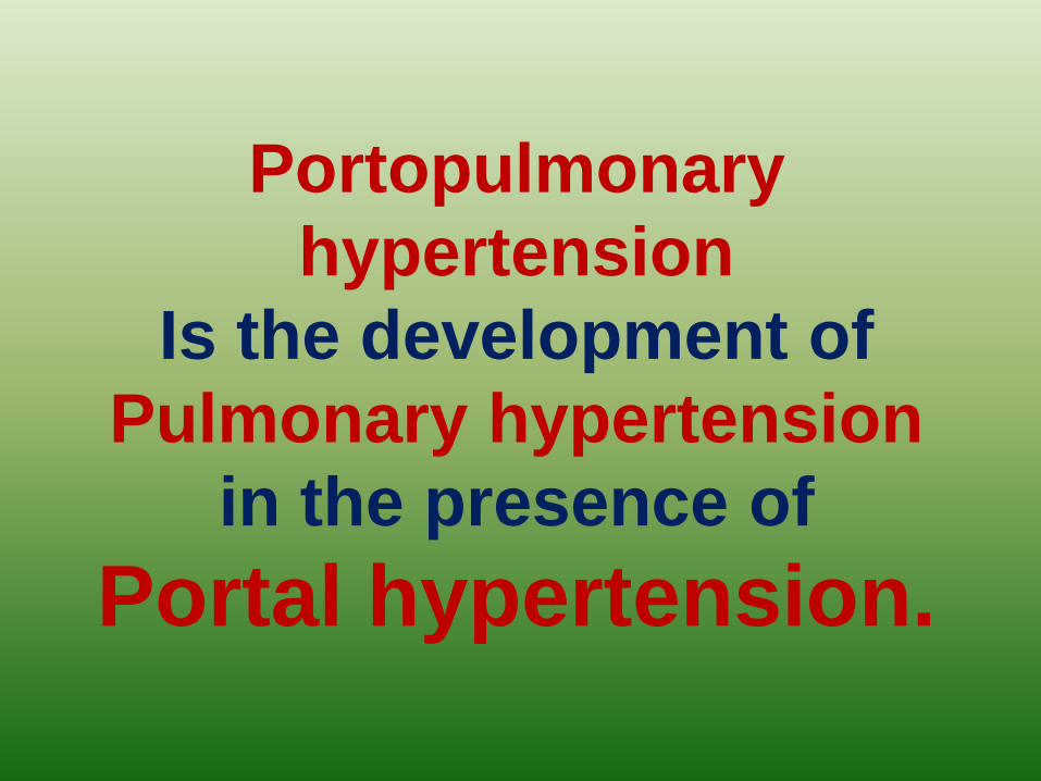
Portopulmonary
hypertension
Is the development of
Pulmonary hypertension
in the presence of
Portal hypertension.

•When should patients with
cirrhosis be referred for liver
transplantation?Patients with cirrhosis should be referred
for transplantation when they have their
first major complication
(ascites, variceal bleeding, hepatic
encephalopathy)
or evidence of significant
Hepatic dysfunction

Hepatopulmonary syndrome is a
mismatch of ventilation and perfusion that
results primarily from vasodilation of
pulmonary capillaries.
Arteriovenous communication in the
lungs and pleura may occur as well. It is
characterized by hypoxia and dyspnea
that worsen with upright position
(orthodeoxia and platypnea,
respectively).
Portopulmonary hypertension is the
development of pulmonary
hypertension in the presence of portal
hypertension.
•When should patients with cirrhosis
be referred for liver transplantation?

Global Critical Carehttps://www.facebook.com/groups/1451610115129555/#!/groups/145161011512
9555/ Wellcome in our new group ..... Dr.SAMIR EL ANSARY


![Correlation Between Hepatitis B Surface Antigen Titers and ... · of liver diseases ranging from acute to chronic hepatitis, cirrhosis, and hepatocellular carcinoma(HCC). In the [1]](https://static.fdocuments.in/doc/165x107/5f0d52b57e708231d439c744/correlation-between-hepatitis-b-surface-antigen-titers-and-of-liver-diseases.jpg)