Hepatic & pancreatic tumors
46
HEPATIC & PANCREATIC TUMORS Dr. Abdul Qadeer MBBS; FCPS; FICS Assistant Professor in Surgery King Faisal University College of Medicine
-
Upload
abdul-qadeer-memon -
Category
Health & Medicine
-
view
113 -
download
1
Transcript of Hepatic & pancreatic tumors

HEPATIC & PANCREATIC
TUMORSDr. Abdul Qadeer
MBBS; FCPS; FICSAssistant Professor in Surgery
King Faisal University College of Medicine

OBJECTIVES
1. Clinical anatomy of liver2. Physiology of liver3. Different types of benign hepatic tumors4. Management of different types of
benign hepatic tumors5. Types of malignant hepatic tumors6. Epidemiology of malignant hepatic
tumors7. Management of different malignant
hepatic tumors

OBJECTIVES (CONTD…)
9. Clinical anatomy of pancreas10. Physiology of pancreas11. Different types of pancreatic tumors12. Management of different types of
pancreatic tumors

1. CLINICAL ANATOMY OF LIVER
Largest organ in the body Situated in the RHC Weight = 1.5 kg in a 70 kg man Covered by a capsule & visceral
peritoneum except ‘bare area’ on its posterior surface
Two lobes, right & left; right lobe = 3/4th, left lobe = smaller

HTTP://WWW.BING.COM/IMAGES/SEARCH?Q=LIVER&VIEW=DETAILV2&ID=1BA9D5EC40003C1AF136FEB6D5E1E9D04936BE1F&SELECTEDINDEX=6&CCID=LW1C%2FQBH&SIMID=608002713506418269&THID=JN.8%2FMSQLIAO3WCNWUAOCJLAG&MODE=OVERLAY&FIRST=1

LIGAMENTS & PERITONEAL REFLECTIONS Left & right triangular ligaments Falciform ligament (remnant of
umbilical vein) Lesser omentum between stomach &
liver, contains the hilar structures Porta hepatis = hilum of the liver Hepatic artery, portal vein & bile duct
are present within the free edge of the lesser omentum

HTTP://WWW.BING.COM/IMAGES/SEARCH?Q=LIVER&VIEW=DETAILV2&ID=90E6A0C9CA2A5FD68DD13F024B76BEB294661C4A&SELECTEDINDEX=11&CCID=LDAZD%2FJL&SIMID=608005522410635859&THID=JN.3HX5EBHLZPOPASIZ%2BZ9NLW&MODE=OVERLAY&FIRST=1

BLOOD SUPPLY OF LIVER
Portal vein = 80% Hepatic artery = 20% Venous drainage = hepatic veins into
IVC Liver regenerates fully after partial
resection

HTTP://WWW.BING.COM/IMAGES/SEARCH?Q=LIVER+BLOOD+SUPPLY&GO=SUBMIT&QS=DS&FORM=QBIDMH

SEGMENTAL ANATOMY OF LIVER
Eight segments (described by Couinaud)
Each segment is a functional unit with a branch of hepatic artery, portal vein & bile duct

HTTP://WWW.BING.COM/IMAGES/SEARCH?Q=LIVER+SEGMENTS&VIEW=DETAILV2&&&ID=763F22D00E8C3A34F4842B60B35F19A26A60295D&SELECTEDINDEX=25&CCID=2JWPX8SP&SIMID=608002305485769828&THID=JN.BU4OPUDUE17FIBMTMKQU4Q

2. FUNCTIONS OF LIVER Maintains core body temperature pH balance & correction of lactic acidosis Synthesis of clotting factors Glucose metabolism, glycolysis &
gluconeogenesis Urea formation from protein catabolism Bilirubin formation from Hb degradation Drug & hormone metabolism & excretion Removal of gut endotoxins & foreign
antigens

3. DIFFERENT BENIGN HEPATIC TUMORS
Hemangioma Hepatic adenoma Focal nodular hyperplasia (FNH)

LIVER HEMANGIOMAS
Having abnormal plexus of veins Often multiple, may be giant Usually found incidentally
(incidentaloma) Usually diagnosed by US CT scan shows characteristic slow
contrast enhancement due to small vessel uptake
Percutaneous biopsy should be avoided Rarely need surgery

HTTP://WWW.BING.COM/IMAGES/SEARCH?Q=LIVER+HEMANGIOMA&GO=SUBMIT&QS=DS&FORM=QBIR

HEPATIC ADENOMA Mostly occur in women of child-bearing
age Associated with sex hormones (OC
pills) CT or MRI shows a well-circumscribed
vascular solid tumor Difficult to differentiate from HCC Biopsy may be necessary May bleed and have malignant
potential Resection is the treatment of choice

HTTP://WWW.BING.COM/IMAGES/SEARCH?Q=LIVER+ADENOMA&GO=SUBMIT&QS=DS&FORM=QBIRMH

FOCAL NODULAR HYPERPLASIA (FNH) It is a focal overgrowth of functioning liver
tissue supported by fibrous stroma Usually middle-aged women US helps to diagnose but may not be able to
discriminate Contrast CT/MRI may show central scarring &
well-vascularized lesion FNH contains both hepatocytes & Kupffer cells A sulphur colloid liver scan may be useful,
since Kupffer cells take up the colloid Does not have malignant potential If diagnosis is confirm, no treatment is required

HTTP://WWW.BING.COM/IMAGES/SEARCH?Q=LIVER+FNH&GO=SUBMIT&QS=DS&FORM=QBIRMH

4. MANAGEMENT OF BENIGN HEPATIC TUMORS
Liver hemangiomas = Rarely require surgery
Hepatic adenoma = Resection is the treatment of choice
FNH = No treatment is required

5. MALIGNANT HEPATIC TUMORS
1. Primary tumors: Hepatocellular carcinoma Cholangiocarcinoma2. Secondary tumors (Metastasis)

HEPATOCELLULAR CARCINOMA (HCC) Primary liver cancer Associated with chronic liver disease
(CLD), due to HBV & HCV Many patients with CLD are now
screened for HCC by serial USS of liver or serum α-FP
Surgical treatment options include:1. Resection of the tumor2. Liver transplant

HTTP://WWW.BING.COM/IMAGES/SEARCH?Q=LIVER+FNH&GO=SUBMIT&QS=DS&FORM=QBIRMH

Choice of surgical option from the above two depends upon:
a. Stage of the underlying liver diseaseb. Site & size of the tumorc. Availability of organ transplantationd. Management of the
immunosuppressed patient

STAGING & CLINICAL ASSESSMENT OF HCC General assessment of patient for
fitness of surgery Severity of underlying liver disease (by
CTP classification or MELD score) Size, number and site of the tumor

Points 1 point each
2 points each
3 points each
Bilirubin (µmol/L)
<34 34-50 >50
Albumin (g/L) >35 22-35 <25
Ascites None Easily controlled
Poorly controlled
Encephalopathy
None Grade I-II Grade III-IV
INR <1.7 1.7-2.2 >2.2
CHILD-TURCOTTE-PUGH (CTP) CLASSIFICATION OF
HEPATOCELLULAR FUNCTION IN CIRRHOSIS
CTP A = 5-6 Points; CTP B = 7-9 Points; CTP C = 10-15 PointsINR = International Normalized Ratio

MODEL FOR END-STAGE LIVER DISEASE (MELD) SCORE
It is a scoring system for assessing the severity of chronic liver disease
It was initially developed to predict death within three months of surgery in patients who had undergone a transjugular intrahepatic portosystemic shunt (TIPS) procedure and was subsequently found to be useful in determining prognosis and prioritizing for receipt of a liver transplant
This score is now used by the United Network for Organ Sharing (UNOS) and Eurotransplant for prioritizing allocation of liver transplants instead of the older CTP score

MODEL FOR END-STAGE LIVER DISEASE (MELD) SCORE
MELD = 3.78×ln[serum bilirubin (mg/dL)] + 11.2×ln[INR] + 9.57×ln[serum creatinine (mg/dL)] + 6.43×aetiology(0: cholestatic or alcoholic, 1- otherwise)
In interpreting the MELD Score in hospitalized patients, the 3 month mortality is:
1. 40 or more — 71.3% mortality2. 30–39 — 52.6% mortality3. 20–29 — 19.6% mortality4. 10–19 — 6.0% mortality5. <9 — 1.9% mortality

USS CXR/CT Bone scan Contrast CT/MRI WHVP (Wedged hepatic venous
pressure); suggests portal hypertension, poor outcome after liver resection in cirrhotic patients
Indocyanine green (ICG) clearance test: for hepatic flow & function
Patients with CTP class A and high ICG clearance are suitable for major liver resection

CHOLANGIOCARCINOMA
Bile duct cancers typically present with painless obstructive jaundice
PSC (Primary Sclerosing Cholangitis) is the cause
Slow growing tumors Klastskin tumors: arise at the
confluence of right & left hepatic ducts, eventually invading the liver parenchyma

INVESTIGATIONS FOR CHOLANGIOCARCINOMA
LFTs: Obstructive jaundice Tumor marker: CA 19-9 may be elevated USS MDR-CT: Multidetector row computed
tomography MRI/MRCP ERCP PTC PET

6. EPIDEMIOLOGY OF MALIGNANT HEPATIC TUMORS
Sixth most common cancer worldwide 5.7% incidence over all cancers Developing countries = 82%, third
most common cancer in men after lung & stomach
HBV & HCV infections account 75% cases of primary liver cancers
http://www.ncbi.nlm.nih.gov/pmc/articles/PMC3036307/

7. MANAGEMENT OF MALIGNANT HEPATIC TUMORS
For HCC: Surgical resection with 1 to 2-cm safety
margin Liver transplantation Non-surgical therapy for advanced disease:a. Transarterial embolization (TAE)b. Transarterial chemoembolization (TACE)c. Percutaneous ethanol ablation (PEA)d. Radiofrequency ablation (RFA) For CC: Whipple procedure

8. CLINICAL ANATOMY OF PANCREAS Exocrine & endocrine pancreas Head, body, tail, uncinate process Main pancreatic duct (Wirsung) Accessory pancreatic duct (Santorini) Islets of Langerhansa) B-cells (65-80%) : Insulinb) A-cells (15-20%) : Glucagonc) D-cells (3-10) : Somatostatind) PP-cells (1%) : Pancreatic
polypeptide cells

9. PHYSIOLOGY OF PANCREAS
In response to meal, secretin from duodenal mucosa: stimulates pancreas to secrete digestive enzymes in an alkaline (pH 8.4) bicarbonate-rich fluid
Cholecystokinin-panceozymin (CCK-PZ) Vagal stimulation: secretomotor

10. DIFFERENT TYPES OF PANCREATIC TUMORS
Chronic pancreatitis may be the main cause
Carcinoma of pancreas: 85% are ductal adenocarcinomas
Serous and mucinous cystadenomas including intraductal papillary mucinous neoplasms (IPMNs)
Lymphangiomas, dermoid cysts

NEURO-ENDOCRINE TUMORS OF PANCREAS
These include: Insulinoma, Gastrinoma, VIPoma, Glucagonoma, Somatostatinoma, Carcinoid, ACTHoma (causing Cushing’s syndrome), GRFoma (causing acromegaly)

11. MANAGEMENT OF DIFFERENT TYPES OF PANCREATIC TUMORS
1/3rd of pancreatic tumors arise in its head
Ampullary carcinoma Causing characteristic painless
obstructive jaundice Pruritus, dark urine, pale stools,
steatorrhea If no jaundice, symptoms are vague
e.g. discomfort, anorexia, weight loss

INVESTIGATIONS CBC, UCE, FBS, RBS, LFTs USS Contrast CT/MRI ERCP ±Transduodenal or Transgastric FNA or
Trucut biopsy performed under EUS guidance
Percutaneous transperitoneal biopsy should be avoided

PREOPERATIVE PREPARATION
In a patient with obstructive jaundice:a. Well hydration to prevent hepatorenal
shut downb. Vitamin K injectionsc. Prophylactic antibiotics

TREATMENT
Curative surgery if possible ± chemotherapy of 5-FU, radiotherapy is not effective
1. Pylorus-preserving panceatoduodenectomy (PPPD)
2. Whipple procedure3. Total pancreatectomy if the disease is
multifocal4. Distal pancreatectomy with splenectomy
+ local lymphadenectomy: tumors of body/tail

PPPD
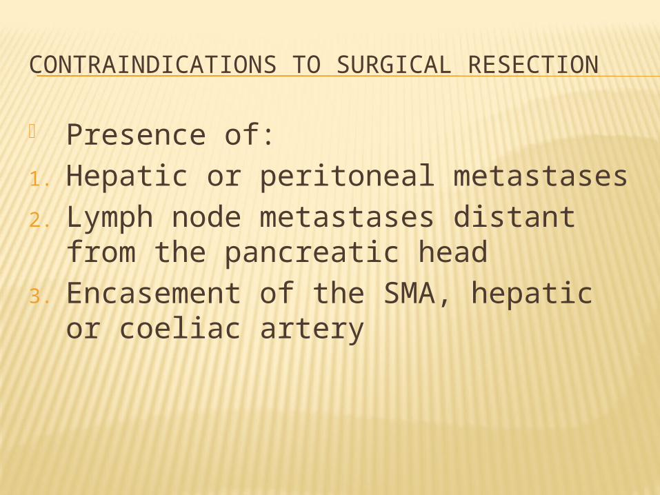
CONTRAINDICATIONS TO SURGICAL RESECTION
Presence of:1. Hepatic or peritoneal metastases2. Lymph node metastases distant from
the pancreatic head3. Encasement of the SMA, hepatic or
coeliac artery

Palliation of pancreatic cancerA.Relieve jaundice & treat biliary sepsis Surgical biliary bypass Stent placed at ERCP or PTC
B.Improve gastric emptying Surgical gastroenterostomy Duodenal stent
C.Pain relief Stepwise escalation of analgesia Coeliac plexus block Transthoracic splanchnicectomy
D.Symptom relief & quality of life Encourage normal activities Enzyme replacement for steatorrhea Treat diabetes
E.Consider chemotherapy

References: Bailey & Love’s Short Practice of Surgery; 26th Edition; Chapters 52, 65, 68