Hemostatic effect of human fibrin glue on bleeding surface of the brain
4
Original article NTSI-Alfred Trauma Research Development Award Paper Hemostatic effect of human fibrin glue on bleeding surface of the brain: An experiment on albino Wistar rats Vivek Agarwal a, *, Vernon Velho a , Kundan S. Gosavi b , Akash A. Khobragade c , Rajendra Aher a , Rachana S. Binayke d a Department of Neurosurgery, Grant Medical College and Sir J. J. Group of Hospitals, India b Department of Anesthesia, Grant Medical College and Sir J. J. Group of Hospitals, India c Department of Pharmacology, Grant Medical College and Sir J. J. Group of Hospitals, India d Department of Pathology, Grant Medical College and Sir J. J. Group of Hospitals, India article info Article history: Received 11 December 2011 Accepted 14 March 2012 Available online 21 April 2012 Keywords: Hemostasis Fibrin glue Animal experiment Brain abstract Background: Achieving and maintaining hemostasis in neurosurgical procedures is critical to the outcome and challenging especially in cases of coagulopathy with diffuse oozing. After trauma to the brain, a cascade of events initiated by tissue factor (TF) or thromboplastin results in a defective coagulation process that even may lead to disseminated intravascular coagulation (DIC). Fibrin glue is sealant made up of fibrinogen and thrombin used for dural defect repair at the base, convexity, anastomosis of the nerve and nerve graft, reinforcing microvascular anastomosis. This study was carried out to determine the hemostatic effect of human fibrin glue on bleeding surface of brain and compare the effect with conventional methods of hemostasis. Method: Thirty (30) white rats (Rattus novergicus) were divided equally into study and control group. After craniotomy and dural opening a stab incision was made on right frontal region of brain. In case group the bleeding was controlled with fibrin glue (average 0.5 ml) and in control group conventional method of hemostasis (cautery, cottonoid patty, and saline wash, surgicel) was used. Both the groups were studied for bleeding time, seizure, neuro- logical deficit, wound complications and mortality. Results: Outcome was assessed as 1) Bleeding in both the groups 2) Complications in both the groups. It was observed that in study group the time taken in hemostasis was signif- icantly less in comparison to the control, No significant difference in the post procedure clinical outcome and inflammatory reaction/gliosis reaction was found in both the groups. Conclusion: Human fibrin glue is simple, easy and safe alternative to conventional methods of hemostasis. Copyright ª 2012, Neurotrauma Society of India. All rights reserved. * Corresponding author. Tel.: þ91 9867647125 (mobile). E-mail addresses: [email protected] (V. Agarwal), [email protected] (V. Velho), [email protected] (K.S. Gosavi), [email protected] (A.A. Khobragade), [email protected] (R. Aher), [email protected] (R.S. Binayke). Available online at www.sciencedirect.com journal homepage: www.elsevier.com/locate/ijnt the indian journal of neurotrauma 9 (2012) 15 e18 0973-0508/$ e see front matter Copyright ª 2012, Neurotrauma Society of India. All rights reserved. doi:10.1016/j.ijnt.2012.04.014
Transcript of Hemostatic effect of human fibrin glue on bleeding surface of the brain

Original article
NTSI-Alfred Trauma Research Development Award Paper
Hemostatic effect of human fibrin glue on bleeding surface ofthe brain: An experiment on albino Wistar rats
Vivek Agarwal a,*, Vernon Velho a, Kundan S. Gosavi b, Akash A. Khobragade c,Rajendra Aher a, Rachana S. Binayke d
aDepartment of Neurosurgery, Grant Medical College and Sir J. J. Group of Hospitals, IndiabDepartment of Anesthesia, Grant Medical College and Sir J. J. Group of Hospitals, IndiacDepartment of Pharmacology, Grant Medical College and Sir J. J. Group of Hospitals, IndiadDepartment of Pathology, Grant Medical College and Sir J. J. Group of Hospitals, India
a r t i c l e i n f o
Article history:
Received 11 December 2011
Accepted 14 March 2012
Available online 21 April 2012
Keywords:
Hemostasis
Fibrin glue
Animal experiment
Brain
a b s t r a c t
Background: Achieving and maintaining hemostasis in neurosurgical procedures is critical to
the outcome and challenging especially in cases of coagulopathy with diffuse oozing. After
trauma to the brain, a cascade of events initiated by tissue factor (TF) or thromboplastin results
inadefectivecoagulationprocess thatevenmayleadtodisseminated intravascularcoagulation
(DIC). Fibrin glue is sealant made up of fibrinogen and thrombin used for dural defect repair at
the base, convexity, anastomosis of the nerve and nerve graft, reinforcing microvascular
anastomosis.Thisstudywascarriedouttodeterminethehemostaticeffectofhumanfibringlue
on bleeding surface of brain and compare the effectwith conventionalmethods of hemostasis.
Method: Thirty (30) white rats (Rattus novergicus) were divided equally into study and control
group. After craniotomy and dural opening a stab incision was made on right frontal region
of brain. In case group the bleeding was controlled with fibrin glue (average 0.5 ml) and in
control group conventional method of hemostasis (cautery, cottonoid patty, and saline
wash, surgicel) was used. Both the groups were studied for bleeding time, seizure, neuro-
logical deficit, wound complications and mortality.
Results: Outcome was assessed as 1) Bleeding in both the groups 2) Complications in both
the groups. It was observed that in study group the time taken in hemostasis was signif-
icantly less in comparison to the control, No significant difference in the post procedure
clinical outcome and inflammatory reaction/gliosis reaction was found in both the groups.
Conclusion: Human fibrin glue is simple, easy and safe alternative to conventional methods
of hemostasis.
Copyright ª 2012, Neurotrauma Society of India. All rights reserved.
* Corresponding author. Tel.: þ91 9867647125 (mobile).E-mail addresses: [email protected] (V. Agarwal), [email protected] (V. Velho), [email protected] (K.S. Gosavi),
[email protected] (A.A. Khobragade), [email protected] (R. Aher), [email protected] (R.S. Binayke).
Available online at www.sciencedirect.com
journal homepage: www.elsevier .com/locate/ i jnt
t h e i n d i a n j o u rn a l o f n e u r o t r a uma 9 ( 2 0 1 2 ) 1 5e1 8
0973-0508/$ e see front matter Copyright ª 2012, Neurotrauma Society of India. All rights reserved.doi:10.1016/j.ijnt.2012.04.014

1. Introduction
Achieving and maintaining hemostasis in neurosurgical proce-
dures is critical to the outcome and challenging, especially in
cases of coagulopathy with diffuse oozing. After trauma to the
brain, a cascade of events initiated by tissue factor (TF) or
thromboplastin results in a defective coagulation process that
evenmay lead to disseminated intravascular coagulation (DIC).1
Fibrin glue has been used as a sealant for repair in situations
such as dural defects at the base and convexity of the brain,
nerve grafts, nerve anastomoses, reinforcing of microvascular
anastomoses, carotico-cavernous fistulae and fixation of bone
fragments.2e6 In addition, it is known to have a hemostatic
potential on thebleedingsurfaceof thebrainwhichmightprove
to be of special interest in cases with defective coagulation and
excessive oozing where conventional methods of hemostasis
are less effective.1 In this study, we have evaluated the hemo-
static action of fibrin glue in comparisonwith the conventional
method of hemostasis i.e. bipolar cautery followed by pressure
with a cottonoid patty and saline wash followed by surgicel.
2. Material and methods
After obtaining the prior approval of the institutional animal
ethics committee, albino Wistar rats of either sex, weighing
250 g on an average, were procured from the central animal
house of Grant Medical College and Sir JJ Group of Hospitals,
Byculla, Mumbai-08. A pilot study conducted on 4 rats found
the mean bleeding time using the conventional methods was
150 (þ57) seconds. To demonstrate a reduction in bleeding
time by at least 60 s using the fibrin glue a sample size of 15
was determined. The animals were randomly divided into two
groups (study and control) of 15 animals each, and were
maintained in clean polypropylene cages with clean husk
bedding which was changed every three days. Food and water
were given ad libitum.
On the day of the procedure, the rats were anesthetized
using intraperitoneal Ketamine (50e100 mg/kg) (Fig. 1).7 Blood
loss was compensated with intraperitoneal Ringers Lactate.
After fixing theheadof rats over a specially designeddissection
table (Figs. 2 and 3), a linear incision was taken over the right
frontal region of the scalp under aseptic conditions. With the
underlying bone exposed, a right frontal craniectomy was
performed using a drill and a fine nibbler and the dura was
exposed just anterior to the coronal suture. After opening the
dura, a stab incision was made over the right frontal lobe and
bleedingwas induced. In the rats of the study group an average
0.5 ml of fibrin glue was applied over the bleeding area. In the
rats of the control group, hemostasis was achieved by
conventionalmethod employingbipolar cautery, pressurewith
cottonoid patty and saline wash and surgicel. Bleeding from
the brain was assessed in form of bleeding time in both the
groups. The incisionwas then closedwith Vicryl 4, 0. Ratswere
givenantibiotics (Cefuroxime IM) andanalgesia, local xylocaine
at surgical site postoperatively and were monitored for up to 3
weeks post procedure for any neurological or behavioral
derangement, seizures or any signs of impending death.8
3. Results
The comparative bleeding times of the rats in the control and
study groups are tabulated in Table 1, and the means
Fig. 1 e Intraperitoneal ketamine injection for general
anesthesia.
Fig. 2 e Rat dissection board.
Fig. 3 e Rat fixed over the dissection board with help of jaw
holder and body clamp.
t h e i n d i a n j o u r n a l o f n e u r o t r a uma 9 ( 2 0 1 2 ) 1 5e1 816
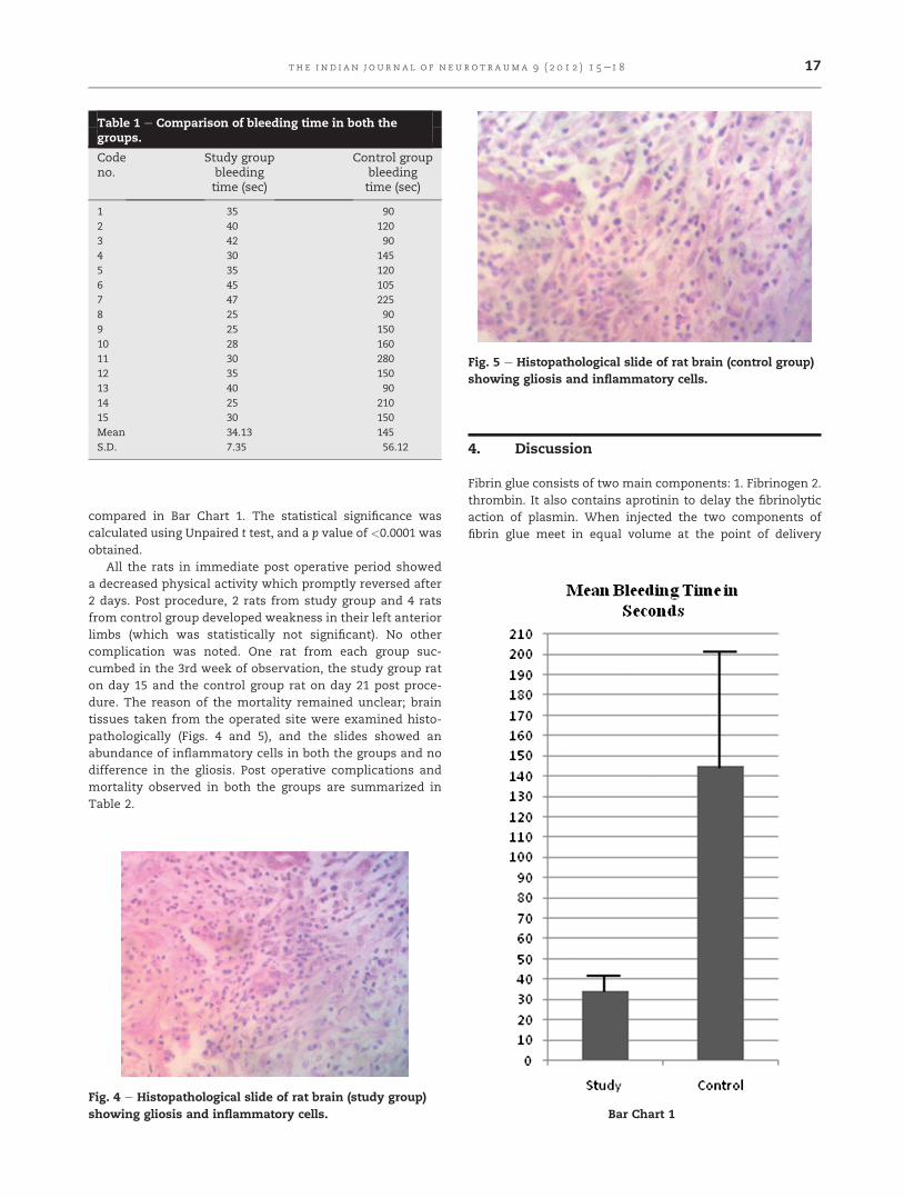
compared in Bar Chart 1. The statistical significance was
calculated using Unpaired t test, and a p value of <0.0001 was
obtained.
All the rats in immediate post operative period showed
a decreased physical activity which promptly reversed after
2 days. Post procedure, 2 rats from study group and 4 rats
from control group developed weakness in their left anterior
limbs (which was statistically not significant). No other
complication was noted. One rat from each group suc-
cumbed in the 3rd week of observation, the study group rat
on day 15 and the control group rat on day 21 post proce-
dure. The reason of the mortality remained unclear; brain
tissues taken from the operated site were examined histo-
pathologically (Figs. 4 and 5), and the slides showed an
abundance of inflammatory cells in both the groups and no
difference in the gliosis. Post operative complications and
mortality observed in both the groups are summarized in
Table 2.
4. Discussion
Fibrin glue consists of two main components: 1. Fibrinogen 2.
thrombin. It also contains aprotinin to delay the fibrinolytic
action of plasmin. When injected the two components of
fibrin glue meet in equal volume at the point of delivery
Table 1 e Comparison of bleeding time in both thegroups.
Codeno.
Study groupbleedingtime (sec)
Control groupbleedingtime (sec)
1 35 90
2 40 120
3 42 90
4 30 145
5 35 120
6 45 105
7 47 225
8 25 90
9 25 150
10 28 160
11 30 280
12 35 150
13 40 90
14 25 210
15 30 150
Mean 34.13 145
S.D. 7.35 56.12
Fig. 4 e Histopathological slide of rat brain (study group)
showing gliosis and inflammatory cells.
Fig. 5 e Histopathological slide of rat brain (control group)
showing gliosis and inflammatory cells.
Bar Chart 1
t h e i n d i a n j o u rn a l o f n e u r o t r a uma 9 ( 2 0 1 2 ) 1 5e1 8 17

where thrombin converts the fibrinogen to fibrin. Depending
on the concentration of the thrombin fibrin glue acts in 2
ways
(1) Fast acting �10 s
(2) Slow acting �60 s
Since it forms coagulum bypassing both the extrinsic and
the intrinsic mechanisms of coagulation it is very helpful in
achieving hemostasis in cases of coagulation disorder.
In cases of diffuse oozing controlling hemostasis with
fibrin glue by spreading over the oozing surface takes lesser
time as in our study, less damage to brain than by cautery, and
less amount of blood loss.
Fibrin glue contains thrombin, a serine protease enzyme,
which has antigenic properties. It might be expected to induce
a variety of pathologic responses, including edema, seizures,
and apoptotic changes.9e12 However, we found no significant
difference in neurological deficit in both the groups and the
histological study was also comparable without significant
difference in inflammation and gliosis. There remains the risk
of transmitting serological disease from pooled and single e
blood donors.
5. Summary
The use of fibrin glue as hemostatic agent over the bleeding
surface of brain study in a controlled rat model demonstrated
that the time taken for hemostasis in the study group was
significantly less in comparison to control.
No significant difference in post operative clinical output
was found in both groups.
No difference in the inflammatory reaction/gliosis reaction
found in both the groups.
6. Conclusion
In our animal model fibrin glue offers a simple, easy and safe
alternative to conventional method of hemostasis on bleeding
surface of the brain.
r e f e r e n c e s
1. Gerald AG. Update on hemostasis: neurosurgery. Surgery.2007;142:55e60.
2. Herter T. Problems of fibrin adhesion of the nerves. NeurosurgRev. 1988;11:249e257.
3. Shaffrey CI, Spotnitz WD, Shaffrey ME, Jane JA. Neurosurgicalapplications of fibrin glue: augmentation of dural closure in134 patients. Neurosurgery. 1990;26:207e210.
4. Krayenbuhl N, Hafez A, Hernesniemi JA, Krisht AF. Tamingthe cavernous sinus: technique of hemostasis using fibringlue. Neurosurgery. 2007;61:52.
5. Fukumoto T, Matsushima Y, Tomita S, Inaba Y. The use offibrin glue in neurosurgical operations. No Shinkei Geka.1985;13:367e373.
6. Schafer M, Klein HJ, Richter HP. Fibrin glue inneurosurgery. Area of use and experiences. Fortschr Med.1985;23:545e547.
7. Book 2. Pharmacology of injectible anesthesia Robert E. Meyerand Richard E. Fish anesthesia and analgesia in laboratoryanimals, Richard E. Fish, Marilyn J. Brown, American collegeof laboratory animal medicine series.
8. Edwards KC, Atkinson RM, Pratt DA, Patterson GG,Wheeldon JM, Foord RD. The toxicology of cefuroxime. Proc RSoc Med. 1977;70:11e18.
9. Kassam A, Nemoto E, Balzer J, et al. Effects of tisseel fibringlue on the central nervous system of nonhuman primates.Ear Nose Throat J. 2004;83:246e248.
10. de Vries J, Menovsky T, van Gulik S, Wesseling P. Histologicaleffects of fibrin glue on nervous tissue: a safety study in rats.Surg Neurol. 2002;57:415e422.
11. Brennan M. Fibrin glue. Blood Rev. 1991;5:240e244.12. Thompson DF, Letassy NA, Thompson GD. Fibrin glue:
a review of its preparation, efficacy, and adverse effectsas a topical hemostat. Drug Intell Clin Pharm. 1988;22:946e952.
Table 2 e Comparison of complications in two groups.
Complication Study group Control group
Neurological deficit 2 4
Seizure Nil Nil
Wound complication Nil Nil
Mortality 1 1
t h e i n d i a n j o u r n a l o f n e u r o t r a uma 9 ( 2 0 1 2 ) 1 5e1 818