Hemodynamics in the Cath lab and ICU - · PDF fileHemodynamics in the Cath lab and ICU Arnold...
56
Hemodynamics in the Cath lab and ICU Arnold Seto, MD, MPA UC-Irvine and Long Beach VA
Transcript of Hemodynamics in the Cath lab and ICU - · PDF fileHemodynamics in the Cath lab and ICU Arnold...
Hemodynamics in the Cath lab and ICU
Arnold Seto, MD, MPA UC-Irvine and Long Beach VA
Presenter
Presentation Notes
2 million PACs placed annually in 1987
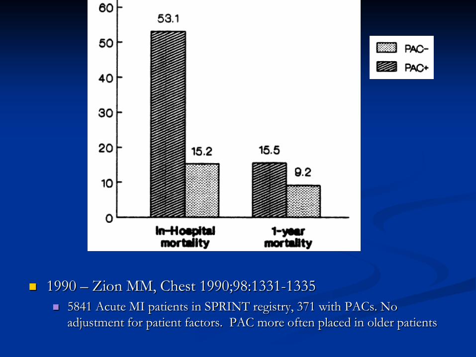
1990 – Zion MM, Chest 1990;98:1331-1335 5841 Acute MI patients in SPRINT registry, 371 with PACs. No
adjustment for patient factors. PAC more often placed in older patients
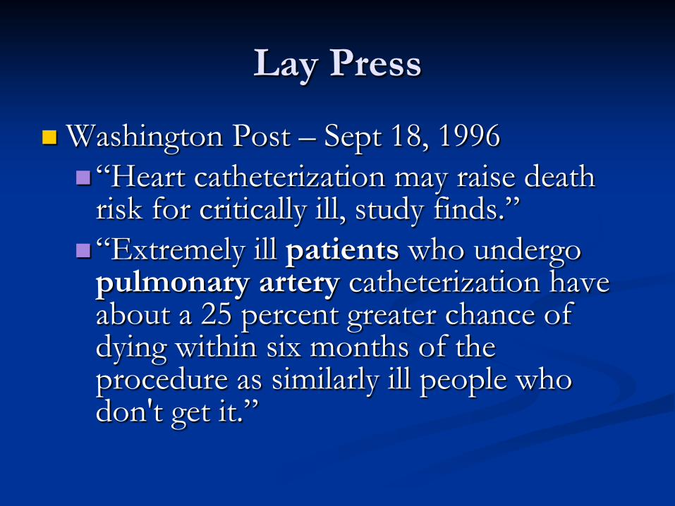
Lay Press
Washington Post – Sept 18, 1996 “Heart catheterization may raise death
risk for critically ill, study finds.” “Extremely ill patients who undergo
pulmonary artery catheterization have about a 25 percent greater chance of dying within six months of the procedure as similarly ill people who don't get it.”
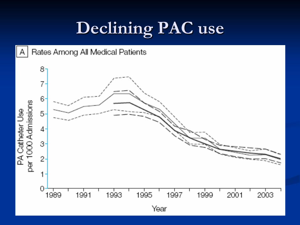
Declining PAC use
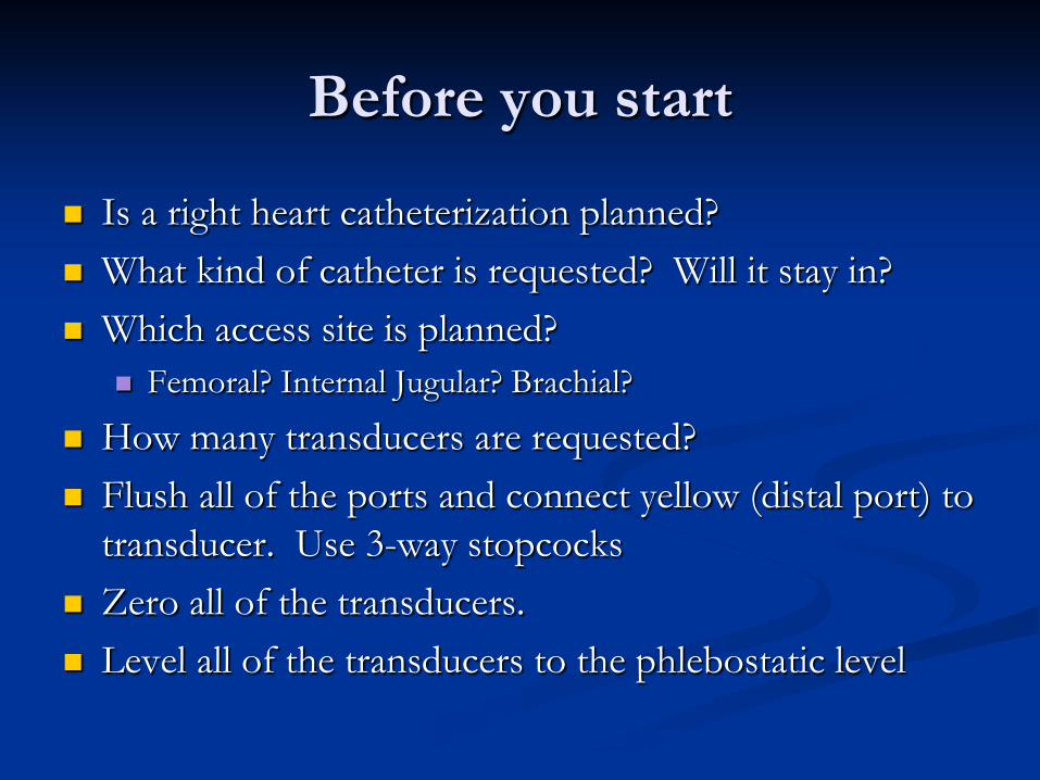
Before you start
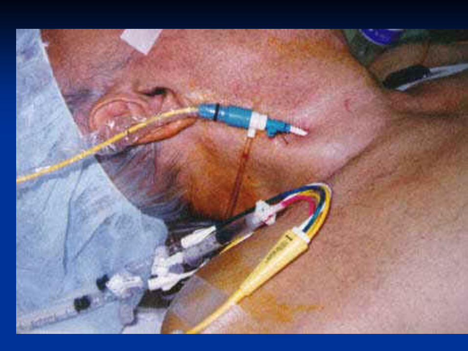
Is a right heart catheterization planned? What kind of catheter is requested? Will it stay in? Which access site is planned?
Femoral? Internal Jugular? Brachial?
How many transducers are requested? Flush all of the ports and connect yellow (distal port) to
transducer. Use 3-way stopcocks Zero all of the transducers. Level all of the transducers to the phlebostatic level
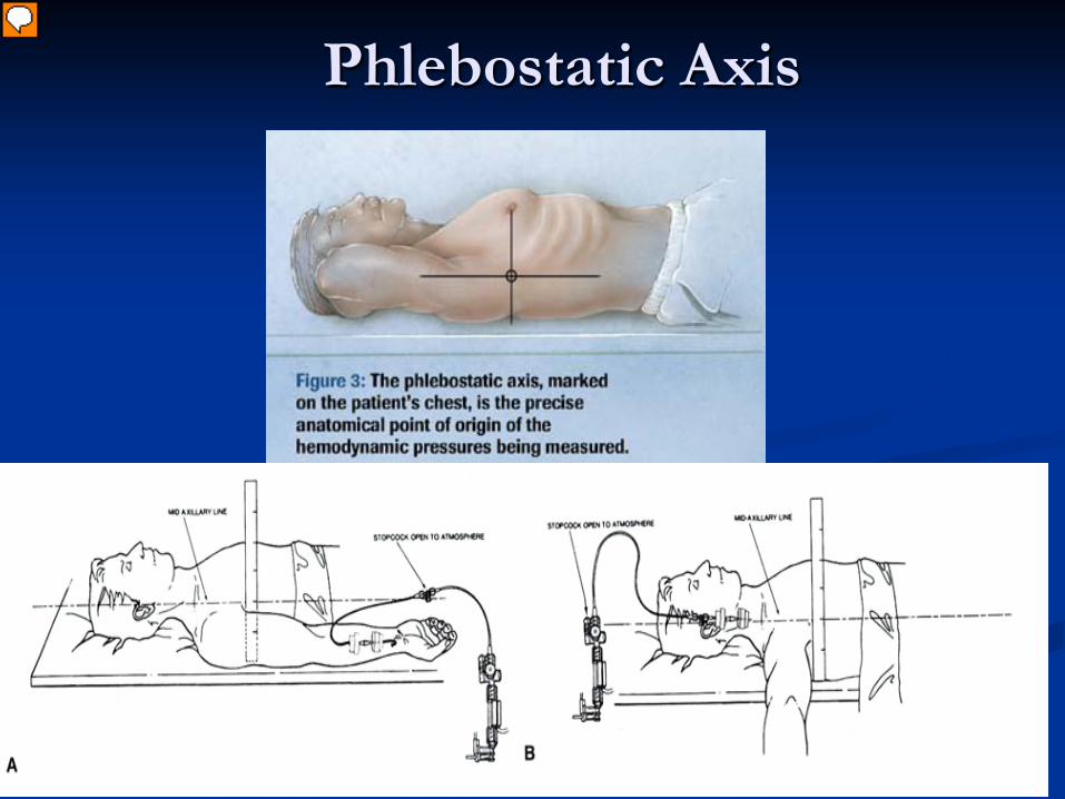
Phlebostatic Axis
Presenter
Presentation Notes
4th IC space, mid axillary line
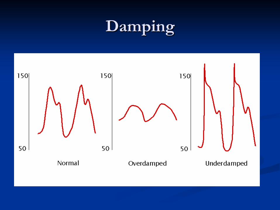
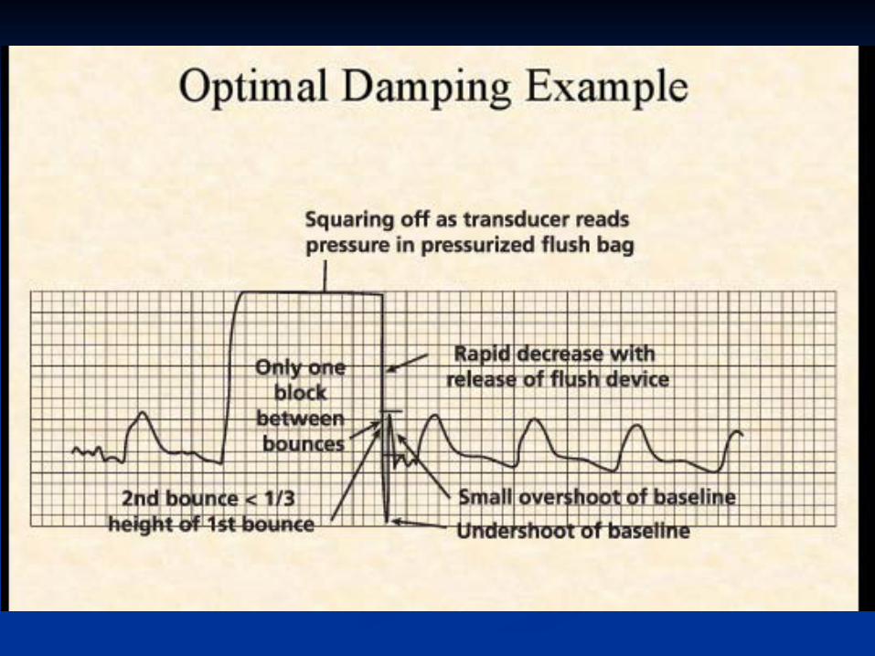
Damping
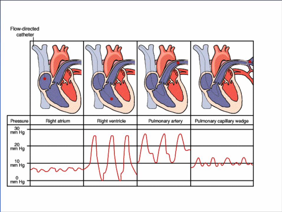
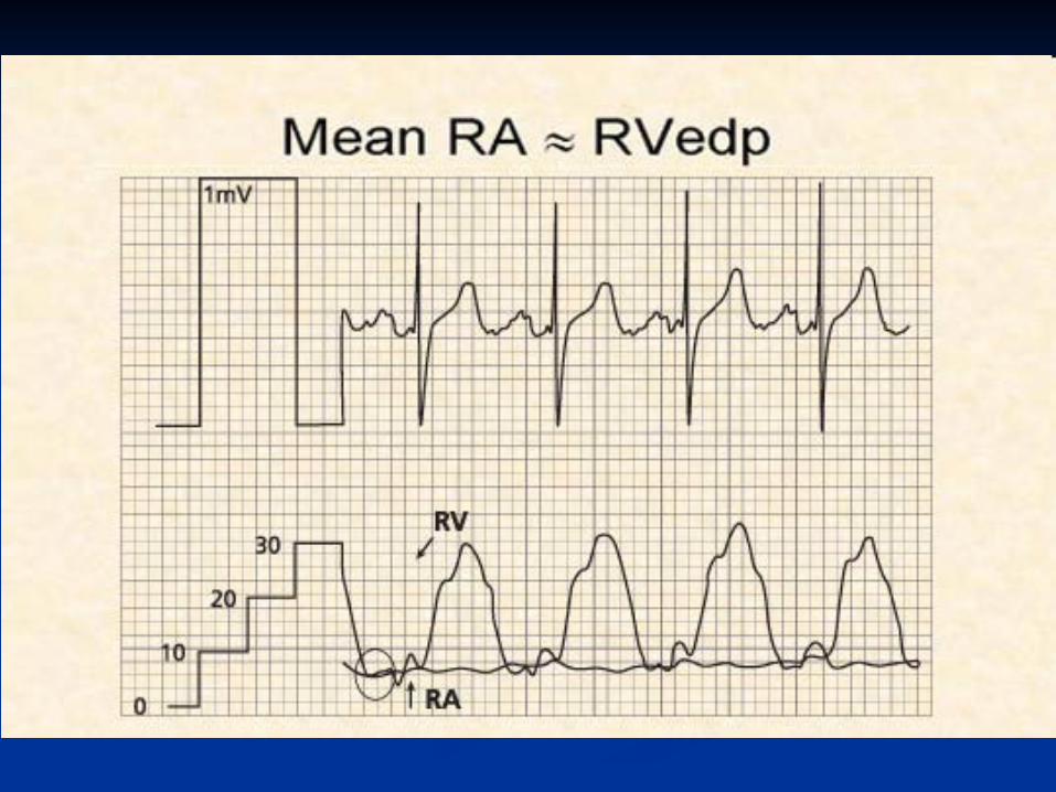
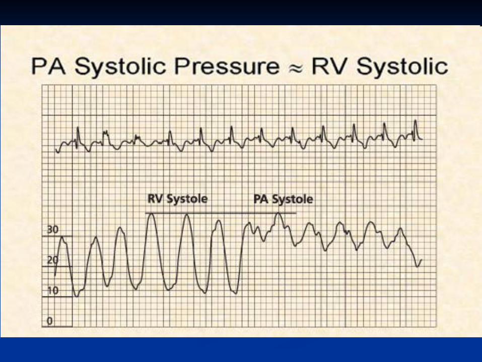
PAC Insertion Waveforms
PAWP=PAOP=PCWP=wedge
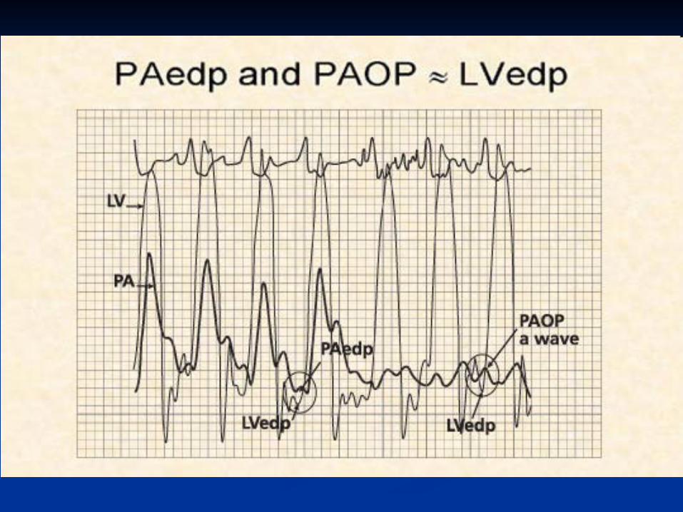

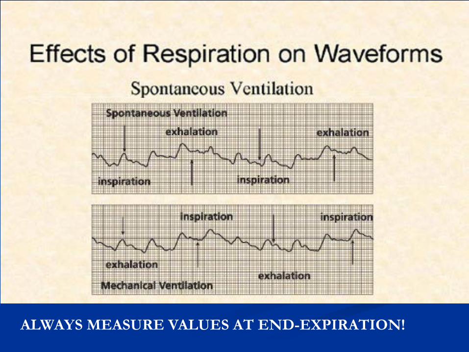
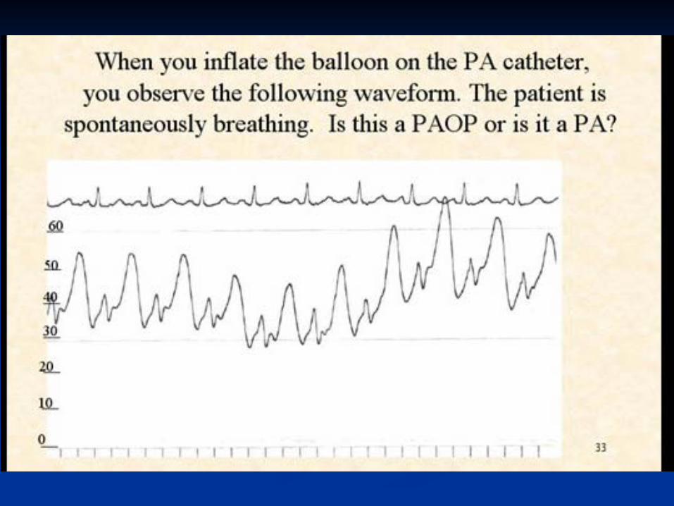
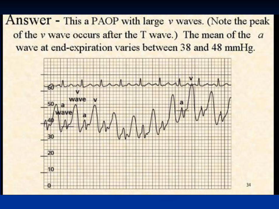
ALWAYS MEASURE VALUES AT END-EXPIRATION!
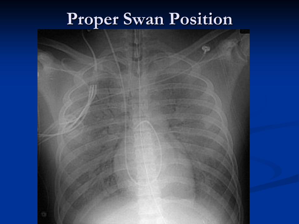
Proper Swan Position
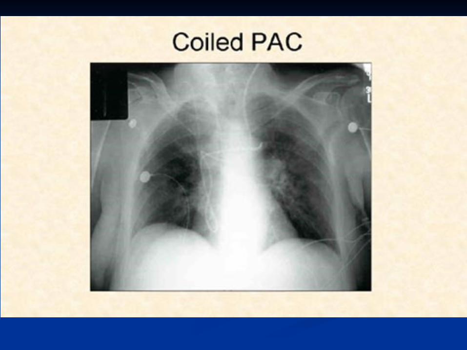
Coiled PAC
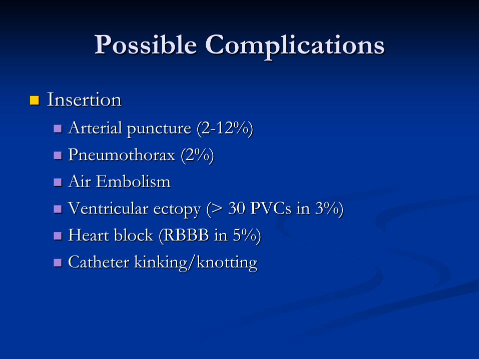
Possible Complications
Insertion Arterial puncture (2-12%) Pneumothorax (2%) Air Embolism Ventricular ectopy (> 30 PVCs in 3%) Heart block (RBBB in 5%) Catheter kinking/knotting
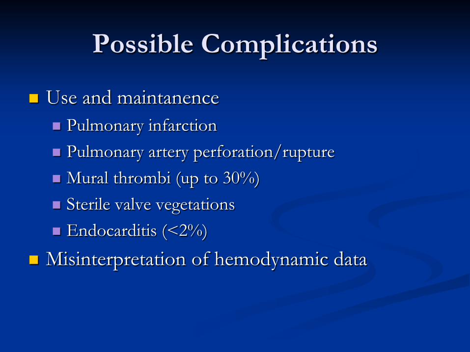
Possible Complications
Use and maintanence Pulmonary infarction Pulmonary artery perforation/rupture Mural thrombi (up to 30%) Sterile valve vegetations Endocarditis (<2%)
Misinterpretation of hemodynamic data
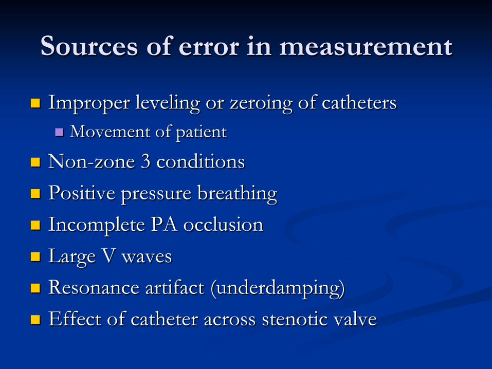
Sources of error in measurement
Improper leveling or zeroing of catheters Movement of patient
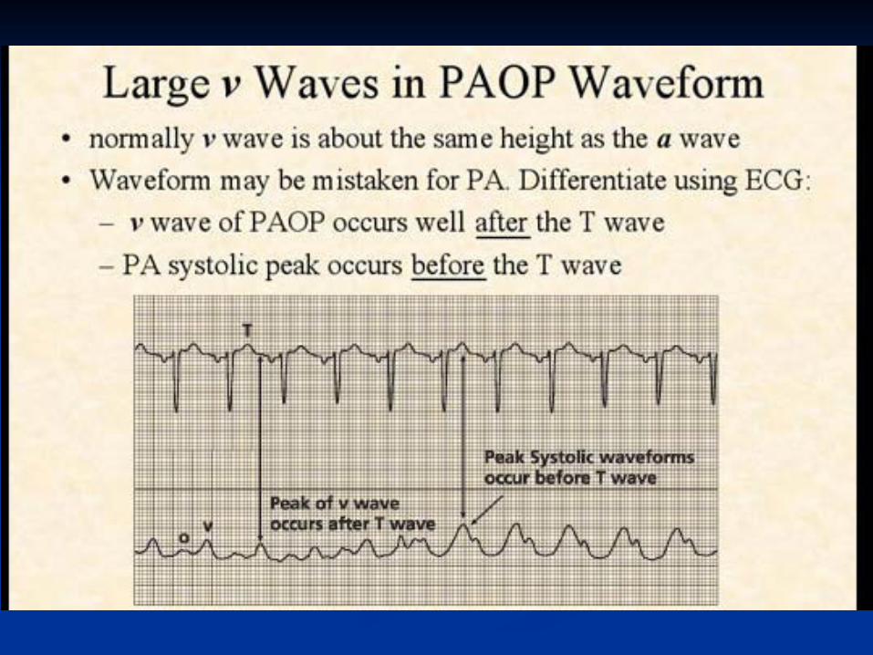
Non-zone 3 conditions Positive pressure breathing Incomplete PA occlusion Large V waves Resonance artifact (underdamping) Effect of catheter across stenotic valve
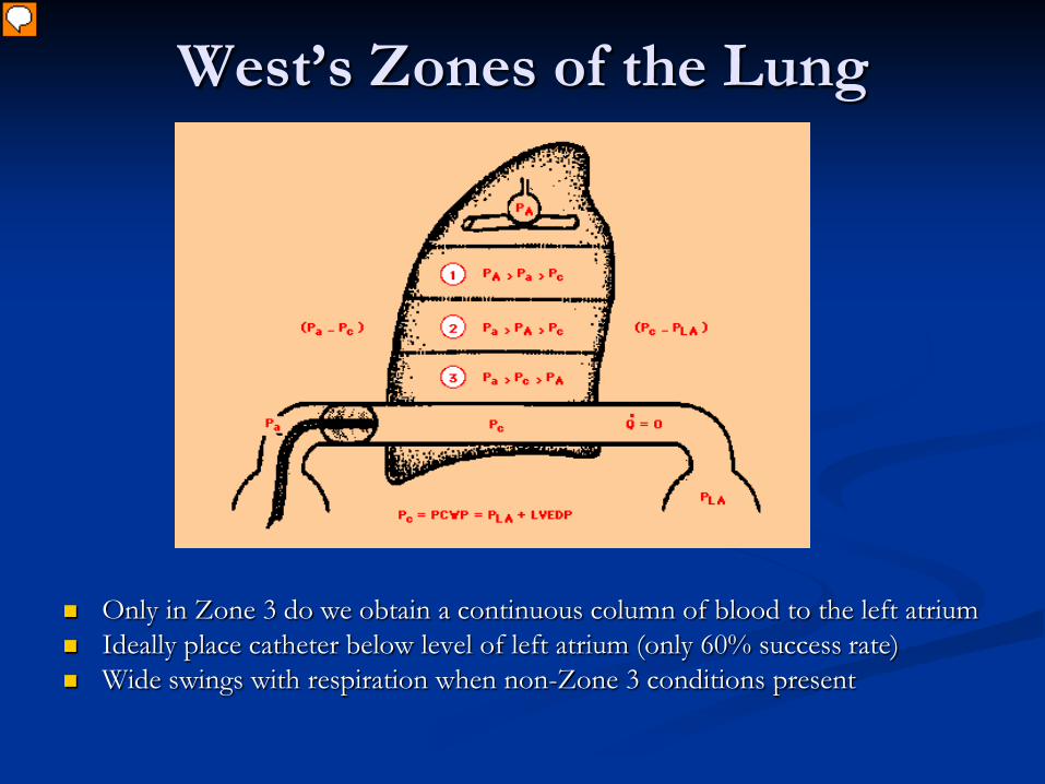
West’s Zones of the Lung
Only in Zone 3 do we obtain a continuous column of blood to the left atrium Ideally place catheter below level of left atrium (only 60% success rate) Wide swings with respiration when non-Zone 3 conditions present
Presenter
Presentation Notes
Only 60% of the time do we get into zone 3, where Pa>Pc>PA and continuous column of blood present.
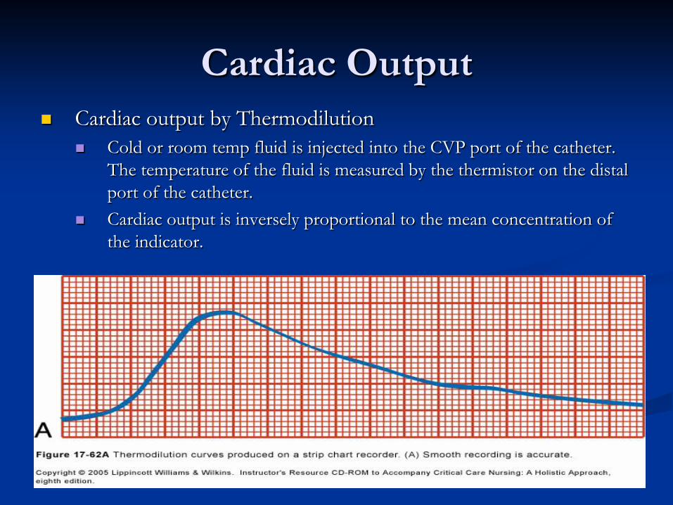
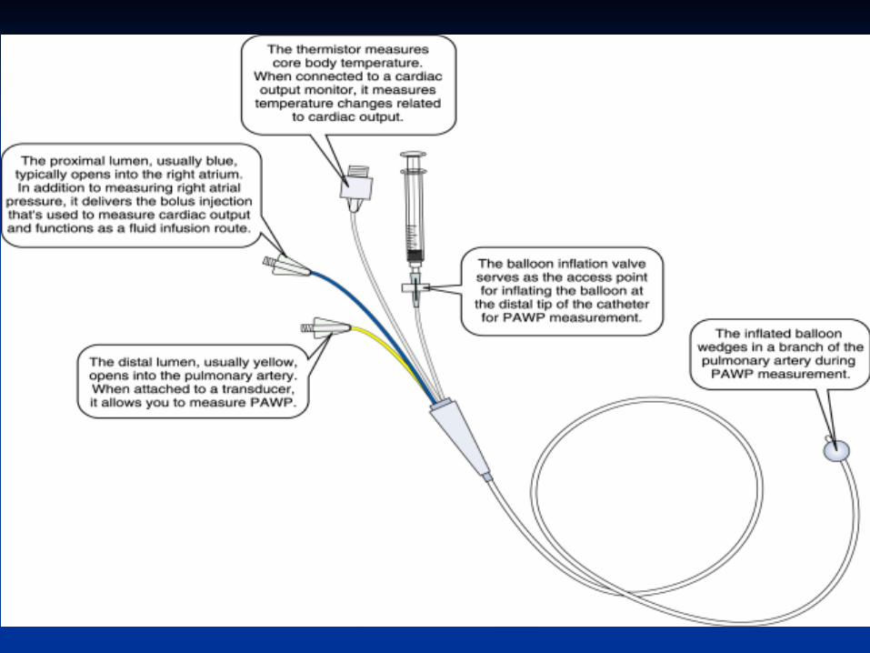
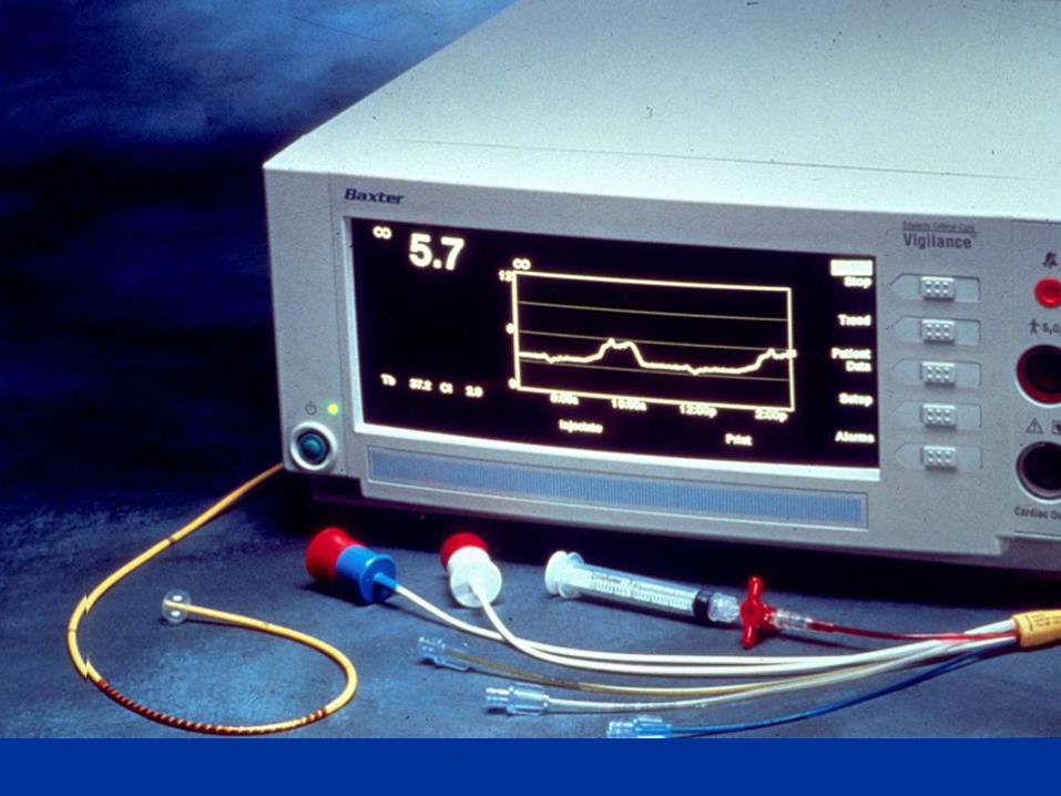
Cardiac Output Cardiac output by Thermodilution
Cold or room temp fluid is injected into the CVP port of the catheter. The temperature of the fluid is measured by the thermistor on the distal port of the catheter.
Cardiac output is inversely proportional to the mean concentration of the indicator.
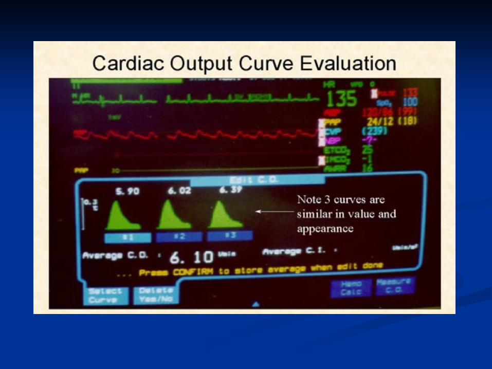
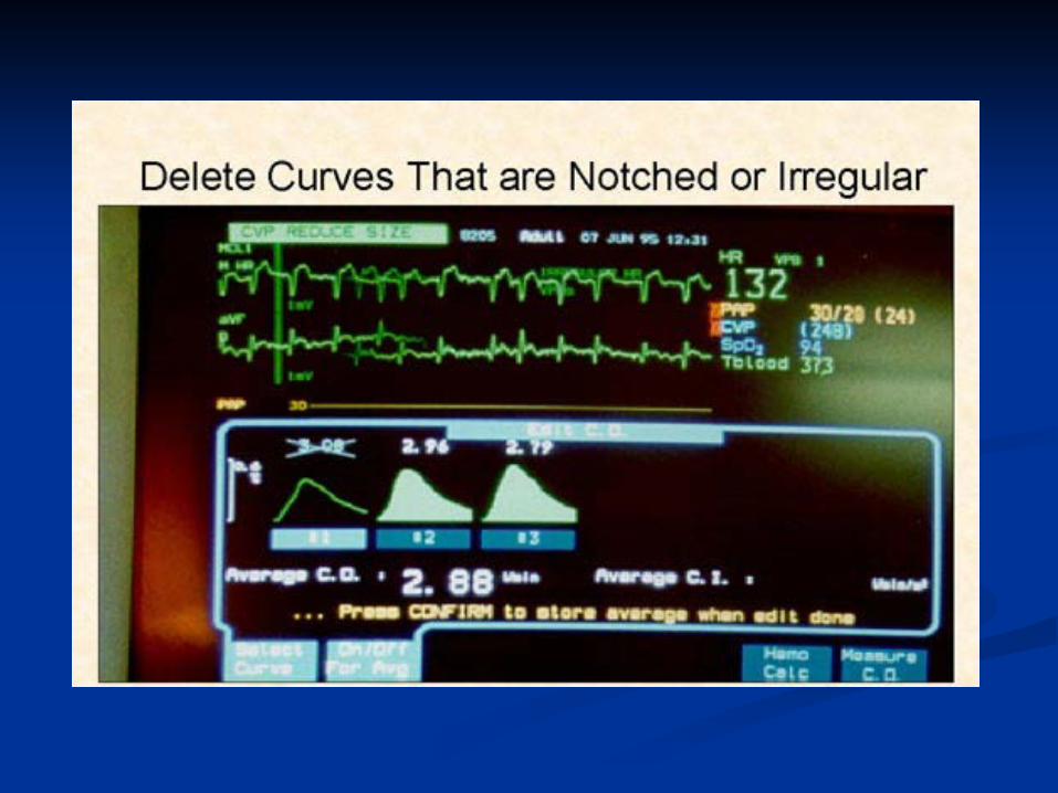
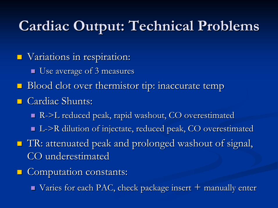
Cardiac Output: Technical Problems
Variations in respiration: Use average of 3 measures
Blood clot over thermistor tip: inaccurate temp Cardiac Shunts:
R->L reduced peak, rapid washout, CO overestimated L->R dilution of injectate, reduced peak, CO overestimated
TR: attenuated peak and prolonged washout of signal, CO underestimated
Computation constants: Varies for each PAC, check package insert + manually enter
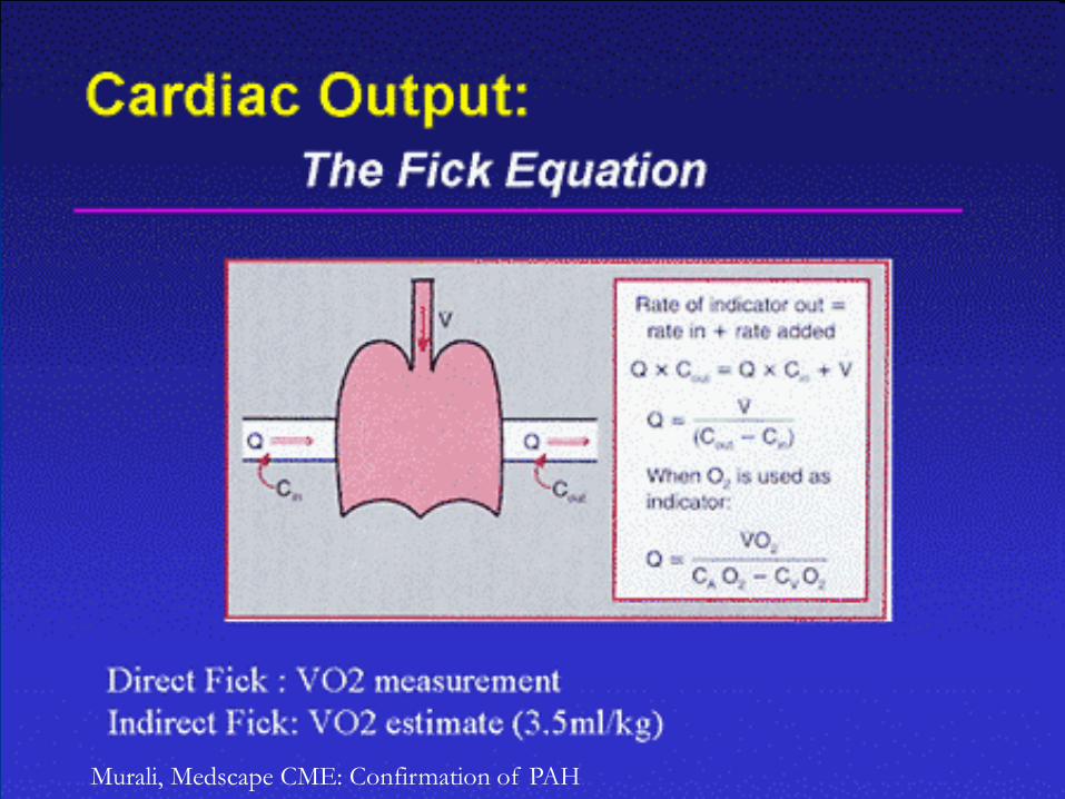
Cardiac Output by Fick
Murali, Medscape CME: Confirmation of PAH
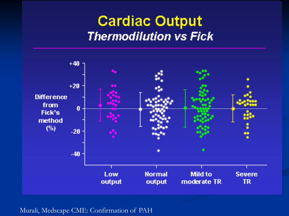
Murali, Medscape CME: Confirmation of PAH
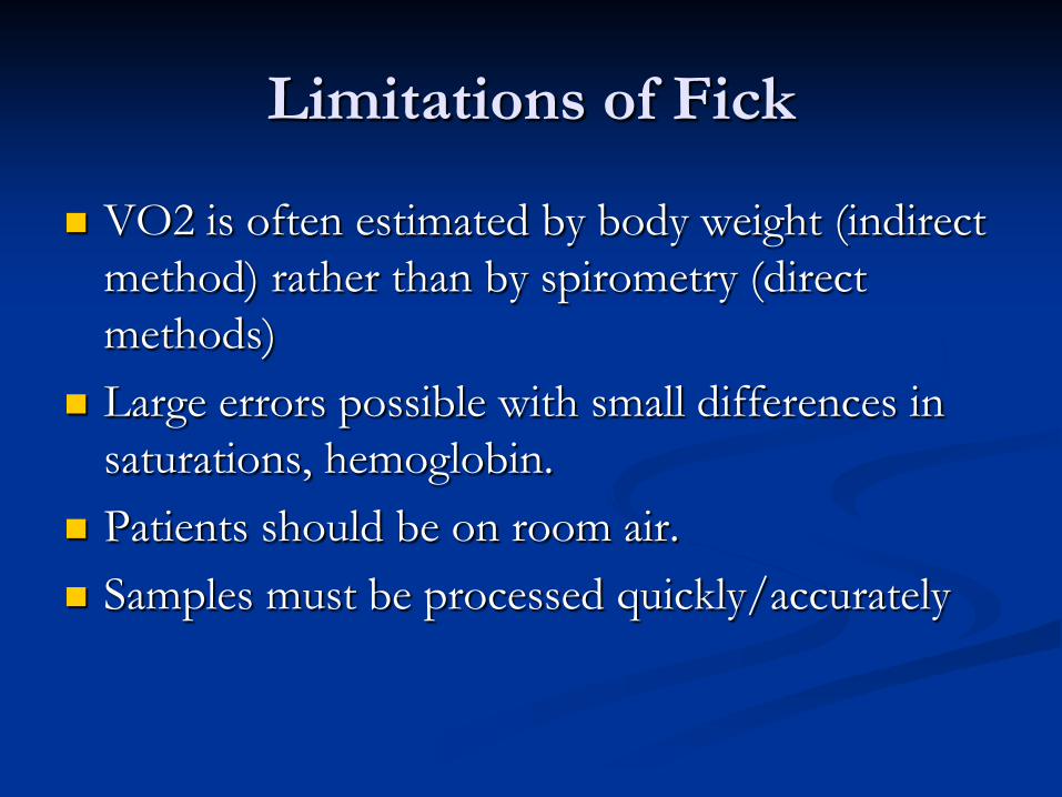
Limitations of Fick
VO2 is often estimated by body weight (indirect method) rather than by spirometry (direct methods)
Large errors possible with small differences in saturations, hemoglobin.
Patients should be on room air. Samples must be processed quickly/accurately
Typical Cath lab hemodynamics: Left and Right heart Cath
Measurements of right heart pressures and hemodynamics
Measurement of LV/PCWP gradient Measurement of LV/Ao gradient Measurement of LV/RV response to inspiration
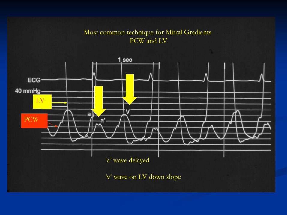
PCW
LV
‘a’ wave delayed
‘v’ wave on LV down slope
Most common technique for Mitral Gradients PCW and LV
BAMC Case #3117: Patient: 61 yo male Dx: 3V CAD filter: 50 Hz/ sample 250 Hz
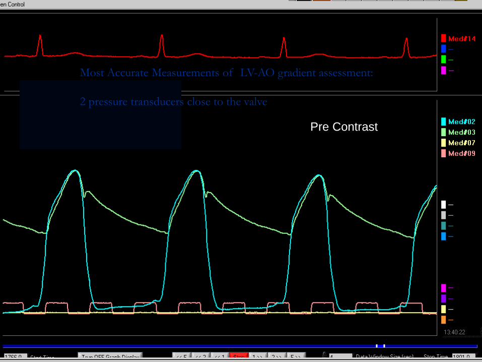
Pre Contrast
Most Accurate Measurements of LV-AO gradient assessment: 2 pressure transducers close to the valve
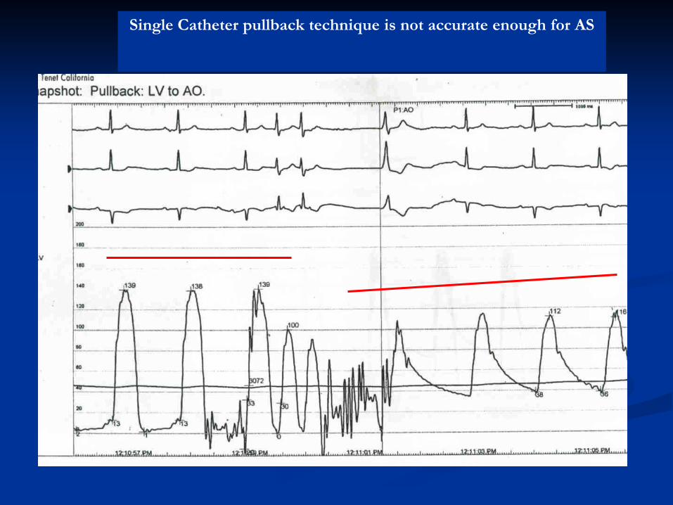
Single Catheter pullback technique is not accurate enough for AS
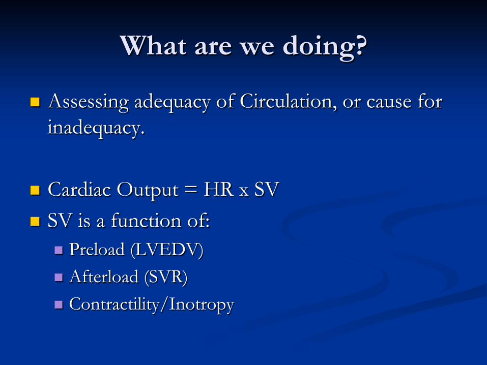
What are we doing?
Assessing adequacy of Circulation, or cause for inadequacy.
Cardiac Output = HR x SV SV is a function of:
Preload (LVEDV) Afterload (SVR) Contractility/Inotropy
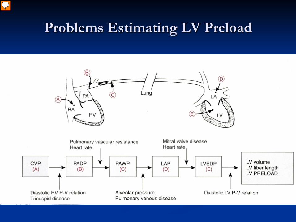
Problems Estimating LV Preload
Presenter
Presentation Notes
There are several problems inherent in estimating LV volume from pressure because diastolic pressure- volume relations are not linear + can change due to disease such as ischemia and diastolic dysfunction; MV and TV disease can also affect the relationship; as can pulmonary disease
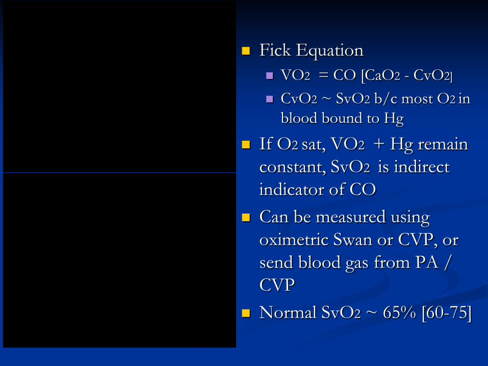
Fick Equation VO2 = CO [CaO2 - CvO2]
CvO2 ~ SvO2 b/c most O2 in blood bound to Hg
If O2 sat, VO2 + Hg remain constant, SvO2 is indirect indicator of CO
Can be measured using oximetric Swan or CVP, or send blood gas from PA / CVP
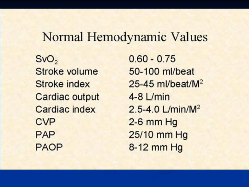
Normal SvO2 ~ 65% [60-75]
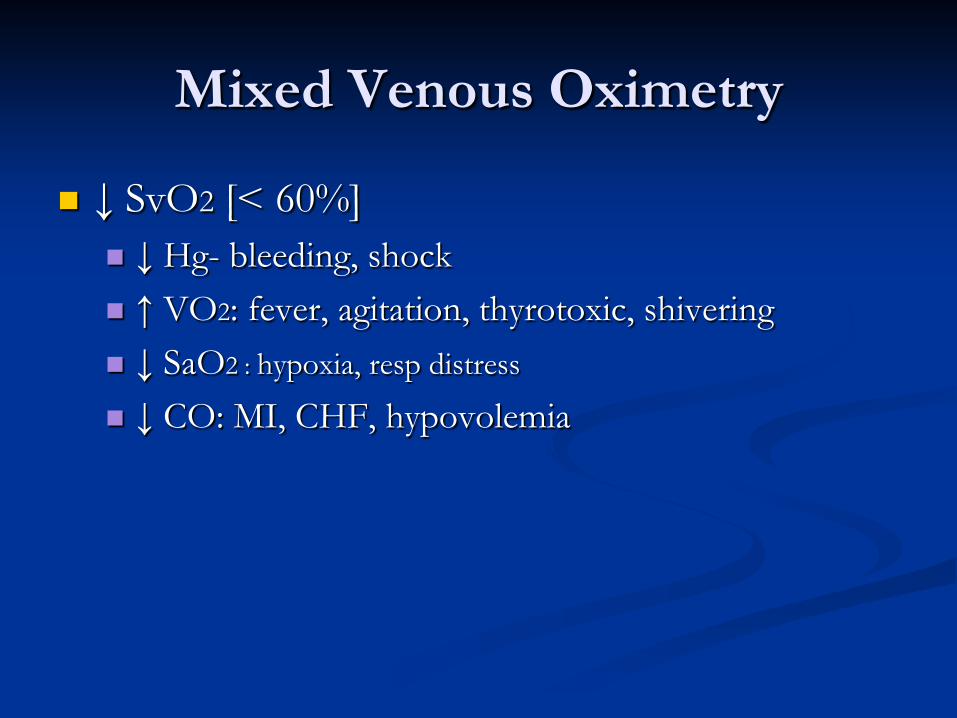
Mixed Venous Oximetry
↓ SvO2 [< 60%] ↓ Hg- bleeding, shock ↑ VO2: fever, agitation, thyrotoxic, shivering ↓ SaO2 : hypoxia, resp distress
↓ CO: MI, CHF, hypovolemia
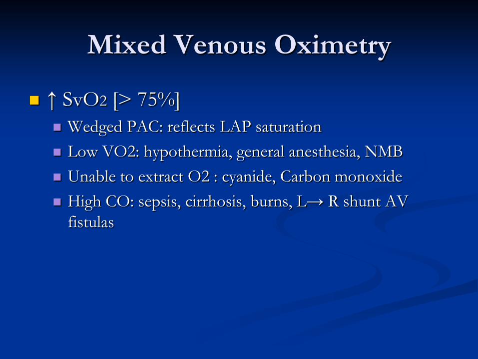
Mixed Venous Oximetry
↑ SvO2 [> 75%] Wedged PAC: reflects LAP saturation Low VO2: hypothermia, general anesthesia, NMB Unable to extract O2 : cyanide, Carbon monoxide High CO: sepsis, cirrhosis, burns, L→ R shunt AV
fistulas
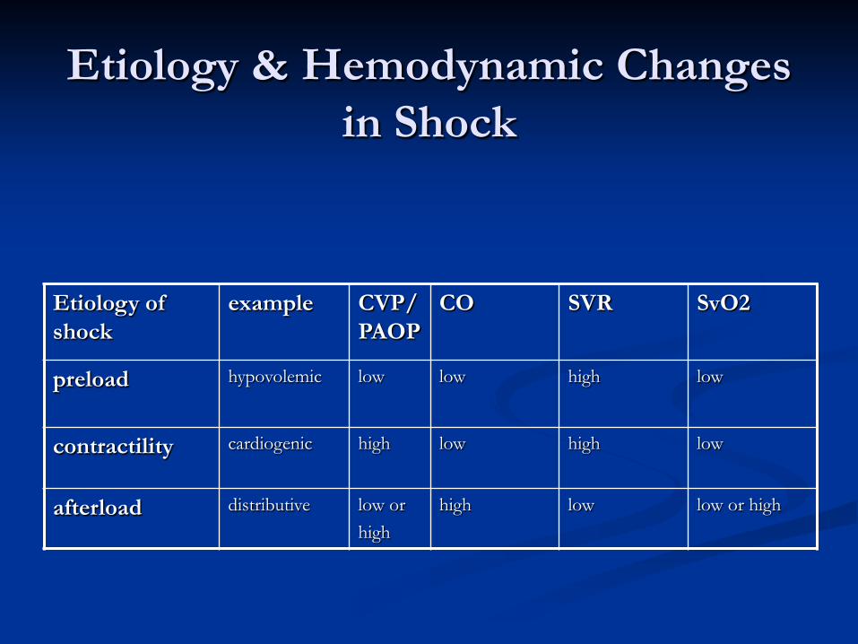
Etiology & Hemodynamic Changes in Shock
Etiology of shock
example CVP/PAOP
CO SVR SvO2
preload hypovolemic low low high low
contractility cardiogenic high low high low
afterload distributive low or high
high low low or high
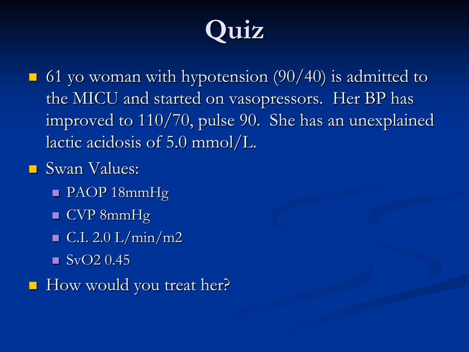
Quiz 61 yo woman with hypotension (90/40) is admitted to
the MICU and started on vasopressors. Her BP has improved to 110/70, pulse 90. She has an unexplained lactic acidosis of 5.0 mmol/L.
Swan Values: PAOP 18mmHg CVP 8mmHg C.I. 2.0 L/min/m2 SvO2 0.45
How would you treat her?
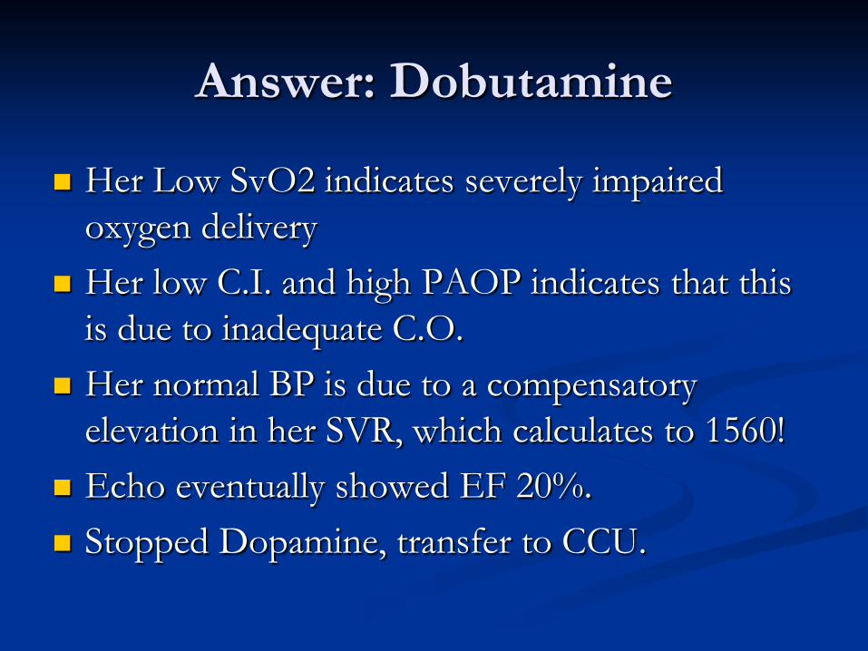
Answer: Dobutamine
Her Low SvO2 indicates severely impaired oxygen delivery
Her low C.I. and high PAOP indicates that this is due to inadequate C.O.
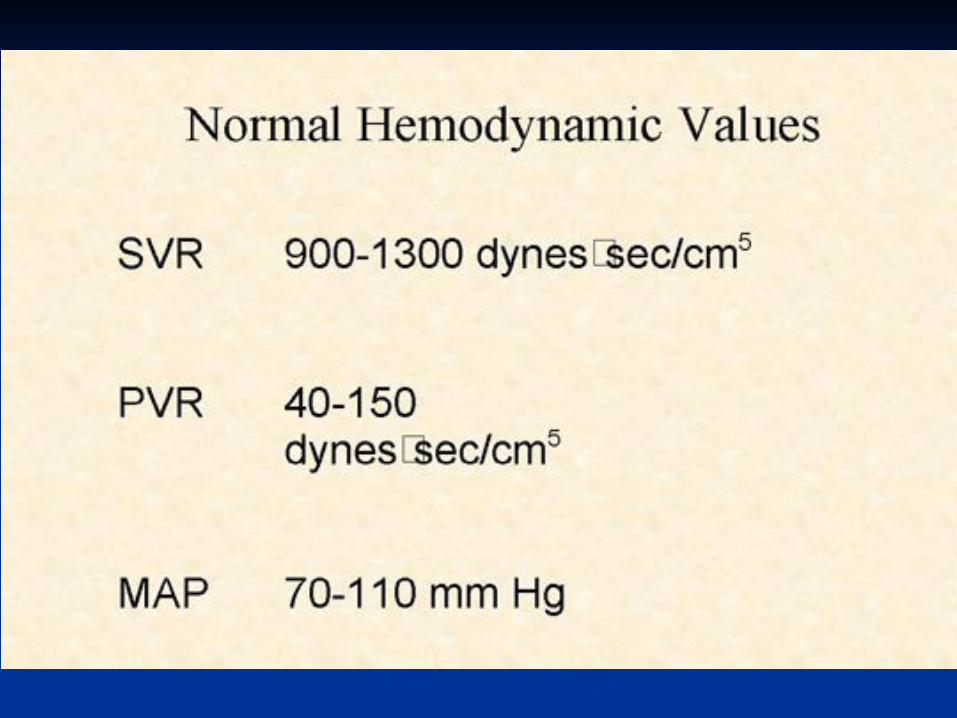
Her normal BP is due to a compensatory elevation in her SVR, which calculates to 1560!
Echo eventually showed EF 20%. Stopped Dopamine, transfer to CCU.
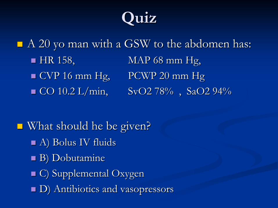
Quiz A 20 yo man with a GSW to the abdomen has:
HR 158 , MAP 68 mm Hg, CVP 16 mm Hg, PCWP 20 mm Hg CO 10.2 L/min, SvO2 78% , SaO2 94%
What should he be given?
A) Bolus IV fluids B) Dobutamine C) Supplemental Oxygen D) Antibiotics and vasopressors
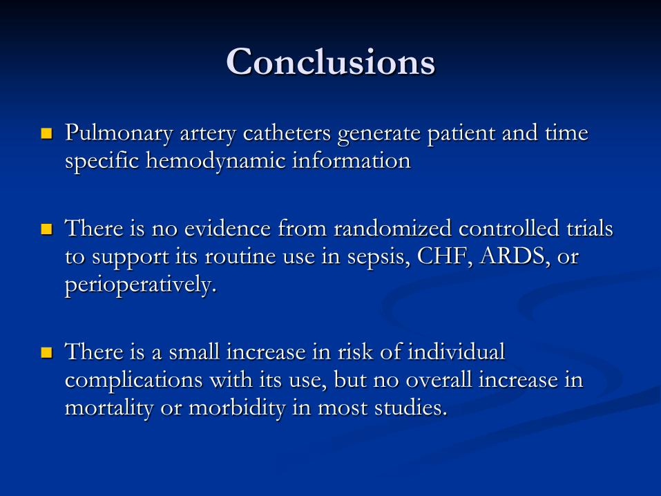
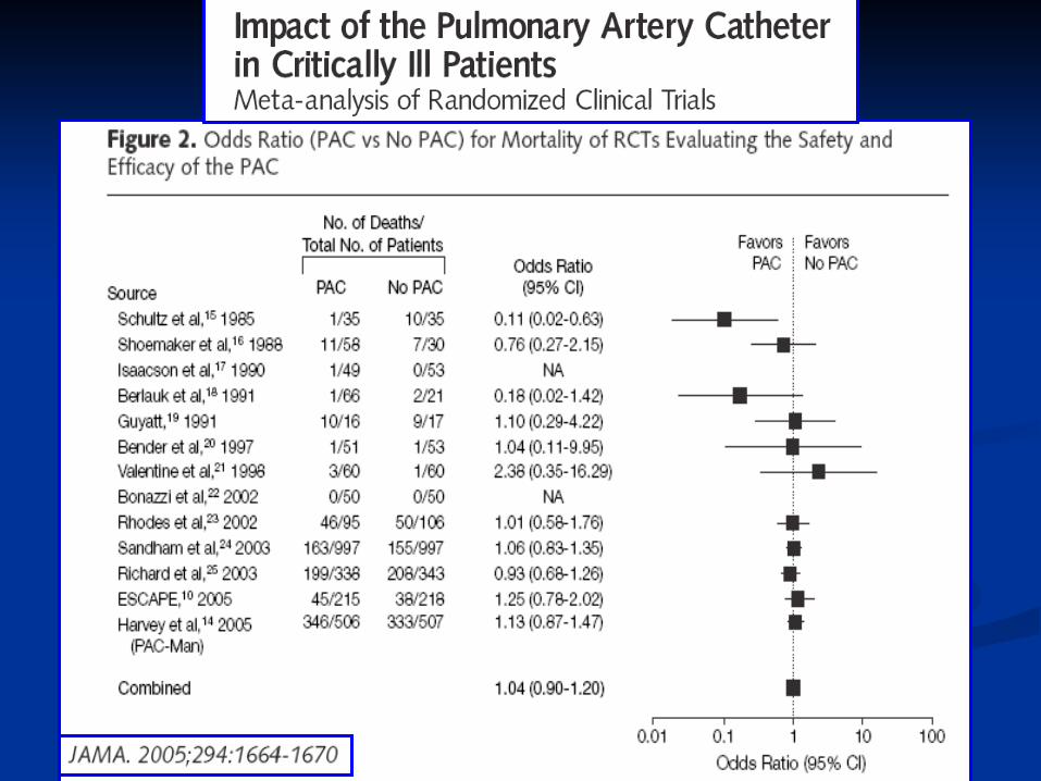
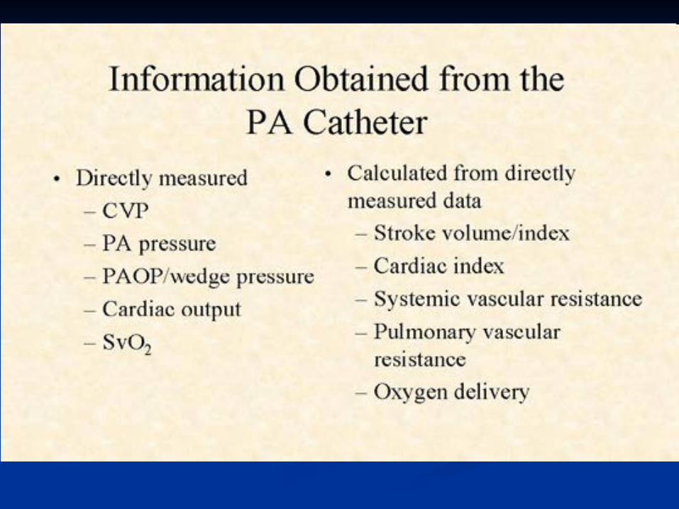
Conclusions
Pulmonary artery catheters generate patient and time specific hemodynamic information
There is no evidence from randomized controlled trials to support its routine use in sepsis, CHF, ARDS, or perioperatively.
There is a small increase in risk of individual complications with its use, but no overall increase in mortality or morbidity in most studies.