Hemodialyzers, including reuse
31
ASN DIALYSIS CURRICULUM ASN Dialysis Advisory Group 0
-
Upload
nguyenthuan -
Category
Documents
-
view
215 -
download
0
Transcript of Hemodialyzers, including reuse

ASN DIALYSIS CURRICULUM
ASN Dialysis Advisory Group
0

Hemodialyzers Alfred K. Cheung •University of Utah, Salt Lake City, UT
Richard A. Ward •University of Louisville, KY
1

Disclosures
• Alfred K. Cheung
• None
• Richard A. Ward
• Research Support:
• Gambro Renal Products
• Rockwell Medical Technologies
• Speaker Honoraria:
• Fresenius
2

Dialyzer Components–Fiber Bundle
3
• The fiber bundle comprises 7–17 x 103 semipermeable hollow fibers that allow solute and fluid transfer between blood and dialysate
• Typical fibers have internal diameter of 200 mm and wall thickness of 30–40 mm. They provide 1.0–2.5 m2 of surface area
• Fibers may incorporate features, such as undulations, to distribute dialysate flow evenly through the bundle
• The fiber bundle is enclosed in an outer housing that forms the dialysate compartment
Fiber bundle Dialyzer housing

Dialyzer Components–Headers
4
• The header is the space enclosed by the end cap and the polyurethane potting material that holds the hollow fibers and forms a barrier between the blood and dialysate compartments
• Headers channel blood from the dialyzer inlet into the membrane fibers, and from the membrane fibers into the dialyzer outlet
• The end caps can be removed from some dialyzers. In those cases, an O-ring is used to form a seal between the end cap and the potting material
End cap
Potting material

Dialyzer Components–Flow Paths
5
• Blood flows through the fiber lumens. Typical clinical blood flow rates are 200–450 ml/min
• Dialysate flows around the external surface of the fibers. Typical dialysate flow rates are 500–800 ml/min
• Blood and dialysate flow in opposite directions (counter-current flow) to maximize diffusive solute transfer
Blood in
Blood out
Dialysate out
Dialysate in

Dialyzer Sterilization
•Early disposable hollow-fiber dialyzers were shipped filled with a formaldehyde solution to maintain sterility
•Later, dialyzers were sterilized with ethylene oxide (ETO) and shipped dry
•The polyurethane potting material used in hollow-fiber dialyzers can act as a reservoir for ETO. Residual ETO could then diffuse into the blood during dialysis, bind to plasma proteins, and induce antibody formation, leading to intradialytic anaphylactoid reactions
•To reduce the risk of these anaphylactoid reactions, ETO has now been largely replaced by alternative methods, such as steam and irradiation, for sterilizing dialyzers
6

Classification Of Dialyzer Membranes
• Dialyzer membranes can be classified based on their:
• Chemical composition
• Cellulosic versus synthetic
• Efficiency of small-solute (based on urea, 60 Da) removal
• Low-efficiency (urea KoA < 450 ml/min) versus high-efficiency (urea KoA > 700 ml/min). Most modern dialyzers are high-efficiency
• Efficiency of large-solute (based on ² 2-microglobulin, 12 kDa) removal
Efficiency of large-solute (based on ² 2-microglobulin, 12 kDa) removal
• Low-flux (² 2M clearance < 10 ml/min) versus high-flux (² 2M clearance > 20 ml/min + ultrafiltration coefficient > 14 ml/hr/mm Hg)
• Water or hydraulic permeability (FDA classification)
• Conventional versus high-permeability
• Biocompatibility
• Biocompatible versus bioincompatible
7

Cellulosic Membranes
8
• Cellulose was the first membrane material widely used for hemodialysis. It is a polymer of cellobiose and occurs in natural materials, such as cotton.
• Cellulose membranes are hydrogels and can be made very thin (6–15 mm dry thickness) while retaining good mechanical strength. They allow high diffusive transport of small molecules (< 200 Da).
8 mm

Cellulosic Membranes
9
•The hydroxyl (OH) groups in cellulose react with serum complement component (C3) activating the alternative pathway of complement
•Products of complement activation, in turn, activate leukocytes, leading to a profound, transient leukopenia during the first hour of dialysis
•Because of concerns that these processes may contribute to dialysis-associated morbidity, unmodified cellulose membranes are now seldom used

Cellulosic Membranes
10
TIME (minutes)
0 60 120 180 240
NE
UT
RO
PH
ILS
/L
0
1000
2000
3000
4000
5000
6000
C3a
desA
rg (
ng/m
L)
0
500
1000
1500
2000
2500
Jacobs AA Jr, et al: Polymorphonuclear leukocyte function during hemodialysis: relationship to complement activation. Nephron 52: 119-124, 1989
Peripheral plasma concentrations of C3adesArg and neutrophil counts during dialysis with a cellulose membrane (adapted from Jacobs AA, et al, Nephron 52:119-124, 1989)

Cellulosic Membranes
11 Jacobs AA Jr, et al: Polymorphonuclear leukocyte function during hemodialysis: relationship to complement activation. Nephron 52: 119-124, 1989

Modified Cellulose Membranes
• Substitution of hydroxyl groups on cellobiose with other chemical moieties reduces the ability of cellulose membranes to activate complement and cause leukopenia
• Moieties that have been used to substitute for cellulose include acetate, diethylaminoethyl (DEAE), benzyl, polyethyleneglycolic, and vitamin E. The resultant membranes are referred to as modified cellulose membranes. Only cellulose diacetate and cellulose triacetate membranes remain in widespread clinical use in the US
• Modified cellulose membranes can be either high-flux or low-flux
12

Synthetic Membranes
• These membranes were made of synthetic polymers and were initially developed for hemodiafiltration
• There may be more than one type of polymer (i.e., alloy) in a given synthetic dialysis membrane that may impart important characteristics (e.g., methallyl sulfonate mixed with polyacrylonitrile and, polyvinylpyrrolidone mixed with polysulfone)
• Synthetic membranes are thick (> 35 m) with cross-sectional structures that were either homogeneous (e.g., AN69®, Hospal) or asymmetric (e.g., polysulfone). The asymmetry refers to a 2-layer structure with an inner thin layer that regulates solute removal and a thick supporting stroma
13
• Synthetic membranes can be either high-flux or low-flux

Membrane Properties–Diffusion Coefficient
• The primary mode for removal of small solutes (e.g., urea) by hemodialysis is diffusion down the concentration gradient between plasma water and dialysate. Transfer of small solutes (e.g., HCO3-) from dialyzate to plasma water also occurs primarily by diffusion
• The rate of diffusion is a function of the thickness and porosity of the membrane and the diffusivity of the solute in the membrane and is expressed as the diffusion coefficient of the membrane for a given solute
• The rate of diffusion is greatest for small molecules. The diffusivity of a solute in a membrane decreases logarithmically as solute size increases
• The rate of diffusion also decreases as membrane thickness increases and porosity decreases
14

Membrane Properties–Diffusion Coefficient
15
SOLUTE RADIUS x 108 (cm)
1 2 3 4 5 6 7 8 9
DIF
FU
SIV
ITY
x 1
06 (cm
2 /sec
)
0.0
0.5
1.0
1.5
2.0
2.5
3.0
UREA
CREATININE
GLUCOSE
VITAMIN B12
URIC ACID
Diffusivity of solutes through a cellulose membrane (adapted from Farrell PC, et al, J Biomed Mater Res 7:275-300, 1973)
Farrell PC, et al: Estimation of the permeability of cellulosic membranes from solute dimensions and diffusivities. J Biomed Mater Res 7: 275-300, 1973

Membrane Properties–Sieving Coefficient
16
• The primary mode for removal of large solutes by hemodialysis is convection, as water containing these solutes flows from plasma to dialysate in response to a hydraulic pressure gradient
• The rate of convection is a function of the water filtration rate, size of the solute, and pore size of the membrane. The ability of a solute to pass through the pores of a membrane is expressed as the sieving coefficient of the membrane for a given solute
• A solute with a sieving coefficient of 1.0 passes freely through the membrane, while the membrane is impermeable to a solute with a sieving coefficient of 0
• Convection provides better removal of large solutes than diffusion because the decrease in sieving coefficient with increasing solute size is less marked than the decrease in diffusion coefficient

Membrane Properties–Sieving Coefficient
17
SOLUTE RADIUS x 108 (cm)
1 2 3 4 5 6 7 8 9
SIE
VIN
G C
OE
FF
ICIE
NT
0.0
0.2
0.4
0.6
0.8
1.0UREA GLUCOSE
SUCROSE
VITAMIN B12
Sieving coefficients for a cellulose membrane. (adapted from Wendt RP, et al, J Memb Sci 5:23-49, 1979)
Wendt RP, et al: Sieving properties of hemodialysis membranes. J Membr Sci 5: 23-49, 1979

Membrane Properties–Hydrophobicity
• The nature of the polymers used in a membrane determines the membrane’s tendency to repel water, referred to as hydrophobicity. In general, cellulose membranes are less hydrophobic while many synthetic polymer membranes are more hydrophobic
• Because polysulfone is so hydrophobic, it is alloyed with hydrophilic polyvinylpyrrolidone (PVP) to decrease the hydrophobicity of the membrane
• Hydrophobic surfaces adsorb serum proteins; adsorption can contribute significantly to low–molecular-weight protein removal by some membranes
• Proteins may adsorb to both the planar surface of the membrane and the inner surface of its pores. The adsorbed protein may reduce diffusive and convective removal of other solutes by effectively reducing the pore size of the membrane
18

Overall Solute Removal
• In practice, solute removal occurs through a combination of diffusion, convection, and adsorption. The relative contributions of the three mechanisms depend on the solute, the membrane, the geometry of the dialyzer, and operating conditions (blood and dialysate flow rates and ultrafiltration rate)
19

Diffusive Solute Removal
• Solute removal by diffusion is limited not only by the membrane, but also by boundary layers that form at the blood-membrane interface and the dialysate-membrane interface
• The overall resistance to diffusive solute removal is the sum of the resistances associated with the membrane and these two boundary layers. The reciprocal of the overall resistance is the mass transfer coefficient (Ko). The diffusive capacity is usually expressed as KoA for a given dialyzer and a given solute, where A is the membrane surface area of the dialyzer
• KoA is generally considered to be largely independent of the blood and dialysate flow rates
• KoA values for urea are usually provided by dialyzer manufacturers. If those KoA values are obtained using aqueous solutions in vitro, they should be discounted by about 20% before being applied clinically when the dialyzer membrane is exposed to blood
20

Clearance
• Diffusive solute removal by a dialyzer is usually described in terms of clearance (K), which is defined as the volume of blood completely cleared of a given solute per unit time, or
• where QBi and QBo are the blood flow rates; CBi and CBo are the solute concentrations at the inlet and outlet of the dialyzer, respectively
• Unlike KoA, clearance is dependent on both the blood and dialysate flow rates. For this reason, it is not a good means of characterizing the innate dialyzer performance
21
Bi
BoBoBiBi
CCQCQ
K−
=
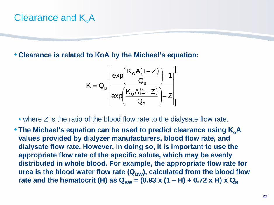
Clearance and KoA
• Clearance is related to KoA by the Michael’s equation:
• where Z is the ratio of the blood flow rate to the dialysate flow rate.
• The Michael’s equation can be used to predict clearance using KoA values provided by dialyzer manufacturers, blood flow rate, and dialysate flow rate. However, in doing so, it is important to use the appropriate flow rate of the specific solute, which may be evenly distributed in whole blood. For example, the appropriate flow rate for urea is the blood water flow rate (QBW), calculated from the blood flow rate and the hematocrit (H) as QBW = (0.93 x (1 – H) + 0.72 x H) x QB
22
( )
( )
−
−
−
−
=Z
QZ1AK
exp
1Q
Z1AKexp
QK
B
O
B
O
B

Convective Solute Removal
• In purely convective therapies, such as hemofiltration, convective clearance (Kconv) is determined by the sieving coefficient for the solute (S) and the filtration flow rate (QF), or
• When convection is combined with diffusion, such as in
hemodiafiltration or hemodialysis with ultrafiltration, the convective and diffusive components of solute removal are not simply additive. Diffusive removal is compromised because filtration decreases the effective blood flow rate, while convection is compromised because diffusion reduces the solute concentration in the blood. In these situations, convection is generally more important for large-solute removal and diffusion is more important for small-solute removal
• S is usually considered to be a fundamental property of the dialyzer membrane for a particular solute when QF is low relative to the blood flow rate. As QF increases relative to the blood flow rate, however, large proteins to which the membrane is impermeable accumulate at the membrane surface in a process called concentration polarization. This protein layer changes S and effectively reduces convective solute removal
23
SQK FConv ×=

Adsorptive Solute Removal
• Adsorption to the dialyzer membrane can be a major mechanism of solute removal for proteins, including ² 2-microglobulin. The importance of adsorption as a mechanism of removal tends to increase relative to diffusive and convective removal as protein size increases
• Adsorption is not a significant mechanism of solute removal for small water-soluble solutes, such as urea, creatinine, and phosphate
• Whether or not a protein is removed by adsorption depends on the physicochemical properties of both the protein and the membrane; membranes fabricated from nominally similar polymers can have quite different adsorptive properties
24
Ultrafiltration Coefficient
• The ultrafiltration coefficient (kUF) is a measure of the water permeability of a membrane and is usually expressed in mL/hr/mm Hg
• For example, if a dialysis machine generated a transmembrane pressure (TMP) of 200 mm Hg, a dialyzer with a kUF of 12 ml/hr/mm Hg would produce an ultrafiltration rate of 12 ml/hr/mm Hg x 200 mm Hg = 2.4 L/hr
• The FDA defines a high-flux dialyzer as one with a kUF e 12 ml/hr/mm Hg and a low-flux dialyzer as one with a kUF < 12 ml/hr/mm Hg
• The FDA classification was important for safety reasons when automated ultrafiltration control devices were not available. With the ultrafiltration control systems installed in contemporary dialysis machines, the kUF of a dialyzer is of little clinical use, although it may give some indication of the sieving coefficients (SC) of the membrane for larger solutes (e.g., ² 2-microglobulin), since dialyzers with a high kUF tend to have high SCs for larger solutes
• The kUF is practically never a limiting factor for fluid removal. The ultrafiltration rate is almost invariability limited by the patient’s tolerance of the rate of fluid removal
25

Dialyzer Biocompatibility–Mechanisms
• Adsorption and transformation of plasma proteins, for example:
• Activation of Hageman factor, triggering the intrinsic coagulation cascade
• Conversion of kininogen to bradykinin as a result of Hageman factor activation by membranes with strongly anionic surfaces. The generated bradykinin accumulates in plasma in the presence of angiotensin converting enzyme (ACE) inhibitors (which is also a kininase)
• Activation of complement C3 and C5, generating anaphylatoxins
• Shearing, adherence, and activation of blood cells; for example, platelets and neutrophils
• Leaching of soluble contaminants (e.g., ethylene oxide) and spallation of insoluble contaminants (e.g., silicone)
26

Dialyzer Biocompatibility–Clinical Factors
• Important clinical bioincompatibility phenomena include:
• Activation of clotting factors and platelets resulting in thrombosis in the dialyzer
• Activation of the complement system, with consequent activation of leukocytes, possibly leading to:
• Acute intra-dialytic pulmonary hypertension
• Chronic low-grade systemic inflammation
• Leukocyte dysfunction
• All dialysis membranes activate complement and leukocytes to some extent. Unmodified cellulosic membranes are potent activators of complement and leukocytes and are generally considered to be bioincompatible
• Generation of bradykinin in the presence of dialysis membrane with anionic surface (and accumulation of the bradykinin in plasma in the presence of ACE inhibitors) causing anaphylactoid reactions
27

Dialyzer Reactions
• On rare occasions, systemic anaphylactoid reactions occur during hemodialysis, with various degrees of intensity
• Manifestations include pruritis, angioedema, chest pain, upper respiratory symptoms, and hypertension and/or hypotension that can culminate in death
• Reactions occur more commonly with a previously unused dialyzer, hence the term “new dialyzer syndrome,” although they can also occur with reused dialyzers
• Leaching of residual ethylene oxide (ETO, used to sterilize dialyzers) from the potting compound into the blood is probably causative in some cases, although other factors, such as complement anaphylatoxins, have also been implicated
• Preventive measures include avoiding dialyzers sterilized with ETO and rigorous rinsing of dialyzers prior to use. Treatment of anaphylactoid reactions is largely symptomatic
• Anaphylactoid reactions induced by bradykinin in the presence of a dialysis membrane with anionic surface charges can be minimized by avoiding those membranes and/or ACE inhibitors
28

Dialyzer Reuse–Techniques
• The primary purpose for reusing dialyzers is economics
• Germicides used to reprocess dialyzers include: formaldehyde, peracetic acid, glutaraldehyde, and heated citric acid. Bleach is sometimes used as an additional cleaning agent
• Deposition of plasma proteins on the dialyzer membrane during previous uses may affect the solute clearance and biocompatibility of a dialyzer
29

Dialyzer Reuse–Consequences
• Different germicides and cleaning agents have different effects on the deposited protein and on the membrane that can affect solute clearance and biocompatibility. For example:
• Complement activation by cellulosic membranes is attenuated when dialyzers are reprocessed with formaldehyde or glutaraldehyde; the complement activating potential is restored if the membrane is first cleaned with bleach.
• Clearance of ² 2-microglobulin decreases when dialyzers with cellulose triacetate membranes are reprocessed with peracetic acid without first being cleaned with bleach. In contrast, ² 2-microglobulin clearance increases when polysulfone membranes containing polyvinylpyrrolidone (PVP) are cleaned with bleach during reprocessing.
• In general, urea clearance decreases by ~2% per 10 reuses for a variety of dialyzers and reprocessing methods.
30


![Value Based Software Reuse Investment - Favaro · Favaro [1996a] has compared several approaches to valuation cited in the literature on software reuse economics, including time to](https://static.fdocuments.in/doc/165x107/5c73dc7e09d3f287228b917b/value-based-software-reuse-investment-favaro-1996a-has-compared-several.jpg)