Heme Uptake Mediated by LHR1 Is Essential for …iai.asm.org/content/81/10/3620.full.pdfHeme Uptake...
7
Heme Uptake Mediated by LHR1 Is Essential for Leishmania amazonensis Virulence Danilo C. Miguel, Andrew R. Flannery, Bidyottam Mittra, Norma W. Andrews Department of Cell Biology and Molecular Genetics, University of Maryland, College Park, Maryland, USA The protozoan parasite Leishmania amazonensis is a heme auxotroph and must acquire this essential factor from the environ- ment. Previous studies showed that L. amazonensis incorporates heme through the transmembrane protein LHR1 (Leishmania Heme Response 1). LHR1-null promastigotes were not viable, suggesting that the transporter is essential for survival. Here, we compared the growth, differentiation, and infectivity for macrophages and mice of wild-type, LHR1-single-knockout (LHR1/ lhr1), and LHR1-complemented (LHR1/lhr1 plus LHR1) L. amazonensis strains. LHR1/lhr1 promastigotes replicated poorly in heme-deficient media and had lower intracellular heme content than wild-type parasites. LHR1/lhr1 promastigotes were also less effective in reducing ferric iron to ferrous iron, a reaction mediated by the heme-containing parasite enzyme LFR1 (Leishma- nia Ferric Reductase 1). LHR1/lhr1 parasites differentiated normally into aflagellated forms expressing amastigote-specific markers but were not able to replicate intracellularly after infecting macrophages. Importantly, the intracellular growth of LHR1/lhr1 amastigotes was fully restored when macrophages were allowed to phagocytose red blood cells prior to infection. LHR1/lhr1 parasites were also severely defective in the development of cutaneous lesions in mice. All phenotypes observed in LHR1/lhr1 L. amazonensis were rescued by expression of episomal LHR1. Our results reveal the importance of efficient heme uptake for L. amazonensis replication and vertebrate host infectivity, reinforcing the potential usefulness of LHR1 as a target for new antileishmanial drugs. L eishmaniasis is a complex disease caused by infection with the protozoan parasite Leishmania. Clinical symptoms vary de- pending on the species, immune status of the host, and region of endemicity and range from cutaneous lesions (localized, mucosal, disseminated, and diffuse) to life-threatening visceral disease (1). The current treatment of leishmaniasis is mainly based on the parenteral administration of toxic and expensive drugs, such as pentavalent antimonials and amphotericin B (2). The proliferative extracellular forms of Leishmania, procyclic promastigotes, replicate within the digestive tract in sand flies. A few days after infecting a sand fly, procyclic promastigotes differ- entiate into metacyclic promastigotes, the infective stages that are inoculated into the dermis of vertebrate hosts during the next blood meal. Once inside the human host, metacyclic promasti- gotes are engulfed by macrophages, where they transform into intracellular amastigotes and multiply within parasitophorous vacuoles (PVs) with properties of phagolysosomes (reviewed in references 3 and 4). Heme is one of many indispensable nutrients that must be scavenged from the phagolysosomal compartment by Leishmania (5). In metazoa, heme biosynthesis occurs through 8 highly con- served chemical reactions, resulting in the insertion of ferrous iron into the protoporphyrin IX ring (6). However, several unicellular organisms, such as bacteria, trypanosomatids (Leishmania and Trypanosoma), and anaerobic protozoan parasites (Giardia, Trichomonas, and Entamoeba), lack several of the enzymes neces- sary for a complete heme-biosynthetic pathway (7, 8). Therefore, in vitro cultivation of trypanosomatids requires addition of a heme source, such as hemin, hematin, or hemoglobin. Heme not only is a crucial component of cytochromes in the respiratory chain, but also functions as an essential cofactor for hemopro- teins, such as the ones involved in the biosynthesis of polyun- saturated fatty acids and sterols (8–10). Earlier studies showed that Leishmania donovani promasti- gotes can internalize and target hemoglobin for degradation through a surface hemoglobin-binding protein (11). Heme up- take was also shown to act as a source of iron in L. infantum (12). However, until recently, the molecular mechanism responsible for direct heme uptake by Leishmania was not understood. Our group has recently clarified this issue by identifying and characterizing LHR1 (Leishmania Heme Response 1), a surface protein with 4 transmembrane domains that directly promotes heme uptake when expressed in Saccharomyces cerevisiae. LHR1 transcripts are increased when promastigotes are cultured in the absence of heme (13), and deletion of one LHR1 allele (the LHR1/lhr1 mutant strain) significantly reduces intracellular heme pools and uptake of zinc mesoporphyrin (ZnMP). Despite several attempts to ob- tain an LHR1-null mutant by replacement of the second LHR1 allele, it was not possible to recover viable parasites. These results suggested that LHR1 is essential for viability in Leishmania ama- zonensis (13). In the present work, we characterized the role of LHR1 by analyzing the biological properties of the LHR1/lhr1 single- knockout strain, such as replication, differentiation, and in vitro and in vivo infectivity. Our results show that parasites lacking one copy of LHR1 are more sensitive to heme deficiency as promasti- gotes and incapable of replicating as amastigotes in macrophages, an effect that is fully rescued when the endosomal pathway of macrophages is loaded with red blood cells, a rich source of heme. Received 31 May 2013 Accepted 5 July 2013 Published ahead of print 22 July 2013 Editor: J. L. Flynn Address correspondence to Norma W. Andrews, [email protected]. Copyright © 2013, American Society for Microbiology. All Rights Reserved. doi:10.1128/IAI.00687-13 3620 iai.asm.org Infection and Immunity p. 3620 –3626 October 2013 Volume 81 Number 10 on June 3, 2018 by guest http://iai.asm.org/ Downloaded from
-
Upload
nguyendiep -
Category
Documents
-
view
215 -
download
1
Transcript of Heme Uptake Mediated by LHR1 Is Essential for …iai.asm.org/content/81/10/3620.full.pdfHeme Uptake...

Heme Uptake Mediated by LHR1 Is Essential for Leishmaniaamazonensis Virulence
Danilo C. Miguel, Andrew R. Flannery, Bidyottam Mittra, Norma W. Andrews
Department of Cell Biology and Molecular Genetics, University of Maryland, College Park, Maryland, USA
The protozoan parasite Leishmania amazonensis is a heme auxotroph and must acquire this essential factor from the environ-ment. Previous studies showed that L. amazonensis incorporates heme through the transmembrane protein LHR1 (LeishmaniaHeme Response 1). LHR1-null promastigotes were not viable, suggesting that the transporter is essential for survival. Here, wecompared the growth, differentiation, and infectivity for macrophages and mice of wild-type, LHR1-single-knockout (LHR1/�lhr1), and LHR1-complemented (LHR1/�lhr1 plus LHR1) L. amazonensis strains. LHR1/�lhr1 promastigotes replicated poorlyin heme-deficient media and had lower intracellular heme content than wild-type parasites. LHR1/�lhr1 promastigotes were alsoless effective in reducing ferric iron to ferrous iron, a reaction mediated by the heme-containing parasite enzyme LFR1 (Leishma-nia Ferric Reductase 1). LHR1/�lhr1 parasites differentiated normally into aflagellated forms expressing amastigote-specificmarkers but were not able to replicate intracellularly after infecting macrophages. Importantly, the intracellular growthof LHR1/�lhr1 amastigotes was fully restored when macrophages were allowed to phagocytose red blood cells prior to infection.LHR1/�lhr1 parasites were also severely defective in the development of cutaneous lesions in mice. All phenotypes observed inLHR1/�lhr1 L. amazonensis were rescued by expression of episomal LHR1. Our results reveal the importance of efficient hemeuptake for L. amazonensis replication and vertebrate host infectivity, reinforcing the potential usefulness of LHR1 as a target fornew antileishmanial drugs.
Leishmaniasis is a complex disease caused by infection with theprotozoan parasite Leishmania. Clinical symptoms vary de-
pending on the species, immune status of the host, and region ofendemicity and range from cutaneous lesions (localized, mucosal,disseminated, and diffuse) to life-threatening visceral disease (1).The current treatment of leishmaniasis is mainly based on theparenteral administration of toxic and expensive drugs, such aspentavalent antimonials and amphotericin B (2).
The proliferative extracellular forms of Leishmania, procyclicpromastigotes, replicate within the digestive tract in sand flies. Afew days after infecting a sand fly, procyclic promastigotes differ-entiate into metacyclic promastigotes, the infective stages that areinoculated into the dermis of vertebrate hosts during the nextblood meal. Once inside the human host, metacyclic promasti-gotes are engulfed by macrophages, where they transform intointracellular amastigotes and multiply within parasitophorousvacuoles (PVs) with properties of phagolysosomes (reviewed inreferences 3 and 4).
Heme is one of many indispensable nutrients that must bescavenged from the phagolysosomal compartment by Leishmania(5). In metazoa, heme biosynthesis occurs through 8 highly con-served chemical reactions, resulting in the insertion of ferrous ironinto the protoporphyrin IX ring (6). However, several unicellularorganisms, such as bacteria, trypanosomatids (Leishmania andTrypanosoma), and anaerobic protozoan parasites (Giardia,Trichomonas, and Entamoeba), lack several of the enzymes neces-sary for a complete heme-biosynthetic pathway (7, 8). Therefore,in vitro cultivation of trypanosomatids requires addition of aheme source, such as hemin, hematin, or hemoglobin. Heme notonly is a crucial component of cytochromes in the respiratorychain, but also functions as an essential cofactor for hemopro-teins, such as the ones involved in the biosynthesis of polyun-saturated fatty acids and sterols (8–10).
Earlier studies showed that Leishmania donovani promasti-
gotes can internalize and target hemoglobin for degradationthrough a surface hemoglobin-binding protein (11). Heme up-take was also shown to act as a source of iron in L. infantum (12).However, until recently, the molecular mechanism responsible fordirect heme uptake by Leishmania was not understood. Our grouphas recently clarified this issue by identifying and characterizingLHR1 (Leishmania Heme Response 1), a surface protein with 4transmembrane domains that directly promotes heme uptakewhen expressed in Saccharomyces cerevisiae. LHR1 transcripts areincreased when promastigotes are cultured in the absence of heme(13), and deletion of one LHR1 allele (the LHR1/�lhr1 mutantstrain) significantly reduces intracellular heme pools and uptakeof zinc mesoporphyrin (ZnMP). Despite several attempts to ob-tain an LHR1-null mutant by replacement of the second LHR1allele, it was not possible to recover viable parasites. These resultssuggested that LHR1 is essential for viability in Leishmania ama-zonensis (13).
In the present work, we characterized the role of LHR1 byanalyzing the biological properties of the LHR1/�lhr1 single-knockout strain, such as replication, differentiation, and in vitroand in vivo infectivity. Our results show that parasites lacking onecopy of LHR1 are more sensitive to heme deficiency as promasti-gotes and incapable of replicating as amastigotes in macrophages,an effect that is fully rescued when the endosomal pathway ofmacrophages is loaded with red blood cells, a rich source of heme.
Received 31 May 2013 Accepted 5 July 2013
Published ahead of print 22 July 2013
Editor: J. L. Flynn
Address correspondence to Norma W. Andrews, [email protected].
Copyright © 2013, American Society for Microbiology. All Rights Reserved.
doi:10.1128/IAI.00687-13
3620 iai.asm.org Infection and Immunity p. 3620–3626 October 2013 Volume 81 Number 10
on June 3, 2018 by guesthttp://iai.asm
.org/D
ownloaded from
As expected, this intracellular-growth defect led to strong inhibi-tion of lesion development in mice. Our data demonstrate for thefirst time that heme acquisition plays a critical role in the estab-lishment of L. amazonensis infections in the mammalian host.
MATERIALS AND METHODSParasite cultivation. L. amazonensis (IFLA/BR/67/PH8) promastigoteswere cultivated at 26°C in medium 199 (Gibco, Invitrogen) supplementedwith 5% penicillin/streptomycin, 0.1% hemin (25 mg/ml in 50% trietha-nolamine), 20% heat-inactivated fetal bovine serum (FBS), 10 mM ade-nine (pH 7.5), and 5 mM L-glutamine. Heme-depleted medium was pre-pared by treating heat-inactivated FBS with 10 mM ascorbic acid for 4 h,followed by extensive dialysis in phosphate-buffered saline (PBS). Hemedepletion was verified by measuring the optical absorbance at 405 nm(14). Aliquots were sterilized through a 0.2-�m filter and stored at �20°C.The single-knockout (LHR1/�lhr1) strain used in this study was obtainedby the replacement of the region containing the LHR1 gene with the hy-gromycin B phosphotransferase (HYG) gene (13). Parasites were main-tained in the presence of hygromycin B (100 �g/ml). The LHR1-comple-mented (LHR1/�lhr1 plus LHR1) strain was obtained after transfection ofLHR1/�lhr1 with the LHR1-3�FLAG episomal expression plasmid (13)and was grown in the presence of hygromycin B (100 �g/ml) andnourseothricin (100 �g/ml). Axenic amastigotes were obtained by mixing5-day cultures (�2 � 106 to 3 � 106/ml) with equal volumes of acidicamastigote medium (M199 supplemented with 0.25% glucose, 0.5%Trypticase, and 40 mM Na succinate, pH 4.5). Culture flasks were kept for16 h at 26°C and then transferred to a 32°C incubator for 3 days. After thisperiod, the cultures were split at a 1:5 ratio. Axenically transformed amas-tigotes were kept for up to 5 days at 32°C.
Measurement of the intracellular heme concentration. The intracel-lular heme concentration was determined using the pyridine hemo-chrome method (15). Promastigotes cultivated in regular or heme-de-pleted M199 were collected by centrifugation, counted, washed twice withPBS, and resuspended in 1 ml of 1 mM Tris-HCl, pH 8.0. The parasiteswere sonicated for 2 min using a Branson digital sonifier at a 30% powersetting in pulses of 5 s intercalated with 5 s of cooling. Aliquots of 840 �lwere transferred to 13- by 100-mm glass tubes containing 100 �l of 1 NNaOH and vortexed for 20 s following addition of 200 �l of pyridinesolution (Sigma-Aldrich) to each sample. The samples were vortexed andthen transferred to a 1-ml cuvette, and a baseline absorbance spectrumwas obtained on a DU 700 Series Beckman Coulter spectrophotometer.Sodium dithionite crystals (2 mg) were added to each sample, and thereduced hemochrome absorbance spectrum between 500 and 600 nm wasacquired after 1 min. Heme concentrations were calculated based on themillimolar extinction coefficient of 20.7 for the difference in absorptionbetween the spectrum peak at 557 nm and the valley at 541 nm.
Ferric reductase activity assay. Reduction of extracellular ferric ironto ferrous iron by the parasites was measured with the cell-impermeablecompound potassium hexacyanoferrate [K3Fe(CN)6] as previously de-scribed (16). In brief, promastigotes from log phase were washed twice inPBS, resuspended at 4 � 106 parasites/ml in regular M199 without hemin,and incubated for 15 h at 26°C for promastigotes. The promastigotes wereharvested by centrifugation and washed twice in PBS, and approximately1 � 108 parasites were resuspended in 1 ml of Hanks balanced salt solu-tion (HBSS) (Gibco, Invitrogen) containing 1 mM K3Fe(CN)6. Reductionof the ferric to the ferrous form of K3Fe(CN)6 was monitored by theabsorbance change at 420 nm. For each time point, 50-�l samples werecentrifuged at 10,000 � g for 5 min and read on a SpectraMax 5e platereader (Molecular Devices). A standard curve of K3Fe(CN)6 was used toconvert readings of the optical density at 420 nm (OD420) into millimolarK3Fe(CN)6. Three independent assays were performed, and the data rep-resent means � standard errors.
Quantitative real-time PCR. Total RNA was isolated from cultures of1 � 108 axenic L. amazonensis amastigotes using a Qiagen RNeasy kit(Qiagen) according to the manufacturer’s instructions. An on-column
DNase digestion step was performed using an RNase-Free DNase Set(Qiagen) to ensure removal of any contaminating DNA. Synthesis ofcDNA was performed using 1 �g total RNA and qScript cDNA Supermix(Quanta Biosciences). The levels of specific mRNA transcripts in individ-ual samples were quantified using 2 �l of each cDNA sample for amplifi-cation with gene-specific primers in PerfeCTa SYBR green FastMix(Quanta Biosciences) according to the manufacturer’s protocol, using aC1000 thermocycler fitted with a CFX96 real-time system (Bio-Rad Lab-oratories). The primer sequences used were as follows: Amastin-like 1LmxM.08.0760 forward, 5=-AGGTGTGATGTGCTGAACGACGAT-3=,and reverse, 5=-ACGGGAGCATCAGGAAGATGATGT-3=; Amastin-like2 LmxM.08.0770 forward, 5=-CATCTTCGTGTACGGCTTTGCGTT-3=,and reverse, 5=-TTCGGTAAGTCACCACCATGAGCA-3=; and P27LmxM.28.0980 forward, 5=-TTACCGCTTCCGCTATAACGTGCT-3=,and reverse, 5=-ACCACACCGTGTAGATCACGACAA-3=. Three techni-cal and three biological replicates of each reaction were performed, andgene expression was normalized to the mRNAs encoding ubiquitin hy-drolase (UbH LmxM.08_29.2300) as described previously (17).
Field emission scanning electron microscopy. Axenically trans-formed amastigotes (1 � 106/ml) were washed twice with PBS and fixed ina solution containing 2.5% glutaraldehyde in 0.1 M cacodylate buffer, pH7.2, for 24 h at 4°C. Cells were allowed to adhere to poly-L-lysine-coatedcoverslips. The attached cells were osmicated, dehydrated in ethanol se-ries, critical point dried from liquid CO2, and coated with gold in a Balzersgold sputtering system. The cells were observed under an Amray 1820Dscanning electron microscope operating at 20.0 kV.
Bone marrow macrophage infection and red blood cell oxidation.C57BL/6 mouse bone marrow macrophages (BMM) were obtained aspreviously described (18). Approximately 4 � 105 BMM were plated onglass coverslips in RPMI medium (Gibco, Invitrogen) supplemented with20% endotoxin-free FBS, 1% penicillin/streptomycin, 2 mM L-glutamine,50 ng/ml human macrophage colony-stimulating factor (M-CSF)(PeproTech) and incubated for 24 h at 37°C, 5% CO2. Red blood cells(RBC) were purified from human blood collected by finger prick usingheparinized capillaries (Ram Scientific Inc.). The RBC fraction waswashed 3 times in PBS, and RBC were oxidized by 4% hematocrit treat-ment using 5 mM ascorbate and 0.4 mM CuSO4 at 37°C for 90 min (19).Then, the RBC were washed 3 times with PBS containing 0.2% EDTA toremove the Cu2�. This step was followed by 3 washes with PBS. RBC werecounted using a hemocytometer and stored at 4°C for up to 2 days. Infec-tions were performed in BMM preexposed or not to 200 RBC per BMMfor 2 h at 37°C, 5% CO2. After washing the cells with PBS, axenic amasti-gotes were added at a ratio of 5 parasites per BMM for 1 h at 34°C. The cellswere washed 3 times with PBS and incubated for 1, 24, 48, and 72 h.Coverslips were then fixed in 4% paraformaldehyde, and BMM and Leish-mania DNA were stained with 10 mg/ml DAPI (4=,6-diamidino-2-phe-nylindole) for 1 h, after permeabilization with 0.1% Triton X-100 for 10min. The number of intracellular parasites was determined by countingthe total macrophages and the total intracellular parasites per microscopicfield (Nikon E200 epifluorescence microscope). Counts were performedin triplicate for each period of infection.
Immunofluorescence. Infected cells fixed with 4% paraformaldehydefor 10 min were washed with ice-cold PBS 3 times and quenched with 15mM NH4Cl for 20 min. Coverslips were washed 3 times with PBS, incu-bated in blocking solution (3% bovine serum albumin [BSA] in PBS) for1 h, and washed 3 times with PBS. Cells were permeabilized using 0.15%saponin-PBS for 15 min and then incubated with rat anti-mouse Lamp-1monoclonal antibody (MAb) (Developmental Studies Hybridoma Bank)diluted in PBS-1% BSA-0.15% saponin for 1 h. After that period, thecoverslips were incubated with anti-rat IgG Alexa Fluor 488 for 1 h. Forparasite staining, coverslips were permeabilized with 0.1% Triton X-100for 10 min and incubated with mouse polyclonal antibodies preparedagainst axenic amastigotes of L. amazonensis (20), followed by anti-mouse IgG Texas Red. All samples were incubated with 10 mg/mlDAPI for nuclear staining. Coverslips were mounted with ProLong
Leishmania Heme Transport Is Required for Virulence
October 2013 Volume 81 Number 10 iai.asm.org 3621
on June 3, 2018 by guesthttp://iai.asm
.org/D
ownloaded from
Gold antifade reagent (Invitrogen). Secondary antibodies were pur-chased from Molecular Probes. Images were acquired through a LeicaSPX5 confocal microscope using a 63�/1.4 objective and processedusing Volocity Suite (PerkinElmer).
Mouse infection assay. Six-week-old female C57BL/6 mice (n � 5 pergroup) were inoculated with 1 � 106 axenic amastigotes of wild-type(WT), LHR1/�lhr1, or LHR1/�lhr1 plus LHR1 L. amazonensis in the lefthind footpad in a volume of 50 �l PBS. Lesion progression was monitoredonce a week by measuring the left and right hind footpads with a caliper(Mitutoyo Corp., Japan). The parasite burden recovered from the infectedtissue was determined after 9 weeks by limiting dilution (21). The Insti-tutional Animal Care and Use Committee at the University of Marylandapproved the animal study protocol.
Statistical analysis. Data were analyzed by two-tailed Student’s t testusing GraphPad Prism software. A result was considered significant at a Pvalue of 0.05.
RESULTS AND DISCUSSIONHeme uptake-deficient promastigotes are more sensitive toheme deficiency. Prior studies showed that cultivation of Leish-
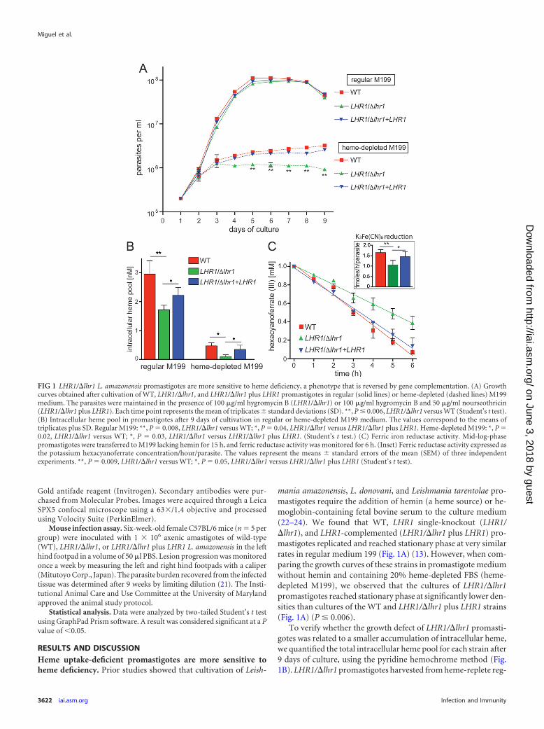
mania amazonensis, L. donovani, and Leishmania tarentolae pro-mastigotes require the addition of hemin (a heme source) or he-moglobin-containing fetal bovine serum to the culture medium(22–24). We found that WT, LHR1 single-knockout (LHR1/�lhr1), and LHR1-complemented (LHR1/�lhr1 plus LHR1) pro-mastigotes replicated and reached stationary phase at very similarrates in regular medium 199 (Fig. 1A) (13). However, when com-paring the growth curves of these strains in promastigote mediumwithout hemin and containing 20% heme-depleted FBS (heme-depleted M199), we observed that the cultures of LHR1/�lhr1promastigotes reached stationary phase at significantly lower den-sities than cultures of the WT and LHR1/�lhr1 plus LHR1 strains(Fig. 1A) (P � 0.006).
To verify whether the growth defect of LHR1/�lhr1 promasti-gotes was related to a smaller accumulation of intracellular heme,we quantified the total intracellular heme pool for each strain after9 days of culture, using the pyridine hemochrome method (Fig.1B). LHR1/�lhr1 promastigotes harvested from heme-replete reg-
FIG 1 LHR1/�lhr1 L. amazonensis promastigotes are more sensitive to heme deficiency, a phenotype that is reversed by gene complementation. (A) Growthcurves obtained after cultivation of WT, LHR1/�lhr1, and LHR1/�lhr1 plus LHR1 promastigotes in regular (solid lines) or heme-depleted (dashed lines) M199medium. The parasites were maintained in the presence of 100 �g/ml hygromycin B (LHR1/�lhr1) or 100 �g/ml hygromycin B and 50 �g/ml nourseothricin(LHR1/�lhr1 plus LHR1). Each time point represents the mean of triplicates � standard deviations (SD). **, P � 0.006, LHR1/�lhr1 versus WT (Student’s t test).(B) Intracellular heme pool in promastigotes after 9 days of cultivation in regular or heme-depleted M199 medium. The values correspond to the means oftriplicates plus SD. Regular M199: **, P � 0.008, LHR1/�lhr1 versus WT; *, P � 0.04, LHR1/�lhr1 versus LHR1/�lhr1 plus LHR1. Heme-depleted M199: *, P �0.02, LHR1/�lhr1 versus WT; *, P � 0.03, LHR1/�lhr1 versus LHR1/�lhr1 plus LHR1. (Student’s t test.) (C) Ferric iron reductase activity. Mid-log-phasepromastigotes were transferred to M199 lacking hemin for 15 h, and ferric reductase activity was monitored for 6 h. (Inset) Ferric reductase activity expressed asthe potassium hexacyanoferrate concentration/hour/parasite. The values represent the means � standard errors of the mean (SEM) of three independentexperiments. **, P � 0.009, LHR1/�lhr1 versus WT; *, P � 0.05, LHR1/�lhr1 versus LHR1/�lhr1 plus LHR1 (Student’s t test).
Miguel et al.
3622 iai.asm.org Infection and Immunity
on June 3, 2018 by guesthttp://iai.asm
.org/D
ownloaded from

ular medium showed a significant decrease in intracellular hemecontent compared to WT (�50% reduction) and LHR1/�lhr1plus LHR1 (�25% reduction) parasites. Cultivation in heme-de-pleted M199 led to markedly reduced levels of intracellular hemein all strains compared to regular medium, but the levels of hemedetected were significantly lower in LHR1/�lhr1 parasites (Fig.1B). This result emphasizes the importance of heme uptake andmaintenance of an intracellular pool for the optimal in vitrogrowth of L. amazonensis promastigotes. The small intracellularheme pools resulting from the reduced heme availability in heme-depleted M199 (Fig. 2B) are likely responsible for the decreasedgrowth rates of WT and LHR1/�lhr1 plus LHR1 strains comparedto regular M199 (Fig. 1A). However, both strains were still able toreach higher densities than the LHR1/�lhr1 single knockout (Fig.1C), indicating that high levels of LHR1 expression allow the par-asites to acquire sufficient heme from the trace amounts remain-ing in heme-depleted culture medium.
L. amazonensis expresses a NADPH-dependent ferric reduc-
tase, LFR1 (16). Since metalloreductases are heme-dependent en-zymes (25), we measured spectrophotometrically the plasmamembrane ferric reductase activity of L. amazonensis promasti-gotes as a reporter for heme availability within the parasites. Re-ductase activity was monitored after promastigote incubation inheme-depleted M199 for 15 h (Fig. 1C). The enzymatic activitiesdetected for WT, LHR1/�lhr1, and LHR1/�lhr1 plus LHR1 pro-mastigotes were 1.65 � 0.14, 1.05 � 0.23, and 1.46 � 0.24 fmol ofhexacyanoferrate/hour/parasite, respectively (P � 0.009; LHR1/�lhr1 versus WT) (Fig. 1C, inset). These results independentlydemonstrate that the lack of one copy of the heme transporterLHR1 in the LHR1/�lhr1 single-knockout strain reduces the avail-ability of intracellular heme that is necessary for assembly of activehemoproteins. Notably, LHR1 add-back complemented theLHR1/�lhr1 single-knockout strain, restoring the ferric reductaseactivity to levels similar to those seen in WT parasites (Fig. 1C).This result emphasizes the interplay between heme and iron up-take in L. amazonensis that is required for parasite infectivity, since
FIG 2 LHR1 deficiency does not affect the generation of axenic amastigotes. Amastigotes of L. amazonensis were obtained axenically (see Materials and Methods)and cultivated for 5 days. (A) The percentage of round aflagellated forms was determined microscopically, and the morphology was analyzed by scanning electronmicroscopy. The values represent the means plus SD of triplicate determinations. Scale bar � 1.0 �m. (B) Transcripts of Amastin-like 1 and 2 (LmxM.08.0760and LmxM.08.0770) and P27 (LmxM.28.0980) were analyzed by quantitative PCR (qPCR) and normalized to the levels of the ubiquitin hydrolase mRNA. Thevalues represent the means and SD of triplicate determinations and are representative of three independent experiments. There were no significant differencesbetween the LHR1/�lhr1 versus WT or LHR1/�lhr1 versus LHR1/�lhr1 plus LHR1 strains (P 0.05; Student’s t test).
Leishmania Heme Transport Is Required for Virulence
October 2013 Volume 81 Number 10 iai.asm.org 3623
on June 3, 2018 by guesthttp://iai.asm
.org/D
ownloaded from

LFR1 is required for generating ferrous iron, the substrate for theiron transporter LIT1, which is required for intracellular replica-tion (16).
Differentiation into infective stages is not affected by dele-tion of one copy of the LHR1 gene. To test whether partial dele-tion of LHR1 interferes with Leishmania differentiation intoinfective stages, we investigated the generation of metacyclic pro-mastigotes by recovering metacyclic forms from stationary-phasecultures using the specific antibody m3A.1. This monoclonal an-tibody specifically agglutinates L. amazonensis promastigotes,yielding purified metacyclic forms in the supernatant (26). Wild-type, LHR1/�lhr1, and LHR1/�lhr1 plus LHR1 strains generatedsimilar percentages of purified metacyclics after 6 days in culture(data not shown).
The transformation of promastigotes into amastigotes was alsoexamined using the axenic amastigote differentiation protocol(see Materials and Methods). The percentages of round aflagel-lated forms for each strain were determined microscopically, andno differences were observed between wild-type, LHR1/�lhr1,and LHR1/�lhr1 plus LHR1 strains (Fig. 2A), suggesting thattransformation into axenic amastigotes is also not affected by thepartial LHR1 deficiency. This conclusion was reinforced by scan-ning electron microscopy examinations of parasite morphology(Fig. 2A, right) and quantification of mRNA levels of three pro-
teins expressed specifically by L. amazonensis amastigotes: Amas-tin-like 1, Amastin-like 2, and P27 (17) (Fig. 2B).
Collectively, our findings indicate that LHR1/�lhr1 parasitesare able to transform into infective amastigote stages at the samelevel observed for WT and add-back strains. The axenically trans-formed amastigotes from all strains were viable, as indicated bytheir redifferentiation into promastigote stages when cultivated inregular M199 at 26°C for up to 7 days (data not shown).
LHR1 single-knockout L. amazonensis is defective in intra-cellular replication in macrophages. Next, we investigatedwhether deletion of one LHR1 copy affected the parasites’ability toinfect and proliferate in BMM. BMM infections were performedwith axenic amastigotes of each strain, and the numbers of intra-cellular parasites were determined after 1, 24, 48, and 72 h. Asexpected, wild-type amastigotes survived intracellularly and in-creased in numbers over time. In sharp contrast, LHR1/�lhr1 par-asites failed to replicate in BMM, and their numbers graduallydecreased, indicating intracellular death. Complementation withLHR1 in the LHR1/�lhr1 plus LHR1 strain fully rescued intracel-lular survival and partially restored the parasites’ ability to repli-cate in BMM (Fig. 3A, Control).
Because LHR1-single knockout parasites contain smalleramounts of intracellular heme (13), we investigated whether de-livering an excess of exogenous hemoglobin to BMM phagolyso-
FIG 3 LHR1/�lhr1 amastigotes are unable to proliferate in BMM. (A) BMM left untreated (Control) or preincubated with red blood cells (RBC) were infectedwith L. amazonensis axenic amastigotes (multiplicity of infection [MOI] � 5) for 1, 24, 48, and 72 h, followed by quantification of intracellular parasites.Infections using WT, LHR1/�lhr1, and LHR1/�lhr1 plus LHR1 amastigotes are shown. The values represent the means and SD of triplicates and are represen-tative of three independent experiments. **, P 0.001; *, P 0.01 (Student’s t test). (B) Immunofluorescence confocal images of BMM infected for 72 h (toprow, control infection; bottom row, BMM preexposed to RBC and then infected with L. amazonensis). Red, anti-Leishmania antibodies; blue, DAPI-stainednuclei; green, lysosomes and PV membranes stained with anti-Lamp-1. The short arrows point to expanded PVs containing replicating parasites; the long arrowsindicate single LHR1/�lhr1 amastigotes in small PVs. Scale bar � 8 �m.
Miguel et al.
3624 iai.asm.org Infection and Immunity
on June 3, 2018 by guesthttp://iai.asm
.org/D
ownloaded from

somal compartments was able to rescue their intracellular growthdefect. Prior studies demonstrated that phagosomes containingphagocytosed particles merge very efficiently with L. amazonensis PVs(27). To this end, BMM were allowed to phagocytose oxidized RBCand were subsequently infected with axenic amastigotes. Quantifica-tion of intracellular parasites over time showed that phagocytosedRBC entirely rescued the growth defect of LHR1/�lhr1 parasites,which replicated in BMM at the same levels observed with the wild-type and LHR1/�lhr1 plus LHR1 strains (Fig. 3A, RBC).
Immunofluorescence experiments revealed that L. amazonen-sis PVs in BMM infected with the LHR1 single-knockout strainwere small and contained single parasites after 72 h. This pheno-type was fully reversed in BMM preloaded with RBC, whichshowed expanded Lamp-1-positive PVs containing multiple rep-licating parasites, as seen for infections with wild-type and LHR1/�lhr1 plus LHR1 strains (Fig. 3B). These results suggest that theheme-enriched environment generated within L. amazonensisPVs through the phagocytosis of oxidized RBC is sufficient tosustain the growth of heme uptake-deficient parasites, possiblythrough alternative low-affinity mechanisms of uptake. This find-ing has important implications for the pathogenesis of Leishmaniainfections, since it indicates that the vigorous process of eryth-rophagocytosis characteristic of macrophages can confer a sur-vival advantage on these parasites. Billions of senescent RBC arecleared daily from circulation by macrophages from the reticu-loendothelial system and degraded in phagolysosomes, wherebreakdown of hemoglobin and cytosolic release of heme occur(28, 29). Free heme within the phagolysosome is then translocatedinto the macrophage cytosol by the heme transporter HRG1(Heme-Responsive Gene 1) (19). The presence of HRG1 on thephagolysosome membrane emphasizes the need for LHR1, theLeishmania-encoded heme transporter, to allow parasites repli-cating within PVs to successfully compete with the host for heme.This intracellular competition for the available heme raises in-triguing questions, such as the possibility that mutations interfer-ing with HRG1 function (19) may affect host susceptibility toLeishmania infections.
The intracellular-growth defect of LHR1/�lhr1 parasites is inagreement with a study that described the inability of L. ama-zonensis axenic amastigotes to proliferate in iron-depleted me-dium, an effect that is reversed after addition of hemoglobin (12).The typhoid fever agent Salmonella enterica exploits the he-mophagocytic ability of BMM to survive during chronic in vivoinfections (30), and heme was also shown to function as a sourceof iron for additional bacterial pathogens residing within macro-phage phagosomes, such as Mycobacterium haemophilum and Lis-teria monocytogenes (31, 32).
LHR1 single-knockout L. amazonensis is defective in cutane-ous-lesion formation in mice. To determine whether deletion ofone LHR1 copy affected the in vivo infectivity of L. amazonensis,axenic amastigotes were injected into the left hind footpads ofC57BL/6 mice, and the development of cutaneous lesions was fol-lowed for 9 weeks. Unlike the wild-type strain (which caused le-sions of 2.7 to 3.3 mm on average during this period), LHR1/�lhr1parasites were not able to induce detectable cutaneous lesions.Importantly, lesion development was restored in the comple-mented LHR1/�lhr1 plus LHR1 strain, albeit at a lower level thanwas observed with wild-type parasites (Fig. 4A). This was not un-expected, since a lack of robust complementation has been fre-quently observed in transgenic Leishmania (33–35). Nine weeks
after infection, the mice were sacrificed, and footpad tissue wascollected for parasite quantification by limiting dilution. The par-asite burden in animals infected with LHR1/�lhr1 parasites waslower than that observed for mice infected with the wild type andthe complemented strain (Fig. 4B). Thus, loss of only one LHR1copy is sufficient to abolish infectivity in L. amazonensis, indicat-ing that a threshold level of the protein must be maintained toallow the parasites to scavenge sufficient heme while replicatingwithin macrophage PVs.
Conclusion. In this study, we characterized in detail the bio-logical properties of a strain of L. amazonensis lacking one copy ofLHR1, which encodes the first heme transporter to be identified intrypanosomatid parasites. We found that a full dose of the proteinis required for promastigote replication under heme deprivationand for assembly of active ferric iron reductase, a heme-contain-ing protein that is required for L. amazonensis infectivity (16).Importantly, we also found that loss of only one LHR1 copy ren-ders the parasites incapable of replicating in macrophages andavirulent for mice. Several attempts to delete the second LHR1allele were unsuccessful, indicating that LHR1 is essential for L.amazonensis viability and a possible useful target for the develop-ment of novel antileishmanial drugs.
Remarkably, our results showed that delivering hemoglobin tothe macrophage PV in the form of phagocytosed RBC fully re-stores intracellular growth of the single-knockout strain. Thisfinding is particularly relevant for our understanding of infectionscaused by the visceralizing species of Leishmania, which cause verysevere disease by replicating in spleen, liver, and bone marrowmacrophages (36). These cells are actively engaged in the recyclingof iron through the phagocytosis and degradation of senescentRBC and therefore have facilitated access to heme (37, 38). Hu-man patients infected with L. amazonensis (39) present a broadspectrum of clinical disease, suggesting that conditions within thehost, which may include variations in the interplay between par-asite and host heme transporters, can influence the course of thedisease. In future studies, it will be important to determine the roleof LHR1-mediated heme acquisition in additional species ofLeishmania and whether potential functional differences in the
FIG 4 LHR1/�lhr1 amastigotes are defective in cutaneous-lesion develop-ment, a phenotype reversed by gene complementation. C57BL/6 female micewere inoculated with 1 � 106 amastigotes of WT, LHR1/�lhr1, or LHR1/�lhr1plus LHR1 L. amazonensis in the left hind footpad. (A) Lesion progression wasrecorded weekly. The values represent the means � SD of 5 mice. (B) Theparasite load in the footpad tissues was quantified 9 weeks after infection (n �5). **, P � 0.007; *, P � 0.011 (Student’s t test).
Leishmania Heme Transport Is Required for Virulence
October 2013 Volume 81 Number 10 iai.asm.org 3625
on June 3, 2018 by guesthttp://iai.asm
.org/D
ownloaded from
transporter will represent an opportunity to investigate its role asa determinant of disease severity in leishmaniasis.
ACKNOWLEDGMENTS
We are grateful to Chau Hyunh for the LHR1 constructs. We also thank IqbalHamza for advice, Cecilia Fernandes for technical assistance with confocalmicroscopy, Juliana Perrone for assistance with animal experiments, and TimMaugel from the Laboratory for Biological Ultrastructure, University ofMaryland, for assistance with scanning electron microscopy.
This work was supported by NIH grant RO1 AI067979 toN.W.A. D.C.M. is the recipient of a Postdoctoral Scholarship from theBrazilian National Council for Scientific and Technological Develop-ment (CNPq/Ciência sem Fronteiras-202079/2011-2).
REFERENCES1. Murray HW, Berman JD, Davies CR, Saravia NG. 2005. Advances in
leishmaniasis. Lancet 366:1561–1577.2. Croft SL, Olliaro P. 2011. Leishmaniasis chemotherapy: challenges and
opportunities. Clin. Microbiol. Infect. 17:1478 –1483.3. Sacks D, Kamhawi S. 2001. Molecular aspects of parasite-vector and
vector-host interactions in leishmaniasis. Annu. Rev. Microbiol. 55:453–483.
4. Ueno N, Wilson ME. 2012. Receptor-mediated phagocytosis of Leishma-nia: implications for intracellular survival. Trends Parasitol. 28:335–344.
5. Naderer T, McConville MJ. 2008. The Leishmania-macrophage interac-tion: a metabolic perspective. Cell Microbiol. 10:301–308.
6. Hamza I, Dailey HA. 2012. One ring to rule them all: trafficking of hemeand heme synthesis intermediates in the metazoans. Biochim. Biophys.Acta 1823:1617–1632.
7. Anzaldi LL, Skaar EP. 2010. Overcoming the heme paradox: heme tox-icity and tolerance in bacterial pathogens. Infect. Immun. 78:4977– 4989.
8. Korený L, Oborník M, Lukeš J. 2013. Make it, take it, or leave it: hememetabolism of parasites. Plos Pathog. 9:e1003088. doi:10.1371/journal.ppat.1003088.
9. Korený L, Lukes J, Oborník M. 2010. Evolution of the haem syntheticpathway in kinetoplastid flagellates: an essential pathway that is not essen-tial after all? Int. J. Parasitol. 40:149 –156.
10. Tripodi KE, Menendez Bravo SM, Cricco JA. 2011. Role of heme andheme-proteins in trypanosomatid essential metabolic pathways. EnzymeRes. 2011:873230. doi:10.4061/2011/873230.
11. Sengupta S, Tripathi J, Tandon R, Raje M, Roy RP, Basu SK, Mukho-padhyay A. 1999. Hemoglobin endocytosis in Leishmania is mediatedthrough a 46-kDa protein located in the flagellar pocket. J. Biol. Chem.274:2758 –2765.
12. Carvalho S, Cruz T, Santarém N, Castro H, Costa V, Tomás AM. 2009.Heme as a source of iron to Leishmania infantum amastigotes. Acta Trop.109:131–135.
13. Huynh C, Yuan X, Miguel DC, Renberg RL, Protchenko O, Hamza I,Andrews NW. 2012. Heme uptake by Leishmania amazonensis is medi-ated by the transmembrane protein LHR1. PLoS Pathog. 8:e1002795. doi:10.1371/journal.ppat.1002795.
14. Zhu Y, Hon T, Ye W, Zhang L. 2002. Heme deficiency interferes with theRas-mitogen-activated protein kinase signaling pathway and expressionof a subset of neuronal genes. Cell Growth Differ. 13:431– 439.
15. Paul KG, Theorell H, Akeson A. 1953. The molar light absorption ofpyridine ferroprotoporphyrin (pyridine haemochromogen). Acta Chem.Scand. 7:1284 –1287.
16. Flannery AR, Huynh C, Mittra B, Mortara RA, Andrews NW. 2011.LFR1 ferric iron reductase of Leishmania amazonensis is essential for thegeneration of infective parasite forms. J. Biol. Chem. 286:23266 –23279.
17. Mittra B, Cortez M, Haydock A, Ramasamy G, Myler PJ, Andrews NW.2013. Iron uptake controls the generation of Leishmania infective formsthrough regulation of ROS levels. J. Exp. Med. 210:401– 416.
18. Becker SM, Delamarre L, Mellman I, Andrews NW. 2009. Differentialrole of the Ca(2�) sensor synaptotagmin VII in macrophages and den-dritic cells. Immunobiology 214:495–505.
19. White C, Yuan X, Schmidt PJ, Bresciani E, Samuel TK, Campagna D,Hall C, Bishop K, Calicchio ML, Lapierre A, Ward DM, Liu P, FlemingMD, Hamza I. 2013. HRG1 is essential for heme transport from the
phagolysosome of macrophages during erythrophagocytosis. Cell Metab.17:261–270.
20. Cortez M, Huynh C, Fernandes MC, Kennedy KA, Aderem A, AndrewsNW. 2011. Leishmania promotes its own virulence by inducing expressionof the host immune inhibitory ligand CD200. Cell Host Microbe 9:463–471.
21. Titus RG, Marchand M, Boon T, Louis JA. 1985. A limiting dilutionassay for quantifying Leishmania major in tissues of infected mice. ParasiteImmunol. 7:545–555.
22. Chang CS, Chang KP. 1985. Heme requirement and acquisition by ex-tracellular and intracellular stages of Leishmania mexicana amazonensis.Mol. Biochem. Parasitol. 16:267–276.
23. Pal JK, Joshi-Purandare M. 2001. Dose-dependent differential effect ofhemin on protein synthesis and cell proliferation in Leishmania donovanipromastigotes cultured in vitro. J. Biosci. 26:225–231.
24. Fritsche C, Sitz M, Weiland N, Breitling R, Pohl HD. 2007. Character-ization of the growth behavior of Leishmania tarentolae: a new expressionsystem for recombinant proteins. J. Basic Microbiol. 47:384 –393.
25. Protchenko O, Shakoury-Elizeh M, Keane P, Storey J, Androphy R,Philpott CC. 2008. Role of PUG1 in inducible porphyrin and heme trans-port in Saccharomyces cerevisiae. Eukaryot. Cell 7:859 – 871.
26. Pinto-da-Silva LH, Fampa P, Soares DC, Oliveira SM, Souto-Padrón T,Saraiva EM. 2005. The 3A1-La monoclonal antibody reveals key featuresof Leishmania (L) amazonensis metacyclic promastigotes and inhibits pro-cyclics attachment to the sand fly midgut. Int. J. Parasitol. 35:757–764.
27. Veras P, de Chastellier C, Rabinovitch M. 1992. Transfer of zymosan(yeast cell walls) to the parasitophorous vacuoles of macrophages infectedwith Leishmania amazonensis. J. Exp. Med. 176:639 – 646.
28. Bratosin D, Mazurier J, Tissier JP, Estaquier J, Huart JJ, Ameisen JC,Aminoff D, Montreuil J. 1998. Cellular and molecular mechanisms ofsenescent erythrocyte phagocytosis by macrophages. A review. Biochimie.80:173–195.
29. Delaby C, Rondeau C, Pouzet C, Willemetz A, Pilard N, Desjardins M,Canonne-Hergaux F. 2012. Subcellular localization of iron and hememetabolism related proteins at early stages of erythrophagocytosis. PLoSOne 7:e42199. doi:10.1371/journal.pone.0042199.
30. Nix RN, Altschuler SE, Henson PM, Detweiler CS. 2007. Hemophago-cytic macrophages harbor Salmonella enterica during persistent infection.PLoS Pathog. 3:e193. doi:10.1371/journalppat.0030193.
31. Saubolle MA, Kiehn TE, White MH, Rudinsky MF, Armstrong D. 1996.Mycobacterium haemophilum: microbiology and expanding clinical andgeographic spectra of disease in humans. Clin. Microbiol. Rev. 9:435– 447.
32. Jin B, Newton SMC, Shao Y, Jiang X, Charbit A, Klebba PE. 2006. Ironacquisition systems for ferric hydroxamates, haemin and haemoglobin inListeria monocytogenes. Mol. Microbiol. 59:1185–1198.
33. Gaur U, Showalter M, Hickerson S, Dalvi R, Turco SJ, Wilson ME,Beverley SM. 2009. Leishmania donovani lacking the Golgi GDP-Mantransporter LPG2 exhibit attenuated virulence in mammalian hosts. Exp.Parasitol. 122:182–191.
34. da Silva MFL, Zampieri RA, Muxel SM, Beverley SM, Floeter-WinterLM. 2012. Leishmania amazonensis arginase compartmentalization in theglycosome is important for parasite infectivity. PLoS One 7:e34022. doi:10.1371/journal.pone.0034022.
35. Oyola SO, Evans KJ, Smith TK, Smith BA, Hilley JD, Mottram JC, KayePM, Smith DF. 2012. Functional analysis of Leishmania cyclopropanefatty acid synthetase. PLoS One 7:e51300. doi:10.1371/journal.pone.0051300.
36. Wilson ME, Jeronimo SMB, Pearson RD. 2005. Immunopathogenesis ofinfection with the visceralizing Leishmania species. Microb. Pathog. 38:147–160.
37. Chowdhury KD, Sen G, Biswas T. 2010. Regulatory role of nitric oxide inthe reduced survival of erythrocytes in visceral leishmaniasis. Biochim.Biophys. Acta 1800:964 –976.
38. Samanta S, Ghoshal A, Bhattacharya K, Saha B, Walden P, Mandal C.2012. Sialoglycosylation of RBC in visceral leishmaniasis leads to en-hanced oxidative stress, calpain-induced fragmentation of spectrin andhemolysis. PLoS One 7:e42361. doi:10.1371/journal.pone.0042361.
39. Barral A, Pedral-Sampaio D, Grimaldi Júnior Momen GH, McMahon-Pratt D, Ribeiro de Jesus A, Almeida R, Badaro R, Barral-Netto M,Carvalho EM. 1991. Leishmaniasis in Bahia, Brazil: evidence that Leish-mania amazonensis produces a wide spectrum of clinical disease. Am. J.Trop. Med. Hyg. 44:536 –546.
Miguel et al.
3626 iai.asm.org Infection and Immunity
on June 3, 2018 by guesthttp://iai.asm
.org/D
ownloaded from


![Carrier-mediated Transport of Oligopeptides in the Human ......Uptake Experiments. Uptake of [‘4C]Gly-Saror cefadroxil by the cul tured cells was examined at 37 Cby the use](https://static.fdocuments.in/doc/165x107/60851c2847e1ab0ad9623c6c/carrier-mediated-transport-of-oligopeptides-in-the-human-uptake-experiments.jpg)


![Transporters for nitrogenous compounds in plants · [49, 50]. In plants, biphasic uptake kinetics were determined consisting of a saturable carrier- mediated system operating at low](https://static.fdocuments.in/doc/165x107/5fd2f80ef613845a9a43af07/transporters-for-nitrogenous-compounds-in-49-50-in-plants-biphasic-uptake-kinetics.jpg)