Hematopoietic bone marrow cells participate in endothelial ...
13
Hematopoietic bone marrow cells participate in endothelial, but not epithelial or mesenchymal cell renewal in adult rats Kathrin I. Odörfer a , Monika Egerbacher b , Nina J. Unger c , Karin Weber c , Angelika Jamnig d , Günter Lepperdinger d , Miriam Kleiter e , Eric P. Sandgren f , Reinhold G. Erben a, * a Institute of Physiology, Pathophysiology and Biophysics, Department of Biomedical Sciences, University of Veterinary Medicine, Vienna, Austria b Institute of Anatomy, Histology and Embryology, Department of Pathobiology, University of Veterinary Medicine, Vienna, Austria c Institute of Physiology, Physiological Chemistry and Animal Nutrition, Ludwig Maximilians University, Munich, Germany d Institute for Biomedical Aging Research, Austrian Academy of Sciences, Innsbruck, Austria e Medical Clinic for Internal Medicine and Infectious Diseases, University Veterinary Medicine, Vienna, Austria f Department of Pathobiological Sciences, School of Veterinary Medicine, University of Wisconsin-Madison, Madison, WI, USA Received: July 31, 2010; Accepted: November 15, 2010 Abstract The extent to which bone marrow (BM) contributes to physiological cell renewal is still controversial. Using the marker human placental alkaline phosphatase (ALPP) which can readily be detected in paraffin and plastic sections by histochemistry or immunohistochemistry, and in ultrathin sections by electron microscopy after pre-embedding staining, we examined the role of endogenous BM in physiological cell renewal by analysing tissues from lethally irradiated wild-type inbred Fischer 344 (F344) rats transplanted (BMT) with unfractionated BM from ALPP-transgenic F344 rats ubiquitously expressing the marker. Histochemical, immunohistochemical and immunoelectron microscopic analysis showed that the proportion of ALPP capillary endothelial cells (EC) profoundly increased from 1 until 6 months after BMT in all organs except brain and adrenal medulla. In contrast, pericytes and EC in large blood vessels were ALPP – . Epithelial cells in kidney, liver, pancreas, intestine and brain were recipient-derived at all time-points. Similarly, osteoblasts, chondrocytes, striated muscle and smooth muscle cells were exclusively of recipient origin. The lack of mesenchymal BM-derived cells in peripheral tissues prompted us to examine whether BMT resulted in engraftment of mesenchymal precursors. Four weeks after BMT, all haematopoietic BM cells were of donor origin by flow cytometric analysis, whereas isolation of BM mesenchymal stem cells (MSC) failed to show engraftment of donor MSC. In conclusion, our data show that BM is an important source of physiological renewal of EC in adult rats, but raise doubt whether reconstituted irradiated rats are an apt model for BM-derived regeneration of mesenchymal cells in peripheral tissues. Keywords: bone marrow transplantation • human placental alkaline phosphatase • endothelial cells • stem cells • physiological cell renewal J. Cell. Mol. Med. Vol 15, No 10, 2011 pp. 2232-2244 © 2011 The Authors Journal of Cellular and Molecular Medicine © 2011 Foundation for Cellular and Molecular Medicine/Blackwell Publishing Ltd doi: 10.1111/j.1582-4934.2010.01216.x Introduction Insights into the mechanisms of physiological cell renewal in adult organs are of fundamental importance for medicine and biology. Earlier reports indicate that bone marrow (BM)-derived precursor cells play a role in cell renewal and repair in many adult organs. For example, female patients receiving sex-mismatched bone marrow transplantation (BMT) show integration of male donor cells into brain as neurons, into heart as cardiomyocytes, into kidney as tubular epithelial cells, into liver as hepatocytes and cholangiocytes, and into lung as epithelial and endothelial cells (EC; reviewed in [1]). Similarly, irradiated mice reconstituted with green fluorescent protein (GFP) or lacZ-labelled BM show partici- pation of BM-derived cells in the renewal of endothelial [2], epithelial [3, 4] and mesenchymal [5] cells in various organs. In addition, it was reported that mobilization of endogenous bone marrow cells (BMC) after acute myocardial infarction contributes to regeneration of cardiac muscle [6]. Collectively, these findings have lead to the concept that circulating BM-derived precursor cells participate in physiological cell renewal and repair in many adult organs. *Correspondence to: Reinhold G. ERBEN, M.D., D.V.M., Institute of Physiology, Pathophysiology and Biophysics, Department of Biomedical Sciences, University of Veterinary Medicine, Veterinaerplatz 1, 1210 Vienna, Austria. Tel.: 43-1-250 77 4550 Fax 43-1-250 77 4599 E-mail: [email protected] brought to you by CORE View metadata, citation and similar papers at core.ac.uk provided by PubMed Central
Transcript of Hematopoietic bone marrow cells participate in endothelial ...

Hematopoietic bone marrow cells participate in endothelial,
but not epithelial or mesenchymal cell renewal in adult rats
Kathrin I. Odörfer a, Monika Egerbacher b, Nina J. Unger c, Karin Weber c, Angelika Jamnig d, Günter Lepperdinger d, Miriam Kleiter e, Eric P. Sandgren f, Reinhold G. Erben a, *
a Institute of Physiology, Pathophysiology and Biophysics, Department of Biomedical Sciences, University of Veterinary Medicine, Vienna, Austria
b Institute of Anatomy, Histology and Embryology, Department of Pathobiology, University of Veterinary Medicine, Vienna, Austriac Institute of Physiology, Physiological Chemistry and Animal Nutrition, Ludwig Maximilians University, Munich, Germany
d Institute for Biomedical Aging Research, Austrian Academy of Sciences, Innsbruck, Austriae Medical Clinic for Internal Medicine and Infectious Diseases, University Veterinary Medicine, Vienna, Austria
f Department of Pathobiological Sciences, School of Veterinary Medicine, University of Wisconsin-Madison, Madison, WI, USA
Received: July 31, 2010; Accepted: November 15, 2010
Abstract
The extent to which bone marrow (BM) contributes to physiological cell renewal is still controversial. Using the marker human placentalalkaline phosphatase (ALPP) which can readily be detected in paraffin and plastic sections by histochemistry or immunohistochemistry,and in ultrathin sections by electron microscopy after pre-embedding staining, we examined the role of endogenous BM in physiologicalcell renewal by analysing tissues from lethally irradiated wild-type inbred Fischer 344 (F344) rats transplanted (BMT) with unfractionatedBM from ALPP-transgenic F344 rats ubiquitously expressing the marker. Histochemical, immunohistochemical and immunoelectronmicroscopic analysis showed that the proportion of ALPP� capillary endothelial cells (EC) profoundly increased from 1 until 6 monthsafter BMT in all organs except brain and adrenal medulla. In contrast, pericytes and EC in large blood vessels were ALPP–. Epithelial cellsin kidney, liver, pancreas, intestine and brain were recipient-derived at all time-points. Similarly, osteoblasts, chondrocytes, striated muscleand smooth muscle cells were exclusively of recipient origin. The lack of mesenchymal BM-derived cells in peripheral tissues promptedus to examine whether BMT resulted in engraftment of mesenchymal precursors. Four weeks after BMT, all haematopoietic BM cells wereof donor origin by flow cytometric analysis, whereas isolation of BM mesenchymal stem cells (MSC) failed to show engraftment of donorMSC. In conclusion, our data show that BM is an important source of physiological renewal of EC in adult rats, but raise doubt whetherreconstituted irradiated rats are an apt model for BM-derived regeneration of mesenchymal cells in peripheral tissues.
Keywords: bone marrow transplantation • human placental alkaline phosphatase • endothelial cells • stem cells • physiological cell renewal
J. Cell. Mol. Med. Vol 15, No 10, 2011 pp. 2232-2244
© 2011 The AuthorsJournal of Cellular and Molecular Medicine © 2011 Foundation for Cellular and Molecular Medicine/Blackwell Publishing Ltd
doi:10.1111/j.1582-4934.2010.01216.x
Introduction
Insights into the mechanisms of physiological cell renewal in adultorgans are of fundamental importance for medicine and biology.Earlier reports indicate that bone marrow (BM)-derived precursorcells play a role in cell renewal and repair in many adult organs.For example, female patients receiving sex-mismatched bone
marrow transplantation (BMT) show integration of male donorcells into brain as neurons, into heart as cardiomyocytes, into kidney as tubular epithelial cells, into liver as hepatocytes andcholangiocytes, and into lung as epithelial and endothelial cells(EC; reviewed in [1]). Similarly, irradiated mice reconstituted withgreen fluorescent protein (GFP) or lacZ-labelled BM show partici-pation of BM-derived cells in the renewal of endothelial [2], epithelial[3, 4] and mesenchymal [5] cells in various organs. In addition, itwas reported that mobilization of endogenous bone marrow cells(BMC) after acute myocardial infarction contributes to regenerationof cardiac muscle [6]. Collectively, these findings have lead to theconcept that circulating BM-derived precursor cells participate inphysiological cell renewal and repair in many adult organs.
*Correspondence to: Reinhold G. ERBEN, M.D., D.V.M., Institute of Physiology, Pathophysiology and Biophysics, Department of Biomedical Sciences, University of Veterinary Medicine,Veterinaerplatz 1, 1210 Vienna, Austria.Tel.: �43-1-250 77 4550Fax �43-1-250 77 4599E-mail: [email protected]
brought to you by COREView metadata, citation and similar papers at core.ac.uk
provided by PubMed Central
J. Cell. Mol. Med. Vol 15, No 10, 2011
2233© 2011 The AuthorsJournal of Cellular and Molecular Medicine © 2011 Foundation for Cellular and Molecular Medicine/Blackwell Publishing Ltd
Furthermore, it was suggested that haematopoietic stem cells maytrans-differentiate into epithelial and also mesenchymal lineagecells [4, 7–9]. However, these interpretations have been ques-tioned on the ground of data indicating methodological problemsinvolved in some of these studies [10–12]. Therefore, the role ofBM in physiological cell renewal is still a matter of controversy.
Recent work from our laboratory has shown that human pla-cental alkaline phosphatase (ALPP) is a highly suitable markerenzyme for histological tracking of genetically labelled cells in alltissues, including hard tissues. In contrast to endogenous heat-labile alkaline phosphatases, ALPP is heat-stable and therefore itsenzymatic activity is retained after heat pre-treatment of paraffinand methylmethacrylate sections [13]. As a consequence, ALPP-labelled cells are easily detectable histologically in the total absenceof background staining. To examine the role of BM in physiologicalcell renewal in various organs, we lethally irradiated 3-month-oldwild-type (wt) Fischer 344 (F344) rats, and reconstituted them withunfractionated BM from R26-ALPP transgenic (ALPP-tg) sex-matched F344 donors. R26-ALPP-tg rats express ALPP in a ubiq-uitous and stable fashion under the R26 promoter, which is a 0.8kb fragment of the ROSA �geo 26 promoter sequence [13–15].ALPP is mainly expressed in the cell membrane, irrespective of thecell type analysed [13, 16], and is developmentally neutral in trans-genic rats and mice [14]. Graft-versus-host reactions can be ruledout a priori in this co-isogeneic BMT model, because F344 rats arean inbred strain. In this sequential study, the reconstituted ratswere followed over a 6-month period after BMT.
Materials and Methods
Animals
All experimental procedures were conducted in compliance with prevailinganimal welfare regulations. Hemizygous male or female R26-F344 ALPP-tgrats were mated with wt F344 rats, and the resulting wt and hemizygous tgoffspring were genotyped as described [14]. Rats were housed in pairs at24�C and a 12 hrs/12 hrs light/dark cycle with free access to tap water andcommercial rat diets (Altromin, Lage, and Ssniff, Soest, Germany).
Lethal irradiation and bone marrow transplantation
Three-month-old wt F344 rats were lethally irradiated with a single dose of8.5 Gy using a cobalt-60 irradiator (Eldorado, Atomic Energy of Canada,Ottawa, Canada), or with a single dose of 8.0 or 9.0 Gy using a linear accel-erator (Siemens Primus, Munich, Germany). Four hours after irradiation,rats were intravenously injected with 4 � 106 unfractionated BMC isolatedfrom sex-matched ALPP-tg co-isogeneic F344 donors. To rule out unsuc-cessful engraftment, injection of freshly prepared tg BMC was repeated 24 hrs after irradiation. For the time course study, groups of four to six ratseach were killed 1, 2, 4 and 6 months after BMT through exsanguinationfrom the abdominal aorta under ketamine/xylazine anaesthesia. For mes-enchymal stem cells (MSC) isolation experiments, animals were killed 4 weeks after BMT.
Flow cytometric detection of ALPP
To determine the degree of chimerism in haematopoietic BMC after BMT,unfractionated BMC were harvested and analysed by fluorescence-activatedcell sorting (FACS) as described [16], using a monoclonal anti-ALPP antibody(Chemicon, Temecula, CA, USA) and rat-adsorbed, fluorescein isothiocyanate(FITC)-labelled goat antimouse IgG antibody (Sigma-Aldrich, Deisenhofen,Germany). The standard curve for determination of the degree of chimerismwas obtained by mixing wt BMC with BMC from ALPP-tg rats at variousknown ratios.
ALPP histology and detection
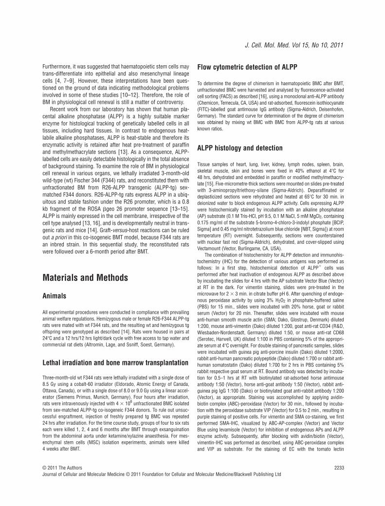
Tissue samples of heart, lung, liver, kidney, lymph nodes, spleen, brain,skeletal muscle, skin and bones were fixed in 40% ethanol at 4�C for 48 hrs, dehydrated and embedded in paraffin or modified methylmethacry-late [15]. Five-micrometre-thick sections were mounted on slides pre-treatedwith 3-aminopropyltriethoxy-silane (Sigma-Aldrich). Deparaffinated ordeplasticized sections were rehydrated and heated at 65�C for 30 min. indeionized water to block endogenous ALPP activity. Cells expressing ALPPwere histochemically stained by incubation with an alkaline phosphatase(AP) substrate (0.1 M Tris-HCl, pH 9.5, 0.1 M NaCl, 5 mM MgCl2, containing0.175 mg/ml of the substrate 5-bromo-4-chloro-3-indolyl phosphate [BCIP,Sigma] and 0.45 mg/ml nitrotetrazolium blue chloride [NBT, Sigma]) at roomtemperature (RT) overnight. Subsequently, sections were counterstainedwith nuclear fast red (Sigma-Aldrich), dehydrated, and cover-slipped usingVectamount (Vector, Burlingame, CA, USA).
The combination of histochemistry for ALPP detection and immunohis-tochemistry (IHC) for the detection of various antigens was performed asfollows: In a first step, histochemical detection of ALPP� cells wasperformed after heat inactivation of endogenous ALPP as described aboveby incubating the slides for 4 hrs with the AP substrate Vector Blue (Vector)at RT in the dark. For vimentin staining, slides were pre-treated in themicrowave for 2 � 3 min. in citrate buffer pH 6. After quenching of endoge-nous peroxidase activity by using 3% H2O2 in phosphate-buffered saline(PBS) for 15 min., slides were incubated with 20% horse, goat or rabbitserum (Vector) for 20 min. Thereafter, slides were incubated with mouseanti-human smooth muscle actin (SMA; Dako, Glostrup, Denmark) diluted1:200, mouse anti-vimentin (Dako) diluted 1:200, goat anti-rat CD34 (R&D,Wiesbaden-Nordenstadt, Germany) diluted 1:50, or mouse anti-rat CD68(Serotec, Harwell, UK) diluted 1:100 in PBS containing 5% of the appropri-ate serum at 4�C overnight. For double staining of pancreatic samples, slideswere incubated with guinea pig anti-porcine insulin (Dako) diluted 1:2000,rabbit anti-human pancreatic polypeptide (Dako) diluted 1:700 or rabbit anti-human somatostatin (Dako) diluted 1:700 for 2 hrs in PBS containing 5%rabbit respective goat serum at RT. Bound antibody was detected by incuba-tion for 0.5–1 hrs at RT with biotinylated rat-adsorbed horse antimouseantibody 1:50 (Vector), horse anti-goat antibody 1:50 (Vector), rabbit anti-guinea pig IgG 1:100 (Dako) or biotinylated goat anti-rabbit antibody 1:200(Vector), as appropriate. Staining was accomplished by applying avidin-biotin complex (ABC)-peroxidase (Vector) for 30 min., followed by incuba-tion with the peroxidase substrate VIP (Vector) for 0.5 to 2 min., resulting inpurple staining of positive cells. For vimentin and SMA co-staining, we firstperformed SMA-IHC, visualized by ABC-AP-complex (Vector) and VectorBlue using levamisole (Vector) for inhibition of endogenous APs and ALPPenzyme activity. Subsequently, after blocking with avidin/biotin (Vector),vimentin-IHC was performed as described, using ABC-peroxidase complexand VIP as substrate. For the staining of EC with the tomato lectin

2234 © 2011 The AuthorsJournal of Cellular and Molecular Medicine © 2011 Foundation for Cellular and Molecular Medicine/Blackwell Publishing Ltd
Lycopersicon esculentum, slides were incubated with the biotinylated lectin[Vector; 1:200 in hydroxethyl-piperazine-ethanesulfonic acid (HEPES) bufferfor 2 hrs at RT] after histochemical ALPP staining and blocking of endoge-nous peroxidase. Visualization was performed with the ABC-peroxidase kitas described above. No counterstaining was carried out, and slides werecover-slipped using aqueous gelatine (Merck, Darmstadt, Germany).
ALPP immuno-electron microscopic analysis
For transmission electron microscopy, tissue samples from heart and kid-ney were fixed in 4% paraformaldehyde for 24 hrs at 4�C. Samples werefrozen in liquid nitrogen with Optimal Cutting Temperature (OCT) com-pound (Sakura Finetek, Zoeterwoude, Netherlands), and 45-�m-thick sec-tions were cut using a cryotome (Leica 1800 CM, Bensheim, Germany).Sections were post-fixed in 4% paraformaldehyde for 1 hrs and rinsed in0.1 M phosphate buffer 3 � 10 min. Peroxidase activity was inhibited by3% H2O2 in PBS for 30 min. and non-specific binding was minimized byincubation with 3% normal goat serum containing 1% bovine serum albu-min (BSA) for 60 min. at RT. Incubation with anti-ALPP (Genetex, Irvine,CA, USA), diluted 1:50 in blocking solution, was carried out at 4�Covernight. As a control, in order to visualize EC in general, sections werestained with anti-human von Willebrand factor (vWF, Dako), 1:600, orCD34 1:50. For negative controls, the primary antibody was omitted.Peroxidase-labelled rabbit PowerVision™ (ImmunoVision Technologies,Burlingame, CA, USA) secondary system was employed for antibodydetection with subsequent DAB staining (Sigma-Aldrich). The reaction wasstopped by rinsing with PBS 3 � 10 min. Sections were placed in 0.1 Mphosphate buffer and kept at 4�C before embedding. Post-fixation was per-formed in 1% osmium tetroxide for 2 hrs at RT followed by dehydrationand incubation in propylene oxide, propylene oxide-epon and subsequentembedding in pure epon 812. Thin sections were stained with lead citrate,and were investigated under a transmission electron microscope (Zeiss EM900; Zeiss, Oberkochen, Germany).
Quantification of endothelial cells in myocardium
The percentage of ALPP� capillary EC was quantified in ALPP-stainedsections in seven optical fields per animal at �400 magnification, evenlydistributed over the left ventricular (five fields) and septal (two fields)myocardium. Using a one-phase exponential model, nonlinear regression analyses were performed with Prism 5.03 (GraphPad Software,Inc., San Diego, CA, USA).
MSC isolation, cultivation and staining
MSC were isolated from long bones of wt, ALPP-tg, and BMT rats, usingan isolation protocol described elsewhere [17]. After 24 hrs, the non-adherent cell fraction was removed by washing twice with D-PBS(Invitrogen, Paisley, UK). After the primary culture had reached confluence,cells were washed twice with D-PBS, and subsequently treated with 0.05%trypsin / 1 mM ethylenediaminetetraacetic acid (EDTA) (Invitrogen) for 5 min. at 37�C. Cells were harvested, washed once in MEM and furtherexpanded. For histochemical staining of cultivated MSC, MSC were seeded at100,000/cm2 in culture flasks or at 1300/cm2 in 12-well plates and cultivatedfor 3 to 7 days until confluency. After fixation with ice-cold acetone-methanol (30/70), plates were washed several times with PBS and endoge-
nous APs were inactivated at 65�C for 30 min. Histochemical staining forheat-stable ALPP was performed with the AP substrate BCIP/NBT asdescribed above. Each cell preparation was stained at least in triplicates,MSC isolated from ALPP-tg and wt donors served as controls.
DNA isolation and Southern blot analysis of cultivated MSC
For DNA isolation, cells were covered with 100 mM Tris-HCl, pH 8.5, 5 mMEDTA, 0.2% SDS, 200 mM NaCl, 100 �g/ml proteinase K and incubated at55�C for 2 hrs. Before phenol/chloroform/iso-amylalcohol (PCI) extraction,debris was removed by centrifugation at 10,000 � g. PCI extraction wasrepeated at least twice in order to yield a clear aqueous phase and inter-phase. Before alcohol precipitation, the solution was extracted once withchloroform/iso-amylalcohol. Large amounts of precipitated DNA wereremoved using a pipette tip, otherwise the precipitate was centrifuged andthe pellet washed in 70% ethanol. The moist pellet was dissolved in 10 mMTris-HCl pH 7.5, 1 mM EDTA by incubation overnight at 55�C and the solu-tion was stored at 4�C. For Southern analysis, 10 �g of genomic DNA wererestricted with 20 units of endonuclease EcoRI (NEB Biolabs, Ipswitch,MA, USA) together with 1 �g/�l RNAseA (Fermentas, St. Leon-Rot,Germany) in an appropriate buffer at 37�C overnight. The DNA was thenseparated by 1% Tris-acetate-EDTA (TAE)/agarose gel electrophoresis, andthe ethidium bromide-stained gel was photo documented under UV irradi-ation. Before transfer to Biodyne® Plus Nylon membrane (Pall Corporation,Dreieich, Germany) in 10� SSPE, the gel was immersed for 15 min. in0.25 N HCl, followed by 30 min. 0.4 N NaOH/0.6 M NaCl, and a 30 min.incubation in 1.5 M NaCl/0.5 M Tris-HCl, pH 7.5. The transferred DNA wasimmobilized onto the membrane by UV irradiation (0.12 J/cm2;Stratalinker, Stratgene, La Jolla, CA, USA). The dried membrane was pre-hybridized for 2 hrs at 65�C with 5� SSC, 5� Denhardt, 1% SDS. Afterreplacement of buffer, a 33P-labelled probe specific to the transgene insertwas hybridized for 18 hrs at 65�C. The membrane was washed (0.2�
SSPE, 0.1% SDS at 65�C) and exposed for 72 hrs to a Fuji imaging plate(MS30170095), which was subsequently scanned by a Fuji Bas 1800-IIphosphoimager (Fuji, Tokyo, Japan).
Results
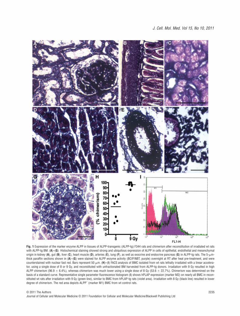
The current study employed two different radiation sources,namely a cobalt-60 source and a linear accelerator. We reportedpreviously that irradiation of F344 rats at a single dose of 8.5 Gyusing a cobalt-60 irradiator followed by transplantation withALPP-tg BM results in full replacement of the haematopoieticcompartment as documented by FACS analysis of BMC [16]. Indose–response experiments with the linear accelerator, we foundin two independent experiments that a single irradiation dose of 9 Gy was necessary to obtain wt F344 rats in which host BMCwere fully replaced by ALPP-tg BMC (Fig. 1H, I).
A prerequisite for our study was that ALPP is ubiquitouslyexpressed in the target tissues to be analysed. Our earlier workhad shown that ALPP is strongly expressed in haematopoieticcells, lung, spleen, lymph nodes and in BM stromal cells such asosteoblasts, osteocytes and chondrocytes in R26-ALPP-tg rats
J. Cell. Mol. Med. Vol 15, No 10, 2011
2235© 2011 The AuthorsJournal of Cellular and Molecular Medicine © 2011 Foundation for Cellular and Molecular Medicine/Blackwell Publishing Ltd
Fig. 1 Expression of the marker enzyme ALPP in tissues of ALPP-transgenic (ALPP-tg) F344 rats and chimerism after reconstitution of irradiated wt ratswith ALPP-tg BM. (A)–(G) Histochemical staining showed strong and ubiquitous expression of ALPP in cells of epithelial, endothelial and mesenchymalorigin in kidney (A), gut (B), liver (C), heart muscle (D), arteries (E), lung (F), as well as exocrine and endocrine pancreas (G) in ALPP-tg rats. The 5-�m-thick paraffin sections shown in (A)–(G) were stained for ALPP enzyme activity (BCIP/NBT, purple) overnight at RT after heat pre-treatment, and werecounterstained with nuclear fast red. Bars represent 50 �m. (H)–(I) FACS analysis of BMC isolated from wt rats lethally irradiated with a linear accelera-tor, using a single dose of 8 or 9 Gy, and reconstituted with unfractionated BM harvested from ALPP-tg donors. Irradiation with 9 Gy resulted in highALPP chimerism (96.9 � 6.4%), whereas chimerism was much lower using a single dose of 8 Gy (53.6 � 22.7%). Chimerism was determined on thebasis of a standard curve. Representative single parameter fluorescence histogram (I) shows hPLAP expression (marker M2) on nearly all BMC in recon-stituted wt rats after irradiation with 9 Gy (green line), similar to BMC from hPLAP-tg rats (violet area). Irradiation with 8 Gy (black line) resulted in lowerdegree of chimerism. The red area depicts ALPP– (marker M1) BMC from wt control rats.
2236 © 2011 The AuthorsJournal of Cellular and Molecular Medicine © 2011 Foundation for Cellular and Molecular Medicine/Blackwell Publishing Ltd
[13, 16]. In the current study, we examined the expression of ALPPin kidney, gut, liver, heart, arteries, lung and pancreas of ALPP-tgrats. We found strong expression of the marker enzyme in endothe-lial, epithelial and mesenchymal cells in all these tissues (Fig. 1A–G).
When we started to examine the occurrence of ALPP� cells invarious organs of irradiated wt rats reconstituted with unfractionatedBM from ALPP-tg donors, the most striking finding was a highnumber of ALPP� endothelial-like cells in capillaries of kidney(Fig. 2A), lung (data not shown), pancreas (Fig. 2B), liver (Fig. 2C)and heart (Fig. 2D–F). In contrast, ALPP labelling of EC was absentin the medulla of adrenal glands (data not shown), in brain (datanot shown) and in large blood vessels such as aorta (Fig. 2G),arteries, or veins (not shown). To explore further the nature of theALPP-labelled endothelial-like cells, we performed co-stainingexperiments. Co-staining of ALPP enzyme activity with tomatolectin (Fig. 2H) or monoclonal mouse anti-CD34 (data not shown)suggested that the majority of the ALPP�, endothelial-like cellswere indeed EC in the heart. However, in kidneys we observedmany ALPP� endothelial-like cells which stained negative fortomato lectin and CD34. In addition, in most organs we foundALPP� cells located in the immediate vicinity to blood vessels(Fig. 2I), staining negative for tomato lectin or CD34, as shownhere for liver. We hypothesized that these cells might representpericytes. In order to answer the question whether EC in lymphcapillaries were ALPP�, we tried to specifically stain lymph ves-sels by using an antibody specific to lymphatic vessel endothelialhyaluronan receptor-1. However, specific immunostaining oflymph vessels failed in our ethanol-fixed tissue samples (data notshown). Similarly, anti-human vWF staining did not work reliablyin our ethanol-fixed paraffin sections (data not shown).
In order to unequivocally document the nature of the ALPP-expressing endothelial- and pericyte-like cells, we developed apre-embedding anti-ALPP immunostaining method for electronmicroscopic analysis. Similar to paraffin histology, semi-thin sec-tions of epon-embedded thick kidney cryosections stained withanti-ALPP antibody by a pre-embedding protocol suggested thatthe BM-derived ALPP� cells represent capillary EC situatedbetween the renal tubuli (Fig. 2J, K). Figure 2L and M shows a rep-resentative ultra-thin section of the immunostained heart with theappropriate negative controls (Fig. 2N, O). The capillary EC shownin Figure 2L and at higher magnification in Figure 2M stainedclearly positive for the marker enzyme, whereas staining wasabsent in the negative controls. In heart and kidney, we found noevidence for ALPP-labelled pericytes or ALPP-labelled EC in lymphcapillaries by immuno-electron microscopy.
Two other observations made in this experiment are notewor-thy. First, the number of labelled capillary EC increased with timeafter transplantation in all analysed tissues, as demonstrated forheart muscle in Figure 2D–F. Under the assumption of a steadystate the increase in labelling index should follow a one-phaseexponential model [18]. Nonlinear regression analysis of theincrease in labelling index over time revealed a half time of 8.36 weeks(Fig. 2P), and a plateau of 24 � 14%. These findings suggest thatabout a fourth of the total capillary EC in the heart are BM derived,and that this myocardial EC compartment undergoes rapid
turnover. Second, in the kidney, BM-derived regeneration of ECwas not uniform, but followed a distinct pattern: Replacement wasmost intense in the cortical regions (Fig. 2Q), in the glomerula(Fig. 2Q), and in the vasa recta of the renal papilla (Fig. 2S), butlow in the medullary regions (Fig. 2R).
Epithelial cells in BMT rats are host-derived
While epithelial cells show strong ALPP expression in ALPP-tg rats(Fig. 1A–C, F–G), we never observed a single ALPP� epithelial cellat any time-point in gut (Fig. 3A), kidney (Fig. 3B), liver (Fig. 3C),skin (Fig. 3D), brain (not shown), lung (Fig. 3E) or pancreas (Fig. 3F–H) in our BMT rat model. The ALPP� cells in these tissueswere EC, leucocytes, and some glial cells in the brain (data notshown). The number of ALPP-labelled Kupffer cells in the liver (datanot shown) and of alveolar macrophages (Fig. 3E) in the lung dis-tinctly increased with time after BMT, showing that there is a highturnover of these tissue-specific macrophages in liver and lung.However, ALPP– CD68� alveolar macrophages could be found even4 months after BMT, suggesting that some of these cells have alifespan exceeding 4 months in the adult rat, even after irradiation(Fig. 3E). Figure 3F to H shows that ALPP� cells found in islets ofLangerhans did not stain positive for insulin (F), somatostatin (G) orpancreatic polypeptide (H), 6 months after BMT.
Lack of BM-derived mesenchymal cells in BMT rats
Osteoblasts, osteocytes and chondrocytes were exclusively ALPP–
in bones of BMT rats until the end of study, i.e. 6 months afterBMT (Fig 4A–C). Histochemical staining of blood vessels (Fig. 4D)and skeletal muscle (Fig. 4E) did not provide evidence for BM-derived, ALPP� smooth muscle cells or muscle fibres. Similarly,when we examined smooth muscle cells in the intestine, onlyextremely few ALPP� cells stained also positive for SMA (Fig. 4F).Most ALPP� cells were located between SMA-stained smoothmuscle cells, and likely represent EC (Fig. 4G).
All cardiomyocytes in BMT animals were ALPP– throughout thestudy. However, we found a substantial number of ALPP� cells nearthe insertion of the heart valves and in the valvular leaflets (Fig. 4H).Some of these ALPP� cells stained positive for vimentin (Fig. 4I),and some also for SMA (data not shown), suggesting differentiationof BM-derived cells into cardiac fibroblasts or myofibroblasts. Inaddition, we observed numerous vimentin� but ALPP– cardiomy-ocyte-like cells (Fig. 4J), also in regions of valvular insertions. In theremaining heart muscle vimentin� and ALPP– cardiomyocytes-likecells were rarely seen (Fig. 4K). These vimentin-expressing car-diomyocyte-like cells might represent early differentiated cardiomy-ocytes, situated especially at sites of peak mechanical stress suchas at the valvular insertion sites. In rare cases, we found SMA andvimentin double positive cardiomyocytes (Fig. 4L). Interestingly, wefound several clusters of ALPP–, vimentin� cells in epicardial areasof the heart basis (Fig. 4M) which may represent a pool of undiffer-entiated endogenous cardiac-resident stem cells.

J. Cell. Mol. Med. Vol 15, No 10, 2011
2237© 2011 The AuthorsJournal of Cellular and Molecular Medicine © 2011 Foundation for Cellular and Molecular Medicine/Blackwell Publishing Ltd

2238 © 2011 The AuthorsJournal of Cellular and Molecular Medicine © 2011 Foundation for Cellular and Molecular Medicine/Blackwell Publishing Ltd
Mesenchymal precursor cells do not engraft in BMT rats
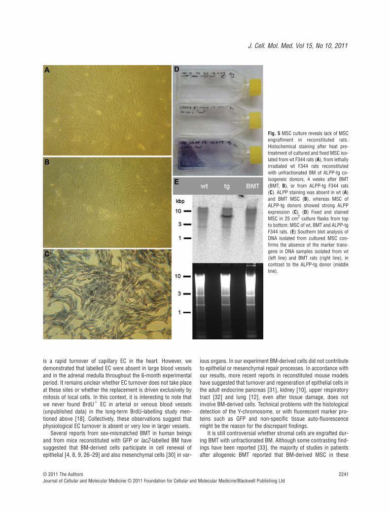
The failure to detect any ALPP�, BM-derived mesenchymal lineage cells in bone or striated and smooth muscle of BMT ratsirrespective of the time after BMT prompted us to ask the questionwhether stromal precursor cells engraft after BMT with unfraction-ated BM. To answer this question, we isolated and cultivated MSCfrom BM of wt, ALPP-tg and BMT rats, 4 weeks after BMT. In linewith our earlier report [13], MSC from ALPP-tg animals showedstrong expression of ALPP as revealed by histochemistry (Fig.5C). MSC cultivated from the BM of wt (Fig. 5A) and also of BMT(Fig. 5B) rats did not show positive ALPP staining. To rule out adown-regulation of marker enzyme expression in ALPP-tg cells ina wt environment, we performed Southern blot analysis of DNAextracted from the MSC cultures. Southern blot analysis clearlydocumented the absence of the ALPP transgene in MSC culturesof BMT rats. These findings indicate that mesenchymal precursorcells do not engraft after lethal irradiation of wt rats and reconsti-tution with unfractionated BM from ALPP-tg donor rats.
Discussion
The goal of the current study was to monitor the renewal ofperipheral tissues by BM-derived cells in adult rats, using amarker protein which can readily be detected in semi-thin paraf-fin and plastic sections by histochemistry or IHC, and in ultrathinsections by transmission electron microscopy after pre-embed-ding immunostaining. In analogy to a plethora of similarlydesigned earlier studies using fluorescent proteins or LacZ asgenetic markers of BM-derived cells, our study was undertakenunder the premise that lethal irradiation and reconstitution with
unfractionated BM would result in engraftment of mesenchymalprecursor cells in the recipient. However, we found that thispremise is wrong, at least in rats. Therefore, we could only exam-ine cellular turnover originating from the haematopoietic BMcompartment. Our data show that the haematopoietic BM com-partment is an important source of EC renewal in capillaries ofmany, but not all organs. However, haematopoietic BMC do notcontribute to physiological regeneration of muscle, bone, carti-lage or epithelial tissues.
The role of BM in EC turnover is a controversial issue. Whileit was reported that BM-derived cells contribute to blood and lymphatic capillary endothelium [19], and to neovascularizationin wound healing [20] or in scar remodelling after myocardialinfarction [21], other authors failed to find BM-derived EC inmouse models of tumour vascularization or revascularization ofischemic tissue [22, 23]. Some mouse studies suggested thatBM-derived cells in blood vessels represent pericytes [22, 23].Using immuno-electron microscopy, we found no evidence ofBM-derived pericytes or lymphatic EC in heart and kidney. Rather,with the exception of circulating blood cells all ALPP-labelledcells were unequivocally capillary EC in blood vessels of heart andkidney. It is currently unclear whether the discrepancies betweensome mouse and our rat study could be explained by species differences in BM-derived EC turnover.
Interestingly, we observed organ-specific turnover patterns ofcapillary EC in our study. Replacement of capillary EC by BM-derived cells was highest in heart and kidney. Low turnover wasseen in brain and adrenal medulla. In accordance with our find-ings, studies in mice transplanted with lacZ-expressing BMreported low values for physiological replacement of EC by BM-derived precursors in skin and brain [24]. In most organs, thenumber of ALPP-labelled EC increased with time, and, with theexception of kidney, showed a homogenous distribution pattern.The reason for the regional differences in renal BM-derived ECturnover is currently unclear. It is known that lethal irradiation
Fig. 2 Light and electron microscopic analysis of tissues from wt F344 rats, lethally irradiated and reconstituted with BM from ALPP-tg F344 donors.ALPP� endothelial-like cells in capillaries are evident in kidney (A), pancreas (B) and liver (C), 2 months after BM transplantation (BMT). The number ofALPP� endothelial-like cells increased with time in all organs as shown for heart tissue 1 (D), 2 (E) and 4 (F) months after BMT. In contrast, ALPP� ECwere absent in large blood vessels such as the Aorta thoracica (G), arteries or veins (not shown). (H) Double staining (arrows) for ALPP by histochem-istry (blue) and for tomato lectin Lycopersicon esculentum (purple) confirmed the endothelial nature of the putative BM-derived capillary EC in themyocardium, 2 months after BMT. (I) ALPP� cells in the vicinity of blood vessels as shown here in liver. Semi-thin serial sections of epon-embedded, 45-�m-thick kidney cryosections stained immunohistochemically against ALPP by a pre-embedding protocol and counterstained with toluidine blue (J) orleft without counterstaining (K, brown DAB immunoprecipitate) show that the anti-ALPP staining (arrows in K) co-localized with capillaries situatedbetween renal tubuli (arrows in J), 6 months after BMT. (L)–(O) Transmission electron microscopy (TEM) of ultra-thin sections of the heart muscle afterpre-embedding anti-ALPP staining clearly showed that the BM-derived ALPP� endothelial-like cells are indeed capillary EC, 6 months after BMT. Highermagnification is shown in (M). Anti-ALPP staining was absent in control sections of hearts from BMT rats when the primary antibody was omitted (N) orin wt controls (O). (P) Nonlinear regression analysis of the time dependent increase in BM-derived EC in the heart muscle revealed a half time of 8.36weeks for these cells. (Q)–(S) In the kidney, EC turnover showed marked inhomogeneity. BM-derived ALPP� EC were more frequent in cortical regions(Q) and in the Vasa recta of the papilla (S) than in medullary areas (R) throughout the study period, as demonstrated here at 2 months after BMT. The 5-�m-thick paraffin sections shown in (A)–(G), (I) and (Q)–(S) were stained for ALPP enzyme activity with BCIP/NBT (purple) overnight at RT after heatpre-treatment, and were counterstained with nuclear fast red. Semi-thin and ultra-thin sections shown in (J)–(O) were immunostained using a mono-clonal anti-ALPP antibody by the pre-embedding protocol described in ‘Methods’. Bars represent 50 �m for light microscopy and 2.5 �m for transmis-sion electron microscopy.

J. Cell. Mol. Med. Vol 15, No 10, 2011
2239© 2011 The AuthorsJournal of Cellular and Molecular Medicine © 2011 Foundation for Cellular and Molecular Medicine/Blackwell Publishing Ltd
damages EC (reviewed in [25]). Therefore, replacement of EC seenat 4 weeks after transplantation could be due to cell damageinduced by irradiation. However, it is unlikely that radiation-induced cell damage can explain the increase in labelled EC at thelater time-points.
In the current study, we calculated a half time of 8.36 weeks forBM-derived EC replacement in cardiac muscle. Using a completely
different methodology, namely assessment of cellular half life byanalysing the decline in labelling index after long-term 5-bromo-2�-deoxyuridine (BrdU) labelling in non-irradiated rats, wereported a cardiac capillary EC half life of 2.2 weeks [18]. Althoughboth methods are not directly comparable because different cellu-lar compartments might be assessed (BrdU labels proliferatingcells irrespective of their origin), they nevertheless show that there
Fig. 3 Lack of BM-derived ALPP�
epithelial cells in irradiated wt F344 ratsreconstituted with unfractionated BMfrom ALPP-tg F344 donors.Histochemical detection of ALPPenzyme activity (BCIP/NBT, purple)reveals absence of BM-derived epithelialcells in gut (A), kidney tubules (B), liver(C), or skin (D), 6 months after BMT. (E)Co-staining of ALPP enzyme activity(blue) and anti-CD68 antibody (purple) inlung sections shows many double posi-tive alveolar macrophages (centre), butalso some CD68� ALPP– macrophagesas indicated by arrows, 4 months afterBMT. Double staining of ALPP enzymeactivity (blue) with anti-insulin (F), anti-somatostatin (G) or anti-pancreaticpolypeptide (H) (purple) demonstrateslack of BM-derived endocrine cells inislets of Langerhans. The paraffin sec-tions shown in (A–D) were stained forALPP enzyme activity with BCIP/NBT(purple) overnight at RT after heat pre-treatment, and were counterstained withnuclear fast red. The paraffin sections in(E–H) were stained for ALPP enzymeactivity using Vector Blue, andimmunostained against CD68, insulin,somatostatin, or pancreatic polypeptideusing Vector VIP (purple) as substrate.Bars represent 50 �m.

2240 © 2011 The AuthorsJournal of Cellular and Molecular Medicine © 2011 Foundation for Cellular and Molecular Medicine/Blackwell Publishing Ltd
Fig. 4 Renewal of mesenchymal cells in irradiated wt F344 rats reconstituted with unfractionated BM from ALPP-tg F344 donors, 4–6 months after BMT.Sections of methylmethacrylate-embedded bones from reconstituted wt F344 rats do not show evidence of ALPP� BM-derived osteoblasts (A, arrows),osteocytes (B, arrows), bone lining cells (B, arrowhead) or chondrocytes (C). In contrast, BMC were strongly positive for the marker enzyme (A, B).Histochemical detection of the marker enzyme in paraffin sections shows absence of BM-derived smooth muscle cells in aorta (D), and absence of ALPP-expressing muscle fibres in skeletal muscle (E). Cells staining double positive (arrowhead) for ALPP activity (blue) and anti- SMA (purple) were rarelypresent in the gut (F). In contrast, BM-derived single ALPP� endothelial-like cells (blue) located between the smooth muscle cells could be frequentlyfound (G, arrows). In the heart, we found numerous ALPP� cells nearby valvular insertion sites and in the valvular leaflets, here at 4 months after BMT(H). Combination of ALPP-staining (blue) with immunohistochemical staining against vimentin (purple) or SMA (not shown) demonstrated that the BM-derived cells in the valvular insertion areas partially also stained positive for vimentin, suggesting a fibroblast-like nature (I, arrowheads). Numerous sin-gle vimentin� cells with cardiomyocyte-like morphology could also be found at the valvular insertion sites, indicated by arrows (J), as well as rarely inthe working myocardium (arrowhead in K), throughout the study period. In rare cases, these vimentin� (purple) cardiomyocyte-like cells stained alsopositive for SMA (blue), possibly reflecting early differentiated cardiomyocytes (L). Moreover, in epicardial areas of the heart base, we found several clus-ters of vimentin� ALPP– cells (M), potentially representing a cardiac resident stem cell pool. The 5-�m-thick paraffin and methylmethacrylate sectionsshown in (A–E) and (H) were stained for ALPP enzyme activity with BCIP/NBT (purple) overnight at RT after heat pre-treatment, and were counterstainedwith nuclear fast red. The paraffin sections in (F–G) and (I–M) were stained for ALPP enzyme activity using Vector Blue, and immunostained againstvimentin or SMA using Vector VIP (purple) or Vector Blue (L) as substrate. Bars represent 50 �m.
J. Cell. Mol. Med. Vol 15, No 10, 2011
2241© 2011 The AuthorsJournal of Cellular and Molecular Medicine © 2011 Foundation for Cellular and Molecular Medicine/Blackwell Publishing Ltd
is a rapid turnover of capillary EC in the heart. However, wedemonstrated that labelled EC were absent in large blood vesselsand in the adrenal medulla throughout the 6-month experimentalperiod. It remains unclear whether EC turnover does not take placeat these sites or whether the replacement is driven exclusively bymitosis of local cells. In this context, it is interesting to note thatwe never found BrdU� EC in arterial or venous blood vessels(unpublished data) in the long-term BrdU-labelling study men-tioned above [18]. Collectively, these observations suggest thatphysiological EC turnover is absent or very low in larger vessels.
Several reports from sex-mismatched BMT in human beingsand from mice reconstituted with GFP or lacZ-labelled BM havesuggested that BM-derived cells participate in cell renewal ofepithelial [4, 8, 9, 26–29] and also mesenchymal cells [30] in var-
ious organs. In our experiment BM-derived cells did not contributeto epithelial or mesenchymal repair processes. In accordance withour results, more recent reports in reconstituted mouse modelshave suggested that turnover and regeneration of epithelial cells inthe adult endocrine pancreas [31], kidney [10], upper respiratorytract [32] and lung [12], even after tissue damage, does notinvolve BM-derived cells. Technical problems with the histologicaldetection of the Y-chromosome, or with fluorescent marker pro-teins such as GFP and non-specific tissue auto-fluorescencemight be the reason for the discrepant findings.
It is still controversial whether stromal cells are engrafted dur-ing BMT with unfractionated BM. Although some contrasting find-ings have been reported [33], the majority of studies in patientsafter allogeneic BMT reported that BM-derived MSC in these
Fig. 5 MSC culture reveals lack of MSCengraftment in reconstituted rats.Histochemical staining after heat pre-treatment of cultured and fixed MSC iso-lated from wt F344 rats (A), from lethallyirradiated wt F344 rats reconstitutedwith unfractionated BM of ALPP-tg co-isogeneic donors, 4 weeks after BMT(BMT, B), or from ALPP-tg F344 rats(C). ALPP staining was absent in wt (A)and BMT MSC (B), whereas MSC ofALPP-tg donors showed strong ALPPexpression (C). (D) Fixed and stainedMSC in 25 cm2 culture flasks from topto bottom: MSC of wt, BMT and ALPP-tgF344 rats. (E) Southern blot analysis ofDNA isolated from cultured MSC con-firms the absence of the marker trans-gene in DNA samples isolated from wt(left line) and BMT rats (right line), incontrast to the ALPP-tg donor (middleline).
2242 © 2011 The AuthorsJournal of Cellular and Molecular Medicine © 2011 Foundation for Cellular and Molecular Medicine/Blackwell Publishing Ltd
patients are of recipient origin [34–37]. The methods used todetermine the origin of stromal cells in these studies ranged fromdemonstration of a Y-chromosome after sex-mismatched BMT[34, 35, 37] to DNA fingerprinting methods [37]. In accordancewith the clinical reports, our data did neither provide evidence forALPP-expressing mesenchymal cells of donor origin in bone orperipheral tissues of BMT rats irrespective of the time-point afterBMT, nor of ALPP� MSC after culture of BM harvested from BMTrats. Along similar lines, Wang et al. [38] showed that in irradiatedmice reconstituted with unfractionated BM of Col1a1-GFP reportertransgenic mice, GFP-expressing cells did not differentiate intoosteocytes. However, other studies in mice reported donor-derived chimerism of MSC after irradiation and transplantation ofunfractionated BMC or MSC [39–41]. The reason for the contrast-ing findings between some mouse experiments and studies inhuman beings and rats remains unclear. However, there is evi-dence that strain-related differences in the sensitivity of murineMSC to irradiation may be involved [40].
Why do stromal precursors, although certainly present inunfractionated BM, not engraft in lethally irradiated human beingsand rats? MSC show relative radioresistance in vitro and may notbe affected by the irradiation regimen [42]. Therefore, the mostlikely explanation is that, in contrast to haematopoietic stem cells,a niche for stromal precursor cells is lacking in BM of irradiatedanimals. Given the fact that the MSC pool remained recipient-derived in our study, how can the occurrence of BM-derivedALPP� fibroblasts in certain organs such as heart or intestine beexplained? Other investigators have also reported the presence ofBM-derived fibroblasts in kidney, valvular leaflets of the heart orskin during wound healing after transplantation of unfractionatedBM or even haematopoietic stem cells in mice [43–45]. Moreover,studies in a mouse model of cardiac fibrosis have provided firmevidence of a blood-borne, BM-derived fibroblast precursor popu-lation of haematopoietic origin, giving rise to -SMA, type I colla-gen, CD34 and CD45� cardiac fibroblasts [46]. Thus, our datacorroborate the existence of a distinct blood-borne population offibroblast precursors of haematopoietic origin also in the rat.
In the current study, we found some ALPP–, vimentin and/orSMA-staining cardiomyocyte-like cells in the heart, especially inareas of high mechanical stress. This finding may implicate that,given the assumption that these vimentin-expressing cells can bedefined as early differentiated cardiomyocytes [47], the heart may
be capable of limited regeneration through differentiation of car-diac stem cells [48]. However, although these repair mechanismsmay be suitable to compensate physiologic cell death especiallyat sites of high mechanical demand, they without doubt fail tocompensate extensive loss of cardiomyocytes after ischemia-reperfusion injury [21]. We demonstrated that in our modelengraftment of donor-derived MSC did not take place. Therefore,we cannot rule out a possible recruitment of BM-derived mes-enchymal precursors in mesenchymal tissue turnover, becausethe recipient-derived BM-MSC could not be tracked in our study.However, a study of heterotopic transplantation of wt rat heartsinto transgenic rats ubiquitously expressing GFP showed thatBM-derived cardiomyocyte turnover represents a negligible eventunder more or less physiologic conditions [49].
In conclusion, our study clearly showed that haematopoieticBMC are an important source of physiological renewal ofendothelial, but not of epithelial or mesenchymal, cells in manyorgans of adult rats. Thus, our data corroborate the notion thatthe major source of endothelial precursor cells in BM is ofhaematopoietic origin [50]. Because mesenchymal precursorcells did not engraft after BMT with unfractionated BM, our studyquestions the usefulness of reconstituted irradiated rats as amodel for evaluating BM-driven regeneration of mesenchymalcells in peripheral tissues.
Acknowledgements
The authors thank Werner Panzer and Siegfried Kosik for help with theirradiation, and Magdalena Helmreich and Waltraud Tschulenk for estab-lishment of protocols for IHC and electron microscopy, respectively. Thisresearch was supported by Deutsche Forschungsgemeinschaft (Er 223/8-1),by the University of Veterinary Medicine Vienna and by the AustrianScience Fund FWF (P 21904-B11). G.L. and A.J. were supported by theAustrian Science Fund FWF (FSP09309).
Conflicts of interest
The authors declare that there are no conflicts of interest.
References
1. Prockop DJ, Gregory CA, Spees JL. Onestrategy for cell and gene therapy: har-nessing the power of adult stem cells torepair tissues. Proc Natl Acad Sci USA.2003; 100: 11917–23.
2. Asahara T, Masuda H, Takahashi T, et al.Bone marrow origin of endothelial progenitorcells responsible for postnatal vasculogene-
sis in physiological and pathological neovas-cularization. Circ Res. 1999; 85: 221–8.
3. Brazelton TR, Rossi FM, Keshet GI, et al.From marrow to brain: expression of neu-ronal phenotypes in adult mice. Science.2000; 290: 1775–9.
4. Lagasse E, Connors H, Al Dhalimy M, et al. Purified hematopoietic stem cells
can differentiate into hepatocytes in vivo.Nat Med. 2000; 6: 1229–34.
5. Ferrari G, Cusella-De Angelis G, et al.Muscle regeneration by bone marrow-derived myogenic progenitors. Science.1998; 279: 1528–30.
6. Orlic D, Kajstura J, Chimenti S, et al.Mobilized bone marrow cells repair the

J. Cell. Mol. Med. Vol 15, No 10, 2011
2243© 2011 The AuthorsJournal of Cellular and Molecular Medicine © 2011 Foundation for Cellular and Molecular Medicine/Blackwell Publishing Ltd
infarcted heart, improving function andsurvival. Proc Natl Acad Sci USA. 2001;98: 10344–9.
7. Ogawa M, Larue AC, Watson PM, et al.Hematopoietic stem cell origin of connec-tive tissues. Exp Hematol. 2010; 38:540–7.
8. Kotton DN, Ma BY, Cardoso WV, et al.Bone marrow-derived cells as progenitorsof lung alveolar epithelium. Development.2001; 128: 5181–8.
9. Lin F, Cordes K, Li L, et al. Hematopoieticstem cells contribute to the regeneration ofrenal tubules after renal ischemia-reperfusioninjury in mice. J Am Soc Nephrol. 2003;14: 1188–99.
10. Lin F, Moran A, Igarashi P. Intrarenalcells, not bone marrow-derived cells, arethe major source for regeneration inpostischemic kidney. J Clin Invest. 2005;115: 1756–64.
11. Quintana-Bustamante O, Alvarez-Barrientos A, Kofman AV, et al.Hematopoietic mobilization in miceincreases the presence of bone marrow-derived hepatocytes via in vivo cell fusion.Hepatology. 2006; 43: 108–16.
12. Kotton DN, Fabian AJ, Mulligan RC.Failure of bone marrow to reconstitutelung epithelium. Am J Respir Cell Mol Biol.2005; 33: 328–34.
13. Unger NJ, Odörfer KI, Weber K, et al.Utility of human placental alkaline phos-phatase as a genetic marker for cell track-ing in bone and cartilage. Histochem CellBiol. 2007; 127: 669–74.
14. Kisseberth WC, Brettingen NT, Lohse JK,et al. Ubiquitous expression of markertransgenes in mice and rats. Dev Biol.1999; 214: 128–38.
15. Erben RG. Embedding of bone samples inmethylmethacrylate: An improved methodsuitable for bone histomorphometry, his-tochemistry, and immunohistochemistry.J Histochem Cytochem. 1997; 45: 307–13.
16. Odörfer KI, Unger NJ, Weber K, et al.Marker tolerant, immunocompetent animalsas a new tool for regenerative medicineand long-term cell tracking. BMC Biotechnol.2007; 7: 30.
17. Farrell E, Byrne EM, Fischer J, et al. Acomparison of the osteogenic potential ofadult rat mesenchymal stem cells culturedin 2-D and on 3-D collagen glycosamino-glycan scaffolds. Technol Health Care.2007; 15: 19–31.
18. Erben RG, Odörfer KI, Siebenhutter M, et al. Histological assessment of cellularhalf-life in tissues in vivo. Histochem CellBiol. 2008; 130: 1041–6.
19. Jiang S, Bailey AS, Goldman DC, et al.Hematopoietic stem cells contribute tolymphatic endothelium. PLoS One. 2008;3: e3812.
20. Bluff JE, Ferguson MW, O’Kane S, et al.Bone marrow-derived endothelial progeni-tor cells do not contribute significantly tonew vessels during incisional wound heal-ing. Exp Hematol. 2007; 35: 500–6.
21. Odörfer KI, Walter I, Kleiter M, et al. Roleof endogenous bone marrow cells in long-term repair mechanisms after myocardialinfarction. J Cell Mol Med. 2008; 12:2867–74.
22. Rajantie I, Ilmonen M, Alminaite A, et al.Adult bone marrow-derived cells recruitedduring angiogenesis comprise precursorsfor periendothelial vascular mural cells.Blood. 2004; 104: 2084–6.
23. Ziegelhoeffer T, Fernandez B, Kostin S,et al. Bone marrow-derived cells do notincorporate into the adult growing vascula-ture. Circ Res. 2004; 94: 230–8.
24. Crosby JR, Kaminski WE, Schatteman G,et al. Endothelial cells of hematopoieticorigin make a significant contribution toadult blood vessel formation. Circ Res.2000; 87: 728–30.
25. Woywodt A, Haubitz M, Buchholz S, et al.Counting the cost: markers of endothelialdamage in hematopoietic stem cell trans-plantation. Bone Marrow Transplant. 2004;34: 1015–23.
26. Korbling M, Katz RL, Khanna A, et al.Hepatocytes and epithelial cells of donororigin in recipients of peripheral-blood stemcells. N Engl J Med. 2002; 346: 738–46.
27. Okamoto R, Yajima T, Yamazaki M, et al.Damaged epithelia regenerated by bonemarrow-derived cells in the human gas-trointestinal tract. Nat Med. 2002; 8:1011–7.
28. Fang TC, Otto WR, Rao J, et al.Haematopoietic lineage-committed bonemarrow cells, but not cloned cultured mes-enchymal stem cells, contribute to regen-eration of renal tubular epithelium afterHgCl 2 -induced acute tubular injury. CellProlif. 2008; 41: 575–91.
29. Krause DS, Theise ND, Collector MI, et al.Multi-organ, multi-lineage engraftment bya single bone marrow-derived stem cell.Cell. 2001; 105: 369–77.
30. Kawada H, Fujita J, Kinjo K, et al.Nonhematopoietic mesenchymal stemcells can be mobilized and differentiateinto cardiomyocytes after myocardialinfarction. Blood. 2004; 104: 3581–7.
31. Lechner A, Yang YG, Blacken RA, et al. No evidence for significant transdif-
ferentiation of bone marrow into pancre-atic beta-cells in vivo. Diabetes. 2004; 53:616–23.
32. Davies JC, Potter M, Bush A, et al. Bonemarrow stem cells do not repopulate thehealthy upper respiratory tract. PediatrPulmonol. 2002; 34: 251–6.
33. Keating A, Singer JW, Killen PD, et al.Donor origin of the in vitro haematopoieticmicroenvironment after marrow transplan-tation in man. Nature. 1982; 298: 280–3.
34. Laver J, Jhanwar SC, O’Reilly RJ, et al.Host origin of the human hematopoieticmicroenvironment following allogeneicbone marrow transplantation. Blood.1987; 70: 1966–8.
35. Simmons PJ, Przepiorka D, Thomas ED,et al. Host origin of marrow stromal cellsfollowing allogeneic bone marrow trans-plantation. Nature. 1987; 328: 429–32.
36. Awaya N, Rupert K, Bryant E, et al.Failure of adult marrow-derived stem cellsto generate marrow stroma after success-ful hematopoietic stem cell transplanta-tion. Exp Hematol. 2002; 30: 937–42.
37. Rieger K, Marinets O, Fietz T, et al.Mesenchymal stem cells remain of hostorigin even a long time after allogeneicperipheral blood stem cell or bone marrowtransplantation. Exp Hematol. 2005; 33:605–11.
38. Wang L, Liu Y, Kalajzic Z, et al.Heterogeneity of engrafted bone-liningcells after systemic and local transplanta-tion. Blood. 2005; 106: 3650–7.
39. Koide Y, Morikawa S, Mabuchi Y, et al.Two distinct stem cell lineages in murinebone marrow. Stem Cells. 2007; 25:1213–21.
40. Rombouts WJ, Ploemacher RE. Primarymurine MSC show highly efficient homingto the bone marrow but lose homing abil-ity following culture. Leukemia. 2003; 17:160–70.
41. Morikawa S, Mabuchi Y, Kubota Y, et al. Prospective identification, isolation,and systemic transplantation of multipo-tent mesenchymal stem cells in murinebone marrow. J Exp Med. 2009; 206:2483–96.
42. Li J, Kwong DL, Chan GC. The effects ofvarious irradiation doses on the growthand differentiation of marrow-derivedhuman mesenchymal stromal cells.Pediatr Transplant. 2007; 11: 379–87.
43. Direkze NC, Forbes SJ, Brittan M, et al.Multiple organ engraftment by bone-mar-row-derived myofibroblasts and fibrob-lasts in bone-marrow-transplanted mice.Stem Cells. 2003; 21: 514–20.

2244 © 2011 The AuthorsJournal of Cellular and Molecular Medicine © 2011 Foundation for Cellular and Molecular Medicine/Blackwell Publishing Ltd
44. Visconti RP, Ebihara Y, Larue AC, et al.An in vivo analysis of hematopoietic stemcell potential: hematopoietic origin of car-diac valve interstitial cells. Circ Res. 2006;98: 690–6.
45. Ishii G, Sangai T, Sugiyama K, et al. Invivo characterization of bone marrow-derived fibroblasts recruited into fibroticlesions. Stem Cells. 2005; 23: 699–706.
46. Haudek SB, Xia Y, Huebener P, et al.Bone marrow-derived fibroblast precur-
sors mediate ischemic cardiomyopathy inmice. Proc Natl Acad Sci USA. 2006; 103:18284–9.
47. Babai F, Musevi-Aghdam J, Schurch W,et al. Coexpression of alpha-sarcomericactin, alpha-smooth muscle actin anddesmin during myogenesis in rat andmouse embryos I. Skeletal muscle.Differentiation. 1990; 44: 132–42.
48. Laugwitz KL, Moretti A, Lam J, et al.Postnatal isl1� cardioblasts enter fully
differentiated cardiomyocyte lineages.Nature. 2005; 433: 647–53.
49. Ausoni S, Zaglia T, Dedja A, et al. Host-derived circulating cells do not signifi-cantly contribute to cardiac regenerationin heterotopic rat heart transplants.Cardiovasc Res. 2005; 68: 394–404.
50. Asahara T, Murohara T, Sullivan A, et al.Isolation of putative progenitor endothelialcells for angiogenesis. Science. 1997; 275:964–7.


![Whole-transcriptome analysis of endothelial to hematopoietic ...genic endothelial cells (ECs [HECs]), the genetic program driving HSC emergence is largely unknown. Here, we use a highly](https://static.fdocuments.in/doc/165x107/608ead5defaeac11651c1f82/whole-transcriptome-analysis-of-endothelial-to-hematopoietic-genic-endothelial.jpg)