Hellenic Crystallographic Association - hecra.gr · Hellenic Crystallographic Association ......
61
1 Hellenic Crystallographic Association 5 th International Conference 24 – 25 September 2010 UNIVERSITY of THESSALY, Katsigra building, Post-office square Larissa, Greece
Transcript of Hellenic Crystallographic Association - hecra.gr · Hellenic Crystallographic Association ......

1
Hellenic Crystallographic Association 5th International Conference
24 – 25 September 2010
UNIVERSITY of THESSALY,
Katsigra building, Post-office square
Larissa, Greece

2
Scientific Committee Maria Calamiotou
Elias Eliopoulos
Stavros Hamodrakas
Athanassios Hountas
Irene Mavridis
Anastassios C. Papageorgiou
Kyriakos Petratos
Socrates Tzartos
Costas Vorgias
Organizing Committee
Demetres Leonidas
George Kontopidis
Maria Kontou
George Papadopoulos
Vicky T. Skamnaki
Sponsors
3
The 5th International Conference of The Hellenic Crystallographic
Association
24-25 September 2010, Larissa University of Thessaly
Programme grid
Friday 24th September 9.00 – 10.00 Registration 10.00 – 10.30 Welcome and opening remarks 10.30 – 12.30 Oral presentations 12.10 – 13.30 Lunch break 13.30 – 15.30 Oral presentations 15.30 – 16.30 Coffee break + Poster Session 16.30 – 17.30 General Assembly of the Hellenic Crystallographic Association members 17.30 – 19.50 Oral presentations
Saturday 25th September
09.00 – end of the day
Voting for the new board of the Hellenic Crystallographic Association
09.30 – 11.30 Oral presentations 11.30 – 12.30 Sponsors’ Presentations 12.30 - 13.30 Lunch break 13.30 – 14.30 Poster Session 14.30 – 17.30 Oral presentations 17.30 – 18.00 Closing Remarks
“Nikos Oikonomakos award”
Sponsored by

4
Friday 24th September Oral presentations OP1. Ligand discovery by finding and filling protein pockets Malcolm Walkinshaw Institute of Structural and Molecular Biology, University of Edinburgh, EDINBURGH EH9 3JR, U.K. E-mail: [email protected] OP2. Structure-guided Design of non-ATP competitive protein kinase inhibitors Campbell McInnes Pharmaceutical and Biomedical Sciences, South Carolina College of Pharmacy, University of South Carolina, Columbia, U.S.A. E-mail: [email protected] OP3. Natural flavonoid Catechin inhibits Glycogen phosphorylase by binding at new allosteric site V.T. Skamnaki1, M. Savvidou2, A. Katsandi2,A.-M. Psarra2, M. Kontou2, D. Kouretas2, D.D. Leonidas2
1Institute of Organic and Pharmaceutical Chemistry, National Hellenic Research Foundation, 48 Vas. Constantinou Ave, Athens 11635, Greece. 2Department of Biochemistry and Biotechnology, University of Thessaly, 26 Ploutonos Str., 41221 Larissa, Greece. E-mail:[email protected] OP4. Water, membranes and adaptation to extreme environments as seen by neutrons Giuseppe Zaccai Institut Laue-Langevin, Grenoble E-mail: [email protected] OP5. CRYSTAL STRUCTURE OF A NEW ENDO-β-1,4-XYLANASE FROM FUSARIUM OXYSPORUM DETERMINED AT 1.9 RESOLUTION M. Dimarogona1,2, E. Topakas2, P. Christakopoulos2 and E. D. Chrysina1 1The National Hellenic Research Foundation, Institute of Organic and Pharmaceutical Chemistry, Athens. 2The National Technical University of Athens, School of Chemical Engineering, Biotechnology Laboratory, Athens. OP6. COMPARATIVE BIOCHEMICAL & STRUCTURAL STUDIES ON TYPE III SECRETION REGULATION SWITCH COMPLEXES. A. D. Gazi, S. Charova, M. Charetidis, M. Ambrazi, N. J. Panopoulos and M. Kokkinidis 1Institute of Molecular Biology & Biotechnology IMBB, Foundation of Research & Technology ? Hellas (FORTH), Nikolaou Plastira 100, Heraklion, Crete, Greece. OP7. EFFECT OF TEMPERATURE ON THE FOLDING AND STABILITY OF FOLDABLE PEPTIDES: A MOLECULAR DYNAMICS APPROACH. P.S. Georgoulia and N.M. Glykos 1Department of Molecular Biology and Genetics, Democritus University of Thrace, University Campus, Dragana, 68100, Alexandroupolis, Greece. OP8. Bacterial protein secretion: the system nanomachine cross-talk G. Gouridis1, 2, K. Chatzi1, 2, M.-F. Sardis1, 2, I. Gelis3, C.G. Kalodimos3, G. Orfanoudaki1, 2, M. Koukaki2, S. Karamanou2 and A. Economou1,2

5
1Institute of Molecular Biology and Biotechnology-FORTH and 2Department of Biology-University of Crete, PO Box 1527, Iraklio, Crete, Greece. 3Chemistry & Chemical Biology, Biomedical Engineering, Rutgers University, 599 Taylor Rd, Piscataway, NJ 08854, USA E-mail: [email protected] OP9. Molecular Dynamics of ACC-1 class C beta-Lactamase and its extended spectrum variant ACC-4 S. D. Kotsakis1, L. S. Tzouvelekis2, E. Tzelepi1, E. Petinaki3, and V. Miriagou1 1Laboratory of Bacteriology, Hellenic Pasteur Institute, 11521 Athens; 2Department of Microbiology, Medical School, University of Athens, 11527 Athens; 3Department of Microbiology, Medical School, University of Thessaly, Larisa, Greece OP10. Ladderane formation in a reactive 3D nanoporous Lanthanide MOF: a single crystal-to-single crystal study S. Skoulika, A. Michaelides, and M. G. Siskos 1Department of Chemistry, University of Ioannina, 45110 Ioannina, Greece OP11. CRYSTAL STRUCTURE OF THE CYCLOPHILIN-A ENZYME FROM AZOTOBACTER VINELANDII E. Christoforides1, M. Dimou2, P. Katinakis2, K. Bethanis1 and M. Karpusas1
1Department of Science, Physics Laboratory, 2Department of Biotechnology, Laboratory of Molecular Biology, Agricultural University of Athens, Iera Odos 75 Athens, Greece OP12. THE CRYSTAL STRUCTURE OF COBALT-SUBSTITUTED PSEUDOAZURIN FROM ALCALIGENES FAECALIS R. Gessmann1, C. Kyvelidou2, M. Papadovasilaki1, and K. Petratos1 1I.M.B.B.-FO.R.T.H., P.O. Box 1385, Heraklion 70013, Greece; 2Department of Biology, University of Crete, P.O. Box 2208, Heraklion 71409, Greece
Saturday 25th September Oral presentations OP13. MEMBRANE PROTEIN CRYSTALLIZATION IN MESO PHASE WITH A VAPOR DIFFUSION SETUP: METHOD DEVELOPMENT AND APPLICATION RESULTS J. Labahn1, J. Kubicek2, R. Schlesinger1, F. Schäfer2, and G. Büldt1 1Institute of Structural Biology and Biophysics (ISB-2), Research Center Jülich, 52428 Jülich, Germany; 2 Qiagen GmbH, Qiagenstr. 1, 40724 Hilden, Germany OP14. ORDER AND DISORDER IN CHROMATIN-ASSOCIATED PROTEINS A.S. Politou Laboratory of Biological Chemistry, University of Ioannina, School of Medicine, GR-45110 Ioannina, Greece and Biomedical Research Institute, Foundation for Research and Technology (BRI-FORTH), GR-45110 Ioannina, Greece

6
Sponsor presentation SP1. Short Wavelength Radiation in Crystallography E. Hovestreydt1, H. Ott1 and J. Graf2
1 Bruker AXS GmbH, Karlsruhe, Germany 2 Incoatec GmbH, Geesthacht, Germany E-Mail: [email protected] SP2. Introduction to Microcalorimetry – bridging the gap between structure and function D. Heep GE Healthcare Oral presentations OP15. BIOLOGICAL STRUCTURES AND MATERIALS AS A SOURCE FOR INSPIRATION FOR THE DESIGN OF NOVEL NANO-BIOMATERIALS Anna Mitraki 1
Department of Materials Science and Technology, c/o Biology Department, University of Crete, and Institute For Electronic Structure and Laser, IESL-FORTH, Vassilika Vouton, 71003 Heraklion, Crete, Greece E-Mail: [email protected] OP16. REFINEMENT AND VALIDATION OF PHOSPHORYLASE KINASE ATP-BINDING SITE INHIBITOR COMPLEXES USING MOLECULAR DYNAMICS AND MM-GBSA CALCULATIONS J.M. Hayes1, V.T. Skamnaki1,2, G. Archontis3, C. Lamprakis1, J. Sarrou1, N. Bischler1, A.-L. Skaltsounis4, S.E. Zographos1, and N.G. Oikonomakos1 1Institute of Organic & Pharmaceutical Chemistry, National Hellenic Research Foundation, 48 Vassileos Constantinou Ave., Athens 11635, Greece; 2Laboratory of Molecular Biophysics and Oxford Centre for Molecular Sciences, Dept. of Biochemistry, Oxford University, Oxford OX1 3QU, U.K.; 3Department of Physics, University of Cyprus, CY1678 Nicosia, Cyprus; 4Division of Pharmacognosy, Dept. of Pharmacy, University of Athens, Panepistimiopolis-Zographou, Athens 15771, Greece E-Mail: [email protected] OP17. LATTICE EFFECTS IN THE NEW FeAs-BASED NdFeAsO0.85 SUPERCONDUCTOR 1M. Calamiotou, 2I. Margiolaki, 1A. Gantis 3E. Siranidi, 4Z.A. Ren, 4Z.X.Zhao, 3E.Liarokapis 1Solid State Physics Department, School of Physics, University of Athens, GR-15784 Athens, Greece. 2ESRF, BP 220, Grenoble, Cedex 9, F-38043 Franc. 3Department of Physics, National Technical University of Athens, 157 80 Athens, Greece. 4Chinese Acad. Sci., Inst. Phys., Natl. Lab. Superconductivity, Beijing 100080, China E-mail: [email protected] OP18. COMPARISON OF STRUCTURAL PARAMETERS AND CLUSTER ANALYSIS OF UREA-, HEAT- AND FORCE INDUCED DENATURATION OF A1, A2 AND A3 DOMAINS OF VON WILLEBRAND FACTOR USING MOLECULAR DYNAMICS TECHNIQUES Stefanos Pentas1, Vaia Stathi2 and Geοrgios E. Papadopoulos2
1Department of Physics of condensed matter, Laboratory of Thin Films Nanosystems and Nanometrology, Aristotle University of Thessaloniki, 2Department of Biochemistry & Biotechnology, University of Thessaly E-mail: [email protected] OP19. SILMOTH CHORION: A FUNCTIONAL, PROTECTIVE AMYLOID Stavros J. Hamodrakas Department of Cell Biology and Biophysics, Faculty of Biology, University of Athens, Panepistimiopolis, Athens 157 01, Greece

7
Friday 26th – Saturday 27th Poster presentations PP1. An overview of diabetes type 2 and glycogen phosphorylase inhibitors. How structure based drug design can prove a valuable therapeutic strategy Kyra-Melinda Alexacou1,2 Spyros E. Zographos,1 Nikos G. Oikonomakos1† and Demetres D. Leonidas3
1Institute of Organic and Pharmaceutical Chemistry, National Hellenic Research Foundation, 48 Vassileos Constantinou Avenue, 11635 Athens, Greece. 2Department of Biology, Chemistry and Pharmacy, Freie Universität Berlin, Takustr. 3, 14195, Berlin, Germany. 3Department of Biochemistry and Biotechnology, University of Thessaly, 26 Ploutonos Str. 41221 Larissa, Greece
PP2. PROTEIN-PROTEIN DOCKING USING THE SHAPE IMPACT DESCRIPTOR. A. Axenopoulos1, P. Daras2, G. Papadopoulos3 and E. Houstis1 1Department of Computer & Communication Engineering, University of Thessaly, Volos, Greece; 2Informatics & Telematics Institute, Centre for Research & Technology Hellas, Thessaloniki, Greece; 3Department of Biochemistry & Biotechnology, University of Thessaly, Larissa, Greece
PP3. CRYSTAL STRUCTURES OF GERANIOL COMPLEXES WITH NATIVE AND PERMETHYLATED β-CYCLODEXTRIN K. Bethanis1, V. Boulaki1, E. Christoforides1, F. Tsorteki1, A. Kokkinou1 and D. Mentzafos1
1Department of Science, Physics Laboratory, 2Department of Biotechnology, Laboratory of Molecular Biology, Agricultural University of Athens, Iera Odos 75 Athens, Greece PP4. COMPLEX OF THE A-SITE rRNA WITH A SMALL MOLECULE OF RIGID BICYCLIC NATURE J. Birtley, G. Kythreoti, E. Saridakis, I. Katsoulis, A. Papakyriakou, I. Mavridis, D. Vourloumis, I. M. Mavridis
Institute of Physical Chemistry, National Center for Scientific Research “Demokritos”, 15310 Aghia Paraskevi, Athens, Greece PP5 . KINETIC AND CRYSTALLOGRAPHIC STUDIES OF GLYCOGEN PHOSPHORYLASE IN COMPLEX WITH D-GLUCOPYRANO-SYLIDENE SPIRO-ISOXAZOLINE DERIVATIVES FOR THE DESIGN OF NEW ANTIDIABETIC DRUGS A.S. Chajistamatiou1,2, D. Gueyrard3, S. Vidal3, J.-P. Praly3, A. Siafaka-Kapadai2, E.D. Chrysina1
1Institute of Organic & Pharmaceutical Chemistry, National Hellenic Research Foundation, 48 Vassileos Constantinou Avenue, Athens, GR-11635, Greece∙, 2Biochemistry Laboratory, Department of Chemistry, National & Kapodistrian University of Athens, Greece, 3Université de Lyon, Institut de Chimie et Biochimie Moléculaires et Supramoléculaires (ICBMS) associé au CNRS, UMR 5246, CPE-Lyon, 43 blvd du 11 Novembre 1918, 69622 Villeurbanne, France PP6. KINETIC AND CRYSTALLOGRAPHIC STUDIES OF POTENTIAL INHIBITORS OF GLYCOGEN PHOPSHORYLASE, A KEY ENZYME FOR THE TREATMENT OF TYPE 2 DIABETES Μ. Chegkazi1,2,3, Α. Pantzou2, D. Sovantzis1,2, T. Hadjiloi1,2, C. Çismas2, A. Siafaka-Kapadai3, A. Gimisis2, Ε. D. Chrysina1 1Institute of Organic and Pharmaceutical Chemistry, The National Hellenic Research Foundation, 48, Vas. Constantinou Av. 116 35 Athens, Greece. 2Organic Chemistry Laboratory, 3Biochemistry Laboratory Department of Chemistry, University of Athens, Panepistimiopolis, 15771, Athens, Greece.
8
PP7. DETERMINATION OF PROTEIN-PROTEIN INTERACTIONS BETWEEN GLUTATHIONE S-TRANSFERASE P1-1 AND C-JUN N-TERMINAL KINASE (JNK-1). L. Chiniadis1, and J. Fernandez-Recio2
1Department of Natural Sciences, Physics lab, Agricultural University of Athens, 11855, 75, Iera Odos street, Athens, Greece 2Life Sciences Department, Barcelona Supercomputing Center, C/ Jordi Girona 29, Barcelona, Spain. PP8. A PRELIMINARY STUDY OF INSECTICIDE BINDING TO GLUTATHIONE S-TRANFERASES OF MALARIA VECTOR MOSQUITOES Sofia Eliopoulou1, Maria Pavlaki2, Dimitra Kalamida2, Vicky Drosou2, Pavlos (Bogos) Agianian1 1Department of Molecular Biology and Genetics (MBG), Democritus University of Thrace, Dragana, 68100 Alexandroupolis. 2Cell Imaging and Biomolecular Interactions (CiBit) Unit, MBG, Democritus University of Thrace, Dragana. E-mail: [email protected] PP9. STRUCTURAL AND KINETIC STUDIES OF A1 AND A2 DOMAINS OF VON WILLEBRAND FACTOR Z. Karoulia1, M. Nomikos2, A. Thanassoulas2, G. Papadopoulos1, T. Choli3, C. Stathopoulos4, D. D. Leonidas1, M. Kontou1
1 Department of Biochemistry and Biotechnology, 41221 Larissa, Greece; 2 Institute of Radioisotopes & Radiodiagnostic Products, NCSR “Demokritos”, 1510 Agia Paraskevi, Attiki, Greece; 3 Department of Biochemistry, Faculty of Chemistry, Aristotele University of Thessaloniki, 54124 Thesaloniki, Greece; 4 Department of Biological Chemistry, Faculty of Medicine, University of Patras, 26504 Patras, Greece PP10. ROP REVISITED: CHANGING A PROTEIN'S OLOGOMERIZATION STATE AND FOLDING MOTIF WITH A SINGLE AMINO ACID SUBSTITUTION. A MOLECULAR DYNAMICS STUDY. M. Kokkinidou1 , P. S. Georgoulia1 and N. M. Glykos1
1Department of Molecular Biology and Genetics, Democritus University of Thrace, University Campus (Dragana), 68100 Alexandroupoli, Greece
PP11. DEVELOPMENT OF A SOFTWARE APPLICATION FOR HANDLING PROTEIN CRYSTALLISATION CONDITIONS AND TRIALS D. Markopoulos1,2, E. Manolakos2, and E.D. Chrysina1 1National Hellenic Research Foundation, Institute of Organic & Pharmaceutical Chemistry, 48 Vassileos Constantinou Ave., 11635 Athens, Greece; 2 National and Kapodistrian University of Athens, Graduate Program “Information Technologies in Medicine and Biology”, Department of Informatics & Telecommunications, Panepistimiopolis, Ilissia, Athens 15784, Greece. PP12. DEVELOPMENT OF AN ELECTRONIC LABORATORY NOTEBOOK FOR STRUCTURE-BASED DRUG DESIGN. E. Mastroleon1,2, E. S. Manolakos2 and E. D. Chrysina1 1National Hellenic Research Foundation, Institute of Organic & Pharmaceutical Chemistry, 48, Vassileos Constantinou Ave. 11635 Athens, Greece 2Graduate Program “Information Technologies in Medicine and Biology”, Department of Informatics & Telecommunications, National and Kapodistrian University of Athens, Panepistimioupolis, Ilissia, 15784 Athens, Greece PP13. NEW CRYSTAL PACKING IN β-CYCLODEXTRIN INCLUSION COMPLEXES I.M. Mavridis, S. D. Chatziefthimiou, E. Hadjoudis Institute of Physical Chemistry, National Center for Scientific Research “Demokritos”, 15310 Aghia Paraskevi, Athens, Greece

9
PP14. SOLID STATE REACTIVITY AND THERMAL STABILITY IN A SERIES OF ORGANIC CO-CRYSTALS. A. Michaelides, S. Skoulika, C. Tsaggaios, V. Dokorou and M. G. Siskos 1Department of Chemistry, University of Ioannina, 45110 Ioannina, Greece PP15. A STRUCTURAL MODEL OF MEMBERS OF THE CPF FAMILY OF CUTICULAR PROTEINS: POSSIBLE ROLE IN M/S DIFFERENTIATION Nikos C. Papandreou1, Vassiliki A. Iconomidou1, Judith H. Willis2 and Stavros J. Hamodrakas1 1 Department of Cell Biology and Biophysics, Faculty of Biology, University of Athens, Athens 157 01, Greece, 2 Department of Cellular Biology, University of Georgia, Athens, GA 30602, USA PP16. THE CRYSTAL STRUCTURE OF RABBIT MUSCLE GLYCOGEN PHOSPHORYLASE b IN COMPLEX WITH N-(β-D-GLUCOPYRANOSYL) GUANIDINOACETAMIDE. P.V. Skourti1,2, D. Loganathan3, A. Siafaka-Kapadai2, E.D. Chrysina1 1Institute of Organic and Pharmaceutical Chemistry, National Hellenic Research Foundation, 48, Vassileos Constantinou Avenue, Athens 11635, Greece, 2Laboratory of Biochemistry Department of Chemistry, National & Kapodistrian University of Athens, Greece, 3Regional Sophisticated Instrumentation Centre, Indian Institute of Technology Madras, Chennai 600 036, India.
PP17. STUDYING BINDING CHARACTERISTICS OF THE ANOPHELES GAMBIAE ODORANT BINDING PROTEINS. T. Thireou1, and E. Eliopoulos1 1Department of Biotechnology, Agricultural University of Athens, 75 Iera Odos, Votanikos, Athens 118-55, Greece
PP18. KINETIC AND MODELING STUDIES OF A NEW SET OF β-D-GLYCOSE PYRIMIDINE DERIVATIVES BINDING AT THE GLYCOGEN PHOSPHORYLASE CATALYTIC SITE V.G. Tsirkone1, A. Katsandi2, S. Manta2, E. Tsoukala2, J.M. Hayes1, M. Kontou2, D. Komiotis2, D.D. Leonidas2
1Institute of Organic and Pharmaceutical Chemistry, National Hellenic Research Foundation, 48 Vas. Constantinou Ave, Athens 11635, Greece. 2Department of Biochemistry and Biotechnology, University of Thessaly, 26 Ploutonos Str., 41221 Larissa, Greece. E-mail: [email protected], [email protected] PP19. AgamOBP1 IS A MOLECULAR TARGET FOR THE DEVELOPMENT OF NOVEL INSECT REPELLENTS. K.E. Tsitsanou1, C.E. Drakou1, A. Thiraiou2, E. Eliopoulos2, K. Iatrou3 and S.E. Zographos1
1Institute of Organic and Pharmaceutical Chemistry, National Hellenic Research Foundation, 48 Vas. Constantinou Ave., 11635 Athens, Greece; 2Department of Agricultural Biotechnology, Agricultural University of Athens; Iera Odos 75, 11855 Athens, Greece; 3Institute of Biology, NCSR “Demokritos”, 15310 Agia Paraskevi, Attiki, Athens, Greece E-mail: [email protected]

10
The 5th International Conference of The Hellenic Crystallographic
Association
24-25 September 2010, Larissa
University of Thessaly
Programme
Friday 24th September 9.00 – 10.00 Registration 10.00 – 10.30 Welcome 10.00 – 10.30 Assoc. Prof. D.D. Leonidas, Organizing Committee
Assoc. Prof. M. Calamiotou, President of the Hellenic Crystallographic Association Prof. V. Bodozoglou, Vice-Rector of University of Thessaly
Chair persons G. Kontopidis, M. Kontou 10.30 – 11.30 M.D. Walkinshaw: Ligand discovery by finding and filling
protein pockets 11.30 – 11.50 C. McInnes: Structure-guided Design of non-ATP competitive
protein kinase inhibitors 11.50 – 12.10 V.T. Skamnaki, M. Savvidou, A. Kantsadi, A.-M. Psarra, M.
Kontou, D. Kouretas, D.D. Leonidas: Natural flavonoid Catechin inhibits Glycogen phosphorylase by binding at new allosteric site
12.10 – 13.30 Lunch break Chair persons A. Hountas, G. Papadopoulos,
13.30 – 14.30 J. Zaccai: Water, membranes and adaptation to extreme environments as seen by neutrons
14.30 – 14.50 M. Dimarogona, E. Topakas, P. Christakopoulos and E. D. Chrysina: Crystal structure of a new endo-β-1,4-xylanase from Fusarium oxysporum determined at 1.9 Å resolution
14.50 – 15.10 A. D. Gazi, S. Charova, M. Charetidis, M. Ambrazi, N. J. Panopoulos and M. Kokkinidis: Comparative Biochemical & Structural studies on type III secretion regulation switch complexes
15.10 – 15.30 P.S. Georgoulia and N.M. Glykos: Effect of temperature on the folding and stability of foldable peptides: a molecular dynamics approach
15.30 – 16.30 Coffee break + Poster Session

11
16.30 – 17.30 General Assembly of the Hellenic Crystallographic Association members
Chair persons I. Mavridis, N. Papandreou 17.30 – 18.30 A. Economou: Bacterial protein secretion: the system
nanomachine cross-talk 18.30 – 18.50 S. D. Kotsakis, L. S. Tzouvelekis, E. Tzelepi, E. Petinaki, and V.
Miriagou: Molecular Dynamics of ACC-1 class C beta-Lactamase and its extended spectrum variant ACC-4
18.50 – 19.10 S. Skoulika, A. Michaelides, and M. G. Siskos: Ladderane formation in a reactive 3D nanoporous Lanthanide MOF: a single crystal-to-single crystal study
19.10 – 19.30 E. Christoforides, M. Dimou, P. Katinakis, K. Bethanis and M. Karpusas: Crystal structure of the cyclophilin-A enzyme from Azotobacter Vinelandii
19.30 - 19.50 R. Gessmann, C. Kyvelidou, M. Papadovasilaki, and K. Petratos: The crystal structure of Cobalt-substituted pseudoazurin from alcaligenes Faecalis
Saturday 25th September
09.00 – end of the day
Voting for the new board of the Hellenic Crystallographic Association
Chair persons M. Calamiotou, S.E. Zographos 09.30 – 10.30 J. Labahn: Membrane protein crystallization in meso phase with
a vapour diffusion setup: Method Development and Application Results
10.30 – 11.30 A. Politou: Order and disorder in chromatin-associated proteins 11.30 -12.00 Sponsor’s Presentations: E. Hovestreydt, H. Ott and J. Graf:
Short wavelength radiation in crystallography 12.00 - 12.30 Sponsor’s Presentations: D. Heep: Introduction to
Microcalorimetry – bridging the gap between structure and function
12.30 - 13.30 Lunch break 13.30 – 14.30 Poster Session Chair persons E. Eliopoulos, K. Petratos
14.30 – 15.30 A. Mitraki: Biological Structures and materials as a source for inspiration for the design of novel nano-biomaterials
15.30 - 15.50 J.M. Hayes, V.T. Skamnaki, G. Archontis, C. Lamprakis, J. Sarrou, N. Bischler, A.-L. Skaltsounis, S.E. Zographos, and N.G. Oikonomakos: Refinement and validation of phosphorylase kinase ATP-binding site inhibitor complexes using molecular dynamics and MM-GBSA calculations
15.50 – 16.10 M. Calamiotou,I. Margiolaki, A. Gantis, E. Siranidi, Z.A. Ren, Z.X.Zhao, E.Liarokapis: Lattice effects in the new FeAs- based NdFeAsO0.85 superconductor
16.10 - 16.30 S. Pentas and G. Papadopoulos: Comparison of structural parameters and cluster analysis of urea-, heat- and force- induced denaturation of A1, A2 and A3 domains of von Willebrand factor using molecular dynamic techniques

12
16.30 – 17.30 S. Hamodrakas: Silmoth chorion: A functional, protective amyloid
17.30 – 18.00 Closing Remarks “Nikos Oikonomakos award”
Sponsored by

13
Friday, 24th September
Oral presentations

14
Ligand discovery by finding and filling protein pockets
Malcolm Walkinshaw Institute of Structural and Molecular Biology, University of Edinburgh, EDINBURGH EH9 3JR, U.K. E-mail: [email protected] The glycolytic pathway is an important drug target for a number of parasite and bacterial infections as well as cancer. A number of enzymes in the pathway are homotetramers and are allosterically activated. Allosteric pockets in proteins provide a relatively rich and unexplored area for biological intervention as potential drug targets. We have undertaken structural studies of human and parasite multimeric enzymes including phosphofructokinase and pyruvate kinase. They are showing unexpected allosteric mechanisms and provide new potential drug-binding pockets. Some of our recent structural results describing these allosteric mechanisms will be presented. Along with the structural work we have been developing a number of computational tools for virtual screening and database mining. For example, an interesting approach to identifying ligand and protein binding sites based on protein surface properties has been developed into a web-accessible computer program called STP (http://opus.bch.ed.ac.uk/stp/). This new algorithm shows over 85% success in predicting binding sites and provides a useful link to our other ligand discovery programs (EDULISS http://eduliss.bch.ed.ac.uk/ , UFSRAT http://opus.bch.ed.ac.uk/ufsrat/ , and LIDAEUS) which are also currently available on the web. The use of these tools to discover novel inhibitors for the trypanosomatid glycolytic pathway and other medically important targets will be described. Morgan, H. P., McNae, I. W., Nowicki, M. W., Hannaert, V., Michels, P. A., Fothergill-Gilmore, L. A. & Walkinshaw, M. D. (2010). Allosteric mechanism of pyruvate kinase from Leishmania mexicana uses a rock and lock model. J. Biol. Chem. 285, 12892-12898. Mehio W., Kemp, GJL, Taylor, P. and Walkinshaw, M.D. (2010) Identification of protein binding surfaces using surface triplet propensities, Bioinformatics (in press)

15
Structure-guided Design of non-ATP competitive protein kinase inhibitors
Campbell McInnes
Pharmaceutical and Biomedical Sciences, South Carolina College of Pharmacy, University of South Carolina, Columbia, SC, 29208 E-mail: [email protected]
An alternative strategy for inhibition of the cyclin dependent kinases in anti-
tumor drug discovery is presented through the substrate recruitment site on the cyclin
positive regulatory subunit. While highly potent peptide and small molecule inhibitors
of CDK2/cyclin A, E substrate recruitment have been reported, little information has
been generated on the determinants of inhibitor binding to the cyclin groove of the
CDK4/cyclin D1 complex. CDK4/cyclin D is a validated anti-cancer drug target and
is continues to be widely pursued in the development of new therapeutics based on
cell cycle blockade. Peptidic inhibitors of CDK4/cyclin D of pRb phosphorylation
have been synthesized, and their complexes with CDK4/cyclin D1 crystal structures
have been generated. Based on available structural information, comparisons of the
cyclin grooves of cyclin A2 and D1 are presented and provide insights into the
determinants for peptide binding and the basis for differential binding and inhibition.
We also describe validation of the unique drug discovery strategy REPLACE for the
identification of protein-protein interaction inhibitors. REPLACE has been
successfully applied to the CDK2/cyclin groove in order to generate non-peptide
fragment replacements for critical amino acid determinants at both the N and C-
termini of the cyclin binding motif. In addition we have further validated this
approach in the identification of partial ligand alternatives for peptidic regions of
inhibitors of the polo-box domain of PLK1, a key regulator of mitosis and an
extensively validated cancer target.

16
Natural flavonoid Catechin inhibits Glycogen phosphorylase by binding at new allosteric site
V.T. Skamnaki1, M. Savvidou2, A. Katsandi2, A.-M. Psarra2, M. Kontou2, D. Kouretas2, D.D. Leonidas2
1Institute of Organic and Pharmaceutical Chemistry, National Hellenic Research Foundation, 48 Vas. Constantinou Ave, Athens 11635, Greece. 2Department of Biochemistry and Biotechnology, University of Thessaly, 26 Ploutonos Str., 41221 Larissa, Greece. [email protected] Type 2 diabetes is characterised by insulin resistance and/or abnormal insulin secretion. Intensive control of blood glucose levels is critical in minimizing the debilitating effects of diabetes and there is a continued search for new compounds to treat this condition. Glycogen phosphorylase (GP) catalyses the degradative phosphorolysis of glycogen to glucose-1-phosphate (glucose-1-P) and offers a potential target for such compounds because of its essential roles in glycogen metabolism and control of liver glucose output [1]. The effect of wine extracts from Greek grape varieties on the enzymatic activity of GP has been investigated. The extracts were found to inhibit GP activity and using X-ray crystallography methods the active ingredient was identified as (-)-catechin. Catechins belong to the group of flavonoids widely distributed in plants and it has been shown that they inhibit glycogen breakdown in primary rat hepatocytes while several polyphenols have been reported to inhibit GP [2, 3]. Kinetic experiments with pure (-)-catechin showed that this compound is a non-competitive inhibitor of the enzyme with Ki=13.9 μM. The structure of the complex of (-)-catechin with the muscle isoform of GP (GPb) was determined by X-ray crystallography at 2.3 Å resolution and revealed that (-)-catechin binds at the new allosteric (indole) binding site of GP. The new allosteric site is currently a target for the development of hypoglycaemic drugs [1]. The binding of catechin at this site has important implications in the design of further potent and specific inhibitors of the enzyme. [1] Oikonomakos, N. G.; Somsak, L. (2008) Curr Opin Investig Drugs 9, 379. [2] Jacobs, S., et al (2006) Mol. Nutr. Food Res. 50, 52 [3] Kamiyama O, et al. (2010) Food Chemistry 122, 1061

17
Water, membranes and adaptation to extreme environments as seen by neutrons Giuseppe Zaccai Institut Laue-Langevin, Grenoble E-mail: [email protected] Neutron scattering, using hydrogen-deuterium labelling to focus on different components in complex systems, is extremely well suited for the measurement not only of structure but also of dynamic fluctuations of proteins, membranes and cellular water. Results revealed molecular mechanisms of adaptation to extreme environments in Bacteria and Archaea, through solvent interactions and dynamics. Frolich, A., Gabel, F., Jasnin, M., Lehnert, U., Oesterhelt, D., Stadler, A. M., Tehei, M., Weik, M., Wood, K. & Zaccai, G. (2009). From shell to cell: neutron scattering studies of biological water dynamics and coupling to activity. Faraday Discuss 141, 117-30; discussion 175-207. Wood, K., Lehnert, U., Kessler, B., Zaccai, G. & Oesterhelt, D. (2008). Hydration dependence of active core fluctuations in bacteriorhodopsin. Biophys J 95, 194-202. Wood, K., Grudinin, S., Kessler, B., Weik, M., Johnson, M., Kneller, G. R., Oesterhelt, D. & Zaccai, G. (2008). Dynamical heterogeneity of specific amino acids in bacteriorhodopsin. J Mol Biol 380, 581-91. Tehei, M., Madern, D., Franzetti, B. & Zaccai, G. (2005). Neutron scattering reveals the dynamic basis of protein adaptation to extreme temperature. J Biol Chem 280, 40974-9. Tehei, M., Franzetti, B., Madern, D., Ginzburg, M., Ginzburg, B. Z., Giudici-Orticoni, M. T., Bruschi, M. & Zaccai, G. (2004). Adaptation to extreme environments: macromolecular dynamics in bacteria compared in vivo by neutron scattering. EMBO Rep 5, 66-70.

18
CRYSTAL STRUCTURE OF A NEW ENDO-β-1,4-XYLANASE FROM FUSARIUM OXYSPORUM DETERMINED AT 1.9 Å RESOLUTION M. Dimarogona1,2, E. Topakas2, P. Christakopoulos2 and E. D. Chrysina1 1The National Hellenic Research Foundation, Institute of Organic and Pharmaceutical Chemistry, Athens. 2The National Technical University of Athens, School of Chemical Engineering, Biotechnology Laboratory, Athens. Hemicelluloses comprise heterogeneous branched and linear polysaccharides that form strong hydrogen bonds with the cellulose fibrils in the plant cell wall. Xylans, the prevalent hemicellulose component, are polysaccharides of β-1,4-linked xylopyranosyl groups decorated with different side chain residues. Endo-β-1,4-xylanases are key enzymes in the hydrolysis of the glycosidic bond between xylose subunits in xylan backbone, yielding short xylooligomers. Due to their critical role in hemicellulose degradation research efforts have been directed in structural studies of xylanases with the aim to shed light in their structure/function relationship. To this direction, a novel endo-β-1,4 xylanase isolated from the filamentous fungus F. oxysporum (FoXyn10a) was isolated and purified to its homogeneity [1]. Diffracting crystals of FoXyn10a were obtained in the tetragonal lattice, spacegroup P41212 with the aid of an Oryx-Nano crystallization robot at 16 oC in the presence of ammonium acetate (0.1 M) and PEG10000 (17% w/v) using the sitting drop vapour diffusion method [2]. X-ray diffraction data were collected at 1.9 Å resolution (100K) using synchrotron radiation at EMBL-Hamburg outstation. The crystal structure of FoXyn10a was determined by molecular isomorphous replacement (Balbes/Phaser) and refined with Refmac as implemented in CCP4 suite. The overall structure of FoXyn10a follows the typical (β/α) fold of the GH10 family (CAZy database) with 5 molecules/a.u (Figure 1). Difference electron density maps, calculated after the first cycles of refinement, coupled with the information derived from multiple sequence alignment of homologous enzymes allowed the identification of additional N-terminal amino acids that were previously unknown. Additional electron density indicated the presence of N-linked oligosaccharides bound to a solvent exposed asparagine residue in all five monomers. [1] P. Christakopoulos et al. Carboh. Res. 302 (1997) 191-195 [2] Dimarogona et al. 33rd FEBS Congress, Abstract book PP3A-18
Figure 1. Structure of FoXyn10a showing the packing of the 5 monomers in the a.u. .

19
COMPARATIVE BIOCHEMICAL & STRUCTURAL STUDIES ON TYPE III SECRETION REGULATION SWITCH COMPLEXES. A. D. Gazi, S. Charova, M. Charetidis, M. Ambrazi, N. J. Panopoulos and M. Kokkinidis 1Institute of Molecular Biology & Biotechnology IMBB, Foundation of Research & Technology – Hellas (FORTH), Nikolaou Plastira 100, Heraklion, Crete, Greece. Type III secretion systems (T3SS) are essential mediators of the interaction of many Gram-negative pathogenic proteobacteria with their human, animal, or plant hosts. T3SSs are multiprotein nanomachines acting in three discrete secretion modes that translocate different types of secretion substrates: i) early substrates are routed to extracellular locations in order to build the T3SS extracellular parts, ii) middle-stage substrates are routed to extracellular locations in order to build the translocation pore in the host cell membrane and iii) late secretion substrates, termed T3SS effectors, are translocated directly into eukaryotic cells to modulate the function of crucial host regulatory processes and trigger a range of highly dynamic cellular responses. Three different classes of T3SS chaperones are recruited in these three discrete secretion modes and a huge re-orientation and reforming of the T3SS gate is probably taking place (1). The HrpG protein from the Pseudomonas syringae pv phaseolicola was found to interact with a T3SS core gate protein participating in the regulation switch from the early to late secretion stages. HrpG also interacts with the HrpJ protein - a homolog of the YopN protein from Yersinia spp. T3SS that was found to participate in the regulation of middle to late secretion equilibrium (1,2). Here we present biochemical and solution structural studies of the HrpG protein and the HrpG-HrpV complex from the plant pathogens P. syringae and Erwinia amylovora. [1] J. E. Deane et al. Cell. Molecul. Lif. Sci. 67 (2009) 1065 [2] A. D. Gazi et al. FEBS J. 275 (2008) 202

20
EFFECT OF TEMPERATURE ON THE FOLDING AND STABILITY OF FOLDABLE PEPTIDES: A MOLECULAR DYNAMICS APPROACH.
P.S. Georgoulia and N.M. Glykos
1Department of Molecular Biology and Genetics, Democritus University of Thrace, University Campus, Dragana, 68100, Alexandroupolis, Greece.
Ab initio design of small foldable peptides has gain a lot of interest due to its direct implications in drug discovery and numerous other biological applications. Temperature is an important factor that not only drives the folding process but is also critical for the stability of such short peptides as the tetra- and penta-peptides. Here we use molecular dynamics simulations with current generation force fields to examine four different temperatures (283K, 298K, 320K, 340K) on a selected set of four tetrapeptides. Those tetrapeptides have been designated as “foldable” after an extensive set of simulations and through the application of a scoring function based on interatomic vector distances, frame-to-frame RMSD matrices and atomic fluctuations. In all cases, we observe a decrease in overall stability of the peptide as the temperature increases, which is mainly due to the increased mobility of the side chains. Successive loop-closure events and changes between folded-unfolded states also tend to occur faster as the temperature increases. However, it is noticeable that for all four peptides, we observe the same cluster of structures regardless the temperature of the simulation. Our ultimate goal is to evaluate our computationally-derived predictions with experimental data that will be obtained using NMR and/or X-ray crystallography. [1] S. Gnanakaran et al. Curr. Opin. Struct. Biol. 13 (2003) 168. [2] J.C. Philips et al. J. Comput. Chem. 26 (2005) 1781.

21
Bacterial protein secretion: the system nanomachine cross-talk
G. Gouridis1, 2, K. Chatzi1, 2, M.-F. Sardis1, 2, I. Gelis3, C.G. Kalodimos3, G. Orfanoudaki1, 2, M. Koukaki2, S. Karamanou2 and A. Economou1,2 1Institute of Molecular Biology and Biotechnology-FORTH and 2Department of Biology-University of Crete, PO Box 1527, Iraklio, Crete, Greece. 3Chemistry & Chemical Biology, Biomedical Engineering, Rutgers University, 599 Taylor Rd, Piscataway, NJ 08854, USA Extra-cytoplasmic polypeptides are usually synthesized as "preproteins" carrying aminoterminal, cleavable signal peptides and secreted across membranes by translocases. The main bacterial translocase comprises the SecYEG protein-conducting channel and the peripheral ATPase motor SecA. Crystallography, NMR and low resolution methods have provided insight in the structure of many of the individual subunits and one of their assembled states. Most proteins destined for the periplasm and beyond are exported post-translationally by SecA. Preprotein targeting to SecA is thought to involve signal peptides and chaperones like SecB. Here we reveal that signal peptides have a novel role beyond targeting: they are essential allosteric activators of the translocase. Upon docking on their binding groove on SecA, signal peptides act in trans to drive three successive states: first, “triggering" that drives the translocase to a lower activation energy state; then "trapping" that engages non-native preprotein mature domains docked with high affinity on the secretion apparatus and, finally, "secretion" during which trapped mature domains undergo multiple turnovers of translocation in segments. A significant contribution by mature domains renders signal peptides less critical in bacterial secretory protein targeting than currently assumed. Rather, it is their function as allosteric activators of the translocase that renders signal peptides essential for protein secretion. A role for signal peptides and targeting sequences as allosteric activators may be universal in protein translocases. Secretion signals in mature domains of preproteins are currently studied using advanced computing tools (e.g. machine learning), signal peptide swapping and mature truncation experiments and are combined with structural biology insight. 1. Karamanou, S., Gouridis, G., Papanikou, E., Sianidis, G., Gelis, I., Keramisanou,
D., Vrontou, E., Kalodimos, C.G. and and Economou, (2007). EMBO J. 26, 2904.
2. Gelis, I., Bonvin, A.M.J.J., Keramisanou, D., Koukaki, M., Gouridis, G., Karamanou, S., Economou, A. and Kalodimos, C.G. (2007) Cell 131, 756.
3. Papanikou, E., Karamanou, S. and Economou, A. (2007) Nat. Rev. Microbiol. 5, 839.
4. Economou, A. (2008) Nature 455, 879. 5. Gouridis, G. Karamanou, S, Gelis, I., Kalodimos, C. and Economou, A. (2009)
Nature 462, 363. 6. Sardis, M.-F. and Economou, A. (2010) Mol. Microbiol 76, 1070.

22
MOLECULAR DYNAMICS OF ACC-1 CLASS C BETA-LACTAMASE AND ITS EXTENDED SPECTRUM VARIANT ACC-4. S. D. Kotsakis1, L. S. Tzouvelekis2, E. Tzelepi1, E. Petinaki3, and V. Miriagou1
1Laboratory of Bacteriology, Hellenic Pasteur Institute, 11521 Athens; 2Department of Microbiology, Medical School, University of Athens, 11527 Athens; 3Department of Microbiology, Medical School, University of Thessaly, Larisa, Greece
ACC enzymes form a distinct cluster of class C beta-lactamases and, apart from ACC-4, hydrolyze ceftazidime and cefotaxime slowly. ACC-4 is a Val211Gly mutant of ACC-1 exhibiting high kcat values against the above substrates [1]. Here, we explored alterations induced by the Val211Gly substitution in the ACC structure through molecular dynamics simulations of ACC-1 and ACC-4 apoenzymes, of ceftazidime (CAZ) acyl-enzyme complexes and of ceftazidime-like boronic acid deacylation transition state analogue (CB4) complexes. Three-dimensional models of the ACC-1 and ACC-4 enzymes were built by homology modeling. Covalent complexes with CAZ and CB4 were constructed by superposition of the best generated models to the crystal structures of AmpCE. coli–CAZ/CB4 complexes. ACC apoenzymes and complexes were simulated in an NPT explicit solvent ensemble for 3 ns [2]. Analysis of atomic fluctuations showed that the ACC-4 enzyme’s active site was more flexible than the ACC-1 both at its apo- and complexed forms (especially the Ω-loop that includes position 211). Principle Component Analysis (PCA) showed that ACC-1 and ACC-4 exhibited a “breathing” like concerted movement that was wider for ACC-4. Comparison of average structures showed a shift in the positions of Tyr221 and Phe120 in ACC-4 that was associated with a movement of the aminothiazole ring of CAZ’s R1 side chain towards the Ω-loop (a movement that is sterically obstructed in ACC-1 by the Val211-Tyr221 surface). These changes affect positioning of the dihydrothiazine ring on the other side of CAZ’s molecule, probably facilitating an unobstructed formation of the tetrahedral deacylation species. The increased flexibility of the active site and the collapse of the Val211-Tyr221 surface induced by the Val211Gly substitution may enhance hydrolysis of cephalosporins with bulky side chains by ACC enzymes through an improved positioning of the aminothiazole-oxyimino substituents. [1] C. C. Papagiannitsis et al. Antimicrob Agents Chemother. (2007) 51: 3763-7 [2] S. D. Kotsakis et al. Antimicrob Agents Chemother. (2010) doi:10.1128/AAC.00771-10

23
LADDERANE FORMATION IN A REACTIVE 3D NANOPOROUS LANTHANIDE MOF: A SINGLE CRYSTAL-TO-SINGLE CRYSTAL STUDY.
S. Skoulika, A. Michaelides, and M. G. Siskos 1Department of Chemistry, University of Ioannina, 45110 Ioannina, Greece
Solid state transformations and reactions of crystalline molecular solids is a
subject of intense current research, reflected by the appearance of many books or reviews in the recent years. A particularly attractive goal is the construction of molecular solids with properly aligned olephin molecules capable of undergoing topochemical [2+2] cycloaddition reactions upon UV irradiation. According to the “topochemical principle” introduced by Schmidt many years ago, such reactions occur when the planes of adjacent double carbon-carbon bonds are parallel with olefin separation not exceeding 4.20 Ǻ. H-bonds, metal coordination and π- π stacking forces are the usual crystal engineering tools for aligning olefin molecules, as mentioned above. In most cases, the reactive units are molecules, molecular salts or co-crystals. Some photoreactive 1D or 2D coordination polymers have also been reported, whereas there are only three examples of photoreactive 3D Metal-Organic Framework solids. However, as far as we know, no nanoporous photoreactive MOFs have been reported so far. Obviously, these solids would offer the possibility of post synthetic modification of the adsorption properties through photoreaction, or, at a more fundamental level, to study the influence of various guests on the rate of photochemical reactions, in a constrained medium. Herein, we present a 3D nanoporous Lanthanide MOF undergoing a photochemical single crystal-to-single crystal (SCSC) photo-cycloaddition reaction with formation of a strained tetracarboxylic [3]- ladderane (both reactants and product are seen in the figure). As far as we know, this is the first example of a SCSC transformation leading to a ladderane.
24
CRYSTAL STRUCTURE OF THE CYCLOPHILIN-A ENZYME FROM AZOTOBACTER VINELANDII
E. Christoforides1, M. Dimou2, P. Katinakis2, K. Bethanis1 and M. Karpusas1
1Department of Science, Physics Laboratory, 2Department of Biotechnology, Laboratory of Molecular Biology, Agricultural University of Athens, Iera Odos 75 Athens, Greece
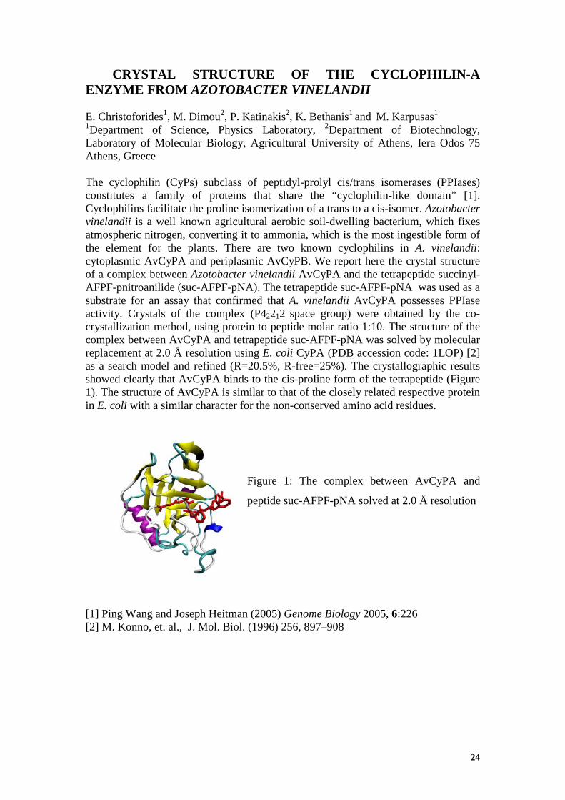
The cyclophilin (CyPs) subclass of peptidyl-prolyl cis/trans isomerases (PPIases) constitutes a family of proteins that share the “cyclophilin-like domain” [1]. Cyclophilins facilitate the proline isomerization of a trans to a cis-isomer. Azotobacter vinelandii is a well known agricultural aerobic soil-dwelling bacterium, which fixes atmospheric nitrogen, converting it to ammonia, which is the most ingestible form of the element for the plants. There are two known cyclophilins in A. vinelandii: cytoplasmic AvCyPA and periplasmic AvCyPB. We report here the crystal structure of a complex between Azotobacter vinelandii AvCyPA and the tetrapeptide succinyl-AFPF-pnitroanilide (suc-AFPF-pNA). The tetrapeptide suc-AFPF-pNA was used as a substrate for an assay that confirmed that A. vinelandii AvCyPA possesses PPIase activity. Crystals of the complex (P42212 space group) were obtained by the co-crystallization method, using protein to peptide molar ratio 1:10. The structure of the complex between AvCyPA and tetrapeptide suc-AFPF-pNA was solved by molecular replacement at 2.0 Å resolution using E. coli CyPA (PDB accession code: 1LOP) [2] as a search model and refined (R=20.5%, R-free=25%). The crystallographic results showed clearly that AvCyPA binds to the cis-proline form of the tetrapeptide (Figure 1). The structure of AvCyPA is similar to that of the closely related respective protein in E. coli with a similar character for the non-conserved amino acid residues.
Figure 1: The complex between AvCyPA and
peptide suc-AFPF-pNA solved at 2.0 Å resolution
[1] Ping Wang and Joseph Heitman (2005) Genome Biology 2005, 6:226 [2] M. Konno, et. al., J. Mol. Biol. (1996) 256, 897–908
25
THE CRYSTAL STRUCTURE OF COBALT-SUBSTITUTED PSEUDOAZURIN FROM ALCALIGENES FAECALIS
R. Gessmann1, C. Kyvelidou2, M. Papadovasilaki1, and K. Petratos1
1I.M.B.B.-FO.R.T.H., P.O. Box 1385, Heraklion 70013, Greece; 2Department of Biology, University of Crete, P.O. Box 2208, Heraklion 71409, Greece
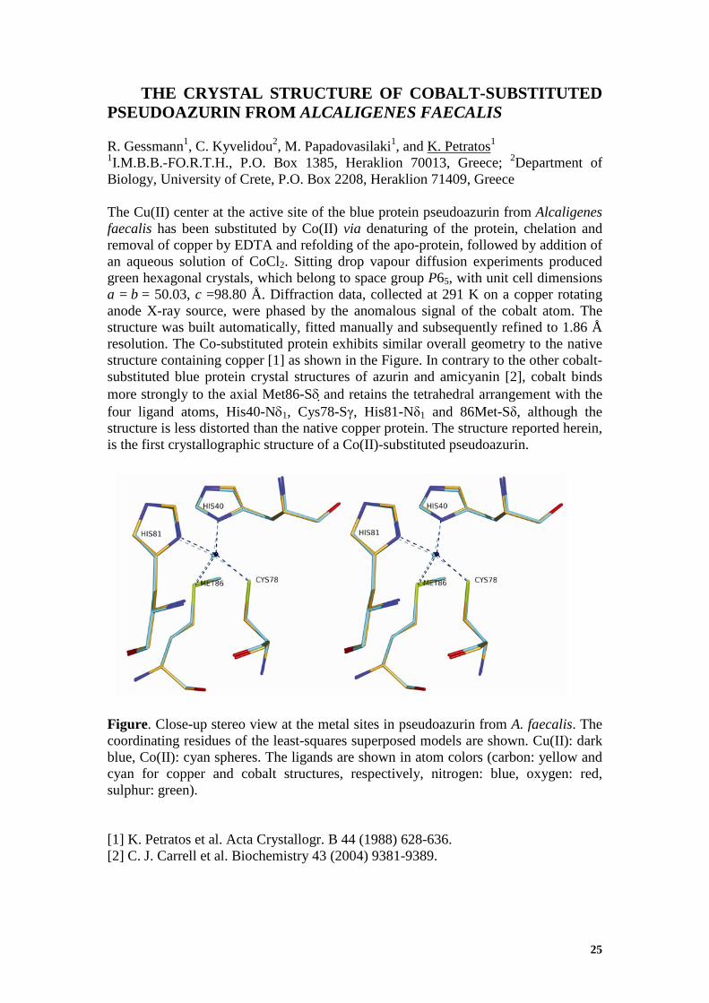
The Cu(II) center at the active site of the blue protein pseudoazurin from Alcaligenes faecalis has been substituted by Co(II) via denaturing of the protein, chelation and removal of copper by EDTA and refolding of the apo-protein, followed by addition of an aqueous solution of CoCl2. Sitting drop vapour diffusion experiments produced green hexagonal crystals, which belong to space group P65, with unit cell dimensions a = b = 50.03, c =98.80 Å. Diffraction data, collected at 291 K on a copper rotating anode X-ray source, were phased by the anomalous signal of the cobalt atom. The structure was built automatically, fitted manually and subsequently refined to 1.86 Å resolution. The Co-substituted protein exhibits similar overall geometry to the native structure containing copper [1] as shown in the Figure. In contrary to the other cobalt-substituted blue protein crystal structures of azurin and amicyanin [2], cobalt binds more strongly to the axial Met86-Sδ and retains the tetrahedral arrangement with the four ligand atoms, His40-Nδ1, Cys78-Sγ, His81-Nδ1 and 86Met-Sδ, although the structure is less distorted than the native copper protein. The structure reported herein, is the first crystallographic structure of a Co(II)-substituted pseudoazurin. Figure. Close-up stereo view at the metal sites in pseudoazurin from A. faecalis. The coordinating residues of the least-squares superposed models are shown. Cu(II): dark blue, Co(II): cyan spheres. The ligands are shown in atom colors (carbon: yellow and cyan for copper and cobalt structures, respectively, nitrogen: blue, oxygen: red, sulphur: green). [1] K. Petratos et al. Acta Crystallogr. B 44 (1988) 628-636. [2] C. J. Carrell et al. Biochemistry 43 (2004) 9381-9389.

26
Saturday, 25th September
Oral presentations

27
MEMBRANE PROTEIN CRYSTALLIZATION IN MESO PHASE WITH A VAPOR DIFFUSION SETUP: METHOD DEVELOPMENT AND APPLICATION RESULTS J. Labahn1, J. Kubicek2, R. Schlesinger1, F. Schäfer2, and G. Büldt1 1Institute of Structural Biology and Biophysics (ISB-2), Research Center Jülich, 52428 Jülich, Germany; 2 Qiagen GmbH, Qiagenstr. 1, 40724 Hilden, Germany For less than hundred membrane proteins their 3-dimensional structure at atomic resolution had been determined. This can be directly traced back to problems in obtaining membrane protein crystals for structural investigations. Landau and Rosenbusch[1] used lipidic meso-phases to accommodate the specific needs of membrane proteins: The lipidic component monoolein self-organizes with water into meso-phases [2] (Fig.1). The cubic phase Pn3m consists of a bi-continuous bilayer that separates two channel systems of aqueous phase. The bilayer is locally 2-dimensional like a cell membrane and therefore allows the incorporation of membrane proteins. But it extends continuously through space and supports therefore diffusion of the protein in three dimensions and crystallization upon dehydration. The vapor diffusion setup allows targeting reproducibly the final hydration level of the meso phase and combines the advantages of vapor diffusion crystallization and in meso crystallization.
An optimized protocol that optionally employs an automatic dispensing system allows a fast screening for crystallization conditions of membrane proteins. Typically crystals with a size of 30 to 70 µm (smallest extension) are obtained. At synchrotrons, crystals typically diffract to 1.2-3.2 Å depending on the protein. [1] E.M. Landau, J.P. Rosenbusch, Proc. Natl. Acad. Sci. USA, 93, 14532 (1996) [2] H. Qiu, M. Caffrey, Biomaterials 21, 223 (2000)
FIG.2: Crystallization of Bacteriorhodopsin (BR): Solid monoolein was hydrated with BR solution, and dehydrated by the vapour diffusion setup to induce crystallization.
FIG.1: Isotherm from monooolein/water phase diagram: The formation of the optically isotropic cubic phases or birefringent lamellar phase upon changing hydration level can be observed with a polarization-microscope.

28
ORDER AND DISORDER IN CHROMATIN-ASSOCIATED PROTEINS
A.S. Politou Laboratory of Biological Chemistry, University of Ioannina, School of Medicine, GR-45110 Ioannina, Greece and Biomedical Research Institute, Foundation for Research and Technology (BRI-FORTH), GR-45110 Ioannina, Greece In eukaryotic nuclei the genetic material is packaged in the form of chromatin. Chromatin is assembled from histones and DNA and in its “open” state allows the accessibility of gene regulatory factors, while in its most condensed state restricts access of the transcriptional machinery to target genes. Chemical modifications of DNA and histones control chromatin remodeling during transcription, mitosis, DNA repair and replication and act individually, sequentially and/or in combination to generate epigenetically heritable gene expression patterns. Distinct structural and functional states of chromatin ranging from “highly active” to “completely silenced" are thus linked with various combinations of histone post-translational modifications, but also with specific nucleosome rearrangements, deposition and/or exchange of histone variants and interactions of chromatin with non-histone regulators. Many protein components of large enzymatic assemblies involved in chromatin remodelling contain specialized protein modules, such as chromo, bromo, Tudor, PWWP and PHD domains. It is thought that each one of these domains interacts with a specific chromatin component, having, ultimately, a distinct effect on chromatin structure and function. However, despite considerable progress in this direction, the functional interaction and the subtle differences between chromatin-associated proteinsremain poorly understood. Our studies of different aspects of epigenetic regulation aim at characterizing the structural determinants involved in this process. More specifically, we have determined the structures and explored the interactions of representative chromatin-associated protein modules. We have also modeled the conformational properties and association tendencies of histone modification patterns. Results obtained so far and emerging information from on-going studies on the structure and energetics of chromatin-related complexes are expected to complement existing functional information and address fundamental questions regarding chromatin organization and regulation of its structure in health and disease.

29
Sponsors presentations

30
Short Wavelength Radiation in Crystallography
E. Hovestreydt1, H. Ott1 and J. Graf2
1 Bruker AXS GmbH, Karlsruhe, Germany, 2 Incoatec GmbH, Geesthacht, Germany. E-Mail: [email protected]
Combining synthetic multilayer mirrors with micro-focus X-ray sources (rotating or
stationary target) has become a standard with in-house X-ray sources for single crystal
diffraction as well as a number of applications in powder diffraction[1]. The
maximum angle of incidence at which a multilayer mirror reflects is significantly
smaller for higher energy radiation, such as Mo-Kα or Ag-Kα radiation than it is for
Cu-Kα radiation. This is why synthetic multilayer mirrors traditionally have been used
for Cu-Kα radiation or softer wavelengths. Modern deposition technology, however,
allows for the reproducible production of high quality multilayer mirrors with smaller
d-spacing. In consequence these mirrors reflect higher energy radiation at larger
angles of incidence [2, 3]. Combined with the latest generation of reliable and stable
micro-focus tubes this provides new high-performance low-power X-ray sources for
shorter wavelengths.
We will present selected results on the use of these high-performance sources. In
particular we will address the use in solid-state, small molecule and high-pressure
crystallography.
[1] J. Wiesmann, C. Hoffmann, J. Graf, C. Michaelsen, Physics Meets Industry (Eds. J. Gegner, F. Haider), New Possibilities for X-ray Diffractometry, Expert Verlag, Renningen. 13 - 20 (2007) [2] C. Michaelsen, J. Wiesmann, C. Hoffmann, A. Oehr, A.B. Storm, L.J. Seijbel, Proc. SPIE, 5193, 211 (2004) [3] M. Schuster, H. Göbel, L. Brügemann, D. Bahr, F. Burgäzy, C. Michaelsen, M. Störmer, P. Ricardo, R. Dietsch, T. Holz, H. Mai, Proc. SPIE, 3767, 183 (1999)
31
Introduction to Microcalorimetry – bridging the gap between
structure and function D. Heep GE Healthcare

32
Saturday, 25th September
Oral presentations

33
BIOLOGICAL STRUCTURES AND MATERIALS AS A SOURCE FOR INSPIRATION FOR THE DESIGN OF NOVEL NANO-BIOMATERIALS Anna Mitraki 1
Department of Materials Science and Technology, c/o Biology Department, University of Crete, and Institute For Electronic Structure and Laser, IESL-FORTH, Vassilika Vouton, 71003 Heraklion, Crete, Greece [email protected]
Natural fibrous proteins include families found in natural materials such as
wool, silk, in tissue components such as collagen, elastin or in virus and bacteriophage adhesins [1]. They have long fascinated scientists and engineers due to their mechanical and elastic properties, and considerable efforts have been made in order to produce artificial materials inspired from these natural proteins. Repetitive sequences, or “building blocks” derived from these fibrous proteins can self-assemble into well-defined structures (wires, tubes etc.) under mild conditions and are relatively inexpensive and easy to manufacture. Moreover, their versatile chemistry as well as their physical and chemical stability makes them good candidates for a wide range of applications. Of particular interest is the possibility of using these peptide nanofibers and nanotubes as templates for the growth of inorganic materials, such as metals, semiconductors, silica, etc. We have been involved for a number of years in the rational design, synthesis and characterization of self-assembling proteins and peptides following identification of building blocks in natural fibrous proteins such as viral fibers. This previous work resulted in the identification of a minimal, octapeptide building block that self-assembles into fibrils. We have recently used these fibrous objects as templates for the growth of inorganic materials [2]. The ability to reliably produce metal-coated fibrils with robust binding of metal nanoparticles is a vital first step towards the exploitation of these fibrils as conducting nanowires with applications in nano-circuitry. I will describe how structural insight and basic biochemical studies, combined with practical integration approaches, can result in concrete materials applications ranging from the nano- to the macro-scale.
[1] A. Mitraki. et al., Advances in Protein Chemistry (2006) 73: 97-124. [2] Kasotakis, E., et al., Biopolymers –Peptide Science (2009) 92: 164-172

34
REFINEMENT AND VALIDATION OF PHOSPHORYLASE KINASE ATP-BINDING SITE INHIBITOR COMPLEXES USING MOLECULAR DYNAMICS AND MM-GBSA CALCULATIONS J.M. Hayes1, V.T. Skamnaki1,2, G. Archontis3, C. Lamprakis1, J. Sarrou1, N. Bischler1, A.-L. Skaltsounis4, S.E. Zographos1, and N.G. Oikonomakos1 1Institute of Organic & Pharmaceutical Chemistry, National Hellenic Research Foundation, 48 Vassileos Constantinou Ave., Athens 11635, Greece; 2Laboratory of Molecular Biophysics and Oxford Centre for Molecular Sciences, Dept. of Biochemistry, Oxford University, Oxford OX1 3QU, U.K.; 3Department of Physics, University of Cyprus, CY1678 Nicosia, Cyprus; 4Division of Pharmacognosy, Dept. of Pharmacy, University of Athens, Panepistimiopolis-Zographou, Athens 15771, Greece Email: [email protected]
Phosphorylase kinase is a key enzyme in the glycogenolysis pathway catalysing the Ca2+ dependent conversion of glycogen phosphorylase b (low activity, low subtrate affinity) to glycogen phosphorylase a (high activity, high substrate affinity). With an aim towards glycogenolysis control in type-2 diabetes, we have investigated via kinetics experiments and computation the potential of indirubin (IC50 > 50 μM), indirubin-3’-oxime (IC50 > 144 nM), KT5720 (Ki = 18.4 nM) and staurosporine (Ki=0.37 nM) as phosphorylase kinase (PhKγtrnc) ATP-binding site inhibitors [1]. Study of the inhibition of phosphorylase kinase by crystallography has proved difficult due to the multimeric nature of the enzyme, with only the ATP-bound complex structure determined but no structures with inhibitors. In such cases, computation provides a valuable alternative. Here, Desmond molecular dynamics simulations are performed to refine the PhKytrnc-inhibitor complexes starting from the ATP-bound receptor structure. MM-GBSA binding free energy calculations are used to validate the refined complexes and explain the kinetics results. Further, the performance of the much less computationally costly Glide docking and induced-fit docking (which includes receptor flexibility using Prime) methods of Schrodinger [2] are compared with the molecular dynamics results. [1] J.M. Hayes et al. Proteins in press [2] Schrödinger Software Suite, Schrödinger, LLC, New York, NY

35
LATTICE EFFECTS IN THE NEW FeAs-BASED NdFeAsO0.85 SUPERCONDUCTOR
1M. Calamiotou, 2I. Margiolaki, 1A. Gantis 3E. Siranidi, 4Z.A. Ren, 4Z.X.Zhao, 3E.Liarokapis. 1Solid State Physics Department, School of Physics, University of Athens, GR-15784 Athens, Greece, 2ESRF, BP 220, Grenoble, Cedex 9, F-38043 France, 3Department of Physics, National Technical University of Athens, 157 80 Athens, Greece, 4Chinese Acad. Sci., Inst. Phys., Natl. Lab. Superconductivity, Beijing 100080, China E-mail: [email protected]
The discovery of superconductivity in FeAs-based layered compounds RFeAsO1-xFx (R=Sm, Nd, Ce, Pr, Gd) [1] and Ba1-xKxFe2As2 [2] belonging to the family of pnictides attracted a lot of experimental and theoretical attention [3]. However the mechanism, which induces superconductivity by doping the parent non superconducting RFeAsO (1111) and BaFe2As2 (122) respectively compounds, is still controversial. Experiments have shown that the geometry of the FeAs4 tetrahedra plays an important role in the superconducting properties. The NdFeAsO0.85 compound is one of the highest Tc (53.5K) pnictides [4] and the tunable oxygen content that leads to the occurrence of superconductivity strongly resembles the situation in cuprate superconductors. We present here recent results of a Synchrotron X-ray Powder Diffraction (SXRPD) study of the NdFeAsO0.85 superconductor that was carried out at the High Resolution ID31 beamline at ESRF. By employing the strategy of thermodiffractograms combined with high statistics scans at selected temperatures we were able to follow the evolution of subtle structural changes as a function of temperature from 10K up to 295K. Anomalies in the geometry of the superconducting FeAs4 coordination tetrahedral have been found that become prominent in the vicinity of 180K, and disappear at the transition temperature Tc [5]. The structural results are discussed in comparison with similar structural and spectroscopic anomalies in other FeAs-based compounds, and in connection to a possible relation to superconductivity. [1] Y. Kamihara et al. J.Am.Chem.Soc.130 (2008) 3296; [2] M. Rotter et al. Phys. Rev. Lett. 101 (2010)107006; [3] C. Cruz et al. Nature 453 (2008) 899; [4] Z.A. Ren et al. Europhys.Lett. 83 (2008) 17002; [5] M. Calamiotou et al. Europhys.Lett., (2010) to be published

36
COMPARISON OF STRUCTURAL PARAMETERS AND CLUSTER ANALYSIS OF UREA-, HEAT- AND FORCE INDUCED DENATURATION OF A1, A2 AND A3 DOMAINS OF VON WILLEBRAND FACTOR USING MOLECULAR DYNAMICS TECHNIQUES
Stefanos Pentas1, Vaia Stathi2 and Geοrgios E. Papadopoulos2
1Department of Physics of condensed matter, Laboratory of Thin Films Nanosystems and Nanometrology, Aristotle University of Thessaloniki, 2Department of Biochemistry & Biotechnology, University of Thessaly. Email: [email protected] Von Willebrand factor is indispensable for the clotting of blood, especially in capillaries of small diameter. Genetic diseases as well as acquired illnesses disrupt its normal function and predispose to haemorrhages. Blood shear stress causes partial unfolding of the vWf a2 domain and renders it vulnerable to ADAMTS13 mediated proteolysis. Renal failure leads to elevated plasma levels of urea, which in turn can enhance the effect of shear stress. The “a” domains were subjected to the combined effect of urea (8 M) and heat (600 K) on the one hand and to the effect of heat alone (600 K) on the other in order to study their relative stabilities and their unfolding pathway using molecular dynamics techniques [2]. Furthermore, steered molecular dynamics has been applied to study the unfolding effect of shear stress. As expected, the a2 domain denatures more rapidly under urea and/or heat than a1 and a3, with a1 domain exhibiting a considerable resistance. An unfolding force of 800 pN was able to expose the site of proteolysis of a2 in ∼2ns, whereas no significant effect was observed in a1 and a3. Principal component analysis identified a five state denaturing mechanism for the a1 and a3 domains and a four state mechanism regarding a2. The differences in the stability and in the denaturing mechanism between the three vWf domains can be attributed to the presence of disulphide bonds and to their position in the tertiary structure of the a1 and a3 domains. Our findings are generally in agreement with experimental studies on the stability of “a” domains in the presence of urea, which however identified less intermediate states [1]. The results of our study support the widely accepted model that proteolysis of a2 by AMADTS13 is facilitated by the unfolding effect (not on a1 and a3) of high shear stress in the blood flow exposing the site of proteolysis in a2. [1] Matthew Auton, Miguel A. Cruz and Joel Moake, Conformational stability and domain unfolding of the Von Willebrand Factor A Domains, J. Mol. Biol. (2007) 366, 986–1000 [2] Kalé L, Skeel R, Bhandarkar M, Brunner R, Gursoy A, Krawetz N, Phillips J, Shinozaki A, Varadarajan K, and Schulten K: NAMD2 (1999) “Greater scalability for parallel molecular dynamics.” J. Comput. Phys. 151: 283-312.

37
SILMOTH CHORION: A FUNCTIONAL, PROTECTIVE AMYLOID STAVROS J. HAMODRAKAS Department of Cell Biology and Biophysics, Faculty of Biology, University of Athens, Panepistimiopolis, Athens 157 01, Greece Amyloid fibrils are associated with more than two-dozen human diseases including Alzheimer’s, Parkinson’s, prions, diabetes type II and many others, collectively called amyloidoses, the so-called “protein conformational diseases”. In striking contrast to the disease-associated amyloids there are also amyloids with native biological activities, that is, functional amyloids. These were discovered after our reported finding (Iconomidou et al., 2000) that silkmoth chorion is a paradigm of a natural protective amyloid. In this talk, I shall shortly review our published work during the past ca. 30 years on silkmoth chorion protein synthetic peptide-analogues, on silkmoth chorion itself and on silkmoth chorion protein structure, that documents rather conclusively that silkmoth chorion proteins form an amyloid structure, after millions of years of molecular evolution, with a mainly protective, but also multifunctional role.

38
Poster presentations
24th - 25th September

39
An overview of diabetes type 2 and glycogen phosphorylase inhibitors. How structure based drug design can prove a valuable therapeutic strategy Kyra-Melinda Alexacou1,2 Spyros E. Zographos,1 Nikos G. Oikonomakos1† and
Demetres D. Leonidas3
1Institute of Organic and Pharmaceutical Chemistry, National Hellenic Research
Foundation, 48 Vassileos Constantinou Avenue, 11635 Athens, Greece, 2Department
of Biology, Chemistry and Pharmacy, Freie Universität Berlin, Takustr. 3, 14195,
Berlin, Germany, 3Department of Biochemistry and Biotechnology, University of
Thessaly, 26 Ploutonos Str. 41221 Larissa, Greece
Diabetes type 2 is a complex disease characterized by altered glucose metabolism and
insulin resistance. Almost half of all people with diabetes type 2 are not aware they
have this life threatening condition, as they can show symptoms years after the onset
of the disease. Glycogen phosphorylase, an allosteric enzyme, plays a pivotal role in
controlling the metabolism of glycogen. It catalyzes the first step in the degradation of
glycogen by releasing glucose-1-phosphate from a long chain of glucose residues.
Various binding sites on the enzyme are known, notably the catalytic, allosteric, new
allosteric and inhibitor sites. By means of kinetic in vitro experiments and X-ray
crystallography experiments, these binding sites were targeted by studying a large
number of glycogen phosphorylase inhibitors as potential hyperglycaemic drugs. In
this project we wished to develop new potent inhibitors by using as scaffolds the
inhibitor N-acetyl-β-D-glucopyranosylamine and oxadiazole derivatives of
glucopyranose. Several groups of compounds were studied including
glucosyltriazolylacetamide, pentacyclic triterpenes, hydroquinone derivatives,
glucopyranosylidene-spiro-iminothiazolones and aldehyde/ketone glucopyranosyl
thiosemicarbazones. These studies have given new insights into fundamental
structural aspects of the enzyme enhancing our understanding of how the enzyme
recognizes and specifically binds ligands, which could be of potential therapeutic
value in the treatment of diabetes type 2.

40
PROTEIN-PROTEIN DOCKING USING THE SHAPE IMPACT DESCRIPTOR A. Axenopoulos1, P. Daras2, G. Papadopoulos3 and E. Houstis1 1Department of Computer & Communication Engineering, University of Thessaly, Volos, Greece; 2Informatics & Telematics Institute, Centre for Research & Technology Hellas, Thessaloniki, Greece; 3Department of Biochemistry & Biotechnology, University of Thessaly, Larissa, Greece The problem of molecular docking involves prediction of a ligand conformation and orientation, also known as pose, within the active site of a receptor. The stability of a pose is a result of several factors (Coulomb forces, hydrogen bonds, Van der Waals forces, hydrophobic interactions). Apart from the physicochemical complementarity, geometric complementarity is also taken into account in several docking algorithms. In this paper, a novel approach for fast protein-protein docking based on geometric complementarity is presented. The complementarity matching is achieved by using an efficient shape descriptor, the Shape Impact Descriptor (SID). SID is applied on small patches extracted from the Solvent Excluded Surface of the molecules. The key property of SID is its rotation invariance, which obviates the need for taking an exhaustive set of rotations for each pair of patches. During the complementarity matching step, the receptor patches are matched with the ligand patches using SID and the most complementary pairs are maintained for the final scoring step. The proposed method is compared with other state-of-the-art geometry-based, rigid-docking approaches [1, 2], demonstrating superior performance. [1] Zujun Shentu, Mohammad Al Hasan, Chris Bystroff and Mohammad J. Zaki, “Context Shapes: Efficient Complementary Shape Matching for Protein-Protein Docking”. Proteins: Structure, Function and Bioinformatics, 70(3):1056-1073. February 2008. [2] R. Chen and Z. Weng. “ZDOCK: An initial-stage protein-docking algorithm”. Proteins: Structure, Function and Genetics, 52:80–87, 2003.
41
CRYSTAL STRUCTURES OF GERANIOL COMPLEXES WITH NATIVE AND PERMETHYLATED β-CYCLODEXTRIN K. Bethanis1, V. Boulaki1, E. Christoforides1, F. Tsorteki1, A. Kokkinou1 and D. Mentzafos1
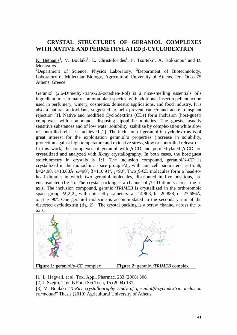
1Department of Science, Physics Laboratory, 2Department of Biotechnology, Laboratory of Molecular Biology, Agricultural University of Athens, Iera Odos 75 Athens, Greece Geraniol (2,6-Dimethyl-trans-2,6-octadien-8-ol) is a nice-smelling essentials oils ingredient, met in many common plant species, with additional insect repellent action used in perfumery, winery, cosmetics, domestic applications, and food industry. It is also a natural antioxidant, suggested to help prevent cancer and acute transplant rejection [1]. Native and modified Cyclodextrins (CDs) form inclusion (host-guest) complexes with compounds disposing lipophilic moieties. The guests, usually sensitive substances and of low water solubility, stabilize by complexation while slow or controlled release is achieved [2]. The inclusion of geraniol in cyclodextrins is of great interest for the exploitation geraniol’s properties (increase in solubility, protection against high temperature and oxidative stress, slow or controlled release). In this work, the complexes of geraniol with β-CD and permethylated β-CD are crystallized and analyzed with X-ray crystallography. In both cases, the host:guest stoichiometry in crystals is 1:1. The inclusion compound, geraniol/β-CD is crystallized in the monoclinic space group P21, with unit cell parameters: a=15.58, b=24.98, c=18.68Å, α=90º, β=110.91º, γ=90º. Two β-CD molecules form a head-to-head dimmer in which two geraniol molecules, distributed in five positions, are encapsulated (fig 1). The crystal packing is a channel of β-CD dimers across the a-axis. The inclusion compound, geraniol/TRIMEB is crystallized in the orthorombic space group P212121, with unit cell parameters: a= 14.903, b= 20.888, c= 27.686Å, α=β=γ=90º. One geraniol molecule is accommodated in the secondary rim of the distorted cyclodextrin (fig. 2). The crystal packing is a screw channel across the b-axis.
Figure 1: geraniol/β-CD complex Figure 2: geraniol/TRIMEB complex [1] L. Hagvall, et al. Tox. Appl. Pharmac. 233 (2008) 308. [2] J. Szejtli, Trends Food Sci Tech, 15 (2004) 137. [3] V. Boulaki “X-Ray crystallography study of geraniol/β-cyclodextrin inclusion compound” Thesis (2010) Agricultural University of Athens.

42
COMPLEX OF THE A-SITE rRNA WITH A SMALL MOLECULE OF RIGID BICYCLIC NATURE J. Birtley, G. Kythreoti, E. Saridakis, I. Katsoulis, A. Papakyriakou, I. Mavridis, D. Vourloumis, I. M. Mavridis
Institute of Physical Chemistry, National Center for Scientific Research “Demokritos”, 15310 Aghia Paraskevi, Athens, Greece The bacterial ribosome is a key target for many natural and semi-synthetic antibiotics, such as aminoglycosides, tetracyclins, and macrolides, which interact preferentially with its RNA components (rRNA). The decoding, or A-site, an internal loop within the 16S rRNA, is the molecular target for natural aminoglycoside antibiotics (Scheme), which interfere with the conformational flexibility of two adenines involved in mRNA decoding, thereby inducing an increased error rate in protein synthesis and ultimately leading to bacterial cell death [1]. Recent crystallographic advances in the field of RNA, along with biological assays of a novel synthetic analog will be presented. The studies aim at improving the pharmacological and resistance profiles of natural aminoglycoside antibiotics, which are used as starting points and thus provide the necessary knowledge, for specifically screening newly developed small molecules [2] against well defined and characterized RNA targets. [1] J. R. Thomas, P. J. Hergenrother, Chemical Reviews, 108 (2008) 1171 [2] I. A Katsoulis, et al. ChemBioChem, 10 (2009) 1969

43
KINETIC AND CRYSTALLOGRAPHIC STUDIES OF GLYCOGEN PHOSPHORYLASE IN COMPLEX WITH D-GLUCOPYRANO-SYLIDENE SPIRO-ISOXAZOLINE DERIVATIVES FOR THE DESIGN OF NEW ANTIDIABETIC DRUGS A.S. Chajistamatiou1,2, D. Gueyrard3, S. Vidal3, J.-P. Praly3, A. Siafaka-Kapadai2, E.D. Chrysina1
1Institute of Organic & Pharmaceutical Chemistry, National Hellenic Research Foundation, 48 Vassileos Constantinou Avenue, Athens, GR-11635, Greece∙, 2Biochemistry Laboratory, Department of Chemistry, National & Kapodistrian University of Athens, Greece, 3Université de Lyon, Institut de Chimie et Biochimie Moléculaires et Supramoléculaires (ICBMS) associé au CNRS, UMR 5246, CPE-Lyon, 43 blvd du 11 Novembre 1918, 69622 Villeurbanne, France The number of people suffering from type 2 diabetes has increased during the last decade worldwide. The enzyme of glycogen phosphorylase (GP) plays a key role in glycogen degradation and controls the release of glucose in blood circulation. Therefore, GP is used as a molecular target for the structure-based design of potential hypoglycaemic drugs for the treatment of type 2 diabetes disease. In particular, the catalytic site of GP has been probed with a large number of glucose analogues1. Recently, D-glucopyranosylidene-spiro-isoxazolines were shown to be potent inhibitors of rabbit muscle GP activity2. To further investigate this new class of compounds, a series of derivatives was synthesized, decorating the aromatic moiety of the lead molecule. The new analogues were assessed in different concentrations for their potency to inhibit the enzyme with kinetic experiments in the direction of glycogen synthesis and their binding mode was studied by X-ray crystallography. The kinetic results indicated that all compounds are competitive inhibitors of GP with high potency (IC50 in μM range). X-ray diffraction data collected at EMBL-Hamburg outstation using synchrotron radiation revealed that the new analogues bind tightly at the catalytic site of the enzyme as clearly indicated by the difference electron density maps. The complex structures of rabbit muscle GP with two of these compounds DCGi37 and DCGi55 will be presented.
Figure 1. 2Fo-Fc difference electron density map showing binding of a D-glucopyranosylidene-spiro-isoxazoline derivative at the catalytic site of RMGPb. [1]. Chrysina E.D. Mini Rev. Med. Chem. (2010) in press [2]. Benltifa M. et al. Bioorg. Med. Chem. 17 (2009) 7368

44
KINETIC AND CRYSTALLOGRAPHIC STUDIES OF
POTENTIAL INHIBITORS OF GLYCOGEN PHOPSHORYLASE, A KEY ENZYME FOR THE TREATMENT OF TYPE 2 DIABETES Μ. Chegkazi1,2,3, Α. Pantzou2, D. Sovantzis1,2, T. Hadjiloi1,2, C. Çismas2, A. Siafaka-Kapadai3, A. Gimisis2, Ε. D. Chrysina1 1Institute of Organic and Pharmaceutical Chemistry, The National Hellenic Research Foundation, 48, Vas. Constantinou Av. 116 35 Athens, Greece. 2Organic Chemistry Laboratory, 3Biochemistry Laboratory Department of Chemistry, University of Athens, Panepistimiopolis, 15771, Athens, Greece. Glycogen phosphorylase (RMGPb) is a molecular target for the treatment of non insulin dependent (type 2) diabetes. This enzyme is responsible in the liver for glycogen degradation to glucose-1 phosphate increasing glucose levels in the bloodstream. Our efforts are focused on the structure-based design of compounds, with stronger inhibitory effect than glucose that stabilize the inactive T-state conformation of the enzyme. We have performed kinetic studies in the direction of glycogen synthesis and collected X-ray diffraction data using SRS at EMBL-Hamburg outstation for a series of β-D-glucopyranosyl N7- and N9- guanine analogues (substituents introduced in C8 of guanine base, Scheme 1). The results showed that β-D-glucopyranosyl N7-guanine analogue is the most potent inhibitor of enzyme activity and binds tightly at the catalytic site of RMGPb. Here we present the crystal structures of RMGPb in complex with all new derivatives determined at ~2.0 Å resolution.
Scheme 1. β-D-glucopyranosyl N7- and N9- guanine analogues [1] L. Somsak et al. Curr. Med. Chem. 15 (2008) 2933-2983 [2] T. Gimisis Mini-Rev. Med. Chem. (2010) in press.

45
DETERMINATION OF PROTEIN – PROTEIN
INTERACTIONS BETWEEN GLUTATHIONE S-TRANSFERASE P1-1 AND C-JUN N-TERMINAL KINASE (JNK-1). L. Chiniadis1, and J. Fernandez-Recio2
1Department of Natural Sciences, Physics lab, Agricultural University of Athens, 11855, 75, Iera Odos street, Athens, Greece 2Life Sciences Department, Barcelona Supercomputing Center, C/ Jordi Girona 29, Barcelona, Spain. Glutathione S-transferases (GST's) are cytosolic enzymes that are members of a large superfamily (mu, pi, alpha, tau) with a diverse function. They catalyze the conjugation of glutathione tripeptide with a wide variety of electrophilic compounds such as toxins, carcinogens and endogenous compounds such as hydroperoxides [1]. JNK-1 belongs to a family of MAPK's (Mitogen-activated protein kinases) that plays a crucial and central role in transducing various extracellular signals into the nuclei. We have applied here docking calculations to examine the interactions between GST P1-1 and JNK-1, which have been observed with other biochemical techniques. We first modelled the loop extension at the C-terminus based on a known X-ray structure, producing five model structures of similar acceptable quality according to TM-scores. Blind docking was performed with pyDock software [3], and restrained docking with pyDockRST [4], with distance restraints based on residues predicted by the Most Interacting Residue method [5], covering all possible combinations of N-terminus and C-terminus interactions of the two proteins. Blind docking predictions confirmed the biochemical results showing that C-terminus of GST P1-1 is interacting with C-terminus of JNK-1. Furthermore, pseudo-energies from restrained docking revealed a significant difference between the energy terms of C-terminus of GST P1-1 binding to C-terminus of JNK 1 and the energy terms of the other terminus domains. Finally, the initial results on molecular dynamics simulations of the complex will be presented.
[1] Wang, T. et al., J Biol Chem, 276, (24), (2001). [2] Adler, V. et al., EMBO Journal, 18, (5), (1999). [3] Cheng, T., Blundell, T.L., Fernandez-Recio, J., Proteins (68), 503, (2007). [4] Chelliah, V., Blundell, T.L., Fernandez-Recio, J., J. Mol. Biol.,(357), 1669, (2006). [5] Papandreou, N., Berezovsky, I.N., Lopes, A., Eliopoulos, E., Chomilier, J., Eur. J. Biochem., (271), 4762, (2004).
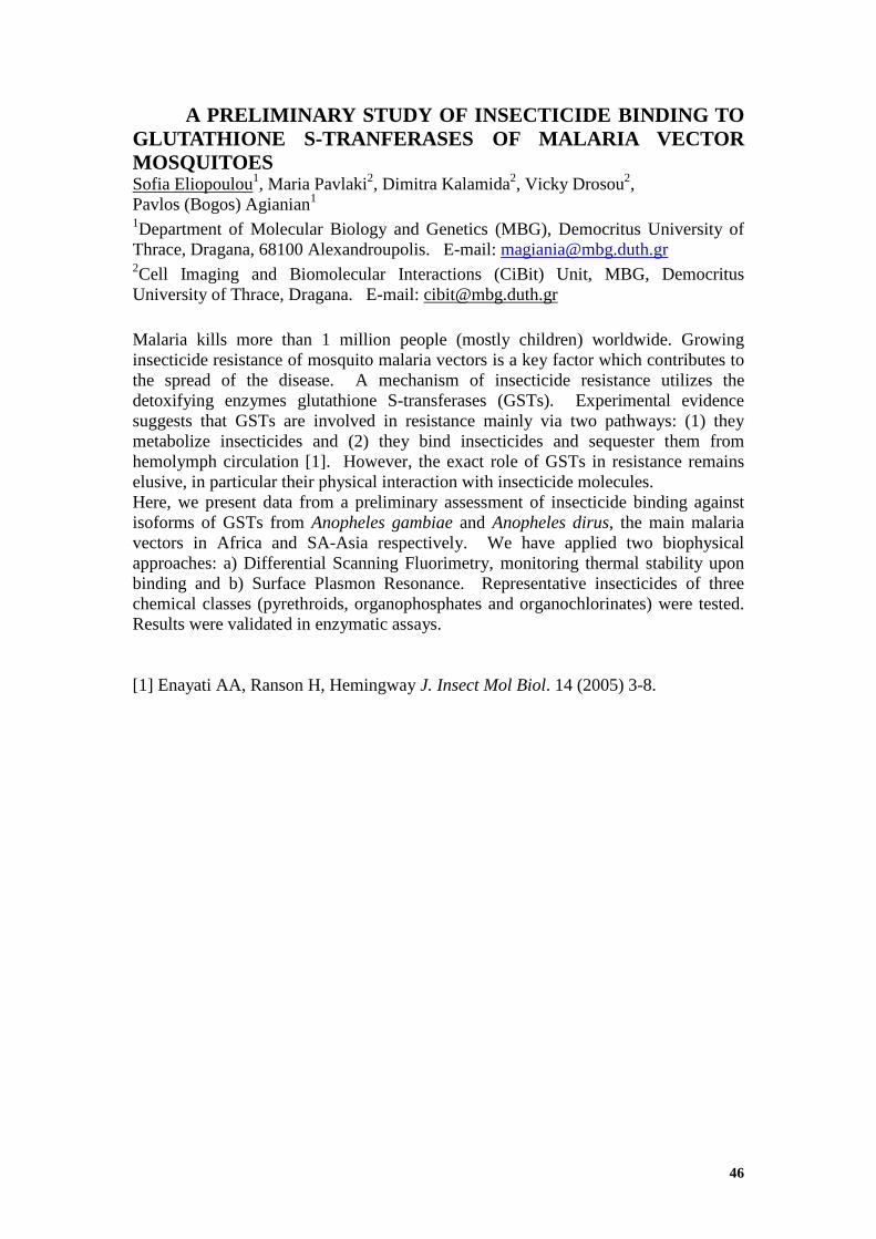
46
A PRELIMINARY STUDY OF INSECTICIDE BINDING TO GLUTATHIONE S-TRANFERASES OF MALARIA VECTOR MOSQUITOES Sofia Eliopoulou1, Maria Pavlaki2, Dimitra Kalamida2, Vicky Drosou2, Pavlos (Bogos) Agianian1 1Department of Molecular Biology and Genetics (MBG), Democritus University of Thrace, Dragana, 68100 Alexandroupolis. E-mail: [email protected] 2Cell Imaging and Biomolecular Interactions (CiBit) Unit, MBG, Democritus University of Thrace, Dragana. E-mail: [email protected]
Malaria kills more than 1 million people (mostly children) worldwide. Growing insecticide resistance of mosquito malaria vectors is a key factor which contributes to the spread of the disease. A mechanism of insecticide resistance utilizes the detoxifying enzymes glutathione S-transferases (GSTs). Experimental evidence suggests that GSTs are involved in resistance mainly via two pathways: (1) they metabolize insecticides and (2) they bind insecticides and sequester them from hemolymph circulation [1]. However, the exact role of GSTs in resistance remains elusive, in particular their physical interaction with insecticide molecules. Here, we present data from a preliminary assessment of insecticide binding against isoforms of GSTs from Anopheles gambiae and Anopheles dirus, the main malaria vectors in Africa and SA-Asia respectively. We have applied two biophysical approaches: a) Differential Scanning Fluorimetry, monitoring thermal stability upon binding and b) Surface Plasmon Resonance. Representative insecticides of three chemical classes (pyrethroids, organophosphates and organochlorinates) were tested. Results were validated in enzymatic assays.
[1] Enayati AA, Ranson H, Hemingway J. Insect Mol Biol. 14 (2005) 3-8.
47
STRUCTURAL AND KINETIC STUDIES OF A1 AND A2 DOMAINS OF VON WILLEBRAND FACTOR Z. Karoulia1, M. Nomikos2, A. Thanassoulas2, G. Papadopoulos1, T. Choli3, C. Stathopoulos4, D.D. Leonidas1, M. Kontou1
1 Department of Biochemistry and Biotechnology, 41221 Larissa, Greece; 2 Institute of Radioisotopes & Radiodiagnostic Products, NCSR “Demokritos”, 1510 Agia Paraskevi, Attiki, Greece; 3 Department of Biochemistry, Faculty of Chemistry, Aristotele University of Thessaloniki, 54124 Thesaloniki, Greece; 4 Department of Biological Chemistry, Faculty of Medicine, University of Patras, 26504 Patras, Greece The multimeric protein von Willebrand factor (vWF) is one of the main players in hemostasis, facilitating thrombus formation at sites of vascular injury by bridging platelets to the exposed subendothelial matrix. Decreased activity of vWF, either due to its insufficient production, or due to the presence of its dysfunctional forms, causes haemorrhagic disposition. However, increased activity of this factor as a result of a disorder in its catabolism increases the danger of thrombosis. The vWF protein contains four types of repeated domains (D1, D2, D', D3, A1, A2, A3, A4, D4, B, C1, C2), that are responsible for different characteristics of molecular binding. The A1–domain (residues 497 – 716) is involved in the binding of vWF to the platelet receptor Ib (GPIb). The A2–domain (residues 717 – 909) is related to the normal feedback of vWF and prevention of blood coagulation. Recent data suggest that the A2 – domain binds to A1, which is thus prevented from binding to the platelets, thus inhibiting platelet thrombus growth [1]. Besides the physiological consequences regarding the control mechanism of vWF – platelet interactions, this finding also open new avenues for the development of novel antithrombotic strategies, directed against the vWF-GPIb axis. In order to use the A2 – domain as a lead compound for the design of new and potent antithrombotic factors, the exact nature of the interaction between the A1 and A2 domains needs to be clarified. The genes for the A1 and A2 domain were amplified by PCR and the products were cloned. The proteins were overexpressed, purified and studied by biophysical methods. The circular dichroism spectra of the two domains mixture suggested a significant secondary structure change, compared to the structures of the isolated domains, which could be interpreted as a generation of a complex between them. The binding between the domains has been confirmed with fluorescence spectroscopy, which enabled us to determine the kinetic data of the interaction [2].
The interaction between A1 and A2 domains has been further studied by a docking procedure followed by molecular dynamics simulations. Three possible models of A1-A2 binding have met the combined biochemical and energetic criteria. In order to definitely identify the binding site of the A1-A2 complex, we planed mutations of those amino acids that are crucial for binding, according to our three models. These mutations were incorporated in the sequence by site directed mutagenesis. The overexpressed mutated proteins will be studied for binding, in order to conclusively define the mode of the two domains interaction.
[1] C. Martin et al. Journal of Thrombosis and Haemostasis, (2007); 5: 1630-70 [2] M. Auton et al. J. Mol. Biol. (2007) 366, 986-1000

48
ROP REVISITED: CHANGING A PROTEIN'S OLOGOMERIZATION STATE AND FOLDING MOTIF WITH A SINGLE AMINO ACID SUBSTITUTION. A MOLECULAR DYNAMICS STUDY. M. Kokkinidou1 , P. S. Georgoulia1 and N. M. Glykos1
1Department of Molecular Biology and Genetics, Democritus University of Thrace, University Campus (Dragana), 68100 Alexandroupoli, Greece Can a simple amino acid-substitution convert a homodimeric protein to a
homotetrameric one with a simultaneous change of the folding motif? Both the
experimental as well as our own computational studies converge to a positive reply to
the above question. We are going to demonstrate that a homotetrameric variant of Rop
in which each monomer has the form of a single α-helix is not only (according to our
computational findings) consistent but it can also account for the available
experimental results [1]. Rop is a homodimeric, antiparallel, 4-α-helical bundle. The
main characteristic of the bundles is the heptad pattern which however in the case of
the Rop molecule is interrupted at the point of the turn. Two main mutations, the 2aa
[2] (which remains dimeric) and the RM6 (which becomes tetrameric), were created
in order to restore the heptad pattern, aiming at the extinction of the turn. It was
hypothesized that the behaviour of 2aa is due to the “wrong” placement of a
hydrophobic aminoacid. By substituting this residue with the more compatible one the
Rop variant 2aaQL was prepared [1] which has been shown by gel filtration [1] to
have tetrameric population. In order to find the structure of this variation we used
homology modeling having RM6 as template structure and we ended-up with two
plausible models. We then used molecular dynamics simulations to check the
consistency and dynamics of these models. The structure of first model cannot be
considered the structure we are looking for because it does not responded to the basic
characteristics of the bundle. On the other hand, the second model is more promising
because by analyzing the simulation we showed that it is more internally consistent
than that of the first. In the future, and if the structure is determined by x-ray
crystallography we could compare the computationally-derived structure with the
experimentally-determined one.
[1] Y. Kyriazidis (2006), Masters Thesis, University of Crete. [2] Vlassi, M. et al. and Kokkinidis, M. (1994), Nat. Struct. Biol., 1: 706-716.

49
DEVELOPMENT OF A SOFTWARE APPLICATION FOR HANDLING PROTEIN CRYSTALLISATION CONDITIONS AND TRIALS D. Markopoulos1,2, E. Manolakos2, and E.D. Chrysina1 1National Hellenic Research Foundation, Institute of Organic & Pharmaceutical Chemistry, 48 Vassileos Constantinou Ave., 11635 Athens, Greece; 2 National and Kapodistrian University of Athens, Graduate Program “Information Technologies in Medicine and Biology”, Department of Informatics & Telecommunications, Panepistimiopolis, Ilissia, Athens 15784, Greece. The new automated high throughput approaches applied in the field of macromolecular crystallization during the last decade have resulted in the generation of a huge amount of information1. To meet the emerging need for handling this information a number of tools has been developed. Our work focuses on the organization of commercial crystallization conditions as well as those derived from a crystallization lab routine in a database to support the crystal optimization process. We have developed a computer software application for handling protein crystallization conditions and trials. The new tool facilitates organization and storage of crystallization related data in a database comprising three main areas of interest: a) commercially available and home-designed screens of crystallization conditions used in each trial, b) Optimization conditions: Keeping record of all the deviations made from the initial crystallization solution in order to achieve the optimum result. c) Evaluation of the crystallization trial results by applying a score (number). In addition, users can make their own search queries and retrieve valuable information for (e.g. crystallization conditions of homologous proteins) the design of further optimization crystallization experiments2 (Figure 1). Additional utilities, such as filing of protein samples allow users submit targeted queries (e.g. crystallization conditions of homologous proteins) and manage such relational database more efficiently.
Figure 1. The Crystallization database manager: results of a query
[1] I.M. Berry et al., Acta Cryst. (2006). D62, 1137–1149 [2] C. E. Kundrot, CMLS, Cell. Mol. Life Sci. Vol. 61, 2004

50
DEVELOPMENT OF AN ELECTRONIC LABORATORY NOTEBOOK FOR STRUCTURE-BASED DRUG DESIGN. E. Mastroleon1,2, E. S. Manolakos2 and E. D. Chrysina1 1National Hellenic Research Foundation, Institute of Organic & Pharmaceutical Chemistry, 48, Vassileos Constantinou Ave. 11635 Athens, Greece 2Graduate Program “Information Technologies in Medicine and Biology”, Department of Informatics & Telecommunications, National and Kapodistrian University of Athens, Panepistimioupolis, Ilissia, 15784 Athens, Greece Dissection of the 3D-structure of macromolecular targets for the design and synthesis of new drugs with high specificity comprises a series of multi-disciplinary approaches in vitro, in silico and in vivo. Recording and maintaining an up-to-date file with the data derived from the methods applied is of great importance, especially after the advanced instrumentation used nowadays and the vast amount of data generated [1]. Our work focuses on the development of a database and a software tool that follows the individual steps of a structure-based drug design approach for the recording, storage and evaluation of the data produced at each stage. More specifically, the user can store data for compounds to be tested as potential drugs, kinetic and crystallographic experiments, as well as results from toxicological, physiological and clinical studies and background information about drugs already launched in the market. The application is web-based, user friendly and implemented in PHP, HTML and SQL, providing a full user management system. Searching with multiple filters, sorting, exporting and saving results, allow the user to handle efficiently the available records (Figure 1). In addition there exist advanced menu options for editing or deleting data and recording experiments explicitly. The tool includes utilities for routine calculations, on-line help and supports connecting to related online databases for data mining.
Figure 1. Preview of results for compounds filed in the database [1] C. Stephan et al. Proteomics 2010, 10, 1230-1249

51
NEW CRYSTAL PACKING IN β-CYCLODEXTRIN INCLUSION COMPLEXES Ι. M. Mavridis, S. D. Chatziefthimiou, E. Hadjoudis Institute of Physical Chemistry, National Center for Scientific Research “Demokritos”, 15310 Aghia Paraskevi, Athens, Greece Cyclodextrins (CDs) are well known water soluble cyclic oligosaccharides, which are used for micro encapsulation of organic molecules inside their relatively apolar cavity. Trials to grow single crystals of the inclusion complexes of βCD with two Schiff bases (anils), derivatives of salicylideneaniline from absolute ethanol resulted in crystals with novel packing: P21, a=15.887(8), b=14.784(12), c=15.29.680 (14) Å, β=103.19(2)o, two βCD monomers in the asymmetric unit (Figure). The lattice has no resemblance to any of lattices of βCD dimers [1]. Refinement did not reveal a guest in a clearly detectable amount, the structure appearing as a βCD-ethanol complex. However, it differs also from any of the three forms, I - III of the βCD/ethanol inclusion complexes [2]. Form I of the latter is a monomeric complex, isomorphous to the structure of the hydrated βCD itself. Forms II and III are dimeric complexes, as most βCD inclusion complexes. The latter forms were derived from trials to crystallise βCD/phenol and βCD/benzoic acid complexes as in the present case. We propose that initially the βCD/anil complexes were formed. Subsequently, the anils were hydrolysed during the long crystallisation process and the hydrolysis products dissolved in ethanol. However, due to the low solubility of βCD in ethanol, the crystals were not destroyed and most of the guest was substituted by ethanol molecules.
[1] P. Giastas et al, Acta Crystallogr. B59 (2003) 287 [2] T. Aree, N. Chaichit, Carbohydr. Res. 343 (2008) 2285

52
SOLID STATE REACTIVITY AND THERMAL STABILITY IN A SERIES OF ORGANIC CO-CRYSTALS. A. Michaelides, S. Skoulika, C. Tsaggaios, V. Dokorou and M. G. Siskos 1Department of Chemistry, University of Ioannina, 45110 Ioannina, Greece
Co-crystals are multiple-component crystals in which two or more molecules that form solid compounds under ambient conditions coexist through hydrogen bonding. They represent a long known but relatively unexplored class of compounds. These solids are very attractive to the pharmaceutical or chemical research because they offer opportunities to modify the chemical or physical properties of the molecules such as solubility and melting point without the need to make or break covalent bonds. Furthermore co-crystallization has recently been used to bring carbon carbon double bonds within reactive distance (3.5-4.2 Ǻ).
We prepared and studied by X-ray crystallography a series of co-crystals between aliphatic or aromatic dicarboxylic acids and trans-1,2-bis(4-pyridyl)ethylene (bpe). In all cases we observed formation of polymeric chains through strong OH…N hydrogen bonds. The structures were further stabilized by aromatic interactions and multiple weak CH…O bonds, resulting in a significant improvement of their thermal stability compared to that of the two components. There is a strong correlation between melting point and crystal packing of the co-crystals. In the case of the co-crystal between fumaric acid and bpe the polymeric chains are stacked in such a way that the two components form distinct columns with olefin distance of only 3.8 Ǻ, offering the possibility of performing two different photochemical [2+2] cycloaddition reactions. However, upon UV irradiation, photochemical reaction takes place only between molecules of bpe. This is due to the fact that bpe is a much better light absorber than fumaric acid.
53
A STRUCTURAL MODEL OF MEMBERS OF THE CPF FAMILY OF CUTICULAR PROTEINS: POSSIBLE ROLE IN M/S DIFFERENTIATION Nikos C. Papandreou1, Vassiliki A. Iconomidou1, Judith H. Willis2 and Stavros J. Hamodrakas1 1 Department of Cell Biology and Biophysics, Faculty of Biology, University of Athens, Athens 157 01, Greece, 2 Department of Cellular Biology, University of Georgia, Athens, GA 30602, USA The physical properties of cuticle are determined by the structure of its two major components, cuticular proteins (CPs) and chitin, and, also, by their interactions. A common consensus region (extended R&R Consensus) found in the majority of cuticular proteins, the CPRs, binds to chitin. Previous work established that β-pleated sheet predominates in the Consensus region and we proposed that it is responsible for the formation of helicoidal cuticle. Remote similarities between CPRs and a lipocalin, bovine plasma retinol binding protein (RBP), led us to suggest an antiparallel β-sheet half-barrel structure as the basic folding motif of the R&R Consensus. There are several other families of cuticular proteins. One of the best defined is CPF. Its four members in Anopheles gambiae are expressed during the early stages of either pharate pupal or pharate adult development, suggesting that the proteins contribute to the outer regions of the cuticle, the epi- and/or exocuticle. These proteins did not bind to chitin in the same assay used successfully for CPRs. Although CPFs are distinct in sequence from CPRs, the same lipocalin could also be used to derive a homology model. For the CPFs, the basic folding motif predicted is an antiparallel β-sheet full-barrel structure. Possible implications of this structure are discussed, in relation to M/S differentiation.

54
THE CRYSTAL STRUCTURE OF RABBIT MUSCLE GLYCOGEN PHOSPHORYLASE b IN COMPLEX WITH N-(β-D-GLUCOPYRANOSYL) GUANIDINOACETAMIDE. P.V. Skourti1,2, D. Loganathan3, A. Siafaka-Kapadai2, E.D. Chrysina1 1Institute of Organic and Pharmaceutical Chemistry, National Hellenic Research Foundation, 48, Vassileos Constantinou Avenue, Athens 11635, Greece, 2Laboratory of Biochemistry Department of Chemistry, National & Kapodistrian University of Athens, Greece, 3Regional Sophisticated Instrumentation Centre, Indian Institute of Technology Madras, Chennai 600 036, India. There is growing evidence that Type 2 diabetes disease is interconnected with other major diseases such as cancer and micro- and macrovascular disease (including cardiovascular disease) by stimulating insulin resistance pathways involved in inflammation, cell proliferation and atherosclerosis. Therefore, long-term studies of glycogen metabolism, one of the major causes of the elevated blood glucose levels reveal that the enzymes implicated in glycogenolysis / gluconeogenesis metabolic pathway could serve as molecular targets for the treatment of type 2 diabetes. Glycogen phosphorylase enzyme (GP) is directly involved in glycogen degradation. The crystal structure of GP in complex with different classes of compounds has been determined with X-ray crystallography in the search of new potent inhibitors1. N-acetyl-β-D-glucopyranosylamine was one of the first glucose analogues studied in complex with GP and employed as lead molecule for further optimisation2. The knowledge obtained from this complex led to the design and synthesis of new derivatives with the aim to improve their inhibitory effect on GP activity. To this end, N-(β-D-glucopyranosyl)guanidinoacetamide was studied by kinetic and crystallographic experiments using SRS at EMBL-Hamburg outstation. The results showed that it is a potent inhibitor of GP and its crystal structure in complex with the enzyme determined at 2.03 Å resolution, indicated that binds tightly at the catalytic site with the amide nitrogen (at C1 position in β-configuration of glucopyranose) forming the ‘characteristic’ hydrogen bond interaction with the backbone His377 O2.
Figure 1. Schematic representation of 2Fo-Fc electron density map showing binding of N-(β-D-glucopyranosyl)guanidinoacetamide at the catalytic site of GP. [1]. L. Somsak et al. Curr. Med. Chem. 15 (2008) 2933 [2]. E.D. Chrysina Mini-Rev. Med. Chem. (2010) In press

55
STUDYING BINDING CHARACTERISTICS OF THE ANOPHELES GAMBIAE ODORANT BINDING PROTEINS. T. Thireou1, and E. Eliopoulos1 1Department of Biotechnology, Agricultural University of Athens, 75 Iera Odos, Votanikos, Athens 118-55, Greece Significant effort towards understanding anopheline mosquito olfaction has been focused on odorant binding proteins (OBPs) that are essential for effective and efficient perception of odour stimuli. Only the OBP1 structure has been experimentally determined (pdb code: 2ERB). The homology models of several members of the OPB family were created using protein structure homology modelling [1]. The derived models were superimposed with the crystal structure of OBP1 using the Dali server [2] and the aminoacid residues comprising the binding pockets of the various OBPs were identified. Structural superposition revealed interesting physicochemical and morphological features that potentially influence binding affinity and recognition. In order to understand ligand-receptor interactions, various ligands such as known insect repellents, volatile components of plant extracts and other organic and synthetic compounds were tested for in silico binding. Additionally we queried PubChem [3] for structurally similar compounds. The binding of those ligands to OBP1 and the model structures of the other OBPs, was studied using the molecular docking program AutoDock with a Lamarckian genetic algorithm (LGA) search method [4]. Docking simulations depicted affinity variations of the selected ligands for the studied OBPs. The above mentioned molecular modelling calculations can be a valuable tool for investigating the mechanisms underlying the specificity of odour recognition and facilitating the identification of new potentially effective ligands. [1] K. Arnold et al. Bioinformatics 22 (2006) 195 [2] H. Hasegawa and L. Holm. Curr. Opin. Struct. Biol. 19 (2009) 341 [3] http://pubchem.ncbi.nlm.nih.gov/ [4] G. Morris et al. Journal of Computational Chemistry 19 (1998) 1639

56
KINETIC AND MODELING STUDIES OF A NEW SET OF β-D-GLYCOSE PYRIMIDINE DERIVATIVES BINDING AT THE GLYCOGEN PHOSPHORYLASE CATALYTIC SITE V.G. Tsirkone1, A. Katsandi2, S. Manta2, E. Tsoukala2, J.M. Hayes1, M. Kontou2, D. Komiotis2, D.D. Leonidas2
1Institute of Organic and Pharmaceutical Chemistry, National Hellenic Research Foundation, 48 Vas. Constantinou Ave, Athens 11635, Greece. 2Department of Biochemistry and Biotechnology, University of Thessaly, 26 Ploutonos Str., 41221 Larissa, Greece. [email protected], [email protected] Regulation of the glycogen metabolism is a therapeutic strategy for blood glucose control in type 2 diabetes. The glycogen phosphorylase (GP) enzyme plays a key role in the glycogenolysis pathway; hence GP has been widely used as a target for compounds that might prevent glycogen breakdown under high glucose conditions [1]. Towards this goal, the large amount of deposited GP-inhibitor complexes to the RCSB Protein Data Bank has facilitated a structure based drug design (SBDD) approach. Modeling can serve as a useful tool in SBDD and the design of better GP inhibitors [2]. Here, kinetics experiments and Glide docking calculations have been performed to determine the inhibitory potential of a new set of β-D-glucose pyrimidine derivatives. Kinetic experiments with rabbit muscle GPb have shown that these compounds are competitive inhibitors of the enzyme with IC50 values range between 6 mM and 6 µM. These values are in agreement with docking results. For the docking, quantum mechanics polarized ligand docking was used, with ligand charge polarization effects in the field of the receptor accounted for using electrostatic potential (ESP) fit charges. The latter method has proved effective in previous binding studies at the GP catalytic site [3,4].
1.Somsák L.; Czifrák K.; Tóth M.; Bokor E.; Chrysina E.D.; Alexacou K.M.; Hayes J.M.; Tiraidis C.; Lazoura E.; Leonidas D.D.; Zographos S.E.; Oikonomakos N.G. (2008) Curr. Med. Chem., 15, 2933.
2.Hayes J.M.; Leonidas D.D. (2010) Mini Rev. Med. Chem., in press. 3.Tsirkone V.G.; Tsoukala E.; Lamprakis C.; Manta S.; Hayes J.M.; Skamnaki V.T.;
Drakou C.; Zographos S.E.; Komioti D.; Leonidas D.D (2010) Biorg. Med. Chem., 18, 3413.
4.Benltifa M.; Hayes J.M.; Vidal S.; Gueyrard D.; Goekjian P.G.; Praly J-P.; Kiziliz G.; Tiraidis C.; Alexacou K.M.; Chrysina E.D.; Zographos S.E.; Leonidas D.D.; Archontis G., Oikonomakos N.G. (2009) Biorg. Med. Chem., 17, 7368.
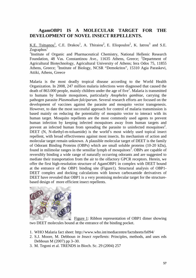
57
AgamOBP1 IS A MOLECULAR TARGET FOR THE DEVELOPMENT OF NOVEL INSECT REPELLENTS. K.E. Tsitsanou1, C.E. Drakou1, A. Thiraiou2, E. Eliopoulos2, K. Iatrou3 and S.E. Zographos1
1Institute of Organic and Pharmaceutical Chemistry, National Hellenic Research Foundation, 48 Vas. Constantinou Ave., 11635 Athens, Greece; 2Department of Agricultural Biotechnology, Agricultural University of Athens; Iera Odos 75, 11855 Athens, Greece; 3Institute of Biology, NCSR “Demokritos”, 15310 Agia Paraskevi, Attiki, Athens, Greece Malaria is the most deadly tropical disease according to the World Health Organization. In 2008, 247 million malaria infections were diagnosed that caused the death of 863.000 people, mainly children under the age of five1. Malaria is transmitted to humans by female mosquitoes, particularly Anopheles gambiae, carrying the pathogen parasite Plasmodium falciparum. Several research efforts are focused on the development of vaccines against the parasite and mosquito vector transgenesis. However, to date the most successful approach for control of malaria transmission is based mainly on reducing the potentiality of mosquito vector to interact with its human target. Mosquito repellents are the most commonly used agents to prevent human infection by keeping infected mosquitoes away from human targets and prevent an infected human from spreading the parasite to uninfected mosquitoes2. DEET (N, N-diethyl-m-toluamide) is the world’s most widely used topical insect repellent, with broad effectiveness against most insects. Its mechanism of action and molecular target remain unknown. A plausible molecular target of DEET is the family of Odorant Binding Proteins (OBPs) which are small soluble proteins (10-20 kDa), found in milimolar ranges in the sensillar lymph of mosquitoes3. OBPs are capable of reversibly binding a wide range of naturally occurring odorants and are suggested to mediate their transportation from the air to the olfactory GPCR receptors. Herein, we offer the first high-resolution structure of AgamOBP1 in complex with DEET bound at the entrance of the OBP1 binding site (Figure1). Structural analysis of OBP1-DEET complex and docking calculations with known carboxamide derivatives of DEET have revealed that OBP1 is a very promising molecular target for the structure-based design of more efficient insect repellents.
Figure 1: Ribbon representation of OBP1 dimer showing
two DEET molecules bound at the entrance of the binding pocket. 1. WHO Malaria fact sheet: http://www.who.int/mediacentre/factsheets/fs094/ 2. S.J. Moore, M. Debboun in Insect repellents: Principles, methods, and uses eds
Debboun M (2007) pp 3–30. 3. M. Tegoni et al. TRENDS in Bioch. Sc. 29 (2004) 257

58
Authors’ index Agianian, P., pg. 46 Alexacou, K-M., pg. 39 Ambrazi, M., pg. 19 Archontis, G., pg. 34 Axenopoulos, A., pg. 40 Bethanis, K., pg. 24, 41 Birtley, J., pg. 42 Bischler, N., pg. 34 Boulaki, V., pg. 41 Büldt, G., pg. 27 Calamiotou, M., pg. 35 Chajistamatiou, A.S., pg. 43 Charetidis, M., pg. 19 Charova, S., pg. 19 Chatzi, K., pg. 21 Chatziefthimiou, S. D. , pg. 51 Chegkazi, Μ., pg. 44 Chiniadis, L., pg. 45 Choli, T., pg. 47 Christakopoulos, P., pg. 18 Christoforides, E., pg. 24, 41 Chrysina, Ε. D., pg. 18,43,44,49,50,54 Çismas, C., pg. 44 Daras, P., pg. 40 Dimarogona, M., pg. 18 Dimou, M., pg. 24 Dokorou, V., pg. 52 Drakou, C.E., pg. 57 Drosou, V., pg. 46 Economou, A., pg. 21 Eliopoulos, E., pg. 55,57 Eliopoulou, S., pg. 46 Fernandez-Recio, J., pg. 45 Gantis, A., pg. 35 Gazi, A.D., pg. 19 Gelis, I., pg. 21 Georgoulia, P.S., pg. 20,48 Gessmann, R., pg. 25 Gimisis, A., pg. 44 Glykos, N.M., pg. 20,48 Gouridis, G., pg. 21 Graf, J, pg. 30 Gueyrard, D., pg. 43 Hadjiloi, T., pg. 44 Hadjoudis, E., pg. 51 Hamodrakas, S.J., pg. 37,53 Hayes, J.M., pg. 34,56 Heep, D., pg. 31
Houstis, E., pg. 40 Hovestreydt, E., pg. 30 Iatrou, K., pg. 57 Iconomidou, V.A., pg. 53 Kalamida, D., pg. 46 Kalodimos, C.G., pg. 21 Karamanou, S., pg. 21 Karoulia, Z., pg. 47 Karpusas, M., pg. 24 Katinakis, P., pg. 24 Katsandi, A., pg. 16, 56 Katsoulis, I., pg. 42 Kokkinidis, M., pg. 19 Kokkinidou, M., pg. 48 Kokkinou, A., pg. 41 Komiotis, D., pg. 56 Kontou, M., pg. 16,47,56 Kotsakis, S.D., pg. 22 Koukaki, M., pg. 21 Kouretas, D., pg. 16 Kubicek, J., pg. 27 Kythreoti, G., pg. 42 Kyvelidou, C., pg. 25 Labahn, J., pg. 27 Lamprakis, C., pg. 34 Leonidas, D.D., pg. 16,39,47,56 Liarokapis, E., pg. 35 Loganathan, D., pg. 54 Manolakos, E., pg. 49 Manolakos, E. S., pg. 50 Manta, S., pg. 56 Margiolaki, I., pg. 35 Markopoulos, D., pg. 49 Mastroleon, E., pg. 50 Mavridis, I., pg. 42 Mavridis, I.M., pg. 42 Mavridis, M., pg. 51 McInnes, C., pg. 15 Mentzafos, D., pg. 41 Michaelides, A., pg. 23,52 Miriagou, V., pg. 22 Mitraki, A., pg. 33 Nomikos, M., pg. 47 Oikonomakos, N.G., pg. 34,39 Orfanoudaki, G., pg. 21 Ott, H., pg. 30 Panopoulos, N.J., pg. 19 Pantzou, Α., pg. 44

59
Papadopoulos, G., pg. 40,47 Papadopoulos, G.E., pg. 36 Papadovasilaki, M., pg. 25 Papakyriakou, A., pg. 42 Papandreou, N.C., pg. 53 Pavlaki, M., pg. 46 Pentas, S., pg. 36 Petinaki, E., pg. 22 Petratos, K., pg. 25 Politou, A.S., pg. 28 Praly, J.-P., pg. 43 Psarra A.-M., pg. 16 Ren, Z.A., pg. 35 Sardis, M.-F., pg. 21 Saridakis, E., pg. 42 Sarrou, J., pg. 34 Savvidou, M., pg. 16 Schäfer, F., pg. 27 Schlesinger, R., pg. 27 Siafaka-Kapadai, A., pg. 43,44,54 Siranidi, E., pg. 35 Siskos, M.G., pg. 23,52 Skaltsounis, A.-L., pg. 34 Skamnaki, V.T., pg. 16,34 Skoulika, S, pg. 23,52 Skourti, P.V., pg. 54 Sovantzis, D., pg.44 Stathi, V., pg. 36 Stathopoulos, C, pg. 47 Thanassoulas, A, pg. 47 Thiraiou, A., pg. 57 Thireou, T., pg. 55 Topakas, E., pg. 18 Tsaggaios, C., pg. 52 Tsirkone, V.G., pg. 56 Tsitsanou, K.E., pg. 57 Tsorteki, F., pg. 41 Tsoukala, E., pg. 56 Tzelepi, E., pg. 22 Tzouvelekis, L. S. pg. 22 V. Dokorou, pg. 52 Vidal, S., pg. 43 Vourloumis, D., pg. 42 Walkinshaw, M., pg. 13 Willis, J.H., pg. 53 Zaccai, G., pg. 17 Zhao, Z.X., pg. 35 Zographos, S.E., pg. 34,39,57

60
NOTES

61