Helical side chain chemistry of a peptoid-based SP-C ...
12
FULL PAPER Helical side chain chemistry of a peptoid-based SP-C analogue: Balancing structural rigidity and biomimicry Nathan J. Brown 1 | Jennifer S. Lin 2 | Annelise E. Barron 2 1 Department of Chemical and Biological Engineering, Northwestern University, Evanston, Illinois 2 Department of Bioengineering, Stanford University, Stanford, California Correspondence Annelise E. Barron, Department of Bioengineering, Stanford University, 443 Via Ortega, Shriram Center, Room 229, Stanford, CA 94305. Email: [email protected] Funding information National Institutes of Health, Grant/Award Numbers: 2 R01 HL067984, Biotechnology Training Program; U.S. Department of Energy, Grant/Award Number: DE-AC02-05CH11231 Abstract Surfactant protein C (SP-C) is an important constituent of lung surfactant (LS) and, along with SP-B, is included in exogenous surfactant replacement therapies for treating respi- ratory distress syndrome (RDS). SP-C's biophysical activity depends upon the presence of a rigid C-terminal helix, of which the secondary structure is more crucial to function- ality than precise side-chain chemistry. SP-C is highly sequence-conserved, suggesting that the β-branched, aliphatic side chains of the helix are also important. Nonnatural mimics of SP-C were created using a poly-N-substituted glycine, or “peptoid,” back- bone. The mimics included varying amounts of α-chiral, aliphatic side chains and α-chi- ral, aromatic side chains in the helical region, imparting either biomimicry or structural rigidity. Biophysical studies confirmed that the peptoids mimicked SP-C's secondary structure and replicated many of its surface-active characteristics. Surface activity was optimized by incorporating both structurally rigid and biomimetic side chain chemistries in the helical region indicating that both characteristics are important for activity. By balancing these features in one mimic, a novel analogue was created that emulates SP-C's in vitro surface activity while overcoming many of the challenges related to natural SP-C. Peptoid-based analogues hold great potential for use in a synthetic, biomi- metic LS formulation for treating RDS. KEYWORDS biomimetic, lung surfactant, peptidomimetics, peptoid, SP-C 1 | INTRODUCTION Premature babies, that is those born with less than 28-32 weeks gesta- tion, have a high incidence of respiratory distress as a result of under- developed lungs and a subsequent deficiency of functional lung surfactant (LS) material, [1,2] the thin mono-/multi-layer coating of lipids and proteins that lines the alveolar surfaces of vertebrate lungs. Infant respiratory distress syndrome (IRDS), once a primary cause of infant mortality in the United States, is now regularly treated with the instilla- tion of exogenous surfactant material into the immature airways. [3] The exogenous surfactant functions in place of the native LS to maintain alveolar patency and to lessen the work of breathing until the patient is able to secrete functional LS. Despite the efficacy of these natural, animal-derived surfactant preparations, the potential for cross-species transfer of infectious agents, high production costs, and batch-to-batch variability continue to be ongoing concerns. [4] Surfactant replacement may benefit the treatment of other respiratory-related disorders for both infants and adults; however, to increase the applicability of surfac- tant replacement, a much greater quantity is needed at a reasonable cost. [5,6] These concerns and limited production potential have prompted the research and development of a synthetic surfactant for- mulation that can function like native LS. [7] However, this endeavor has been more daunting than originally anticipated, largely due to the ABBREVIATIONS: FM, fluorescence microscopy; IRDS, infant respiratory distress syndrome; LC, liquid condensed; LE, liquid expanded; LS, lung surfactant; LWSB, Langmuir-Wilhelmy surface balance; PBS, pulsating bubble surfactometer; SP-C, surfactant protein C. Received: 16 November 2018 Revised: 15 March 2019 Accepted: 18 March 2019 DOI: 10.1002/bip.23277 Biopolymers. 2019;e23277. wileyonlinelibrary.com/journal/bip © 2019 Wiley Periodicals, Inc. 1 of 12 https://doi.org/10.1002/bip.23277
Transcript of Helical side chain chemistry of a peptoid-based SP-C ...

F U L L P A P E R
Helical side chain chemistry of a peptoid-based SP-C analogue:Balancing structural rigidity and biomimicry
Nathan J. Brown1 | Jennifer S. Lin2 | Annelise E. Barron2
1Department of Chemical and Biological
Engineering, Northwestern University,
Evanston, Illinois
2Department of Bioengineering, Stanford
University, Stanford, California
Correspondence
Annelise E. Barron, Department of
Bioengineering, Stanford University, 443 Via
Ortega, Shriram Center, Room 229, Stanford,
CA 94305.
Email: [email protected]
Funding information
National Institutes of Health, Grant/Award
Numbers: 2 R01 HL067984, Biotechnology
Training Program; U.S. Department of Energy,
Grant/Award Number: DE-AC02-05CH11231
Abstract
Surfactant protein C (SP-C) is an important constituent of lung surfactant (LS) and, along
with SP-B, is included in exogenous surfactant replacement therapies for treating respi-
ratory distress syndrome (RDS). SP-C's biophysical activity depends upon the presence
of a rigid C-terminal helix, of which the secondary structure is more crucial to function-
ality than precise side-chain chemistry. SP-C is highly sequence-conserved, suggesting
that the β-branched, aliphatic side chains of the helix are also important. Nonnatural
mimics of SP-C were created using a poly-N-substituted glycine, or “peptoid,” back-
bone. The mimics included varying amounts of α-chiral, aliphatic side chains and α-chi-
ral, aromatic side chains in the helical region, imparting either biomimicry or structural
rigidity. Biophysical studies confirmed that the peptoids mimicked SP-C's secondary
structure and replicated many of its surface-active characteristics. Surface activity was
optimized by incorporating both structurally rigid and biomimetic side chain chemistries
in the helical region indicating that both characteristics are important for activity.
By balancing these features in one mimic, a novel analogue was created that emulates
SP-C's in vitro surface activity while overcoming many of the challenges related to
natural SP-C. Peptoid-based analogues hold great potential for use in a synthetic, biomi-
metic LS formulation for treating RDS.
K E YWORD S
biomimetic, lung surfactant, peptidomimetics, peptoid, SP-C
1 | INTRODUCTION
Premature babies, that is those born with less than 28-32 weeks gesta-
tion, have a high incidence of respiratory distress as a result of under-
developed lungs and a subsequent deficiency of functional lung
surfactant (LS) material,[1,2] the thin mono-/multi-layer coating of lipids
and proteins that lines the alveolar surfaces of vertebrate lungs. Infant
respiratory distress syndrome (IRDS), once a primary cause of infant
mortality in the United States, is now regularly treated with the instilla-
tion of exogenous surfactant material into the immature airways.[3] The
exogenous surfactant functions in place of the native LS to maintain
alveolar patency and to lessen the work of breathing until the patient is
able to secrete functional LS. Despite the efficacy of these natural,
animal-derived surfactant preparations, the potential for cross-species
transfer of infectious agents, high production costs, and batch-to-batch
variability continue to be ongoing concerns.[4] Surfactant replacement
may benefit the treatment of other respiratory-related disorders for
both infants and adults; however, to increase the applicability of surfac-
tant replacement, a much greater quantity is needed at a reasonable
cost.[5,6] These concerns and limited production potential have
prompted the research and development of a synthetic surfactant for-
mulation that can function like native LS.[7] However, this endeavor has
been more daunting than originally anticipated, largely due to the
ABBREVIATIONS: FM, fluorescence microscopy; IRDS, infant respiratory distress syndrome;
LC, liquid condensed; LE, liquid expanded; LS, lung surfactant; LWSB, Langmuir-Wilhelmy
surface balance; PBS, pulsating bubble surfactometer; SP-C, surfactant protein C.
Received: 16 November 2018 Revised: 15 March 2019 Accepted: 18 March 2019
DOI: 10.1002/bip.23277
Biopolymers. 2019;e23277. wileyonlinelibrary.com/journal/bip © 2019 Wiley Periodicals, Inc. 1 of 12
https://doi.org/10.1002/bip.23277

difficulty in producing or mimicking the hydrophobic proteins of LS,
surfactant proteins B and C (SP-B and SP-C).
Although SP-B and SP-C represent only a small portion of LS
(1-2 wt%), SP-B is critical for biophysical function[8] and SP-C is
important for normal postnatal lung function.[9] LS, a complex bioma-
terial composed of approximately 10% proteins and 90% lipids, is
essential for normal respiration. In the airways, this dynamic film func-
tions to: (1) form an interfacial surfactant layer by rapidly adsorbing to
the alveolar air-liquid interface, (2) prevent alveolar collapse by greatly
reducing the interfacial surface tension upon compression to near-
zero values (expiration), and (3) reduce the maximum surface tension
and diminish the work of breathing by efficiently respreading upon
expansion (inhalation).[10] Dipalmitoyl phosphatidylcholine (DPPC),
the primary lipid component of LS, along with the other saturated
phospholipids, are the main surface tension-reducing entities in
LS. The same biophysical properties that allow saturated phospho-
lipids to achieve very low surface tensions also prevents them from
rapidly reabsorbing and respreading upon expansion.[11] Adding fluid,
unsaturated, or neutral phospholipids to LS improves respreading and
slightly enhances LS adsorption to the air-liquid interface, but conse-
quently increases minimum surface tension.[11,12] The addition of the
hydrophobic proteins, SP-B and SP-C, to the lipid portion greatly
enhances surfactant adsorption, stability, and recycling of the lipid
film.[13,14] The inclusion of these surfactant-specific proteins is, there-
fore, necessary for proper respiration, as their omission results in
lethal respiratory failure.[15–18]
SP-C is a helical and extraordinarily hydrophobic, 35-amino acid
long protein that is highly sequenced-conserved among all mammalian
species.[2,19] Its 37-Å-long helical region is capable of traversing the
lipid bilayer and associates and interacts with the interior of phospho-
lipid acyl chains.[7] In addition, the N-terminal region of natural SP-C
contains two palmitoylated cysteines at positions 5 and 6 that are
thought to play a key role in maintaining the association between
SP-C and associated phospholipids with the interfacial surfactant film
at very high levels of compression, acting as a hydrophobic “anchor”
for the excluded surfactant material and aiding in the reincorporation
of this material during expansion.[7,20] Palmitoylation has also been
shown to be vital in maintaining the rigid α-helical structure of
SP-C.[21] The important biophysical activities of SP-C and its inclusion
in animal-derived surfactants suggest that the protein is a critical con-
stituent of a synthetic surfactant preparation; however, large-scale
production is exceedingly difficult due to its highly hydrophobic
nature.[4] Although SP-C is relatively small and lacks any tertiary struc-
ture, the native protein and sequence-identical analogues are difficult
to handle at all stages of experimentation. The poly-valyl helix is
composed entirely of aliphatic residues with β-branched side chains
that spontaneously convert into β-sheet aggregate structures with
reduced surface activity in the absence of phospholipids.[22–24] The
difficulties associated with native SP-C have led researchers to use a
number of different strategies to overcome its metastable secondary
structure and aggregation propensity by producing SP-C mimics in
heterologous systems, creating synthetic peptide analogues, and
synthesizing nonnatural peptidomimetics.[3,4,25–28]
In designing an appropriate peptidomimetic, it is important to first
consider what characteristics are essential to create a functional and
more manageable SP-C analogue. SP-C structure-function studies
have revealed certain necessary molecular features that retain SP-C's
functionality. SP-C's extreme hydrophobicity, the longitudinally
amphipathic patterning of hydrophobic and polar residues, and, most
importantly, the maintenance of SP-C's rigid, helical secondary struc-
ture are all crucial characteristics.[29–34] Many of SP-C's surface-active
properties are facilitated by the valyl-rich helical region, which approx-
imates the thickness of a DPPC bilayer.[35] Several studies of synthetic
mimics have focused on this region and found that the α-helical con-
formation, rather than the exact side chain chemistry, is of importance
for capturing SP-C's surface-active properties.[29,30,32,36,37] Therefore,
it can be hypothesized that the requisite SP-C molecular parameters
can be preserved with alternative yet equivalent side-chain structures,
thereby simplifying the production and handling of SP-C analogues.
Table 1 summarizes the key aspects of surface activity that can be
evaluated in vitro for SP-C analogues by using complementary experi-
mental techniques.
One approach to mimicking SP-C is the utilization of poly-
N-substituted glycines or “peptoids.”[30,38,39] Peptoids are structurally
similar to peptides, but the side chains are instead attached to the
amide nitrogens rather than to the α-carbons. Peptoids are resistant
TABLE 1 Elements of surface activity
Key aspect ofsurface activity Technique Ideal features of mimic
Monolayer-phase
behavior and lipid
respreading
LWSB i. Early lift-off (>100 Å2/
molecule)
ii. Biomimetic plateau
region between 40 and
55 mN/m
iii. High maximum surface
pressure near 72 mN/m
iv. No significant hysteresis
between successive
surface area
compressions
Surface film morphology
as a function of
surface pressure
FM i. LC domain size ~25 μm2
ii. Bright film domains
appear upon film
compression
Rapid adsorption to
air-water interface
Static PBS Reach surface tension <25
mN/m in <1 min
Reduce and control
surface tension as a
function of surface
area
Dynamic
PBS
i. Low (near zero)
minimum surface
tension with minimal
compression (low
compressibility)
ii. Low maximum surface
tension
iii. No significant hysteresis
between successive
compression-expansion
loops
2 of 12 BROWN ET AL.

to protease degradation and are more biostable than peptides as a
result of this modified backbone.[40] They are also simple and cost-
effective to synthesize relative to peptides.[41] The peptoid backbone
is achiral and lacks backbone hydrogen bond donors; however, pep-
toids with α-chiral, sterically bulky side chains are capable of assuming
extraordinarily stable, handed helices.[42–44] Peptoids are excellent can-
didates for mimicking bioactive molecules that rely on helical structure
to properly function, such as the hydrophobic proteins of LS, because
peptoids are able to form such stable helices.[39,45–47] These helical
structures are similar to a polyproline type I helix and have ~3 residues
per turn with a helical pitch of ~6 Å.[48,49] Notably, many of the same
design strategies used in the development of SP-C peptide-based ana-
logues are also applicable to peptoid-based analogues.[39,50] Similar to
the peptide-based analogues, peptoid-based analogues containing a
more rigid, aromatic-based helix display superior SP-C-like behaviors in
comparison to peptoid-based analogues containing a more biomimetic,
aliphatic-based helix, which suggests that the overall secondary struc-
ture of SP-C is the more important feature to mimic relative to the
exact side chain chemistry.[30,32]
Despite the superior surface activity of the aromatic-based
mimics, the aliphatic-based mimics displayed some desirable proper-
ties, such as a lower maximum surface tension during dynamic cycling,
indicating favorable interactions between the branched aliphatic side
chains and the lipid acyl chains. The preservation of these interactions
may be functionally important considering that the SP-C poly-valyl
helix is conserved among all species studied thus far, but it is still
unclear as to whether or not this is simply an adaption to the
extremely hydrophobic lipid environment or instead one of functional
necessity.
To investigate the helical side chain properties further, a group of
peptoid-based mimics were created and characterized to optimize the
molecular features of both the α-chiral, aromatic and the α-chiral, ali-
phatic side chains (Figure 1). Specifically, the designed mimics contain
varying amounts of aromatic and aliphatic residues in the 14-residue
helical region (i.e., all-aromatic, 10 aromatic/4 aliphatic, and 5 aro-
matic/9 aliphatic side chains), imparting two molecular characteristics
to one mimic by obtaining structural rigidity from the aromatic side
chains and side chain biomimicry (i.e., mimicking valine structure) from
the aliphatic side chains. We find that increasing the aliphatic content
in the helical region incrementally increases the in vitro surface activity
of the peptoid mimics, causing a reduction in maximum surface
tension during dynamic cycling. With the incorporation of approxi-
mately one-third aromatic side chains for structural rigidity and two-
thirds aliphatic side chains for side chain biomimicry in the helical
region, the extents of rigidity and biomimicry were balanced and opti-
mized, resulting in a mimic that displays superior surface activity than
mimics composed solely of either aromatic or aliphatic side chains. To
further improve the surface activity of the most promising mimic, two
alkyl chains were introduced in the N-terminal region, following previ-
ous work.[51] The amide-linked C-18 alkyl chains mimic the structure
and hydrophobicity of the palmitoyl chains of SP-C that are responsi-
ble for important surface-active properties.[20,35] Alkylation further
improved the peptoid mimic's surface activity, resulting in a surfac-
tant film with comparable in vitro surface activities to a natural SP-
C-containing formulation.
2 | MATERIALS AND METHODS
2.1 | Materials
Peptoid synthesis reagents, primary amines, and palmitic acid (PA) were
purchased from Sigma-Aldrich (Milwaukee, WI). Fmoc-protected pro-
line and Rink amide resin were obtained from NovaBiochem (San
Diego, CA). Organic solvents for sample synthesis, purification, and
preparation (HPLC-grade or better) were purchased from Fisher Scien-
tific (Pittsburgh, PA). Synthetic phospholipids DPPC and palmitoyloleoyl
phosphatidylglycerol (POPG) were purchased from Avanti Polar Lipids
(Alabaster, AL) and used as received. Texas-Red 1,2-dihexadecanoyl-sn-
glycero-3-phosphoethanolamine, triethylammonium salt (TR-DHPE)
was obtained from Molecular Probes (Eugene, OR). The native SP-C
was a gift from Prof. Jesus Perez-Gil and was extracted from porcine
LS utilizing the methodology of Perez-Gil et al.[52]
2.2 | Peptoid synthesis
The SP-C mimics (Table 2) were synthesized on a 433A ABI Peptide
Synthesizer (Foster City, CA) on solid support, using the two-step
submonomer protocol as previously described.[53] The crude products
were purified by RP-HPLC on a Waters (Milford, MA) system with a
Vydac C4 column and a linear gradient of 40%-90% solvent B in sol-
vent A over 80 minutes (solvent A = 0.1% TFA in water and solvent
B = 0.1% TFA in isopropanol). The final purity of the peptoids was
determined by analytical RP-HPLC to be >97%. The correct molar
masses were confirmed by electrospray ionization mass spectrometry
(ESI/MS).
2.3 | Circular dichroism spectroscopy
Circular dichroism (CD) measurements were conducted using a Jasco
model 715 spectropolarimeter (Easton, MD) with ~60 μM peptoid in
methanol. CD spectra were acquired using a quartz cylindrical cell
(Hellma model 121-QS, Forest Hills, NY) with a path length of
0.02 cm, using a scan rate of 100 nm/min. CD spectra represent the
average of 40 successive spectral accumulations. Data are reported inF IGURE 1 Comparison of peptoid monomer side chain structures(rigid vs biomimetic)
BROWN ET AL. 3 of 12

per-residue molar ellipticity (deg cm2/dmol), as calculated per mole of
amide groups present and normalized by the molar concentration of
peptoid.
2.4 | Langmuir-Wilhelmy surface balance andfluorescent microscopy
Surface pressure-area isotherms were obtained using a home-built
Langmuir-Wilhelmy surface balance (LWSB) with two straight Teflon
barriers as previously described.[47] For each experiment, the sub-
phase was filled with ~300 mL buffered subphase (150 mM NaCl,
5 mM CaCl2, and 10 mM HEPES at pH 6.90) and heated to 37 �C. A
Wilhelmy surface balance (Reigler & Kirstein, Berlin, Germany) was
then calibrated and used to monitor the surface pressure as the area
of the trough was either expanded or compressed. The surfactant
material in an organic solution was spread at the air-liquid interface
using a syringe, and solvent was allowed to evaporate for 10 minutes.
The barriers were then compressed at a rate of 30 mm/min. Experi-
ments were repeated at least six times with highly repeatable results.
Fluorescence microscopic (FM) images were obtained by using a
Nikon MM40 compact microscope stand with a 100 W mercury lamp
(Tokyo, Japan) in conjunction with the LWSB. Fluorescence was
detected by a Dage-MTI three-chip color camera (Dage-MTI, Michi-
gan City, IN) in conjunction with a generation II intensifier (Fryer,
Huntley, IL). Samples were spiked with 0.5 mol% of a fluorescently
labeled lipid, TR-DHPE, for detection. Inclusion of the headgroup-
labeled lipid at this concentration did not alter surfactant film
morphology as previously shown.[54] FM experiments were also
performed on aqueous buffered subphase at 37 �C with a barrier speed
of 5 mm/min. Lipid domain coverage and sizes were calculated using
the ImageJ software (location) application.[55]
2.5 | Pulsating bubble surfactometer
Static and dynamic characterization of surfactant film properties were
conducted on a modified PBS (General Transco, Largo, FL) as previ-
ously described,[56] using an imaging system to accurately track bub-
ble size and shape throughout the experiment. The lipid mixture
(DPPC:POPG:PA, 68:22:9 [by weight]) was dissolved in chloroform:
methanol (3:1) alone or with 1.6 mol% SP-C additive, equivalent to
10 wt% total protein (or protein mimic) content (i.e., total protein con-
tent of the final surfactant formulation), an SP-C concentration and
TABLE 2 Structure of peptoid-based SP-C analogues
4 of 12 BROWN ET AL.

lipid formulation successfully identified as optimal, and tested in
previous in vitro and in vivo studies.[25,38] The PBS samples were
then dried under vacuum and resuspended in an aqueous buffer
solution (150 mM NaCl, 5 mM CaCl2, and 10 mM HEPES at
pH 6.90) to a lipid concentration of 1.0 mg/mL. Samples were then
loaded into the PBS sample chamber using a modified leak-free
methodology and placed on the PBS instrument at 37 �C.[57] A bub-
ble with a radius of 0.4 mm was then formed and an imaging acqui-
sition system was used to determine the bubble size. For static
adsorption experiments, trans-film bubble pressure was recorded
as a function of time while holding the bubble radius static for
20 minutes. Dynamic measurements of surface tension as a func-
tion of bubble surface area were subsequently collected by cycling
the bubble radius between approximately 0.4 mm and 0.55 mm at
an oscillation frequency of 20 cycles/min (the adult respiratory
rate) for 10 minutes. PBS experiments were repeated a minimum of
six times for each preparation.
2.6 | Statistical analysis
One-way analysis of variance with post hoc Tukey-Kramer multiple
comparison testing was used to analyze the results (P < 0.05).
3 | RESULTS AND DISCUSSION
3.1 | Peptoid design and rationale
SP-C's biophysical activity is dominated by the presence of an
extremely hydrophobic, rigid helix that contains only β-branch amino
acids, predominately valine, leucine, and isoleucine. However, the
replacement of SP-C's helix with the α-helical, transmembrane seg-
ment of bacteriorhodopsin does not alter SP-C's surface activity, indi-
cating that the exact side-chain chemistry of this region is less
important than the overall secondary structure of this region.[32,58] An
analogous finding was observed in peptoid-based analogues of SP-C,
in which a rigid, aromatic helix better replicated the surface activity
of a synthetic SP-C peptide than a more biomimetic, less structur-
ally rigid aliphatic helix.[30] Despite the superiority of the aromatic-
based helix, the aliphatic-based mimic displayed some favorable
surface-active properties, including a lower maximum surface ten-
sion during dynamic cycling. This suggests a favorable interaction
between the lipid acyl chains and the aliphatic side chains of the
peptoid mimic.
To combine features of both side-chain chemistries in one
mimic, varying amounts of α-chiral, aliphatic (Nssb) and α-chiral,
aromatic (Nspe) monomers were incorporated into the helical
region of a peptoid-based SP-C mimic. This strategy attempted to
maintain stable helicity with the presence of the aromatic Nspe side
chains, while enhancing the degree of close biomimicry with the
incorporation of the isoleucine-like side chains of the aliphatic Nssb
residues. As both side chains are α-chiral, overall helical peptoid
secondary structure is the predominant conformation of the back-
bone; yet with these differences in sequence come differences in
structural dynamics. Previous fundamental studies of oligopeptoid
secondary structure by CD and 2D-NMR[48,49,59] have shown that
Nspe-rich peptoid helices are stabilized by interactions between
the aromatic rings in the side chains with backbone carbonyls,
whereas Nssb-rich helices, which lack those interactions, are more
dynamic, alternating more frequently between cis-amide and trans-
amide-dominated backbone conformers according to 2D-NMR,
while still being overall helical as seen by CD.[48,49,59] To state this
more simply, Nspe-rich peptoids are more strongly helical in struc-
ture (form a more rigid helix), whereas Nssb-rich peptoids will have
a more biomimetic chemical structure, but more flexible helical
structures.
The designed peptoid SP-C mimics are depicted in Table 2. Each
of the analogues was designed to retain the longitudinally amphi-
pathic patterning of polar and nonpolar residues in SP-C as well as its
helical secondary structure. The SP-C mimics comprised an achiral N-
terminal region with side-chain structures analogous to those of
human SP-C, and a hydrophobic, α-chiral helical region, emulating
the helical secondary structure of native SP-C. The helical region
in these analogues contained entirely aromatic residues (Mimic C),
one-third aliphatic, isoleucine-like residues (Mimic CIle1), or two-
thirds aliphatic residues (Mimic CIle2), creating a range from the
most rigid (Mimic C) to the most biomimetic (Mimic CIle2).
Because of the 3-residue periodicity of peptoid helices, the α-chi-
ral, aliphatic, and aromatic residues were positioned to align
facially in the helical region so that Mimic CIle1 contained one ali-
phatic face and Mimic CIle2 contained two aliphatic faces. An
entirely aliphatic mimic was not investigated, as previous studies
have shown that this mimic was inferior overall in comparison to
the aromatic-based mimic.[30] Two N-terminal alkyl chains were
also incorporated into Mimic CIle2, resulting in Mimic di-pCIle2.
These amide-linked C-18 alkyl chains mimic the length and hydro-
phobicity of the palmitoyl chains of SP-C and have been found to
positively impact the surface activity of a purely aromatic peptoid-
based SP-C analogue.[51]
3.2 | Circular dichroism
SP-C's highly helical secondary structure in solution is a critical feature
to capture in a synthetic mimic, as differences in helical content
greatly affect SP-C's biophysical surface activity.[32] The secondary
structure must also be stable over time in solution, which would sig-
nificantly lessen the difficulty associated with handling the metastable
protein. CD was conducted in methanol at ~60 μM and room temper-
ature to characterize the secondary structure of the peptoid mimics
(Figure 2). All the peptoid-based mimics exhibited spectral features
consistent with helical structure. The spectrum for Mimic C was quali-
tatively similar to that of a peptide α-helix, with an intense maximum
at ~192 nm and a double minimum at ~205 nm and ~220 nm, indicat-
ing a polyproline type-I-like peptoid helix and similar to other peptoids
with chiral, aromatic side chains.[42,44,48,59] Increasing the aliphatic
content in the helical region resulted in a CD spectrum that was pro-
gressively similar to a typical polyproline type I peptide helix. The CD
BROWN ET AL. 5 of 12

spectra for Mimics CIle1, CIle2, and di-pCIle2 contained a minimum at
~220 nm that became progressively weaker in this order, and the local
minimum at ~205 nm was gradually shifted toward shorter wave-
lengths. A local maximum was also observed at ~208 nm. The degree
of spectral shift coincided with the amount of helix-inducing aliphatic
side chains present within Mimic CIle2 and Mimic di-pCIle2, and these
mimics displayed spectra that were most similar to a polyproline type
I helix. The presence of the alkyl chains in the peptoid analogue di-
pCIle2 decreased the minimum at ~220 nm, but otherwise, the CD
spectrum was very similar to that of the unalkylated mimic (Mimic
CIle2) and did not notably affect the secondary structure of the pep-
toid mimic. Overall, the peptoid mimics were all structured and helical
in solution.
3.3 | Langmuir-Wilhelmy surface balance
To determine the influence of the peptoid analogues on the mono-
layer phase behavior of a lipid film, SP-C mimics were added at
1.6 mol% to an optimized lipid formulation consisting of DPPC:POPG:
PA (68:22:9, by weight). This formulation closely mimics the behaviors
of the lipid portion of LS.[38,60] The resulting surfactant formulations
were analyzed on the LWSB as previously described.[47] The surface
pressure-area LWSB isotherms at 37 �C for the lipids alone, lipids with
natural SP-C, and lipids with the peptoid analogues are shown in
Figure 3.
In typical LWSB isotherms of LS, the material is initially spread at
the air-liquid interface in the gaseous phase, where there are few
interactions between molecular species and no change in surface
pressure. As the trough area available to the molecules is decreased
with compression of the barriers, the molecules are in closer proximity
and start interacting with each other, forming a uniform liquid
expanded (LE) phase. The initial increase in surface pressure (decrease
in surface tension) observed at this point is termed “lift-off.” An early
lift-off (>100 Å2/molecule) is an important characteristic of LS.[61]
Further compression of the interfacial surfactant layer causes
increased interactions amongst the surfactant species, and an increase
in surface pressure leading to the coexistence of the LE and liquid
condensed (LC) phases. As the surface layer is compressed further, a
biomimetic plateau region in the isotherm is observed between
40 and 55 mN/m. This is likely due to the reversible removal and
rearrangement of the material from the interface, forming a metasta-
ble surfactant layer, which is more pronounced in the presence of SP-
F IGURE 2 Circular dichroism (CD) spectra of the peptoid-basedSP-C mimics (Mimics C, CIle1, CIle2, and di-pCIle2) showingqualitatively similar characteristics of peptoid helices. As the aliphaticcontent is increased, the CD spectra display features that areprogressively similar to a polyproline type I peptide helix. Spectrawere acquired in methanol at a concentration of ~60 μM at roomtemperature
F IGURE 3 Langmuir-Wilhelmy surface balance (LWSB) studies at 37 �C. (A) Surface pressure-area isotherms obtained for DPPC:POPG:PA(68:22:9, by weight) alone, with 1.6 mol% SP-C, and with 1.6 mol% Mimic C; (B) lipids with 1.6 mol% Mimic CIle1, Mimic CIle2, and Mimic di-pCIle2. Isotherms were collected on a buffered subphase (150 mM NaCl, 10 mM HEPES, 5 mM CaCl2, pH 6.90)
6 of 12 BROWN ET AL.
C.[62,63] Eventually, the surface layer reaches a state of compression
where the interfacial layer can no longer accommodate a further
reduction in surface area without being excluded from the interface.
LS has a very high maximum surface pressure near 72 mN/m,
corresponding to a low surface tension of ~0 mN/m.
Figure 3A shows that the LWSB isotherm for the lipid only formu-
lation has a lift-off point of ~85 Å2/molecule. The slope of the iso-
therm was relatively small up to a surface pressure of ~12 mN/m,
indicating high compressibility that is typical of the LE phase. The
slope of the isotherm increased as the surface layer was further com-
pressed from 12 to ~48 mN/m, indicating a less compressible
film.[64,65] A slight shift in the isotherm at ~50 mN/m and a collapse
pressure of ~72 mN/m was observed upon further compression of
the lipid film. Introducing porcine SP-C to the lipid formulation dra-
matically altered the surface pressure-area isotherm characteristics.
The lift-off shifted to a greater molecular area (~97 Å2/molecule vs
~85 Å2/molecule), which indicates increased surface activity due to
the existence of SP-C in the interfacial surfactant layer. At lower sur-
face pressures, the isotherm was similar to the lipids alone, but shifted
toward a larger molecular area. A pronounced biomimetic plateau
region beginning at ~42 mN/m was seen after further compression.
This is likely due to the reversible removal of lipid and protein from
the surface layer, creating a surface-associated, surfactant reser-
voir.[62] This surfactant reservoir may explain how SP-C can interact
with lipids, allowing for low surface tension upon compression, and
respreading rapidly on expansion.[62,66] After additional compression
of the SP-C containing formulation, a similar collapse pressure of ~72
mN/m was observed.
Adding Mimic C to the lipid formulation resulted in a LWSB iso-
therm that is nearly identical to the SP-C formulation, with a similar
lift-off point of ~96 Å2/molecule and a pronounced plateau at ~43
mN/m (Figure 3A). Adding one aliphatic face to the helical region,
Mimic CIle1, similarly altered the surfactant film characteristics of the
lipid formulation, but with some variation in the plateau region
(Figure 3B). Lift-off is also shifted to greater molecular area, ~95 Å2/
molecule, than compared to lipids alone; however, the plateau region
is different than that observed for formulations containing either SP-
C or Mimic C. The Mimic CIle1 formulation displayed an altered pla-
teau region from surface pressures ~43 to ~50 mN/m, the extent of
which is as equally pronounced as the plateau region for formulations
SP-C and Mimic C. Adding Mimic CIle2 and Mimic di-pCIle2 also
resulted in a similar isotherm with a lift-off of ~95 Å2/molecule for
both formulations. The plateau region of these isotherms is more simi-
lar to the SP-C and Mimic C-containing films, with only one kink in
the isotherm occurring at ~43 mN/m for Mimic CIle2 and ~47 mN/m
for Mimic di-pCIle2. The increase in the plateau pressure for the
alkylated mimic is consistent with studies of an alkylated, aromatic
peptoid mimic of SP-C.[51] The formulations containing Mimic CIle2
and di-pCIle2 also had a high collapse pressure of ~72 mN/m.
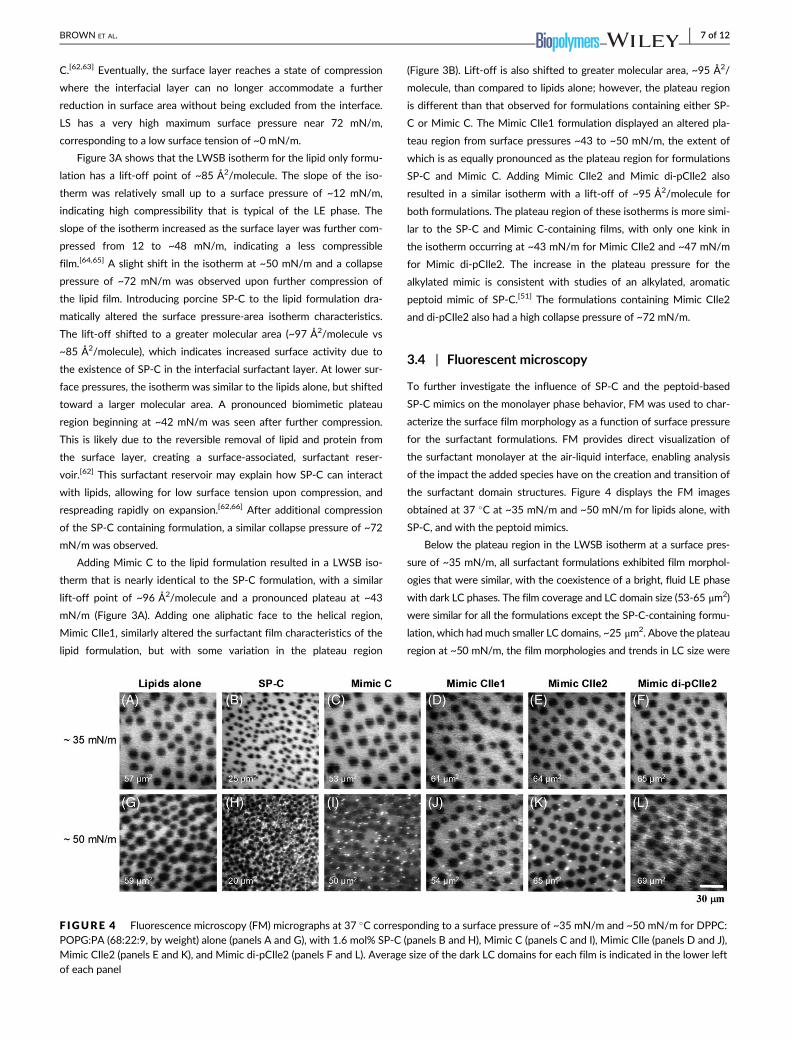
3.4 | Fluorescent microscopy
To further investigate the influence of SP-C and the peptoid-based
SP-C mimics on the monolayer phase behavior, FM was used to char-
acterize the surface film morphology as a function of surface pressure
for the surfactant formulations. FM provides direct visualization of
the surfactant monolayer at the air-liquid interface, enabling analysis
of the impact the added species have on the creation and transition of
the surfactant domain structures. Figure 4 displays the FM images
obtained at 37 �C at ~35 mN/m and ~50 mN/m for lipids alone, with
SP-C, and with the peptoid mimics.
Below the plateau region in the LWSB isotherm at a surface pres-
sure of ~35 mN/m, all surfactant formulations exhibited film morphol-
ogies that were similar, with the coexistence of a bright, fluid LE phase
with dark LC phases. The film coverage and LC domain size (53-65 μm2)
were similar for all the formulations except the SP-C-containing formu-
lation, which had much smaller LC domains, ~25 μm2. Above the plateau
region at ~50 mN/m, the film morphologies and trends in LC size were
F IGURE 4 Fluorescence microscopy (FM) micrographs at 37 �C corresponding to a surface pressure of ~35 mN/m and ~50 mN/m for DPPC:POPG:PA (68:22:9, by weight) alone (panels A and G), with 1.6 mol% SP-C (panels B and H), Mimic C (panels C and I), Mimic CIle (panels D and J),Mimic CIle2 (panels E and K), and Mimic di-pCIle2 (panels F and L). Average size of the dark LC domains for each film is indicated in the lower leftof each panel
BROWN ET AL. 7 of 12
similar to those seen at ~35 mN/m except that bright, vesicle-like pro-
trusions were now present for all of the surfactant formulations exclud-
ing the lipids alone. These protrusions likely corresponded to removal of
surfactant material from the interface and the formation of a surface-
associated surfactant reservoir. Their formation in the SP-C and SP-C
mimic-containing films is also supported by the pronounced plateau
region in the LWSB isotherms. All the peptoid-containing films displayed
similar morphology to the SP-C film although the native protein was
better able to condense the size of the LC domains.
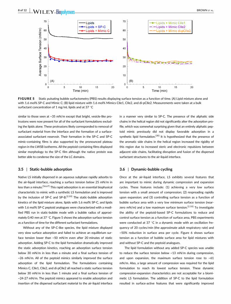
3.5 | Static-bubble adsorption
Native LS initially dispersed in an aqueous subphase rapidly adsorbs to
the air-liquid interface, reaching a surface tension below 25 mN/m in
less than a minute.[56,67] This rapid adsorption is an essential biophysical
characteristic to mimic with a synthetic LS formulation and is improved
by the inclusion of SP-C and SP-B.[2,10] The static-bubble adsorption
kinetics of the lipid mixture alone, lipids with 1.6 mol% SP-C, and lipids
with 1.6 mol% SP-C peptoid analogues were characterized with a modi-
fied PBS run in static-bubble mode with a bubble radius of approxi-
mately 0.40 mm at 37 �C. Figure 5 shows the adsorption surface tension
as a function of time for the different surfactant formulations.
Without any of the SP-C-like species, the lipid mixture displayed
very slow surface adsorption and failed to achieve an equilibrium sur-
face tension lower than ~50 mN/m even after 20 minutes of static
adsorption. Adding SP-C to the lipid formulation dramatically improved
the static adsorption kinetics, reaching an adsorption surface tension
below 30 mN/m in less than a minute and a final surface tension of
~26 mN/m. All of the peptoid mimics similarly improved the surface
adsorption of the lipid formulation. The formulations containing
Mimics C, CIle1, CIle2, and di-pCIle2 all reached a static surface tension
below 30 mN/m in less than 1 minute and a final surface tension of
~24-27 mN/m. The peptoid mimics appeared to enable adsorption and
insertion of the dispersed surfactant material to the air-liquid interface
in a manner very similar to SP-C. The presence of the aliphatic side
chains in the helical region did not significantly alter the adsorption pro-
file, which was somewhat surprising given that an entirely aliphatic pep-
toid mimic previously did not display favorable adsorption in a
synthetic lipid formulation.[30] It is hypothesized that the presence of
the aromatic side chains in the helical region increased the rigidity of
this region due to increased steric and electronic repulsions between
adjacent side chains, facilitating disruption and fusion of the dispersed
surfactant structures to the air-liquid interface.
3.6 | Dynamic-bubble cycling
Once at the air-liquid interface, LS exhibits several features that
are important to mimic during dynamic compression and expansion
cycles. These features include: (1) achieving a very low surface
tension with a small amount of compression; (2) respreading rapidly
upon expansion; and (3) controlling surface tension as a function of
bubble surface area with a very low minimum surface tension (near-
zero mN/m) and a low maximum surface tension.[2,10] To investigate
the ability of the peptoid-based SP-C formulations to reduce and
control surface tension as a function of surface area, PBS experiments
were conducted at 37 �C in a dynamic mode with an oscillation fre-
quency of 20 cycles/min (the approximate adult respiratory rate) and
~50% reduction in surface area per cycle. Figure 6 shows surface
tension as a function of bubble surface area for lipid mixtures with
and without SP-C and the peptoid analogues.
The lipid formulation without any added SP-C species was unable
to reduce the surface tension below ~13 mN/m during compression,
and upon expansion, the maximum surface tension rose to ~61
mN/m. Also, a large amount of compression was required for the lipid
formulation to reach its lowest surface tension. These dynamic
compression-expansion characteristics are not acceptable for a biomi-
metic LS formulation. The addition of SP-C to the lipid formulation
resulted in surface-active features that were significantly improved
F IGURE 5 Static pulsating bubble surfactometry (PBS) results displaying surface tension as a function of time. (A) Lipid mixture alone andwith 1.6 mol% SP-C and Mimic C; (B) lipid mixture with 1.6 mol% Mimics CIle1, CIle2, and di-pCIle2. Measurements were taken at a bulksurfactant concentration of 1 mg/mL lipids and at 37 �C
8 of 12 BROWN ET AL.

relative to the lipid formulation alone. Both the minimum and maximum
surface tensions were significantly lower, <1 mN/m and ~39 mN/m,
respectively. The surfactant film with SP-C was also much less com-
pressible, requiring less compression to reach a low surface tension.
The addition of the peptoid-based SP-C mimics to the lipid formula-
tion also resulted in an improvement of the lipid formulation's surface-
active features during dynamic cycling with all formulations reaching
near-zero surface tension. The presence of Mimic C caused a reduction
in the maximum and minimum surface tensions to ~53 mN/m and <1
mN/m, respectively. These improvements were significant over the
lipid formulation, but the maximum surface tensions were inferior rela-
tive to the formulation containing native SP-C. Adding one aliphatic
face into the helical region (Mimic CIle1) resulted in a compression-
expansion loop that was very similar to Mimic C, but with the maximum
surface tension slightly reduced to ~50 mN/m. The introduction of a
second α-chiral, aliphatic face into the helical region (Mimic CIle2) fur-
ther improved the dynamic compression-expansion film behavior,
resulting in a maximum surface tension of ~47 mN/m. The reduction in
maximum surface tension indicated that the inclusion of aliphatic side
chains in the helical region promoted favorable interactions between
the hydrophobic helices of the peptoid analogues and the lipid acyl
chains. This interaction may increase the aliphatic-containing peptoid
mimics' association with excluded surfactant material through increased
lipid affinity, better enabling the reincorporation of this material upon
expansion, and reducing the maximum surface tension. The presence of
the α-chiral, aromatic side chains was previously found to be necessary
for activity in peptoid-based mimics, as their removal led to a surfactant
film that required a significant amount of compression to reach a low
surface tension.[30] By creating a mimic, Mimic CIle2, that utilizes
features of both aromatic and aliphatic side chain chemistries, the
structural and biomimetic requirements of the hydrophobic helical
region were balanced, and the surface activity of the SP-C mimic was
thus optimized. It is possible that an all-aromatic mimic may be easily
excluded from insertion into the lipid bilayer, whereas some aromatic
character may allow a mimic to keep its rigidity but stay inserted due to
the aliphatic chains.
Despite the much-improved surface activity, Mimic CIle2 did not
fully replicate the very low maximum surface tension exhibited by the
SP-C-containing surfactant film (Figure 6). This is likely due to the pres-
ence of the two palmitoyl chains in positions 5 and 6 in the N-terminal
region of natural SP-C that promote interactions with the phospho-
lipids. When two alkyl chains were added to the N-terminal region of
Mimic CIle2 (Mimic di-pCIle2) to mimic this palmitoylation, it was found
that the maximum surface tension during dynamic cycling was dramati-
cally reduced to ~39 mN/m (Figure 6B). Lift-off area did not change
though (Figure 3B), demonstrating that the alkylated chains were well-
incorporated into the film. Both the maximum and minimum surface
tensions of the Mimic di-pCIle2 formulation were comparable to the
SP-C formulation, with no statistically significant difference between
maximum surface tension values, making Mimic di-pCIle2 a promising
candidate for use in a biomimetic LS formulation.
4 | CONCLUSIONS
The C-terminal hydrophobic helix of SP-C has been shown to greatly
influence SP-C's many reported interactions and surface activities in a
phospholipid environment; however, because of the high content of
F IGURE 6 Dynamic pulsating bubble surfactometry (PBS) results displaying surface tension as a function of surface area at an oscillationfrequency of 20 cycles/min, after 5 minutes of initial cycling. (A) Lipid mixture alone and with 1.6 mol% SP-C and Mimic C. (B) Lipid mixtures with1.6 mol% Mimics CIle1, CIle2, and di-pCIle2. Measurements were taken at a bulk surfactant concentration of 1 mg/mL lipids and at 37 �C. Loopdirections are clockwise with expansion followed by compression. A one-way analysis of variance with post hoc Tukey-Kramer multiplecomparison testing was used to analyze maximum surface tension results. All groups were significantly different (P < 0.05) from each otherexcept for Lipids + SP-C and Lipids + Mimic di-pCIle2
BROWN ET AL. 9 of 12

β-branched amino acids, the helix is metastable in solution and prone to
misfolding and aggregating into inactive, non-helical conformations.[24,58]
One strategy to address this challenge is by mimicking SP-C with
poly-N-substituted glycines or “peptoids.”[30,38,39] Peptoids with
α-chiral, sterically bulky side chains are able to adopt stable, handed
helices,[42–44] making them excellent candidates for biomimicry of the
hydrophobic proteins of LS.[39,45–47] In prior work, peptoid mimics utiliz-
ing structurally rigid, aromatic residues in the helical region were found
to have superior SP-C-like behaviors than more biomimetic analogues
containing a less structurally rigid, aliphatic helix.[30] However, the
aliphatic-based peptoid mimics did display a lower maximum surface
tension during dynamic cycling. This suggests a potential interaction
between the lipid acyl chains and the aliphatic side chains of the peptoid
helix. To investigate this possibility, a set of peptoid-based SP-C ana-
logues was created, incorporating both aromatic and aliphatic residues
in the helical region to combine the structural features of the aromatic
residues and biomimetic features of the aliphatic residues in one SP-C
mimic, while still retaining the helical secondary structure.
CD spectroscopy studies of the synthetic analogues showed that
all the peptoid-based mimics were structured (helical) in solution. The
surface activities of these analogues in a surfactant lipid film were
characterized in vitro using LWSB, FM, and PBS and compared to
native porcine SP-C. All the peptoid analogues displayed favorable
surface-active features when combined with a synthetic lipid formula-
tion, and overall, the results from LWSB, FM, and static PBS experi-
ments were similar among the mimics. However, increasing the
aliphatic content of the helical region increased the surface activity of
the peptoid mimics by incrementally decreasing the maximum surface
tension. The surfactant containing Mimic CIle2 displayed the lowest
maximum surface tension during dynamic cycling while retaining a
low compressibility due to the aromatic side chains. These studies rev-
ealed that both the side chain chemistry and the rigid secondary struc-
ture of the helical region were important components of an analogue
of SP-C that contains Ile-like side chains. A structurally rigid helix
resulted in a less compressible film, requiring less compression to
reach a minimum surface tension whereas side-chain biomimicry dra-
matically reduced the maximum surface tension of a dynamically
cycled film in a stepwise fashion. To further improve the biomimicry
and activity of Mimic CIle2, two alkyl chains were included in the N-
terminal region. These amide-linked alkyl chains were similar to the
palmitoyl chains of natural SP-C. This modification dramatically
reduced the maximum surface tension of the surfactant formulation
and was comparable to natural SP-C, the first time this milestone has
been achieved by a peptoid mimic. Therefore, the structural and bio-
mimetic features of SP-C in this peptoid analogue were enhanced by
combining the optimal molecular and structural characteristics of both
the helical C-terminal region and the amphipathic N-terminal region
into one design.
Additional characterization experiments would investigate peptoid
orientation in membranes using X-ray reflectivity (XR) studies.[68]
Other recent work has revealed that the activity of both SP-B and SP-
C surfactant proteins is related with the formation of supramolecular
protein complexes, and in future work using both types of mimics, this
could be investigated.[69] Techniques such as analytical ultracentrifu-
gation (AUC) can be used to evaluate peptoid oligomerization. The
presence of aromatic side chains would likely facilitate the formation
of self-associated peptoid oligomers.
As these peptoid mimics are further developed for in vivo applica-
tions in the lung, a clearer understanding of the peptoid mechanism of
clearance will be important. While the fate of these mimics in the lung
has yet to be established, prior work on helical, amphipathic, antimi-
crobial peptoids used in vivo to treat an intraperitoneal infection dem-
onstrated that these peptoids were well-tolerated (i.e., no apparent
acute negative effects and no apparent immune response).[70] Similarly,64Cu-labeled antimicrobial peptoids when delivered systemically per
oral, intravenously, or intraperitoneally generally exhibited higher
in vivo stability and tissue accumulation and slower elimination in com-
parison to peptides.[71] Although we expect that our peptoid mimics
will be more resistant to protease degradation compared to peptides,
further studies are needed to determine in vivo fate of the entire lipid-
peptoid complex; similar to a recent carbon 13-labeled DPPC study
comparing the in vivo metabolism of poractant alfa to a synthetic,
peptide-based surfactant, CHF5633.[72] Recently, a mono-alkylated
(single Nocd residue) variant of Mimic C was tested in an in vivo rat
model of acute lung injury.[25] The peptoid-based surfactants were
comparable to an animal-derived surfactant (BLES) in terms of physio-
logical and biochemical outcomes. In fact, the SP-C mimic alone formu-
lation appeared to perform slightly better than BLES in terms of blood
oxygen level and Alveolar-arterial (A-a) gradient. Future in vivo studies
would test other promising SP-C mimic designs (e.g., Mimic di-pCIle2)
for efficacy. Overall, peptoid-based analogues hold great potential for
use in a synthetic, biomimetic LS formulation for treating respiratory
distress-related disorders.
ACKNOWLEDGMENTS
We thank Prof. Jesus Perez-Gil for the gift of porcine SP-C as well as
Dr. Mark Johnson and Dr. Ronald Zuckermann for their assistance.
Also, NJB is grateful to Dr. Michelle Dohm and Dr. Shannon Seurynck-
Servoss for their assistance and insight. NJB acknowledges support
from the NIH Biotechnology Training Program and US National Insti-
tutes of Health (Grant 2 R01 HL067984). We also acknowledge use of
the Keck Biophysics facility at Northwestern University for CD mea-
surements. The Molecular Foundry has also provided support for this
project; work at the Molecular Foundry was supported by the Office
of Science, Office of Basic Energy Sciences, of the U.S. Department of
Energy under Contract No. DE-AC02-05CH11231.
CONFLICT OF INTEREST
The authors declare no competing interests.
ORCID
Annelise E. Barron https://orcid.org/0000-0002-0735-6873
10 of 12 BROWN ET AL.

REFERENCES
[1] M. E. Avery, J. Mead, AMA J. Dis. Child 1959, 97(5, Part 1), 517.[2] R. H. Notter, Lung Surfactants: Basic Science and Clinical Applications,
Vol. 149, Marcel Dekker, Inc, New York 2000.[3] I. Mingarro, D. Lukovic, M. Vilar, J. Perez-Gil, Curr. Med. Chem. 2008,
15(4), 393.
[4] T. Curstedt, J. Johansson, Neonatology 2006, 89(4), 336.[5] R. Spragg, Am. J. Respir. Cell. Mol. Biol. 2007, 37(4), 377.[6] R. G. Spragg, J. F. Lewis, H. Walmrath, J. Johannigman, G. Bellingan,
P. Laterre, M. C. Witte, G. A. Richards, G. Rippin, F. Rathgeb,
D. Hafner, F. J. H. Taut, W. Seeger, N. Engl. J. Med. 2004, 351(9), 884.[7] N. J. Brown, J. Johansson, A. E. Barron, Acc. Chem. Res. 2008, 41(10),
1409.
[8] T. E. Weaver, J. J. Conkright, Annu. Rev. Physiol. 2001, 63, 555.[9] L. M. Nogee, A. E. Dunbar 3rd. , S. E. Wert, F. Askin, A. Hamvas,
J. A. Whitsett, N. Engl. J. Med. 2001, 344(8), 573.[10] J. Johansson, T. Curstedt, B. Robertson, Pediatr. Pathol. Mol. Med.
2001, 20(6), 501.[11] R. Veldhuizen, K. Nag, S. Orgeig, F. Possmayer, Biochim. Biophys. Acta
1998, 1408(2–3), 90.[12] L. Gomez-Gil, D. Schurch, E. Goormaghtigh, J. Perez-Gil, Biophys. J.
2009, 97(10), 2736.
[13] S. B. Hall, A. R. Venkitaraman, J. A. Whitsett, B. A. Holm,
R. H. Notter, Am. Rev. Respir. Dis. 1992, 145(1), 24.[14] Z. Wang, S. B. Hall, R. H. Notter, J. Lipid Res. 1996, 37(4), 790.[15] J. C. Clark, S. E. Wert, C. J. Bachurski, M. T. Stahlman, B. R. Stripp,
T. E. Weaver, J. A. Whitsett, Proc. Natl. Acad. Sci. U. S. A. 1995, 92
(17), 7794.
[16] L. M. Nogee, D. E. de Mello, L. P. Dehner, H. R. Colten, N. Engl.
J. Med. 1993, 328(6), 406.
[17] L. M. Nogee, S. E. Wert, S. A. Proffit, W. M. Hull, J. A. Whitsett, Am.
J. Respir. Crit. Care Med. 2000, 161(3 Pt 1), 973.
[18] D. K. Vorbroker, S. A. Profitt, L. M. Nogee, J. A. Whitsett, Am.
J. Physiol. 1995, 268(4 Pt 1), L647.
[19] N. J. Foot, S. Orgeig, S. Donnellan, T. Bertozzi, C. B. Daniels, J. Mol.
Evol. 2007, 65(1), 12.[20] A. Gonzalez-Horta, D. Andreu, M. R. Morrow, J. Perez-Gil, Biophys. J.
2008, 95(5), 2308.[21] Z. D. Wang, O. Gurel, J. E. Baatz, R. H. Notter, J. Biol. Chem. 1996,
271(32), 19104.
[22] Y. Kallberg, M. Gustafsson, B. Persson, J. Thyberg, J. Johansson,
J. Biol. Chem. 2001, 276(16), 12945.[23] T. Szyperski, G. Vandenbussche, T. Curstedt, J. M. Ruysschaert,
K. Wuthrich, J. Johansson, Protein Sci. 1998, 7(12), 2533.[24] M. Gustafsson, J. Thyberg, J. Naslund, E. Eliasson, J. Johansson, FEBS
Lett. 1999, 464(3), 138.[25] A. M. Czyzewski, L. M. McCaig, M. T. Dohm, L. A. Broering, L. J. Yao,
N. J. Brown, M. K. Didwania, J. S. Lin, J. F. Lewis, R. Veldhuizen,
A. E. Barron, Sci. Rep. 2018, 8(1), 6795.[26] R. H. Notter, R. Gupta, A. L. Schwan, Z. Wang, M. G. Shkoor,
F. J. Walther, PeerJ 2016, 4, e2635.[27] C. W. Bae, S. H. Chung, Y. S. Choi, Yonsei Med. J. 2016, 57(1), 203.[28] M. Seehase, J. J. Collins, E. Kuypers, R. K. Jellema, D. R. Ophelders,
O. L. Ospina, J. Perez-Gil, F. Bianco, R. Garzia, R. Razzetti,
B. W. Kramer, PLoS One 2012, 7(10), e47631.[29] A. Almlen, G. Stichtenoth, B. Robertson, J. Johansson, T. Curstedt,
Neonatology 2007, 92(3), 194.[30] N. J. Brown, C. W. Wu, S. L. Seurynck-Servoss, A. E. Barron, Biochem-
istry 2008, 47(6), 1808.[31] L. Creuwels, E. H. Boer, R. A. Demel, L. M. G. Vangolde, H. P. Haagsman,
J. Biol. Chem. 1995, 270(27), 16225.
[32] J. Johansson, G. Nilsson, R. Stromberg, B. Robertson, H. Jornvall,
T. Curstedt, Biochem. J. 1995, 307, 535.[33] J. Johansson, T. Szyperski, K. Wuthrich, FEBS Lett. 1995, 362(3), 261.
[34] T. Takei, Y. Hashimoto, T. Aiba, K. Sakai, T. Fujiwara, Biol. Pharm. Bull.
1996, 19(10), 1247.
[35] I. Plasencia, K. M. Keough, J. Perez-Gil, Biochim. Biophys. Acta 2005,1713(2), 118.
[36] G. Nilsson, M. Gustafsson, G. Vandenbussche, E. Veldhuizen,
W. J. Griffiths, J. Sjovall, H. P. Haagsman, J. M. Ruysschaert, B. Robertson,T. Curstedt, J. Johansson, Eur. J. Biochem. 1998, 255(1), 116.
[37] T. Takei, Y. Hashimoto, E. Ohtsubo, K. Sakai, H. Ohkawa, Biol. Pharm.
Bull. 1996, 19(12), 1550.[38] Seurynck-Servoss, N. J. Brown, M. T. Dohm, C. W. Wu, A. E. Barron,
Colloids Surf. B Biointerfaces 2007, 57(1), 37.[39] C. W. Wu, S. L. Seurynck, K. Y. Lee, A. E. Barron, Chem. Biol. 2003, 10
(11), 1057.
[40] S. M. Miller, R. J. Simon, S. Ng, R. N. Zuckermann, J. M. Kerr,
W. H. Moos, Drug Dev. Res. 1995, 35(1), 20.[41] A. M. Czyzewski, A. E. Barron, Aiche J. 2008, 54(1), 2.[42] K. Kirshenbaum, A. E. Barron, R. A. Goldsmith, P. Armand,
E. K. Bradley, K. T. Truong, K. A. Dill, F. E. Cohen, R. N. Zuckermann,
Proc. Natl. Acad. Sci. U. S. A. 1998, 95(8), 4303.[43] T. J. Sanborn, C. W. Wu, R. N. Zuckerman, A. E. Barron, Biopolymers
2002, 63(1), 12.[44] C. W. Wu, T. J. Sanborn, K. Huang, R. N. Zuckermann, A. E. Barron,
J. Am. Chem. Soc. 2001, 123(28), 6778.[45] S. L. Seurynck-Servoss, M. T. Dohm, A. E. Barron, Biochemistry 2006,
45(39), 11809.
[46] M. T. Dohm, S. L. Seurynck-Servoss, J. Seo, R. N. Zuckermann,
A. E. Barron, Biopolymers 2009, 92(6), 538.
[47] S. L. Seurynck, J. A. Patch, A. E. Barron, Chem. Biol. 2005, 12(1), 77.[48] P. Armand, K. Kirshenbaum, R. A. Goldsmith, S. Farr-Jones, A. E. Barron,
K. T. Truong, K. A. Dill, D. F. Mierke, F. E. Cohen, R. N. Zuckermann,
E. K. Bradley, Proc. Natl. Acad. Sci. U. S. A. 1998, 95(8), 4309.[49] C. W. Wu, K. Kirshenbaum, T. J. Sanborn, J. A. Patch, K. Huang,
K. A. Dill, R. N. Zuckermann, A. E. Barron, J. Am. Chem. Soc. 2003,125(44), 13525.
[50] N. J. Brown, S. L. Seurynck, C. W. Wu, M. Johnson, A. E. Barron,
Biophys. J. 2005, 88(1), 576a.[51] N. J. Brown, M. T. Dohm, J. Bernardino de la Serna, A. E. Barron,
Biophys. J. 2011, 101(5), 1076.
[52] J. Perezgil, A. Cruz, C. Casals, Biochim. Biophys. Acta 1993, 1168(3), 261.
[53] R. N. Zuckermann, J. M. Kerr, S. B. H. Kent, W. H. Moos, J. Am. Chem.
Soc. 1992, 114(26), 10646.[54] F. Bringezu, J. Q. Ding, G. Brezesinski, J. A. Zasadzinski, Langmuir
2001, 17(15), 4641.[55] M. D. Abramoff, P. J. Magelhaes, S. J. Ram, Biophotonics Int. 2004, 11
(7), 36.
[56] S. L. Seurynck, N. J. Brown, C. W. Wu, K. W. Germino, E. K. Kohlmeir,
E. P. Ingenito, M. R. Glucksberg, A. E. Barron, M. Johnson, J. Appl.
Physiol. 2005, 99(2), 624.[57] G. Putz, J. Goerke, H. W. Taeusch, J. A. Clements, J. Appl. Physiol.
1994, 76(4), 1425.[58] A. Clercx, G. Vandenbussche, T. Curstedt, J. Johansson, H. Jornvall,
J. M. Ruysschaert, Eur. J. Biochem. 1995, 229(2), 465.[59] C. W. Wu, T. J. Sanborn, R. N. Zuckermann, A. E. Barron, J. Am. Chem.
Soc. 2001, 123(13), 2958.[60] Y. Tanaka, T. Takei, T. Aiba, K. Masuda, A. Kiuchi, T. Fujiwara, J. Lipid
Res. 1986, 27(5), 475.
[61] C. Alonso, T. Alig, J. Yoon, F. Bringezu, H. Warriner, J. A. Zasadzinski,
Biophys. J. 2004, 87(6), 4188.[62] D. Y. Takamoto, M. M. Lipp, A. von Nahmen, K. Y. Lee, A. J. Waring,
J. A. Zasadzinski, Biophys. J. 2001, 81(1), 153.[63] C. Alonso, A. Waring, J. A. Zasadzinski, Biophys. J. 2005, 89(1), 266.
[64] K. Y. C. Lee, A. Gopal, A. von Nahmen, J. A. Zasadzinski,J. Majewski, G. S. Smith, P. B. Howes, K. Kjaer, J. Chem. Phys. 2002,116(2), 774.
BROWN ET AL. 11 of 12

[65] J. Ding, H. E. Warriner, J. A. Zasadzinski, Phys. Rev. Lett. 2002, 88(16),168102.
[66] A. vonNahmen, M. Schenk, M. Sieber, M. Amrein, Biophys. J. 1997,72(1), 463.
[67] E. M. Scarpelli, E. David, M. Cordova, A. J. Mautone, Am. J. Perinatol.
1992, 9(5–6), 414.[68] N. P. Chongsiriwatana, J. A. Patch, A. M. Czyzewski, M. T. Dohm,
A. Ivankin, D. Gidalevitz, R. N. Zuckermann, A. E. Barron, Proc. Natl.
Acad. Sci. U. S. A. 2008, 105(8), 2794.[69] E. J. Cabre, M. Martinez-Calle, M. Prieto, A. Fedorov, B. Olmeda,
L. M. S. Loura, J. Perez-Gil, J. Biol. Chem. 2018, 293(24), 9399.
[70] A. M. Czyzewski, H. Jenssen, C. D. Fjell, M. Waldbrook,
N. P. Chongsiriwatana, E. Yuen, R. E. Hancock, A. E. Barron, PLoS
One 2016, 11(2), e0135961.
[71] J. Seo, G. Ren, H. Liu, Z. Miao, M. Park, Y. Wang, T. M. Miller,
A. E. Barron, Z. Cheng, Bioconjug. Chem. 2012, 23(5), 1069.
[72] J. Madsen, M. H. Panchal, R. A. Mackay, M. Echaide, G. Koster,
G. Aquino, N. Pelizzi, J. Perez-Gil, F. Salomone, H. W. Clark,
A. D. Postle, J. Lipid Res. 2018, 59(10), 1880.
How to cite this article: Brown NJ, Lin JS, Barron AE. Helical
side chain chemistry of a peptoid-based SP-C analogue:
Balancing structural rigidity and biomimicry. Biopolymers.
2019;e23277. https://doi.org/10.1002/bip.23277
12 of 12 BROWN ET AL.