Helical antimicrobial polypeptides with radial amphiphilicity · Helical antimicrobial polypeptides...
6
Helical antimicrobial polypeptides with radial amphiphilicity Menghua Xiong a,1 , Michelle W. Lee b,1 , Rachael A. Mansbach c , Ziyuan Song a , Yan Bao d , Richard M. Peek Jr. e , Catherine Yao a , Lin-Feng Chen d , Andrew L. Ferguson a,f,2 , Gerard C. L. Wong b,2 , and Jianjun Cheng a,2 a Department of Materials Science and Engineering, University of Illinois at Urbana–Champaign, Urbana, IL 61801; b Department of Bioengineering, Department of Chemistry and Biochemistry, California NanoSystems Institute, University of California, Los Angeles, CA 90095; c Department of Physics, University of Illinois at Urbana–Champaign, Urbana, IL 61801; d Department of Biochemistry, University of Illinois at Urbana–Champaign, Urbana, IL 61801; e Division of Gastroenterology, Department of Medicine and Cancer Biology, Vanderbilt University School of Medicine, Nashville, TN 37232; and f Department of Chemical and Biomolecular Engineering, University of Illinois at Urbana–Champaign, Urbana, IL 61801 Edited by Alexander M. Klibanov, Massachusetts Institute of Technology, Cambridge, MA, and approved September 18, 2015 (received for review April 23, 2015) α-Helical antimicrobial peptides (AMPs) generally have facially am- phiphilic structures that may lead to undesired peptide interac- tions with blood proteins and self-aggregation due to exposed hydrophobic surfaces. Here we report the design of a class of cationic, helical homo-polypeptide antimicrobials with a hydro- phobic internal helical core and a charged exterior shell, possess- ing unprecedented radial amphiphilicity. The radially amphiphilic structure enables the polypeptide to bind effectively to the negatively charged bacterial surface and exhibit high antimicrobial activity against both gram-positive and gram-negative bacteria. Moreover, the shielding of the hydrophobic core by the charged exterior shell decreases nonspecific interactions with eukaryotic cells, as evi- denced by low hemolytic activity, and protects the polypeptide backbone from proteolytic degradation. The radially amphiphilic polypeptides can also be used as effective adjuvants, allowing im- proved permeation of commercial antibiotics in bacteria and en- hanced antimicrobial activity by one to two orders of magnitude. Designing AMPs bearing this unprecedented, unique radially amphi- philic structure represents an alternative direction of AMP develop- ment; radially amphiphilic polypeptides may become a general platform for developing AMPs to treat drug-resistant bacteria. antimicrobial peptide | α-helix | polypeptides | radial amphiphilicity | bacteria A ntimicrobial peptides (AMPs) typically contain ∼40–60 amino acids, consisting of both cationic and hydrophobic amino acids. They adopt various secondary structures (e.g., α-helix) and can kill a range of bacteria. As AMPs target generic and necessary lipid components of bacterial membranes (1, 2) and depend less on specific bacterial metabolic status (3–6), development of re- sistance has been slow. Because of this feature, AMPs have attracted significant attention as potential antimicrobial agents clinically. Among all AMPs developed, the α-helical peptides are the most heavily investigated and generally are facially amphi- philic (FA) in structure with the cationic and hydrophobic amino acids separated to opposite faces of the helix. This structure correlates well to antimicrobial activity (6–10). Recent work has shown how amino acid content of AMPs enables this activity via specific types of membrane curvature generation (11–13). Despite extensive effort, the commercial development of AMPs has seen limited success, in part due to drawbacks native to pep- tides. Although much has been learned from fundamental studies on AMP mechanisms (1, 2, 14, 15), precise, quantitative pre- dictions of an AMP’s activity, therapeutic index, and antimicro- bial activity compared with off-target eukaryotic cytotoxicity are currently impossible. Designing new AMP-related antibiotics relies on sequence-controlled peptide synthesis and parameter optimi- zation, which is expensive and labor-intensive. Another drawback of most AMP-derived antibiotics is their poor stability in biological systems. LL-37 and magainin, for example, can be degraded by proteases in several minutes in blood circulation and lose their antimicrobial activities (16, 17). Finally, an important drawback of AMPs is related to their FA structure with an exposed hy- drophobic helix face (Fig. 1A), which leads to undesired poly- peptide interactions with blood proteins and self-aggregation. AMP helix bundles have been reported to cause substantially reduced antimicrobial activity (18, 19). There has been significant interest in developing AMP ana- logs, such as β-peptides (20–23), α/β-peptides (24, 25), peptoids (26), and aromatic oligomers (15, 27–30). These compounds have demonstrated improved stability over conventional AMPs. They are in general sequence specific and often require solid- phase peptide synthesis, thereby sharing similar advantages and development drawbacks as AMPs. There has also been growing interest in synthetic polymer-based AMP mimics bearing both cationic and hydrophobic groups, which can be prepared through cost-effective polymerization processes. For example, simplified polymeric AMP analogs have been developed, including poly (methacrylamides) (31), poly(β-lactams) (14, 32–35), polypeptides (36–38), poly(norborenes) (39, 40), and poly(carbonates) (41, 42). These compounds are considerably less expensive than peptides, and much work is being done to optimize them. Herein, we envision a fundamentally different design of AMPs with radially amphiphilic (RA) structure (Fig. 1B) rather than FA structure (Fig. 1A). This class of antimicrobial homo-polypeptides Significance We developed antimicrobial polypeptides (AMPs) with un- precedented radial amphiphilicity. Unlike typical AMPs charac- terized by facial amphiphilicity or biomimetic antimicrobial polymers with randomly distributed charged and hydrophobic groups, this class of AMPs is made up of homo-polypeptides that feature a radially amphiphilic (RA) structure and adopt a stable α-helical conformation with a hydrophobic helical core and a charged exterior shell, which is formed by flexible hydrophobic side chains with terminal charge group. The RA polypeptides appear to offer several advantages over conventional AMPs with regard to stability against proteases and simplicity of de- sign. They also exhibit high antibacterial activity against both gram-negative and gram-positive bacteria and low hemolytic activity. This design may become a general platform for de- veloping AMPs to treat drug-resistant bacteria. Author contributions: M.X., M.W.L., G.C.L.W., and J.C. designed research; M.X., M.W.L., R.A.M., Z.S., Y.B., and A.L.F. performed research; M.X., M.W.L., R.M.P., L.-F.C., A.L.F., G.C.L.W., and J.C. analyzed data; and M.X., M.W.L., C.Y., A.L.F., G.C.L.W., and J.C. wrote the paper. The authors declare no conflict of interest. This article is a PNAS Direct Submission. 1 M.X. and M.W.L. contributed equally to this work. 2 To whom correspondence may be addressed. Email: [email protected], [email protected]. edu, or [email protected]. This article contains supporting information online at www.pnas.org/lookup/suppl/doi:10. 1073/pnas.1507893112/-/DCSupplemental. www.pnas.org/cgi/doi/10.1073/pnas.1507893112 PNAS | October 27, 2015 | vol. 112 | no. 43 | 13155–13160 CHEMISTRY
Transcript of Helical antimicrobial polypeptides with radial amphiphilicity · Helical antimicrobial polypeptides...

Helical antimicrobial polypeptides withradial amphiphilicityMenghua Xionga,1, Michelle W. Leeb,1, Rachael A. Mansbachc, Ziyuan Songa, Yan Baod, Richard M. Peek Jr.e,Catherine Yaoa, Lin-Feng Chend, Andrew L. Fergusona,f,2, Gerard C. L. Wongb,2, and Jianjun Chenga,2
aDepartment of Materials Science and Engineering, University of Illinois at Urbana–Champaign, Urbana, IL 61801; bDepartment of Bioengineering,Department of Chemistry and Biochemistry, California NanoSystems Institute, University of California, Los Angeles, CA 90095; cDepartment ofPhysics, University of Illinois at Urbana–Champaign, Urbana, IL 61801; dDepartment of Biochemistry, University of Illinois at Urbana–Champaign,Urbana, IL 61801; eDivision of Gastroenterology, Department of Medicine and Cancer Biology, Vanderbilt University School of Medicine, Nashville,TN 37232; and fDepartment of Chemical and Biomolecular Engineering, University of Illinois at Urbana–Champaign, Urbana, IL 61801
Edited by Alexander M. Klibanov, Massachusetts Institute of Technology, Cambridge, MA, and approved September 18, 2015 (received for review April23, 2015)
α-Helical antimicrobial peptides (AMPs) generally have facially am-phiphilic structures that may lead to undesired peptide interac-tions with blood proteins and self-aggregation due to exposedhydrophobic surfaces. Here we report the design of a class ofcationic, helical homo-polypeptide antimicrobials with a hydro-phobic internal helical core and a charged exterior shell, possess-ing unprecedented radial amphiphilicity. The radially amphiphilicstructure enables the polypeptide to bind effectively to the negativelycharged bacterial surface and exhibit high antimicrobial activityagainst both gram-positive and gram-negative bacteria. Moreover,the shielding of the hydrophobic core by the charged exterior shelldecreases nonspecific interactions with eukaryotic cells, as evi-denced by low hemolytic activity, and protects the polypeptidebackbone from proteolytic degradation. The radially amphiphilicpolypeptides can also be used as effective adjuvants, allowing im-proved permeation of commercial antibiotics in bacteria and en-hanced antimicrobial activity by one to two orders of magnitude.Designing AMPs bearing this unprecedented, unique radially amphi-philic structure represents an alternative direction of AMP develop-ment; radially amphiphilic polypeptides may become a generalplatform for developing AMPs to treat drug-resistant bacteria.
antimicrobial peptide | α-helix | polypeptides | radial amphiphilicity |bacteria
Antimicrobial peptides (AMPs) typically contain ∼40–60 aminoacids, consisting of both cationic and hydrophobic amino acids.
They adopt various secondary structures (e.g., α-helix) and cankill a range of bacteria. As AMPs target generic and necessarylipid components of bacterial membranes (1, 2) and depend lesson specific bacterial metabolic status (3–6), development of re-sistance has been slow. Because of this feature, AMPs haveattracted significant attention as potential antimicrobial agentsclinically. Among all AMPs developed, the α-helical peptides arethe most heavily investigated and generally are facially amphi-philic (FA) in structure with the cationic and hydrophobic aminoacids separated to opposite faces of the helix. This structurecorrelates well to antimicrobial activity (6–10). Recent work hasshown how amino acid content of AMPs enables this activity viaspecific types of membrane curvature generation (11–13).Despite extensive effort, the commercial development of AMPs
has seen limited success, in part due to drawbacks native to pep-tides. Although much has been learned from fundamental studieson AMP mechanisms (1, 2, 14, 15), precise, quantitative pre-dictions of an AMP’s activity, therapeutic index, and antimicro-bial activity compared with off-target eukaryotic cytotoxicity arecurrently impossible. Designing new AMP-related antibiotics relieson sequence-controlled peptide synthesis and parameter optimi-zation, which is expensive and labor-intensive. Another drawbackof most AMP-derived antibiotics is their poor stability in biologicalsystems. LL-37 and magainin, for example, can be degraded byproteases in several minutes in blood circulation and lose their
antimicrobial activities (16, 17). Finally, an important drawbackof AMPs is related to their FA structure with an exposed hy-drophobic helix face (Fig. 1A), which leads to undesired poly-peptide interactions with blood proteins and self-aggregation.AMP helix bundles have been reported to cause substantiallyreduced antimicrobial activity (18, 19).There has been significant interest in developing AMP ana-
logs, such as β-peptides (20–23), α/β-peptides (24, 25), peptoids(26), and aromatic oligomers (15, 27–30). These compoundshave demonstrated improved stability over conventional AMPs.They are in general sequence specific and often require solid-phase peptide synthesis, thereby sharing similar advantages anddevelopment drawbacks as AMPs. There has also been growinginterest in synthetic polymer-based AMP mimics bearing bothcationic and hydrophobic groups, which can be prepared throughcost-effective polymerization processes. For example, simplifiedpolymeric AMP analogs have been developed, including poly(methacrylamides) (31), poly(β-lactams) (14, 32–35), polypeptides(36–38), poly(norborenes) (39, 40), and poly(carbonates) (41, 42).These compounds are considerably less expensive than peptides,and much work is being done to optimize them.Herein, we envision a fundamentally different design of AMPs
with radially amphiphilic (RA) structure (Fig. 1B) rather than FAstructure (Fig. 1A). This class of antimicrobial homo-polypeptides
Significance
We developed antimicrobial polypeptides (AMPs) with un-precedented radial amphiphilicity. Unlike typical AMPs charac-terized by facial amphiphilicity or biomimetic antimicrobialpolymers with randomly distributed charged and hydrophobicgroups, this class of AMPs is made up of homo-polypeptides thatfeature a radially amphiphilic (RA) structure and adopt a stableα-helical conformation with a hydrophobic helical core and acharged exterior shell, which is formed by flexible hydrophobicside chains with terminal charge group. The RA polypeptidesappear to offer several advantages over conventional AMPswith regard to stability against proteases and simplicity of de-sign. They also exhibit high antibacterial activity against bothgram-negative and gram-positive bacteria and low hemolyticactivity. This design may become a general platform for de-veloping AMPs to treat drug-resistant bacteria.
Author contributions: M.X., M.W.L., G.C.L.W., and J.C. designed research; M.X., M.W.L., R.A.M.,Z.S., Y.B., and A.L.F. performed research; M.X., M.W.L., R.M.P., L.-F.C., A.L.F., G.C.L.W., andJ.C. analyzed data; and M.X., M.W.L., C.Y., A.L.F., G.C.L.W., and J.C. wrote the paper.
The authors declare no conflict of interest.
This article is a PNAS Direct Submission.1M.X. and M.W.L. contributed equally to this work.2To whom correspondence may be addressed. Email: [email protected], [email protected], or [email protected].
This article contains supporting information online at www.pnas.org/lookup/suppl/doi:10.1073/pnas.1507893112/-/DCSupplemental.
www.pnas.org/cgi/doi/10.1073/pnas.1507893112 PNAS | October 27, 2015 | vol. 112 | no. 43 | 13155–13160
CHEM
ISTR
Y

features a hydrophobic helical core covered with cationic groupsin all radial directions of the helix. Because the charged groupsform the outer shell of the polypeptides, shielding the hydro-phobic helical core, these peptides should have minimal hydro-phobic force induced self-aggregation and reduced interactionwith blood proteins. With such RA structure, the polypeptidebackbone amide bonds should also be well protected and areexpected to have improved stability against proteolytic degrada-tion compared with a typical α-helical AMP.
ResultsRA Polypeptide Displays High Antibacterial Activity and Selectivity.We developed a class of helical, charged polypeptides with longhydrophobic side chains and terminal charge groups that dem-onstrated remarkable helical stability against changes in pH, salts,temperature, and various denaturing conditions (43–45). Weadopted this architecture to design RA polypeptides and ex-plored their applications as AMPs. PHLG-BIm (Fig. 1C) was aspecific cationic α-helical polypeptide with RA structure. It wassynthesized through ring-opening polymerization of amino acidN-carboxyanhydrides (NCAs) (46), followed by amination with1-methylbenzimidazole (SI Appendix, Scheme S1). With a sepa-ration of 11 σ-bonds between the backbone and positive charges,PHLG-BIm, with DP of 40 and 28, adopts α-helical conformation.The helical conformation of polypeptides was evident from thecharacteristic double minima at 208 and 222 nm in the circulardichroism (CD) spectra at a concentration of 0.40 mg/mL in water(Fig. 1D). The corresponding nonhelical PHDLG-BIm40 was
synthesized through the polymerization of DL-NCA to study theeffect of helical structure on the antibacterial activity of polypeptide.Our molecular simulations provide molecular-level theoretical
support for the RA structure. A simulation snapshot in Fig. 1Eillustrates the cationic side chains forming a hydrophilic shellaround the hydrophobic aliphatic side chains and helical core.Simulation movies are presented in Movies S1–S3. Discountingthe two terminal residues to eliminate end effects, the peptidebackbone is close to the ideal helix, RMSDhelix = (0.04 ± 0.01) nm,with a radius rhelix = (0.232 ± 0.002) nm and twist γhelix = (100 ± 1)°also nearly ideal (ridealhelix = 0.23 nm, γidealhelix = 100°). The meanper residue molar ellipticity at 222 nm computed from oursimulations using Dichro-Calc (comp.chem.nottingham.ac.uk/cgi-bin/dichrocalc/bin/getparams.cgi) (47) of [θ]222 nm = (−21 ±1) × 103 degrees (deg) cm2/dmol is in excellent agreement withthe experimental value of −23 × 103 deg cm2/dmol for PHLG-BIm40 and −19 × 103 deg cm2/dmol for PHLG-BIm28 reported inFig. 1D. To quantify the RA structure, we computed the prob-ability distribution of the side chain N12 atoms after aligning thepeptide backbone to a reference structure and discarding thefour side chains at the termini to eliminate end effects (Fig. 1 Fand G) (48). We also measured the probability distribution ofside chain lengths from the Cα to the N12 atom. The mode of thedistribution is 1.24 nm, and the mean value is 1.15 nm with a95% CI of [0.90, 1.33] nm (SI Appendix, Fig. S3). The result wasfurther confirmed by nuclear overhauser effect (NOE) spec-troscopy, with no NOE detected between protons around the ni-trogen atoms on the benzimidazole (N12) (d, b, b′, c, c′, e) and
Fig. 1. Schematic illustration of (A) FA AMP and (B) RA AMP. (C) The chemical structure of PHLG-BIm. (D) CD spectra of PHLG-BIm40, PHDLG-BIm40, and PHLG-BIm28 in aqueous solution at pH = 7. (E) RA peptide structure predicted from all-atom molecular dynamics simulations. Representative peptide snapshot fromour simulation trajectory rendered using VMD. For clarity of viewing, the water molecules have been removed and the backbone and side chains colored inred and gray, respectively. (F) The 3D probability distribution of the side chain N12 atoms around the α-helical backbone. A fly-around of this image ispresented in Movie S3. (G) A 2D projection of the 3D probability distribution in F onto a plane perpendicular to the long axis of the peptide. The probabilitydensity lies in the range 0–0.83 nm−2 and 20 evenly spaced contours plotted. (H) The antibacterial and hemolytic activity of polypeptides. The antibacterialactivity of polypeptides was determined using MIC. The hemolytic activity of polypeptides was determined by HC50 (50% hemolytic concentration) value.(I) The stability of RA polypeptide PHLG-BIm40 when incubated with trypsin, pronase, elastase from P. aeruginosa or elastase from human leukocytes for 8 h.
13156 | www.pnas.org/cgi/doi/10.1073/pnas.1507893112 Xiong et al.

protons around Cα (h, i, g) (SI Appendix, Fig. S5). Because thepositive charge in the cationic side chains resides primarily in thetermini, these distributions are a proxy for the positive chargedistribution, providing strong support for an RA structure.The antibacterial activity of PHLG-BIm was evaluated by
the minimal inhibitory concentration (MIC) of the polypeptideagainst bacteria (49). With an RA structure, PHLG-BIm40showed strong antibacterial activity against both gram-negativebacteria, DH5α and MG1655, and gram-positive bacteria,ATCC12608 and ATCC11778, with MIC values of 3.3, 26.1,13.1, and 13.1 μM, respectively (Fig. 1E). The helical PHLG-BIm40 showed higher antibacterial activity than the nonhelicalPHDLG-BIm40, with MIC values 16, 4, 4, and 4 times loweragainst DH5α, MG1655, ATCC12608, and ATCC11778, re-spectively. Length was also found to influence the antimicrobialactivity of the RA polypeptides, as PHLG-BIm28, with DP of 28,showed lower antimicrobial activity compared with PHLG-BIm40. We also tested the antimicrobial activity of RA poly-peptides against other bacterial strains, including clinicallyisolated Helicobacter pylori strains (B107, J291, J99, J99-AF, J99-A9, and J99-A11) (50, 51) and drug-resistant strains (Methicillin-resistant Staphylococcus aureus, NRS382, NRS383, NRS384).Among those H. pylori strains, J99-AF, J99-A9, and J99-A11 areclarithromycin-resistant strains. The RA polypeptides showed highantibacterial activity against these clinically isolated and drug-resistant strains. More interestingly, the antimicrobial activity of theRA polypeptide remained stable against DH5α and ATCC12608 inthe presence of different salts (physiological concentrations of150 mM NaCl, 1 mM MgCl2, and 2.5 mM CaCl2), and mucin, themain component of mucosa (SI Appendix, Table S2). The MICvalues of RA polypeptide against DH5α and ATCC12608 de-creased in the presence of human serum, fetal bovine serum (FBS),plasma, and artificial tears in comparison with RA polypeptide-onlytreatment. The results demonstrate that the RA polypeptides arestable in serum and plasma, and polyanionic compounds do notdramatically affect their antimicrobial activity. The decreased MICin serum and plasma may be attributed to the serum complementsystem, which provides innate defense against microbial infections.PHLG-BIm40 exhibited low hemolytic activity with an HC50 (50%hemolytic concentration) value higher than 104.6 μM, which in-dicates a high selectivity of >32 (defined as HC50/MIC), as opposedto a selectivity of >2 for PHDLG-Blm40 against DH5α bacterialcells. It is important to point out that the RA polypeptide is notjust bacteriostatic but bacteriocidal. Nearly 100% killing of allfour bacterial species was observed at their respective MIC ordouble MIC within 2 h (SI Appendix, Fig. S6). In addition, the RApolypeptides showed concentration-dependent antimicrobial kill-ing in medium and in conditions with NaCl, human serum, plasma,and artificial tears (SI Appendix, Fig. S7).Although many AMPs with high antibacterial activity have
been developed, the application of AMPs is usually limited bythe short durations of activity due to their rapid digestion byendogenous proteases (5, 16, 17, 52, 53). The RA polypeptides,with densely packed hydrophobic side chains forming a hydro-phobic cortex that can protect the amide bonds of the poly-peptide backbone, in principle should be more stable againstproteolysis compared with typical AMPs. We incubated PHLG-BIm40 with trypsin, pronase, and elastase from Pseudomonasaeruginosa or elastase from human leukocytes for 8 h and ana-lyzed polypeptide degradation by HPLC. PHLG-BIm40 exhibitedexcellent proteolytic stability and experienced almost no degra-dation by the proteases (Fig. 1F), whereas LL-37, a positivecontrol peptide, was readily degraded under the same conditions(SI Appendix, Fig. S8). Furthermore, after 8 h of protease ortrypsin treatment, the antibacterial activity of PHLG-BIm40remained unchanged, as demonstrated by having the same MICvalue against DH5α as the untreated RA polypeptide (3.3 μM).
RA Polypeptide Kills Bacteria by Directly Disrupting the Bacterial CellMembrane. We find that the RA PHLG-BIm kills bacteria by di-rectly disrupting the bacterial cell membrane in a manner similar
to typical AMPs, through vesicle leakage, bacterial membranepermeabilization, and bacteria morphology assays. We firstinvestigated the membrane-disruptive activity of helical PHLG-BIm40 and nonhelical PHDLG-BIm40 polypeptides on anionicliposomes 1-palmitoyl-2-oleoyl-sn-glycero-3-phosphoethanolamine(POPE)/1-palmitoyl-2-oleoyl- sn-glycero-3-phospho-(1’-rac-glycerol)(POPG) and neutral liposomes 1,2-dioleoyl-sn-glycero-3-phos-phocholine (DOPC)/1-palmitoyl-2-oleoyl-sn-glycero-3- phosphocho-line (POPC), which were used to model phosphatidylethanolamine(PE)-rich bacteria and eukaryotic cell membranes, respectively.At the same concentration, PHLG-BIm40 induced greater dyeleakage from both anionic and neutral liposomes than PHDLG-BIm40, suggesting that the helical polypeptide has higher mem-brane disruption capability (Fig. 2A). PHLG-BIm40 also causedmore leakage from the anionic liposomes than the neutral lipo-somes, which is well-correlated with the observed selectivityagainst bacterial over mammalian cells. The leakage results alsoshowed the capability of PHLG-BIm to permeabilize model bac-teria membranes rich in negative intrinsic curvature-forming lipids.We next used flow cytometry to evaluate permeabilization ofPHLG-BIm through bacterial membranes. MG1655 bacteriacells were incubated with PHLG-BIm40 and propidium iodide(PI), a membrane impermeable dye. The total number ofPI-containing bacteria cells was greater for those treated with
SYT
O9
PI
PHLG-BIm40PHDLG-BIm40Control
Control PHDLG-BIm40 PHLG-BIm40
0 2 4 60
30
60
90
120
Dye
leak
age
(%)
Concentration ( M)
DOPC/POPC (PHDLG-BIm40)DOPC/POPC (PHLG-BIm40)POPE/POPG (PHDLG-BIm40)POPE/POPG (PHLG-BIm40)
1 2 3 4 5 6 70
25
50
75
100
% o
f PI p
ositi
ve c
ells
2, PHDLG-BIm40 (0.8 M)
4, PHDLG-BIm40 (1.6 M)
6, PHDLG-BIm40 (3.3 M)
1, PI only
3, PHLG-BIm40 (0.8 M)
5, PHLG-BIm40 (1.6 M)
7, PHLG-BIm40 (3.3 M)
****
**
A B
C
D
Fig. 2. PHLG-BIm40 kills bacteria by directly disrupting the bacterial cellmembrane. (A) Extent of calcein efflux in neutral vesicles (DOPC/POPC) andnegatively charged vesicles (DOPE/DOPG) after treatment with PHDLG-BIm40
(nonhelical, lacking radially amphiphilic structure) or PHLG-BIm40 (helicalwith radially amphiphilic structure) at various concentrations for 1 h. (B) Flowcytometry analysis of propidium iodide (PI) uptake after incubation with freePI, PI with PHDLG-BIm40, or PI with PHLG-BIm40 at various concentrations. Allof the data are represented as average ± SD and analyzed by Student t test(**P ≤ 0.01). (C) The fluorescence microscopy of stained Escherichia coliMG1655 in the absence and presence of PHDLG-BIm40 and PHLG-BIm40
(3.3 μM). (Scale bar, 50 μm.) (D) SEM images of MG1655 after treatment withPBS, PHDLG-BIm40, or PHLG-BIm40. (Scale bar, 1 μm.)
Xiong et al. PNAS | October 27, 2015 | vol. 112 | no. 43 | 13157
CHEM
ISTR
Y
PHLG-Blm40 than PHDLG-BIm40 and increased with higher con-centration of RA polypeptide (Fig. 2B). An uptake study analyzedby fluorescence imaging provided additional evidence of enhancedmembrane activity of PHLG-BIm40, which permeabilized MG1655bacterial cell membranes more effectively than PHDLG-BIm40when the bacterial cells were coincubated with polypeptide and dye(PI and SYTO9) (Fig. 2C). Using scanning electron microscopy, weobserved drastic changes and damage of the bacterial membranesafter incubation with PHLG-BIm40, whereas PHDLG-BIm40 min-imally affected bacteria morphology (Fig. 2D). Taken together, theresults indicate that membrane disruption and permeation are animportant component of the antimicrobial activity of PHLG-BIm.To examine in detail the root causes of selective membrane
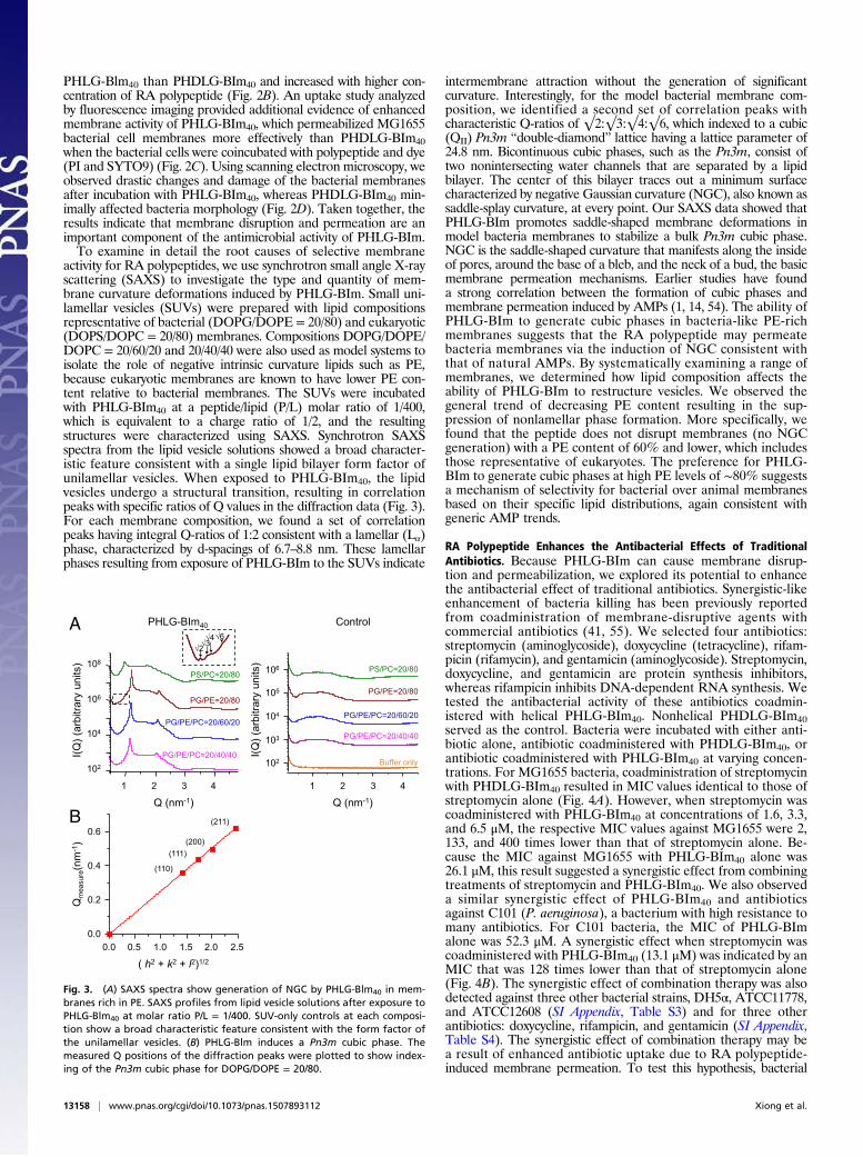
activity for RA polypeptides, we use synchrotron small angle X-rayscattering (SAXS) to investigate the type and quantity of mem-brane curvature deformations induced by PHLG-BIm. Small uni-lamellar vesicles (SUVs) were prepared with lipid compositionsrepresentative of bacterial (DOPG/DOPE = 20/80) and eukaryotic(DOPS/DOPC = 20/80) membranes. Compositions DOPG/DOPE/DOPC = 20/60/20 and 20/40/40 were also used as model systems toisolate the role of negative intrinsic curvature lipids such as PE,because eukaryotic membranes are known to have lower PE con-tent relative to bacterial membranes. The SUVs were incubatedwith PHLG-BIm40 at a peptide/lipid (P/L) molar ratio of 1/400,which is equivalent to a charge ratio of 1/2, and the resultingstructures were characterized using SAXS. Synchrotron SAXSspectra from the lipid vesicle solutions showed a broad character-istic feature consistent with a single lipid bilayer form factor ofunilamellar vesicles. When exposed to PHLG-BIm40, the lipidvesicles undergo a structural transition, resulting in correlationpeaks with specific ratios of Q values in the diffraction data (Fig. 3).For each membrane composition, we found a set of correlationpeaks having integral Q-ratios of 1:2 consistent with a lamellar (Lα)phase, characterized by d-spacings of 6.7–8.8 nm. These lamellarphases resulting from exposure of PHLG-BIm to the SUVs indicate
intermembrane attraction without the generation of significantcurvature. Interestingly, for the model bacterial membrane com-position, we identified a second set of correlation peaks withcharacteristic Q-ratios of √2:√3:√4:√6, which indexed to a cubic(QII) Pn3m “double-diamond” lattice having a lattice parameter of24.8 nm. Bicontinuous cubic phases, such as the Pn3m, consist oftwo nonintersecting water channels that are separated by a lipidbilayer. The center of this bilayer traces out a minimum surfacecharacterized by negative Gaussian curvature (NGC), also known assaddle-splay curvature, at every point. Our SAXS data showed thatPHLG-BIm promotes saddle-shaped membrane deformations inmodel bacteria membranes to stabilize a bulk Pn3m cubic phase.NGC is the saddle-shaped curvature that manifests along the insideof pores, around the base of a bleb, and the neck of a bud, the basicmembrane permeation mechanisms. Earlier studies have founda strong correlation between the formation of cubic phases andmembrane permeation induced by AMPs (1, 14, 54). The ability ofPHLG-BIm to generate cubic phases in bacteria-like PE-richmembranes suggests that the RA polypeptide may permeatebacteria membranes via the induction of NGC consistent withthat of natural AMPs. By systematically examining a range ofmembranes, we determined how lipid composition affects theability of PHLG-BIm to restructure vesicles. We observed thegeneral trend of decreasing PE content resulting in the sup-pression of nonlamellar phase formation. More specifically, wefound that the peptide does not disrupt membranes (no NGCgeneration) with a PE content of 60% and lower, which includesthose representative of eukaryotes. The preference for PHLG-BIm to generate cubic phases at high PE levels of ∼80% suggestsa mechanism of selectivity for bacterial over animal membranesbased on their specific lipid distributions, again consistent withgeneric AMP trends.
RA Polypeptide Enhances the Antibacterial Effects of TraditionalAntibiotics. Because PHLG-BIm can cause membrane disrup-tion and permeabilization, we explored its potential to enhancethe antibacterial effect of traditional antibiotics. Synergistic-likeenhancement of bacteria killing has been previously reportedfrom coadministration of membrane-disruptive agents withcommercial antibiotics (41, 55). We selected four antibiotics:streptomycin (aminoglycoside), doxycycline (tetracycline), rifam-picin (rifamycin), and gentamicin (aminoglycoside). Streptomycin,doxycycline, and gentamicin are protein synthesis inhibitors,whereas rifampicin inhibits DNA-dependent RNA synthesis. Wetested the antibacterial activity of these antibiotics coadmin-istered with helical PHLG-BIm40. Nonhelical PHDLG-BIm40served as the control. Bacteria were incubated with either anti-biotic alone, antibiotic coadministered with PHDLG-BIm40, orantibiotic coadministered with PHLG-BIm40 at varying concen-trations. For MG1655 bacteria, coadministration of streptomycinwith PHDLG-BIm40 resulted in MIC values identical to those ofstreptomycin alone (Fig. 4A). However, when streptomycin wascoadministered with PHLG-BIm40 at concentrations of 1.6, 3.3,and 6.5 μM, the respective MIC values against MG1655 were 2,133, and 400 times lower than that of streptomycin alone. Be-cause the MIC against MG1655 with PHLG-BIm40 alone was26.1 μM, this result suggested a synergistic effect from combiningtreatments of streptomycin and PHLG-BIm40. We also observeda similar synergistic effect of PHLG-BIm40 and antibioticsagainst C101 (P. aeruginosa), a bacterium with high resistance tomany antibiotics. For C101 bacteria, the MIC of PHLG-BImalone was 52.3 μM. A synergistic effect when streptomycin wascoadministered with PHLG-BIm40 (13.1 μM) was indicated by anMIC that was 128 times lower than that of streptomycin alone(Fig. 4B). The synergistic effect of combination therapy was alsodetected against three other bacterial strains, DH5α, ATCC11778,and ATCC12608 (SI Appendix, Table S3) and for three otherantibiotics: doxycycline, rifampicin, and gentamicin (SI Appendix,Table S4). The synergistic effect of combination therapy may bea result of enhanced antibiotic uptake due to RA polypeptide-induced membrane permeation. To test this hypothesis, bacterial
Fig. 3. (A) SAXS spectra show generation of NGC by PHLG-Blm40 in mem-branes rich in PE. SAXS profiles from lipid vesicle solutions after exposure toPHLG-Blm40 at molar ratio P/L = 1/400. SUV-only controls at each composi-tion show a broad characteristic feature consistent with the form factor ofthe unilamellar vesicles. (B) PHLG-Blm induces a Pn3m cubic phase. Themeasured Q positions of the diffraction peaks were plotted to show index-ing of the Pn3m cubic phase for DOPG/DOPE = 20/80.
13158 | www.pnas.org/cgi/doi/10.1073/pnas.1507893112 Xiong et al.
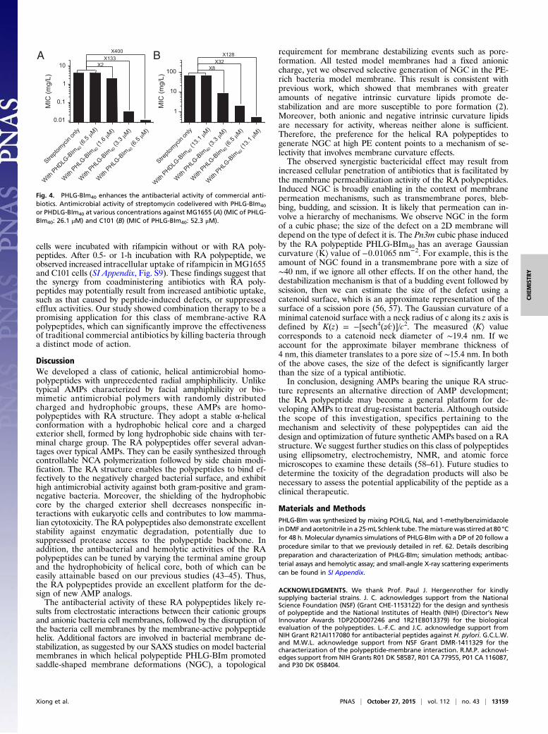
cells were incubated with rifampicin without or with RA poly-peptides. After 0.5- or 1-h incubation with RA polypeptide, weobserved increased intracellular uptake of rifampicin in MG1655and C101 cells (SI Appendix, Fig. S9). These findings suggest thatthe synergy from coadministering antibiotics with RA poly-peptides may potentially result from increased antibiotic uptake,such as that caused by peptide-induced defects, or suppressedefflux activities. Our study showed combination therapy to be apromising application for this class of membrane-active RApolypeptides, which can significantly improve the effectivenessof traditional commercial antibiotics by killing bacteria througha distinct mode of action.
DiscussionWe developed a class of cationic, helical antimicrobial homo-polypeptides with unprecedented radial amphiphilicity. Unliketypical AMPs characterized by facial amphiphilicity or bio-mimetic antimicrobial polymers with randomly distributedcharged and hydrophobic groups, these AMPs are homo-polypeptides with RA structure. They adopt a stable α-helicalconformation with a hydrophobic helical core and a chargedexterior shell, formed by long hydrophobic side chains with ter-minal charge group. The RA polypeptides offer several advan-tages over typical AMPs. They can be easily synthesized throughcontrollable NCA polymerization followed by side chain modi-fication. The RA structure enables the polypeptides to bind ef-fectively to the negatively charged bacterial surface, and exhibithigh antimicrobial activity against both gram-positive and gram-negative bacteria. Moreover, the shielding of the hydrophobiccore by the charged exterior shell decreases nonspecific in-teractions with eukaryotic cells and contributes to low mamma-lian cytotoxicity. The RA polypeptides also demonstrate excellentstability against enzymatic degradation, potentially due tosuppressed protease access to the polypeptide backbone. Inaddition, the antibacterial and hemolytic activities of the RApolypeptides can be tuned by varying the terminal amine groupand the hydrophobicity of helical core, both of which can beeasily attainable based on our previous studies (43–45). Thus,the RA polypeptides provide an excellent platform for the de-sign of new AMP analogs.The antibacterial activity of these RA polypeptides likely re-
sults from electrostatic interactions between their cationic groupsand anionic bacteria cell membranes, followed by the disruption ofthe bacteria cell membranes by the membrane-active polypeptidehelix. Additional factors are involved in bacterial membrane de-stabilization, as suggested by our SAXS studies on model bacterialmembranes in which helical polypeptide PHLG-BIm promotedsaddle-shaped membrane deformations (NGC), a topological
requirement for membrane destabilizing events such as pore-formation. All tested model membranes had a fixed anioniccharge, yet we observed selective generation of NGC in the PE-rich bacteria model membrane. This result is consistent withprevious work, which showed that membranes with greateramounts of negative intrinsic curvature lipids promote de-stabilization and are more susceptible to pore formation (2).Moreover, both anionic and negative intrinsic curvature lipidsare necessary for activity, whereas neither alone is sufficient.Therefore, the preference for the helical RA polypeptides togenerate NGC at high PE content points to a mechanism of se-lectivity that involves membrane curvature effects.The observed synergistic bactericidal effect may result from
increased cellular penetration of antibiotics that is facilitated bythe membrane permeabilization activity of the RA polypeptides.Induced NGC is broadly enabling in the context of membranepermeation mechanisms, such as transmembrane pores, bleb-bing, budding, and scission. It is likely that permeation can in-volve a hierarchy of mechanisms. We observe NGC in the formof a cubic phase; the size of the defect on a 2D membrane willdepend on the type of defect it is. The Pn3m cubic phase inducedby the RA polypeptide PHLG-BIm40 has an average Gaussiancurvature ⟨K⟩ value of −0.01065 nm−2. For example, this is theamount of NGC found in a transmembrane pore with a size of∼40 nm, if we ignore all other effects. If on the other hand, thedestabilization mechanism is that of a budding event followed byscission, then we can estimate the size of the defect using acatenoid surface, which is an approximate representation of thesurface of a scission pore (56, 57). The Gaussian curvature of aminimal catenoid surface with a neck radius of c along its z axis isdefined by K(z) = −[sech4(z∕c)]/c2. The measured ⟨K⟩ valuecorresponds to a catenoid neck diameter of ∼19.4 nm. If weaccount for the approximate bilayer membrane thickness of4 nm, this diameter translates to a pore size of ∼15.4 nm. In bothof the above cases, the size of the defect is significantly largerthan the size of a typical antibiotic.In conclusion, designing AMPs bearing the unique RA struc-
ture represents an alternative direction of AMP development;the RA polypeptide may become a general platform for de-veloping AMPs to treat drug-resistant bacteria. Although outsidethe scope of this investigation, specifics pertaining to themechanism and selectivity of these polypeptides can aid thedesign and optimization of future synthetic AMPs based on a RAstructure. We suggest further studies on this class of polypeptidesusing ellipsometry, electrochemistry, NMR, and atomic forcemicroscopes to examine these details (58–61). Future studies todetermine the toxicity of the degradation products will also benecessary to assess the potential applicability of the peptide as aclinical therapeutic.
Materials and MethodsPHLG-BIm was synthesized by mixing PCHLG, NaI, and 1-methylbenzimidazoleinDMFand acetonitrile in a 25-mL Schlenk tube. Themixturewas stirredat 80 °Cfor 48 h. Molecular dynamics simulations of PHLG-BIm with a DP of 20 follow aprocedure similar to that we previously detailed in ref. 62. Details describingpreparation and characterization of PHLG-BIm; simulation methods; antibac-terial assays and hemolytic assay; and small-angle X-ray scattering experimentscan be found in SI Appendix.
ACKNOWLEDGMENTS. We thank Prof. Paul J. Hergenrother for kindlysupplying bacterial strains. J. C. acknowledges support from the NationalScience Foundation (NSF) (Grant CHE-1153122) for the design and synthesisof polypeptide and the National Institutes of Health (NIH) (Director’s NewInnovator Awards 1DP2OD007246 and 1R21EB013379) for the biologicalevaluation of the polypeptides. L.-F.C. and J.C. acknowledge support fromNIH Grant R21AI117080 for antibacterial peptides against H. pylori. G.C.L.W.and M.W.L. acknowledge support from NSF Grant DMR-1411329 for thecharacterization of the polypeptide-membrane interaction. R.M.P. acknowl-edges support from NIH Grants R01 DK 58587, R01 CA 77955, P01 CA 116087,and P30 DK 058404.
0.01
0.1
1
10
)L/gm(
CIM
X2X133
X400
1
10
100
MIC
(mg/
L)
X8X32
X128A B
Fig. 4. PHLG-BIm40 enhances the antibacterial activity of commercial anti-biotics. Antimicrobial activity of streptomycin codelivered with PHLG-BIm40
or PHDLG-BIm40 at various concentrations against MG1655 (A) (MIC of PHLG-BIm40: 26.1 μM) and C101 (B) (MIC of PHLG-BIm40: 52.3 μM).
Xiong et al. PNAS | October 27, 2015 | vol. 112 | no. 43 | 13159
CHEM
ISTR
Y

1. Schmidt NW, Wong GC (2013) Antimicrobial peptides and induced membrane cur-vature: Geometry, coordination chemistry, and molecular engineering. Curr OpinSolid State Mater Sci 17(4):151–163.
2. Yang L, et al. (2008) Mechanism of a prototypical synthetic membrane-active anti-microbial: Efficient hole-punching via interaction with negative intrinsic curvaturelipids. Proc Natl Acad Sci USA 105(52):20595–20600.
3. Hurdle JG, O’Neill AJ, Chopra I, Lee RE (2011) Targeting bacterial membrane function:an underexploited mechanism for treating persistent infections. Nat Rev Microbiol9(1):62–75.
4. Engler AC, et al. (2012) Emerging trends in macromolecular antimicrobials to fightmulti-drug-resistant Infections. Nano Today 7(3):201–222.
5. Hancock RE, Sahl HG (2006) Antimicrobial and host-defense peptides as new anti-infective therapeutic strategies. Nat Biotechnol 24(12):1551–1557.
6. Brogden KA (2005) Antimicrobial peptides: Pore formers or metabolic inhibitors inbacteria? Nat Rev Microbiol 3(3):238–250.
7. Zasloff M (2002) Antimicrobial peptides of multicellular organisms. Nature 415(6870):389–395.
8. Shai Y (1999) Mechanism of the binding, insertion and destabilization of phospholipidbilayer membranes by alpha-helical antimicrobial and cell non-selective membrane-lytic peptides. Biochim Biophys Acta 1462(1-2):55–70.
9. Breukink E, de Kruijff B (1999) The lantibiotic nisin, a special case or not? BiochimBiophys Acta 1462(1-2):223–234.
10. Matsuzaki K, et al. (1998) Relationship of membrane curvature to the formation ofpores by magainin 2. Biochemistry 37(34):11856–11863.
11. Dürr UHN, Sudheendra US, Ramamoorthy A (2006) LL-37, the only human member ofthe cathelicidin family of antimicrobial peptides. Biochim Biophys Acta 1758(9):1408–1425.
12. Zasloff M (1987) Magainins, a class of antimicrobial peptides from Xenopus skin:Isolation, characterization of two active forms, and partial cDNA sequence of a pre-cursor. Proc Natl Acad Sci USA 84(15):5449–5453.
13. Schmidt NW, et al. (2011) Criterion for amino acid composition of defensins andantimicrobial peptides based on geometry of membrane destabilization. J Am ChemSoc 133(17):6720–6727.
14. Lee MW, et al. (2014) Two interdependent mechanisms of antimicrobial activity allowfor efficient killing in nylon-3-based polymeric mimics of innate immunity peptides.Biochim Biophys Acta 1838(9):2269–2279.
15. Yang L, et al. (2007) Synthetic antimicrobial oligomers induce a composition-dependent topological transition in membranes. J Am Chem Soc 129(40):12141–12147.
16. Strömstedt AA, Pasupuleti M, Schmidtchen A, Malmsten M (2009) Evaluation ofstrategies for improving proteolytic resistance of antimicrobial peptides by usingvariants of EFK17, an internal segment of LL-37. Antimicrob Agents Chemother 53(2):593–602.
17. Meng H, Kumar K (2007) Antimicrobial activity and protease stability of peptidescontaining fluorinated amino acids. J Am Chem Soc 129(50):15615–15622.
18. Chen Y, et al. (2005) Rational design of alpha-helical antimicrobial peptides withenhanced activities and specificity/therapeutic index. J Biol Chem 280(13):12316–12329.
19. Bahar AA, Ren D (2013) Antimicrobial peptides. Pharmaceuticals (Basel) 6(12):1543–1575.
20. Hamuro Y, Schneider JP, DeGrado WF (1999) De novo design of antibacterial β-pep-tides. J Am Chem Soc 121(51):12200–12201.
21. Porter EA, Weisblum B, Gellman SH (2002) Mimicry of host-defense peptides by un-natural oligomers: Antimicrobial beta-peptides. J Am Chem Soc 124(25):7324–7330.
22. Porter EA, Wang X, Lee HS, Weisblum B, Gellman SH (2000) Non-haemolytic beta-amino-acid oligomers. Nature 404(6778):565–565.
23. Liu D, DeGrado WF (2001) De novo design, synthesis, and characterization of anti-microbial beta-peptides. J Am Chem Soc 123(31):7553–7559.
24. Schmitt MA, Weisblum B, Gellman SH (2007) Interplay among folding, sequence, andlipophilicity in the antibacterial and hemolytic activities of alpha/beta-peptides. J AmChem Soc 129(2):417–428.
25. Schmitt MA, Weisblum B, Gellman SH (2004) Unexpected relationships betweenstructure and function in alpha,beta-peptides: Antimicrobial foldamers with hetero-geneous backbones. J Am Chem Soc 126(22):6848–6849.
26. Patch JA, Barron AE (2003) Helical peptoid mimics of magainin-2 amide. J Am ChemSoc 125(40):12092–12093.
27. Tew GN, et al. (2002) De novo design of biomimetic antimicrobial polymers. Proc NatlAcad Sci USA 99(8):5110–5114.
28. Tew GN, Clements D, Tang H, Arnt L, Scott RW (2006) Antimicrobial activity of anabiotic host defense peptide mimic. Biochim Biophys Acta 1758(9):1387–1392.
29. Tang H, Doerksen RJ, Jones TV, Klein ML, Tew GN (2006) Biomimetic facially amphi-philic antibacterial oligomers with conformationally stiff backbones. Chem Biol 13(4):427–435.
30. Liu D, et al. (2004) Nontoxic membrane-active antimicrobial arylamide oligomers.Angew Chem Int Ed Engl 43(9):1158–1162.
31. Palermo EF, Sovadinova I, Kuroda K (2009) Structural determinants of antimicrobialactivity and biocompatibility in membrane-disrupting methacrylamide random co-polymers. Biomacromolecules 10(11):3098–3107.
32. Mowery BP, Lindner AH, Weisblum B, Stahl SS, Gellman SH (2009) Structure-activityrelationships among random nylon-3 copolymers that mimic antibacterial host-defense peptides. J Am Chem Soc 131(28):9735–9745.
33. Mowery BP, et al. (2007) Mimicry of antimicrobial host-defense peptides by randomcopolymers. J Am Chem Soc 129(50):15474–15476.
34. Liu R, et al. (2013) Nylon-3 polymers with selective antifungal activity. J Am Chem Soc135(14):5270–5273.
35. Liu R, et al. (2014) Structure-activity relationships among antifungal nylon-3 polymers:Identification of materials active against drug-resistant strains of Candida albicans.J Am Chem Soc 136(11):4333–4342.
36. Zhou C, et al. (2010) High potency and broad-spectrum antimicrobial peptides syn-thesized via ring-opening polymerization of alpha-aminoacid-N-carboxyanhydrides.Biomacromolecules 11(1):60–67.
37. Engler AC, et al. (2011) Effects of side group functionality and molecular weight onthe activity of synthetic antimicrobial polypeptides. Biomacromolecules 12(5):1666–1674.
38. Chin W, et al. (2013) Biodegradable broad-spectrum antimicrobial polycarbonates:Investigating the role of chemical structure on activity and selectivity. Macromolecules46(22):8797–8807.
39. Gabriel GJ, et al. (2009) Comparison of facially amphiphilic versus segregatedmonomers in the design of antibacterial copolymers. Chemistry 15(2):433–439.
40. Colak S, Nelson CF, Nüsslein K, Tew GN (2009) Hydrophilic modifications of an am-phiphilic polynorbornene and the effects on its hemolytic and antibacterial activity.Biomacromolecules 10(2):353–359.
41. Ng VW, Ke X, Lee AL, Hedrick JL, Yang YY (2013) Synergistic co-delivery of mem-brane-disrupting polymers with commercial antibiotics against highly opportunisticbacteria. Adv Mater 25(46):6730–6736.
42. Nederberg F, et al. (2011) Biodegradable nanostructures with selective lysis of mi-crobial membranes. Nat Chem 3(5):409–414.
43. Yin L, et al. (2013) Supramolecular self-assembled nanoparticles mediate oral deliveryof therapeutic TNF-α siRNA against systemic inflammation. Angew Chem Int Ed Engl52(22):5757–5761.
44. Lu H, et al. (2011) Ionic polypeptides with unusual helical stability. Nat Commun 2:206.45. Gabrielson NP, et al. (2012) Reactive and bioactive cationic α-helical polypeptide
template for nonviral gene delivery. Angew Chem Int Ed Engl 51(5):1143–1147.46. Lu H, Cheng J (2007) Hexamethyldisilazane-mediated controlled polymerization of
alpha-amino acid N-carboxyanhydrides. J Am Chem Soc 129(46):14114–14115.47. Bulheller BM, Hirst JD (2009) DichroCalc–circular and linear dichroism online.
Bioinformatics 25(4):539–540.48. (2014) MATLAB (The MathWorks, Natick, MA).49. Andrews JM (2002) Determination of minimum inhibitory concentrations. J Antimicrob
Chemother 49(6):1049–1050.50. Peek RM, Jr, et al. (1999) Helicobacter pylori strain-specific genotypes and modulation
of the gastric epithelial cell cycle. Cancer Res 59(24):6124–6131.51. Israel DA, et al. (2001) Helicobacter pylori genetic diversity within the gastric niche of
a single human host. Proc Natl Acad Sci USA 98(25):14625–14630.52. Findlay B, Zhanel GG, Schweizer F (2010) Cationic amphiphiles, a new generation of
antimicrobials inspired by the natural antimicrobial peptide scaffold. AntimicrobAgents Chemother 54(10):4049–4058.
53. Brogden NK, Brogden KA (2011) Will new generations of modified antimicrobialpeptides improve their potential as pharmaceuticals? Int J Antimicrob Agents 38(3):217–225.
54. Hu K, et al. (2013) A critical evaluation of random copolymer mimesis of homoge-neous antimicrobial peptides. Macromolecules 46(5):1908–1915.
55. Eckert R, et al. (2006) Enhancement of antimicrobial activity against pseudomonasaeruginosa by coadministration of G10KHc and tobramycin. Antimicrob AgentsChemother 50(11):3833–3838.
56. Boucrot E, et al. (2012) Membrane fission is promoted by insertion of amphipathichelices and is restricted by crescent BAR domains. Cell 149(1):124–136.
57. Schmidt NW, Mishra A, Wang J, DeGrado WF, Wong GC (2013) Influenza virus A M2protein generates negative Gaussian membrane curvature necessary for budding andscission. J Am Chem Soc 135(37):13710–13719.
58. Schmidtchen A, et al. (2011) Membrane selectivity by W-tagging of antimicrobialpeptides. Biochim Biophys Acta 1808(4):1081–1091.
59. Ringstad L, et al. (2008) An electrochemical study into the interaction betweencomplement-derived peptides and DOPC mono- and bilayers. Langmuir 24(1):208–216.
60. Bechinger B (1999) The structure, dynamics and orientation of antimicrobial peptidesin membranes by multidimensional solid-state NMR spectroscopy. Biochim BiophysActa 1462(1-2):157–183.
61. García-Sáez AJ, Chiantia S, Salgado J, Schwille P (2007) Pore formation by a Bax-derivedpeptide: Effect on the line tension of the membrane probed by AFM. Biophys J 93(1):103–112.
62. Mansbach RA, Ferguson AL (2015) Machine learning of single molecule free energysurfaces and the impact of chemistry and environment upon structure and dynamics.J Chem Phys 142(10):105101.
13160 | www.pnas.org/cgi/doi/10.1073/pnas.1507893112 Xiong et al.