


HEAT SHOCK FACTOR 1 (HSF1) DOWNREGULATES X … · heat shock factor 1 (hsf1) downregulates x-linked...
27
HEAT SHOCK FACTOR 1 (HSF1) DOWNREGULATES X-LINKED INHIBITOR OF APOPTOSIS PROTEIN-ASSOCIATED FACTOR 1 (XAF 1) THROUGH TRANSCRIPTIONAL REGULATION Jide Wang 1,2 , He Hua 2 , Lifen Yu 2 , Harry Hua-xiang Xia 2 , Marie C.M. Lin 3 , Qing Gu 2 , Ming Li 3 , Bing Zou 2 , Xiaomeng An 3 , Bo Jiang 1 , Hsiang-Fu Kung 3 , Benjamin C.Y. Wong 2 . From the 1 Institute for Digestive Medicine, Nanfang Hospital, Southern Medical University, Guangzhou, China; 2 Department of Medicine, and 3 Institute of Molecular Biology, University of Hong Kong, Hong Kong. Running title: HSF1 down-regulates XAF1 transcription Address correspondence to: Benjamin CY Wong, Department of Medicine, University of Hong Kong, Queen Mary Hospital, Hong Kong. Phone: 852-2855-4541, Fax: 852-2872-5828; E-mail: [email protected] Studies have implicated the role of heat-shock transcription factors 1 (HSF1) in repressing transcription of some non-heat shock genes. XIAP-associated factor 1 (XAF1) was an IAP interacting protein with effect of antagonizing the cytoprotective role of XIAP. XAF1 expression was lower in gastrointestinal cancers than in normal tissues with mechanism unclear. In the present study, we showed that gastrointestinal cancer tissues expressed higher level of HSF1 than matched normal tissues. The expression of XAF1 and HSF1 was negatively correlated in GI cancer cell lines. Stress stimuli, including heat, hypo-osmolarity and H 2 O 2 significantly suppressed the expression of XAF1 while the alteration of HSF1 expression negatively correlated with XAF1 expression. We cloned varying length of 5’-flanking region of XAF1 gene into luciferase reporter vector and evaluated their promoter activities. A transcription silencer was found between -592nt and -1414nt region that were rich of nGAAn/nTTCn elements. A high affinity and functional HSF1 binding element within the -862/-821 nt region was determined by electrophoretic mobility shift assay and chromatin immunoprecipitation assay. Inactivation of this “heat-shock element” by either site-directed mutation or a HSF1 inhibitor, pifithrin-α, restored the promoter activity of the silencer structure. Moreover, pretreatment with antioxidants suppressed HSF1 binding activity, increased the transcriptional activity and expression of XAF1. These findings suggested that endogenous stress pressure in cancer cells sustained the high level expression of HSF1 and subsequently suppressed XAF1 expression, implicating the synergized effect of two anti-apoptotic protein families, HSP and IAPs, in cytoprotection under stress circumstance. Heat shock proteins (HSPs) are conserved molecules present in all prokaryotes and eukaryotes (1, 2). The expression of these 1 http://www.jbc.org/cgi/doi/10.1074/jbc.M505890200 The latest version is at JBC Papers in Press. Published on November 21, 2005 as Manuscript M505890200 Copyright 2005 by The American Society for Biochemistry and Molecular Biology, Inc. by guest on June 3, 2018 http://www.jbc.org/ Downloaded from
Transcript of HEAT SHOCK FACTOR 1 (HSF1) DOWNREGULATES X … · heat shock factor 1 (hsf1) downregulates x-linked...

HEAT SHOCK FACTOR 1 (HSF1) DOWNREGULATES X-LINKED INHIBITOR OF APOPTOSIS PROTEIN-ASSOCIATED FACTOR 1
(XAF 1) THROUGH TRANSCRIPTIONAL REGULATION Jide Wang1,2, He Hua2, Lifen Yu2, Harry Hua-xiang Xia2, Marie C.M. Lin3, Qing Gu2, Ming
Li3, Bing Zou2, Xiaomeng An3, Bo Jiang1, Hsiang-Fu Kung3, Benjamin C.Y. Wong2. From the 1Institute for Digestive Medicine, Nanfang Hospital, Southern Medical University,
Guangzhou, China; 2Department of Medicine, and 3Institute of Molecular Biology, University of Hong Kong, Hong Kong.
Running title: HSF1 down-regulates XAF1 transcription Address correspondence to: Benjamin CY Wong, Department of Medicine, University of Hong Kong, Queen Mary Hospital, Hong Kong. Phone: 852-2855-4541, Fax: 852-2872-5828; E-mail: [email protected]
Studies have implicated the role of heat-shock transcription factors 1 (HSF1) in repressing transcription of some non-heat shock genes. XIAP-associated factor 1 (XAF1) was an IAP interacting protein with effect of antagonizing the cytoprotective role of XIAP. XAF1 expression was lower in gastrointestinal cancers than in normal tissues with mechanism unclear. In the present study, we showed that gastrointestinal cancer tissues expressed higher level of HSF1 than matched normal tissues. The expression of XAF1 and HSF1 was negatively correlated in GI cancer cell lines. Stress stimuli, including heat, hypo-osmolarity and H2O2 significantly suppressed the expression of XAF1 while the alteration of HSF1 expression negatively correlated with XAF1 expression. We cloned varying length of 5’-flanking region of XAF1 gene into luciferase reporter vector and evaluated their promoter activities. A transcription silencer was found between -592nt and -1414nt region that were rich of
nGAAn/nTTCn elements. A high affinity and functional HSF1 binding element within the -862/-821 nt region was determined by
electrophoretic mobility shift assay and chromatin immunoprecipitation assay. Inactivation of this “heat-shock element” by either site-directed mutation or a HSF1
inhibitor, pifithrin-α, restored the promoter activity of the silencer structure. Moreover, pretreatment with antioxidants suppressed HSF1 binding activity, increased the transcriptional activity and expression of XAF1. These findings suggested that endogenous stress pressure in cancer cells sustained the high level expression of HSF1 and subsequently suppressed XAF1 expression, implicating the synergized effect of two anti-apoptotic protein families, HSP and IAPs, in cytoprotection under stress circumstance.
Heat shock proteins (HSPs) are conserved molecules present in all prokaryotes and eukaryotes (1, 2). The expression of these
1
http://www.jbc.org/cgi/doi/10.1074/jbc.M505890200The latest version is at JBC Papers in Press. Published on November 21, 2005 as Manuscript M505890200
Copyright 2005 by The American Society for Biochemistry and Molecular Biology, Inc.
by guest on June 3, 2018http://w
ww
.jbc.org/D
ownloaded from

proteins is very low under normal physiological conditions and can be induced by stress factors including physiological (growth factors, oxidative stress, and hormonal stimulation), environmental (heat shock, heavy metals, and ultraviolet radiation), or pathological (inflammation, autoimmune reactions, and viral, bacteriological, or parasitic infections) stimuli (3, 4). Some stress factors, such as oxidative stress, have been considered as a tumorigenic agents at low concentrations (5, 6). The main function of HSPs is to operate as intracellular chaperones for aberrantly folded or mutated proteins and to provide cytoprotection against the stress conditions. For this reason, the presence of a cellular stress response in cancer cells reduces their sensitivity to chemical stress caused by insufficient tumor perfusion of chemotherapeutic agents (2).
Heat-shock transcription factors (HSFs or HSTFs) were originally characterized as regulators of the expression of heat-shock protein, through binding to specific sequences (“heat-shock element”, HSE), typically a pentanucleotide nGAAn structure oriented in inverted dyad repeats (7, 8). The HSF family consists of three members in human, namely HSF1, HSF2, and HSF4. HSF1 is specifically responsible for the stress-mediated HSP induction. In unstressed cells, HSF1 is present in the cytoplasm either as a monomer or forming heteromeric complexes. Upon treatment with stress inducers, HSF1 homotrimerizes, translocates to the nucleus and binds the HSE for its transactivation capacity (9, 10). Recent studies have shown that HSF1 can also act as a negative
regulator of certain non-heat shock genes,
including interleukin (IL)-1β, c-fos and TNF-α (11-13).
Inhibitors of apoptosis (IAPs) constitute another family of anti-apoptotic proteins. They were identified in baculoviruses where they function to prevent the death of infected host cells (14). XIAP is a potent member of IAPs that expressed in all adult and fetal tissues with the exception of peripheral blood leukocytes. XIAP binds directly to caspases and functions as a competitive inhibitor of caspase catalytic function (15).
Yeast two-hybrid studies identified a XIAP-interacting N-terminal zinc-finger (ZF) protein designated XIAP-associated factor-1 (XAF1) (16). The incubation of recombinant
XIAP with caspase-3 in the absence or presence of XAF1 demonstrated that XAF1 blocked the inhibitory activity of XIAP for caspase-3, and co-expression of XAF1 and XIAP inhibited XIAP-dependent caspase-3 suppression (17). XAF1 has been implicated as a tumor suppressor
because its expression was lower in tumor cells than in normal tissues, and transient expression
of XAF1 sensitized tumor cells to the pro-apoptotic effects of etoposide as well as TRAIL (17, 18). In gastrointestinal (GI) cancers, Byun reported gastric cancer tissues expressed lower level of XAF1 than normal tissues (23). However, few studies have focused on the regulation of XAF1. In this paper, we described the presence of a high affinity HSF1 binding sequence within the 5’-flanking region of XAF1 gene. GI cancer cells expressed high level of HSF1, which enhanced cell survival under stress stimulation through negatively regulating XAF1
2
by guest on June 3, 2018http://w
ww
.jbc.org/D
ownloaded from

expression.
Experimental Procedures
Primers, Oligonucleotides, and Probes - All
oligonucleotides were synthesized by Proligo, Singapore. Table 1 (see below) shows the sequences of each oligonucleotide used for RT-PCR, 5’-Rapid Amplification of 5’-cDNA Ends (5’-RACE), electrophoretic mobility shift assays (EMSA), luciferase construction, site-directed mutagenesis and chromatin immunoprecipitation (ChIP).
Reagents - Catalase (Cat),
N-Acetyl-L-cysteine (NAC), and pifithrin-α (PFA) were purchased from Sigma (St. Louis, MO, USA). Goat anti-human XAF1 (C-16), goat anti-human actin (I-19), normal goat IgG, goat anti-human HSTF1 (C-19) and HRP-conjugated anti-goat IgG were all purchased from Santa Cruz Biotechnology (Santa-Cruz, CA, USA).
Tissue Specimens and Human Cell Lines - Three pairs of gastric and 9 pairs of colon cancer and their adjacent normal tissues were obtained from patients by surgical resection in the Nanfang Hospital (Guangzhou, China). All colon tissues were from sporadic colon cancer patients. Tissue specimens were snap-frozen in liquid N2 and stored at -70°C until used. Tissue slices were subjected to histopathological review,
and tumor specimens composed of at least 80% carcinoma cells were chosen for molecular analysis. Gastric cancer cell lines AGS, Kato-III and colon cancer cell line SW1116, HT-29,
Lovo and Colo205 were obtained from American Type Culture Collection (ATCC, Rockville, MD, USA). Gastric cancer cell line MKN45 and BCG823 were maintained by our lab and described in the previous study (21). They were all maintained in RPMI1640 (Life Technologies, Inc., Gaithersburg, MD, USA) supplemented with 10% fetal bovine serum and
100µg/ml streptomycin and 100u/ml penicillin in a humidified incubator at 37°C with an atmosphere of 5% CO2. For stress treatment, the cells were incubated in complete medium at 42°C (Heat stress, HS) or in hypo-osmotic (HO) medium for 30 minutes, followed by culture in normal medium at 37°C for 24 h. The hypo-osmotic medium contained 67% of complete medium and 33% of sterile double distilled water with the osmolarity of about 209 mOsm/kg. For oxidative stress, the cells were
exposed to 200µM of H2O2 for various timepoints.
Transient Transfection - For transient
transfection, 4µg of pcDNA3.1 construct encoding HSF1 (pcDNA3.1/HSF1, kindly
provided by Dr. RE Kingston) mixed with 250 µl of serum and antibiotics free medium containing
10 µl of LipofectAMINE2000 reagent for 20 min at room temperature. The mixtures were overlaid onto monolayers of cells seeded in 6 well tissue culture plate pre-incubated under serum-free conditions. After 4h incubation at 37 °C, the DNA/liposome complex was replaced with complete medium without antibiotics and cultivated at 37 °C. Whole cell lysates were prepared 48 h later to evaluate the protein
3
by guest on June 3, 2018http://w
ww
.jbc.org/D
ownloaded from

expression. Immunoblotting - The whole cell lysates were
prepared with lysis buffer (20 mM Tris–HCl, 1 mM EDTA, 1 mM EGTA, 1 mM sodium vanadate, 0.2 mM phenylmethylsulfonyl fluoride, 0.5% NP-40, 1 µg/ml leupeptin, 1µg/ml aprotinin, and 1µg/ml pepstatin A). To prepare protein sample for tissue specimens, homogenization was performed in protein lysis buffer. The protein concentration was determined by bicinchoninic acid assay (BCA protein assay kit, Pierce, Rockford, IL, USA) with bovine serum albumin (Sigma) as the standard. Equal aliquots of total cell lysates (30
µg) were solubilized in sample buffer and electrophoresed on denaturing SDS-PAGE gel (5% stacking gel and 12% separating gel). The proteins were then transferred to polyvinylidene difluoride membranes (Millipore, Bedford, MA, USA)). Non-specific binding was blocked with 10 mM pH 7.6 Tris-HCl buffer saline plus 0.05% Tween-20 containing 2% skimmed milk. The blots were probed with primary anti-human XAF1 antibody for 1 h at room temperature followed by the HRP-conjugated anti-goat secondary antibody. Goat anti-human actin antibody (1:1000) was used as internal control. Antigen-antibody complexes were visualized by the enhanced chemiluminescence (ECL) system (Amersham Biosciences, Little Chalfont Buckinghamshire, England, UK).
5’-Rapid Amplification of 5’-cDNA Ends (5’-RACE) - To extend the cDNA transcript, 5’- extension PCR were performed by using SMART
RACE cDNA Amplification Kit (Clontech, Mountain View, CA, USA) with Human Colon 5’-STRETCH PLUS cDNA Library (Clontech) as the template as previously described (19). Briefly, the first round of touchdown PCR was performed using HotStart Taq polymerase (Qiagen, Hilden, Genmany) with adaptor primer 1 (AP1) provided by the kit and the XAF1 gene-specific reverse primer 1 (GSP1). The PCR product was separated in a 1% gel. Because no intensified PCR product was found under UV, a pair of nested primer was
used to re-amplify the PCR product using adaptor primers 2 (AP2) provided by the kit and XAF1 GSP2. Both GSP1 and GSP2 were located at the exon 2 of XAF1 gene. The sequences of primers were listed in Table 1. The condition of touchdown PCR was as follows: 94Cْ, 30 min; 5 cycles of 94 ْC, 30sec and 72 ْC, 4min; 5 cycles of 94 ْ C, 30sec and 70Cْ, 4min; 25 cycles of 94 ْ C, 30sec and 68 ْ C, 4min. The PCR product was separated in a 1% gel. DNA was isolated using a GFX PCR DNA and Gel Band Purification Kit (Amersham, Biosciences) and cloned into a pGEMT-T cloning vector (Promega, Madison, WI, USA). Plasmid DNAs were purified using a commercial kit (Promega) and sequenced using the ABI PRISM 377 DNA Sequencer (Applied
Biosystems), according to the manufacturer’s instructions.
Generation of XAF1-promoter-luciferase
Constructs - Genomic DNA was isolated from cancer cell by proteinase K digestion and sequential phenol extraction. To locate the regulatory promoter of XAF1, five DNA segments that shared the same proximal site and different
4
by guest on June 3, 2018http://w
ww
.jbc.org/D
ownloaded from

distal sites were obtained by PCR amplification. The distal sites located at -1414, -920, -592, -254, and -107 nt, respectively and the proximal primer located at -42bp to -20bp of XAF1 gene. The upstream nucleotide adjacent to the translation starting ATG codon was here defined as -1 (20). Kpn I site was added into the 5’ terminus of all of the forward primers and Xho I site was added into the reverse primer. The primers used were listed in the Table 1. Genomic DNA of AGS cell was used as the template for PCR amplification with Hotstart Taq polymerase. PCR products were visualized on 1% agarose gels by ethidium bromide staining and purified using GFX PCR DNA and Gel Band Purification Kit (Amersham). After digestion of both the pGL3 basic vector (Promega) and the PCR products with Kpn I and Xho I, the purified products were inserted in the forward orientation upstream of a luciferase reporter gene of pGL3 basic vector to generate pLuc-1414, pLuc-920, pLuc-592, pLuc-254 and pLuc-107 constructs.
XAF1 Promoter-luciferase Reporter Expression
- For luciferase assay, the cells were seeded into 24 well plates to 70%-80% confluence and transfected with the various pLuc constructs by Lipofectamine 2000 as previously described (21). pRL-CMV (Promega) was used to normalize the
reporter gene activity. 0.8 µg of pLuc plasmids and 0.008 µg of pRL-CMV vector were mixed with 50 µl of serum and antibiotics free medium containing 4 µl of LipofectAMINE2000 reagent for 20 min at room temperature. The mixtures were overlaid onto monolayers of the various cell lines pre-incubated under serum-free conditions.
After 4h incubation at 37 °C, the DNA/liposome complex was replaced with complete medium without antibiotics and cultivated for an additional 48 h at 37 °C. Cells were solubilized in 1X passive lysis buffer (Promega) and scraped with a
rubber policeman, and mixed with 50 µl of luciferase assay reagent (Promega). The firefly and renilla luciferase activities were measured using the Dual-Luciferase reporter assay system (Promega) with a model TD-20/20 Luminometer (EG&G BERTHOLD, Australia). Firefly luciferase activity value was normalized to renilla activity value. Promoter activity was presented as the fold of relative luciferase unit (RLU) comparing to basic vector control. RLU= values of firefly luciferase unit/ values of renilla luciferase unit.
Preparation of Cytoplasmic and Nuclear Extract - After treatment, cells were resuspended in 400 µl of buffer A (containing 10 mM Hepes at pH 7.9, 1.5 mM MgCl2, 10 mM KCl, 0.5 mM DTT, 0.5 mM PMSF, 1 µg/ml leupeptin, 1µg/ml aprotinin, and 1µg/ml pepstatin A), lysed with
12.5 µl of 10% Nonide P-40, and centrifuged at 12,000g for 10min at 4ºC. The supernatant was collected and used as the cytoplasmic extracts.
The nuclei pellet was re-suspended in 40 µl of buffer B (20mM Hepes, pH 7.9, containing 1.5 mM MgCl2, 450 mM NaCl, 25% glycerol, 0.2 mM EDTA, 0.5 mM DTT, 0.5 mM PMSF, 1 µg/ml leupeptin, 1µg/ml aprotinin, 1µg/ml pepstatin A), agitated for 60 min at 4 ºC, and the nuclear debris was spun down at 20,000g for 15min. The supernatant (nuclear extract) was collected and stored at -80º C until ready for
5
by guest on June 3, 2018http://w
ww
.jbc.org/D
ownloaded from

analysis. Protein concentrations were determined with BCA Protein Assay Kit.
Electrophoretic Mobility Shift Assay (EMSA) -
Double strand DNA probes were labeled with
5µCi of γ-32P-ATP (PerkinElmer Life and Analytical Sciences) using T4 polynucleotide kinase (Promega). The labeled oligonucleotides
were separated from the free γ-32P-ATP using a column (Bio-Rad, Hercules, CA) according to the manufacturer’s instructions. For EMSA, total reaction mixtures containing 10 mM Tris/HCl (pH 7.5), 1 mM MgCl2, 0.5 mM DTT, 0.5 mM EDTA,
50 mM NaCl, 4% glycerol and 50µg of poly(dI-dC)-poly(dI-C)/ml, were incubated with
3 µg of nuclear extracts and various unlabelled competing oligonucleotides for 10 min at room
temperature, followed by addition of 1µl [(0.5~2)X105 c.p.m.] of 32P-end-labelled oligonucleotides. Samples were separated by electrophoresis on 8% non-denaturing polyacrylamide gel, with detection of radioactive bands by autoradiography for 16~24 h at -80 °C.
siRNA Transfection - The siRNA duplexes consisted of 21 base pairs with a 2-base deoxynucleotide overhang (Proligo, Singapore). The sequences of the HSF1 siRNA were as follows (sense strand): siRNA 1, GAUGGCGGCGGCCAUGCUGdTdT. The control siRNA, GL2 (CGUACGCGGAAUACUUCGA) was directed against the luciferase gene. The cells were transfected with siRNA duplexes using Oligofectamine (Invitrogen Life Technologies, Carlsbad, CA, USA) according to the
manufacturer's instructions.
Chromatin Immunoprecipitation (ChIP) Assay - The ChIP assays were performed according to the protocol provided by the ChIP assay kit (Upstate cell signaling solutions, Lake Placid NY, USA). Briefly, cells were treated with 1% formaldehyde to crosslink proteins to DNA. After washing, the cell pellets were re-suspended in lysis buffer and sonicated to yield an average DNA size of 500 bp. Sonicated extracts were subsequently clarified by centrifugation and diluted with ChIP dilution buffer. 20 µl of the diluted lysates was left as the input control. Other lysates were pre-cleared with protein A Agarose/Salmon sperm DNA, then divided into two fractions with incubated with 5 µg of normal goat IgG or goat anti-human HSTF-1 antibody each. Protein A Agarose/Salmon sperm DNA was added to each fractions and rotated at 4ºC. After thoroughly washing, immunoprecipitated products were eluted using elution buffer. The crosslinked DNA-protein complexes were reversed by heating at 65ºC. DNA was purified by phenol/chloroform extraction and ethanol precipitation. Quantitation of the DNA from the XAF1 promoter regions was determined by PCR using gene-specific primers as described in table 1. Hotstart PCR amplification was performed using either immunoprecipitated DNA, a control with goat IgG or chromatin input that had not been immunoprecipitated. To assure linear amplification of DNA, pilot PCR reactions
6
by guest on June 3, 2018http://w
ww
.jbc.org/D
ownloaded from

were performed initially to determine the optimal PCR conditions. In general, samples were heated at 95ºC for 30 min, followed by 34 cycles of 95ºC for 1 min, 55ºC for 1 min and 72ºC for 1 min. After cycling, samples were incubated at 72ºC for 7 min to permit completion of primer extension.
RESULTS
Gastric and colon cancer expressed higher
level of HSF1 than normal tissues - We detected HSF1 expression in 3 pairs of gastric (Fig 1, panel A) and 9 pairs of colon (Fig 1, panel B) cancer and matched normal tissues by immunoblotting assay. All of the gastric cancer tissues and 7 out of 9 colon cancer tissues expressed higher level of HSF1 than normal tissues. To evaluate the activity of HSF1, double strand DNA probe consensus to the heat shock element (HSE) sequence of human HSP70 promoter (HSE/consensus, Fig 4B) was labeled with 32P, EMSA was carried out to detect its binding to the whole cell lysates of tissue specimens. It showed that 5 of 7 cancer tissues (GT1, GT2, CT1, CT2, CT4) displayed higher binding activity than matched normal tissues (Fig 1C). Owing to HSF1 expression and activity reflected cellular stress status, these results inferred that cancer cells have encountered higher stress pressure than normal cells.
Site-directed Mutagenesis - The QuikChange
Site-Directed Mutagenesis Kit (Stratagene, La Jalla, CA, USA) was used to generate constructs with mutation of HSF-binding elements. Briefly, pLuc-920 construct was PCR-amplified in the elongation process using Pfu DNA polymerase and primers (Table 1) with the mutation of the predicted HSF1 binding elements. The incorporation of oligonucleotide primers generated a mutated plasmid containing staggered nicks. The product was then treated with DpnI endonuclease, specific for methylated and hemimethylated DNA, hence the parental DNA template was digested (since DNA originating from E. coli is usually dam methylated). The nicked vector DNA carrying the desired mutations was proliferated in Epicurian coli XL1-Blue supercompetent cells. Plasmid DNA was isolated and sequenced to verify the prospected mutated sequence.
XAF1 expression inversely correlated with HSF1 in GI cancer cell lines - To elucidate the correlation between XAF1 and HSF1 in cancer cells, we first checked their expression in GI cancer cell lines by immunoblotting. As shown in figure 2A, negative correlation was found between these two proteins in 6 out of 7 cell lines except gastric cancer cell line MKN45. To confirm the down-regulation of XAF1 by HSF1, we next transfected AGS and Lovo cells with pCDNA3.1-HSF1 expressing vector and
Statistical Analysis - Results obtained from
triplicate luciferase experiments were expressed as the mean ± SD. RLU with different treatments were compared using a two-tailed Student’s t test and considered significant if p value was less than 0.05.
7
by guest on June 3, 2018http://w
ww
.jbc.org/D
ownloaded from

detected XAF1 expression. We showed that over-expression of HSF1 down-regulated XAF1 expression in both cell lines (Fig 2B). Thirdly, we suppressed HSF1 expression by RNA interference (Fig 2C). Consequently, XAF1 expression was up-regulated (Fig 2C). These findings indicated that the low level expression of XAF1 in cancer cells might be attributed to the high expression of HSF1 and stress pressure.
To test the effect of HSF1 activator (stress stimuli) on XAF1 expression, we then treated gastric cancer cell Kato-III, which constitutively
express XAF1, with oxidation (200µM of H2O2), hypo-osmosis (HO for 30 min) or heat stress (HS, 42ºC for 30min). We showed that stress stimuli up-regulated HSF1 and down-regulated XAF1 expression (Fig 2D).
Location of the regulatory promoter of XAF1 gene - To investigate the putative role of HSF1 in down-regulation of XAF1 expression through transcriptional regulation, the transcription starting site (TSS) of XAF1 gene was determined by 5’- RACE assay. No visible band was found after the first round of touchdown amplification using AP1 and GSP1 primers, whereas a 96-bp fragment was obtained from nested PCR reaction using AP2/GSP2 primer pair. After cloning the 96-bp PCR product into pGEM-T, 12 clones were sequenced using the vector primers, SP6 and T7 promoter sequences. Only three clones contained the adapter sequence and all of these clones mapped the 5’ end of the mRNA to an adenine 26 nucleotides upstream of the ATG initiator codon. A schematic diagram of the 5’-UTR region of
XAF1 gene was shown in Figure 3 A. No typical TATA box was found within this region. Other clones contained XAF1 sequence but no adapter sequence, and for that reason we did not believe that these contained the true 5’ ends. The phenomenon was most likely the result of incomplete reverse transcription. Based on the above findings, we believed that XAF1 has a single TSS located 26bp upstream of the ATG initiator codon.
To determine the regulatory promoter region of XAF1 gene, truncated 5’-flanking sequences extending up to -1414 nt of XAF1 gene (Table 1, Fig 3B) were inserted in forward orientation upstream of a luciferase reporter gene (pGL3 vector) to generate pLuc constructs. The fold of RLU induced was evaluated after transient transfection into AGS/SW1116 cells. As shown in figure 3C, the RLU induction of pLuc-1414, pLuc-920, pLuc-592, pLuc-254 and pLuc-107 were 8.3±1.4, 2.3±0.4, 29.2±2.9, 10.2±0.9 and 16.2±1.2, respectively. Highest RLU was observed for pLuc-592 indicating the presence of cis-enhancing element(s) between -26 to -592nt. However, transfection of the longer XAF1 5’-flanking sequences, pLuc-920, resulted in a significant decrease in transcription activity, thus implicating the presence of a potential repressor element(s) between -592 and -1414nt. Both cell lines have the similar pattern of transcription activities with different values respected to individual constructs.
Identification of HSF1 binding sequence in XAF1 promoter - The high affinity binding sequence for HSF1 comprises a minimum of two
8
by guest on June 3, 2018http://w
ww
.jbc.org/D
ownloaded from

nGAAn/nTTCn elements arranged as an inverted dyad repeat (Fig 4A). Figure 4B showed the sequence of the heat shock response element (HSE) from the human HSP70 promoter (HSE/consensus) (22). However, single nGAAn (nTTCn) elements are also functional in the
transcription regulation of TNF-α and RANK Ligand gene (13). Based on DNA sequence analysis, two putative HSE, -1008 to -982 and -862 to -821, that were rich of pentanucleotide nGGAn (nTTCn) sequences, were present between -592 and -1414nt region of the XAF1 5’-flanking region (Fig 4C).
To test the binding capacity of each nGAAn/nTTCn -containing sequence in the regulatory promoter/5’-flanking region of XAF1 gene, EMSA was performed using the following three-step strategy. First, the capacity of a 100-fold excess of unlabelled oligonucleotides (-1008/-982, -862/-821 and HSE/consensus) to compete and block binding to HSE/consensus was analyzed. Double strand HSE/consensus was radiolabeled and used as probe with the nuclear extracts from AGS cells exposed to 42 °C for 30 min as a source of HSF1. It was found that -862/-821 but not -1008/-982 oligonucleotide blocked HSF1 specific binding. Competition for binding by -862/-821 oligonucleotide was as complete as that of a comparable concentration of HSE/consensus itself (Fig. 5A). Secondly, to confirm that HSF1 bound with high affinity to the -862/-821 sequence, we repeated the EMSA analysis using each -1008/-982 and -862/-821 oligonucleotide as radiolabeled probe (Fig. 5B). Of the XAF1 sequences studied, typical doublet bands were only observed with -862/-821
oligonucleotide and completely blocked by the excessive unlabelled cold probe, indicating the specificity of binding reaction. Thirdly, to further define the role of -862/-821 sequence in stress response, nuclear extract of AGS cells with or without stress stimulations were extracted and bound to radiolabeled -862/-821 oligonucleotide. As shown in figure 5C, HS, HO and oxidative stress increased the binding capacity of -862/-821 probe and the specific bands were completely blocked by unlabeled oligonucleotides. Therefore, we concluded that a high affinity HSE existed in
-862/-821 region. To substantiate the activity of this HSE in vivo, we performed ChIP assay using specific antibody against HSF1. Normal goat IgG was used as the negative control. DNA associated with the chromatin immunoprecipitated by these antibodies was then amplified by PCR with primers specific for the putative HSE region of XAF1 promoter. As expected, no DNA fragments were detected when normal IgG was used (Fig 5D, lane 1, 4, 7, 10). In contrast, DNA fragments with the expected size were detected using anti-HSF1 antibody in AGS cells (Fig 5D, lane 2). In addition, we showed that HS, HO and oxidative stress increased the amount of immunoprecipitated DNA (Fig 5D, Lane 2, 5, 8, 11). These findings suggested that the -862/-821 sequence of XAF1 gene contained a high affinity HSE (HSE/XAF1) for HSF1.
Upregulation of XAF1 expression by inactivation of HSF1 binding - To clarify the function of the -862/-821 sequence in the repression of XAF1 transcription, we abrogated
9
by guest on June 3, 2018http://w
ww
.jbc.org/D
ownloaded from

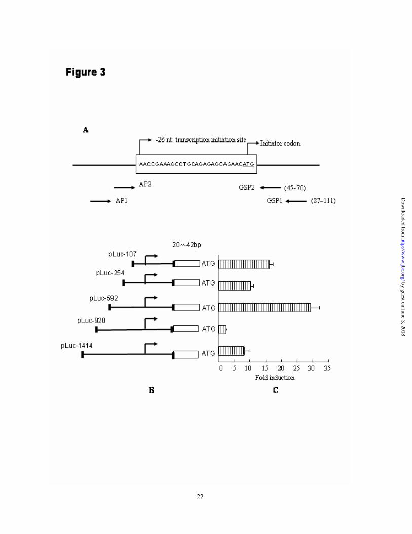
this binding activity of HSE/XAF1 by introducing the GAA to CCC and TTC to GGG mutation into pLuc-920. As the typical HSF1 binding element consisted of inverted dyad repeats of nGAAn/nTTCn motif, two mutant constructs were generated with type 1 mutating the outer pair of GAA/TTC elements and type 2 mutating the inner pair of GAA/TTC elements (Fig 6A). After verifying the prospected mutation by DNA sequencing, the mutant pGL3 constructs were transiently transfected into AGS and SW1116 cells. Transcription activity was evaluated and compared with the wildtype construct. As indicated in Fig 6B, RLU induction of wildtype, type 1 and type 2 mutant pLuc-920 constructs were 3.04±0.52, 2.74±0.36 and 13.54±0.36 in AGS cell and 3.15±0.22,
3.08±0.48 and 16.44±0.33 in SW1116 cell. Type 2 but not the type 1 mutation increased the transcription activity of pLuc-920 significantly (p <0.05 comparing with the wildtype control). To further define binding capacity of the inner GAA/TTC sequence, EMSA assay was carried out to examine the specific binding of wildtype and type 2 mutant oligonucleotides (Fig 6C). Wildtype but not the type 2 mutant probe bound to the nuclear extract of AGS cells effectively. These findings indicated that the inner GAA/TTC sequences in -862/-821 region contributed to HSF1 binding and repression of XAF1 transcription.
Moreover, pifithrin-α (PFA), a novel defined inhibitor of HSF1 (24), was applied to examine its effect on the transcription activity of truncated XAF1 promoter constructs. SW1116 cells were transiently transfected with pLuc-592,
pLuc-920 and pLuc-1414 followed by
incubation with 15µM of PFA. Cells were lysed and assayed for luciferase activity. As shown in figure 6D, treatment with PFA did not or only slightly change the transcription activity of pLuc-592 constructs. However, it increased the transcription activity of pLuc-1414 and pLuc-920 significantly (p <0.05). This finding proved the role of HSF1 binding in repression of XAF1 transcription.
Anti-oxidants overcome the suppression of XAF1 expression - Reactive oxygen substance (ROS) has been implicated as an etiologic factor in numerous diseases, including cancer. ROS can originate exogenously from agents that generate oxygen free radicals, and endogenously, for example, as a result of normal cellular metabolism, such as mitochondrial oxidative phosphorylation (25-27). It has been reported that H2O2 could stimulate binding of HSF1 to the heat shock element (HSE) (28-30). To verify the role of HSF1 in suppressing XAF1 expression, AGS and SW1116 cells were
exposed to 200µM of H2O2 in the presence or absence of antioxidants, N-Acetyl-L-cysteine (NAC) (20mM) or Catalase (CAT) (1000 U/ml) for 12 h. Nuclear extracts were incubated with radio-labeled -862/-821 probe. EMSA were carried out to examine the specific HSF1 binding activity. As shown in Fig 7A, both NAC and CAT were able to suppress HSF1 binding activities. Regarding to transcription activity, AGS cells transiently transfected with pLuc-920 were exposed to NAC or CAT to suppress the putative activation of HSF1. Transcription
10
by guest on June 3, 2018http://w
ww
.jbc.org/D
ownloaded from

activities of pLuc-920 treated with dissolvent control, NAC and CAT were 3.85±0.5,
6.44±0.16 and 11.4±0.24, respectively.
Antioxidants increased transcription activity of pLuc-920 by 50% to 150% (Fig 7B, p <0.05 comparing to non-treatment control). Moreover, pretreatment of AGS cells with antioxidants up-regulated XAF1 protein expression (Fig 7C). These findings revealed that stress factors such as ROS suppressed transcription of XAF1 mediated by the interaction between HSF1 and HSE within the regulatory promoter region.
DISCUSSION
Bcl-2, HSP and IAPs are three anti-apoptotic
family proteins. The crosstalk between HSP and IAPs was confirmed by the finding that HSP90 positively modulated the expression and function of survivin in cancer cells via binding to the conserved baculovirus IAP repeat structure (32). Our present study provides a new insight into the interaction between HSP and IAPs that HSF1 down-regulated IAP interacting protein, XAF1, through transcription regulation. It was well known that cancer cells have encountered higher level of stress pressure, both exogenous and endogenous (25, 26). Many cancers like sporadic colon cancers might be originated from inflammation such as inflammatory bowel diseases in which higher oxidative metabolites were produced by both infiltrated neutrophils and colon epithelial cells (33). The subsequent induction of stress-associated proteins including HSPs and MAPKs will promote cell transformation (34,
35). As the pivotal transcription factor that stimulates HSP proteins, the expression profile of HSF1 in GI cancers has seldom been studied. By using matched normal and cancerous gastric and colon tissues, we showed cancer tissues expressed higher level of HSF1 than normal tissues. This result was consistence with Cen’s finding that HSF1 expression was increased in 86% (30/35) of sporadic colon cancer patients demonstrated by cDNA microarray assays (36). EMSA showed cancer tissues possessed stronger HSF1 binding activity than their normal counterparts. These data indicated that GI cancer cells had high level and active form of HSF1 protein than normal tissues.
Inhibitor of apoptosis proteins, which is partly attributable to adaptive stress response, is commonly found in cancer cells (1-3). XAF1, a newly identified antagonist of XIAP, has been identified as a tumor suppressor. Expression of XAF1 in cancer cells, including gastric cancers, was lower than that of normal tissues (16, 22). Our findings that HSF1 as well as oxidative, hypo-osmotic and heat stress down-regulated XAF1 expression in GI cancer cell lines suggested that XAF1 was a stress-associated gene with its expression being negatively regulated during stress response.
Stress activated survival response included the induction of anti-apoptotic proteins. Yet, if the exposure to a specific stress is excessive, cell death will occur, either by necrosis or apoptosis (3, 37-38). It is reported when cells are exposed to low H2O2 concentration, they develop resistance to subsequent challenges with high
concentrations of the same agent that would
11
by guest on June 3, 2018http://w
ww
.jbc.org/D
ownloaded from

otherwise be lethal (38). In this study, the concentration of H2O2, the temperature of HS and the osmolarity used were all within the tolerable or physiological range (39-41). Their effect on repression of XAF1, and/or induction of other IAP proteins such as survivin reflects a novel survival mechanism of cancer cells.
As a key stress-associated transcription factor, HSF1 exerts both activating and suppressing effect on different target genes. Albeit the consensus HSF1 binding DNA sequence within the promoter of HSPs is the contiguous inverted dyad repeats of pentanucleotide nGAAn (HSE) (22), the capacities of the cis-formed dyad repeats of nGAAn or incontiguous nGAAn/nTTCn binding to HSF1 have been confirmed in all genes repressed by HSF1.
These genes include IL-1β, c-fos, TNF-α and RANK Ligand (11-13). The HSE in RANK Ligand located in -1275~ -2kb region.
To search for the transcription regulatory element, we first demonstrated the transcription initiation site of XAF1 gene by 5’-RACE. It is located in -26nt adenosine upstream of ATG initiator. Through searching DBTSS database (http://dbtss.hgc.jp/index.html) in which most genes possessed multiple TSS due to the different assay other than RACE utilized, we found XAF1 transcription starting region was located at -4nt to -40nt. Thus, we cloned the 5’-flanking sequence containing part of 5’-UTR sequence and defined them as regulatory promoter of XAF1. A putative repressor or silencer sequence was eventually located between -592 to -920nt by dual luciferase reporter assay. Two segments rich of
nGAAn/nTTCn contigs between -592 to -1414nt region were found. Competitive EMSA assay excluded the specific HSF1 binding capacity of -1008/-982 segment, hence, demonstrated -862/-821 segment as a high affinity HSE. In accordance with luciferase result, this segment located at the proposed transcription silencer region. On the other hand, this region contained two pairs of inverted dyad repeats of nGAAn/nTTCn motifs. Further site-mutagenesis strategy implicated the inner pair of nGAAn/nTTCn contig as functional HSF1 binding element (HSE/XAF1) that responsible for repression of XAF1 transcription.
To further validate HSF1 binding-mediated suppression of XAF1 transcription, we used a
HSF1 inhibitor (24), pifithrin-α, to inactivate cytosolic HSF1 protein. Pifithrin-α can also suppresses p53 activity, however, no typical P53 binding element is determined within the 5’-flanking region of XAF1. We showed pretreatment with this inhibitor eliminated the effect of transcription repressor within XAF1 regulatory promoter in unstressed cancer cell, suggesting endogenous intracellular stress pressure maintained the transcriptional inhibition of XAF1 gene in cancer cells.
Reactive oxygen species (ROS) was the predominant endogenous stressor of cancer cells. Bittinger et al. reported that melanoma cells produced large amounts of superoxide anions without stimulants, which was implicated in metastasis by promoting endothelial injury (42). ROS also plays a central role in the modulation of HSF1 activation since it was not only one of the stressors that could activate HSF1 but also be
12
by guest on June 3, 2018http://w
ww
.jbc.org/D
ownloaded from

increased by many other cellular stresses that lead to HSF activation (28,30,43). Therefore, we evaluated the effects of antioxidants on XAF1 regulation. We found both N-Acetyl-L-cysteine and Catalase were able to suppress HSE/XAF1 binding activity, to abrogate transcription inhibition, and to induce XAF1 expression. Because XAF1 was a pro-apoptotic gene and its over-expression suppressed cell growth (data not shown), our findings were consistent to previous observation that a moderate level of intracellular ROS was important to maintain appropriate redox
balance and to stimulate cancer cell proliferation (44,45).
In summary, GI cancer cells expressed high level of HSF1. It mediated stress stimuli-induced down-regulation of XAF1 via interaction with a HSE within the 5’-flanking region. This mechanism may contribute to the low expression of XAF1 in cancer cells and preventing them from apoptosis. For the first time, our findings define XAF1 as a novel stress-associated gene that negatively modulates cancer cell growth.
13
by guest on June 3, 2018http://w
ww
.jbc.org/D
ownloaded from

REFERENCES
1. Feder, M. E., Hofmann, G. E. (1999) Annu. Rev. Physiol. 61, 243-282 2. Sarto, C., Binz, P. A., Mocarelli. P. (2000) Electrophoresis. 21, 1218-1226 3. Carper, S. W., Duffy, J. J., Gerner, E. W. (1987) Cancer Res. 47, 5249-5255 4. Trautinger, F., Kindas-Mugge, I., Knobler, R. M., Honigsmann, H. (1996) J. Photochem. Photobiol.
B. 35, 141-148 5. Wang, D., Kreutzer, D. A., Essigmann, J. M. (1998) Mutat. Res. 400, 99-115 6. Wei, H. (1992) Med. Hypotheses. 39, 267-270 7. Kroeger, P. E., Morimoto, R. I. (1994) Mol. Cell. Biol. 14, 7592-7603 8. Wang, Y., Morgan, W. D. (1994) Nucleic Acids Res. 22, 3113-3118 9. Mivechi, N. F., Shi, X. Y., Hahn, G. M. (1995) J. Cell Biochem. 59, 266-280 10. Pirkkala, L., Nykanen, P., Sistonen, L. (2001) FASEB J. 15, 1118-1131 11. Cahill, C. M., Waterman, W. R., Xie, Y., Auron, P. E., Calderwood, S. K. (1996) J. Biol. Chem. 271,
24874-24879 12. Xie, Y., Zhong, R., Chen, C., Calderwood, S. K. (2003) J. Biol. Chem. 278, 4687-4698 13. Singh, I. S., He, J. R., Calderwood, S., Hasday, J. D. (2002) J. Biol. Chem. 277, 4981-4988. 14. Hay, B. A. (2000) Cell Death Differ. 7, 1045-1056. 15. Arnt, C. R., Chiorean, M. V., Heldebrant, M. P., Gores, G. J., Kaufmann, S. H. (2002) J. Biol. Chem.
277, 44236-44243 16. Fong, W. G., Liston, P., Rajcan-Separovic, E., St Jean, M., Craig, C., Korneluk, R. G. (2000)
Genomics. 70, 113-122. 17. Liston, P., Fong, W. G., Kelly, N. L., Toji, S., Miyazaki, T., Conte, D., Tamai, K., Craig, C. G.,
McBurney, M. W., Korneluk, R. G.. (2001) Nat. Cell. Biol. 3, 128-133 18. Leaman, D. W., Chawla-Sarkar, M., Vyas, K., Reheman, M., Tamai, K., Toji, S., Borden, E. C. (2002)
J. Biol. Chem. 277, 28504-28511 19. Syagailo, Y. V., Okladnova, O., Reimer, E., Grassle, M., Mossner, R., Gattenlohner, S., Marx, A.,
Meyer, J., Lesch, K. P. (2002) Gene. 294, 259-268 20. den Dunnen, J. T., Antonarakis, S. E. (2001) Hum. Genet. 109, 121-124 21. Wong, B. C., Jiang, X. H., Lin, M. C., Tu, S. P., Cui, J. T., Jiang, S. H., Wong, W. M., Yuen, M. F.,
Lam, S. K., Kung, H. F. (2004) Gastroenterology. 126, 136-147 22. Wu, B. J., Kingston, R. E., Morimoto, R. I. (1986) Proc. Natl. Acad. Sci. U S A. 83, 629-633 23. Byun, D. S., Cho, K., Ryu, B. K., Lee, M. G., Kang, M. J., Kim, H. R., Chi, S. G. (2003) Cancer Res.
63, 7068-7075 24. Komarova, E. A., Neznanov, N., Komarov, P. G., Chernov, M. V., Wang, K., Gudkov, A. V. (2003) J.
14
by guest on June 3, 2018http://w
ww
.jbc.org/D
ownloaded from

Biol. Chem. 278, 15465-15468 25. Szatrowski, T. P., and Nathan, C. F. (1991) Cancer Res. 51, 794-798 26. Hileman, E. O., Liu, J., Albitar, M., Keating, M. J., Huang, P. (2004) Cancer Chemother. Pharmacol.
53, 209–219 27. Lee, B. R., Um, H. D. (1999) Exp. Cell. Res. 248, 430-438 28. Ahn, S. G., Thiele, D. J. (2003) Genes Dev. 17, 516-528 29. Ozaki, M., Deshpande, S. S., Angkeow, P., Suzuki, S., Irani, K. (2000) J. Biol. Chem. 275,
35377-35383 30. Nishizawa, J., Nakai, A., Matsuda, K., Komeda, M., Ban, T., Nagata, K. (1999) Circulation. 99,
934-41 31. Garrido, C., Gurbuxani, S., Ravagnan, L., Kroemer, G. (2001) Biochem. Biophys. Res. Commun.
286, 433-442 32. Fortugno, P., Beltrami, E., Plescia, J., Fontana, J., Pradhan, D., Marchisio, P. C., Sessa, W. C., Altieri,
D. C. (2003) Proc. Natl. Acad. Sci. U S A. 100, 13791-13796 33. Munkholm, P. (2003) Aliment Pharmacol Ther. Suppl 2, 1-5 34. Garrido, C., Fromentin, A., Bonnotte, B., Favre, N., Moutet, M., Arrigo, A. P., Mehlen, P., Solary, E.
(1998) Cancer Res. 58, 5495-5499 35. Liao, Q., Guo, J., Kleeff, J., Zimmermann, A., Buchler, M. W., Korc, M., Friess, H. (2003)
Gastroenterology. 124, 1830-1845. 36. Cen, H., Zheng, S., Fang, Y. M., Tang, X. P., Dong, Q. (2004) World J. Gastroenterol. 10, 3122-3126 37. Franek, W. R., Horowitz, S., Stansberry, L., Kazzaz, J. A., Koo, H. C., Li, Y., Arita, Y., Davis, J. M.,
Mantell, A. S., Scott, W., Mantell, L. L.(2001) J. Biol. Chem. 276, 569-575 38. Kim, D. K., Cho, E. S., Seong, J. K., Um, H. D. (2001) J. Cell Sci. 114, 4329-4334 39. Caruccio, L., Bae, S., Liu, A. Y., Chen, K. Y. (1997) Biochem. J. 327, 341-347 40. Huang, L. E., Caruccio, L., Liu, A. Y., Chen, K. Y. (1995) Biochem. J. 307, 347-352 41. Hatayama, T., Fujimoto, S., Sakai, K. (1997) Biol. Pharm. Bull. 20, 605-612 42. Bittinger, F., Gonzalez-Garcia, J. L., Klein, C. L., Brochhausen, C., Offner, F., Kirkpatrick, C. J.
(1998) Melanoma Res. 8, 381-387 43. Bruce, J. L., Price, B. D, Coleman, C. N., Calderwood, S. K. (1993) Cancer Res. 53, 12-15 44. Muzio, G., Salvo, R. A., Trombetta, A., Autelli, R., Maggiora, M., Terreno, M., Dianzani, M. U.,
Canuto, R. A. (1999) Lipids. 34, 705-711 45. Tanaka, T., Akatsuka, S., Ozeki, M., Shirase, T., Hiai, H., Toyokuni, S. (2004) Oncogene. 23,
3980-3989
15
by guest on June 3, 2018http://w
ww
.jbc.org/D
ownloaded from

FOOTNOTES
This study is supported by the Simon KY Lee Gastroenterology Research Fund, Queen Mary Hospital, and the Gastroenterological Research Fund, University of Hong Kong. pcDNA3.1/HSF1 expressing construct was kindly provided by Dr. R.E. Kingston at Department of Molecular Biology, Massachusetts General Hospital, USA.
Key words: XIAP- associated factor 1, heat shock factor 1, stress, transcription, Cancer, Promoter The abbreviations used are: XAF1, XIAP-associated factor 1; HSP, heat shock protein; IAP, inhibitor of apoptosis; XIAP, X-linked IAP protein; HSF, heat shock factor; HSE, heat shock element; HS, heat shock; HO, hypo-osmosis; Cat, Catalase; NAC, N-Acetyl-L-cysteine; PFA, pifithrin-α; RACE, Rapid Amplification of cDNA Ends; EMSA, Electrophoretic Mobility Shift Assay, TSS, transcription starting site; GSP, gene specific primer; AP, adaptor primer; RLU, relative luciferase unit; ChIP, chromatin immunoprecipitation.
FIGURE LEGENDS
Fig. 1. GI cancer expressed higher level of HSF1 than normal tissues. Immunoblotting for HSF1 expression in matched gastric (A) and colon (B) normal and tumor tissues. GN1-GN3, gastric normal tissue; GT1-GT3, gastric tumor tissue; CN1-CN9, colon normal tissue; CT1-CT9, colon tumor tissue. These figures were representatives of two independent experiments. C, HSE/consensus oligonucleotide was labeled with 32P, EMSA was carried out to detect its binding to the whole cell lysate of tissue specimens. This figure was representative of three independent experiments. Fig. 2. XAF1 expression inversely correlated with HSF1 in GI cancer cells. A, Immunoblotting assay for HSF1 and XAF1 expression in GI cancer cell lines. B, AGS and Lovo cells were transfected with empty vector or pcDNA3.1-HSF1 construct for 48 h. HSF1 and XAF1 expressions were detected by
immunoblotting. C, AGS cell was transfected without or with GL2 (control) or HSF1 siRNA for 48 h, HSF1 and XAF1 expressions were detected by immunoblotting with actin as the internal control. This experiment was repeated twice in both AGS and Lovo cells with identical finding. D, Kato-III cell was treated with 200µM of H2O2 for 24 h, heat stress (HS, 42ºC) or hypo-osmosis (HO) for 30 minutes followed by culture in standard medium for 24 h. HSF1 and XAF1 expression were detected by immunoblotting. These figures were the representatives of two to three independent experiments.
16
by guest on June 3, 2018http://w
ww
.jbc.org/D
ownloaded from

Fig. 3. Location of the regulatory promoter region of XAF1 gene. A, Diagram of the location of XAF1 gene specific primer (GSP) and TSS of XAF1 gene determined by 5’-RACE assay. B, location of the truncated 5’-flanking DNA segments of XAF1 gene cloned into pGL3 basic plasmid. The upstream nucleotide adjacent to the translation initiator ATG codon was defined as -1. C, AGS cells were transiently transfected with various pLuc constructs for 24 h. The firefly and renilla luciferase activities were measured. Promoter activity was presented as the fold induction of relative luciferase unit (RLU) comparing to pGL3 basic vector. RLU= values of firefly luciferase unit/ values of renilla luciferase unit. This result was expressed as the mean of three independent experiments ± standard deviation. Fig. 4. Analysis of 5’-flanking sequence of XAF1 gene. A, Consensus sequence of HSE. B, HSE sequence presented in human HSP70 promoter. C, Two nGAAn (nTTCn)-rich sequences presented in the 5’-flanking region of XAF1 gene. The putative HSF1-binding elements were underlined and the nGAAn (nTTCn) motifs were highlighted. Fig. 5. Identification of HSF1 binding sequence in XAF1 gene. A, Double strand HSE/consensus DNA probe was labeled with 32P and bound to the nuclear extracts of heat-treated AGS cells with or without pre-incubation with 100-fold excess of each oligonucleotide (-1008/-982, -862/-821 and HSE/consensus). DNA binding activity was determined by EMSA. B, Double strand oligonucleotides consensus to the -1008/-982 and -862/-821 sequences were labeled with 32P and bound to the nuclear extract of heat-treated AGS cells in absence or presence of 100-fold excess of unlabeled probe (cold probe). C, Double strand -862/-821 oligonucleotide was labeled with 32P and bound to the nuclear extract of AGS cells with various treatments. These figures represented one of three independent experiments with
similar findings. D, Chromatin immunoprecipitation (ChIP) analysis of HSE/XAF1 element from untreated (lane 1-3), H2O2 (Lane 4-6), heat shock (HS, lane 7-9) and hypo-osmosis (HO, lane 10-12) -induced AGS cells using antibody specific for HSF1 (Lane 2, 5, 8, 11) or goat IgG control (lanes 1, 4, 7, 10). Input chromatins were presented in Lane 3, 6, 9, 12. This experiment was repeated twice in both AGS and Lovo cells and the result was repeatable. Fig. 6. Upregulation of XAF1 expression by inactivation of HSF1 binding. A, Location of site-directed mutations of -862/-821 sequences of pLuc-920 constructs, the mutant bases were highlighted. B, Wildtype and mutant pLuc-920 constructs were transiently transfected into AGS and SW1116 cells. Luciferase activities were measured. The result was expressed as the mean of four independent experiments ± standard deviation. *, p <0.05 vs wildtype control. C, Double strand oligonucleotides consensus to wildtype or mutant 2 (MT2) sequences were labeled with 32P and bound to the nuclear extract of AGS cells. This experiment was repeated twice with identical finding. D, SW1116 cells were
transiently transfected with pLuc-1414, pLuc-920 and pLuc-592 followed by the incubation with 15µM
17
by guest on June 3, 2018http://w
ww
.jbc.org/D
ownloaded from

of pifithrin-α (PFA) or dissolvent control. Cells were lysed and assayed for luciferase activity. The result was expressed as the mean of three independent experiments ± standard deviation #, p <0.05 vs control.
Fig. 7. Anti-oxidants restored XAF1 expression. A, AGS cells were exposed to 200µM of H2O2 in the presence or absence of antioxidants, NAC (20mM) or Cat (1000 U/ml) for 12 h. Nuclear extracts were incubated with radio-labeled -862/-821 probe. This figure was the representative of two independent experiments. B, AGS cells transfected with pLuc-920 construct were exposed to NAC (20mM) or Cat (1000 U/ml) and assayed for luciferase activity. The fold induction of RLU was expressed as the mean of three independent experiments ± standard deviation. #, p <0.05 vs control. C. AGS cells were treated with or without antioxidants for 24 h, XAF1 expression was detected by immunoblotting. This experiment was repeated twice with consistent finding.
18
by guest on June 3, 2018http://w
ww
.jbc.org/D
ownloaded from

Experiment Name Position or orientation
Sequence (5’-3’)
Reverse -42~-20 CCGCTCGAGTTCGGTTGAGTTTCGTTTCTTGC
Forward107 -90~-107 GGGGTACCGATCTCCTCCCTCCCTGAA
Forward 254 --235~-254 GGGGTACCCAGCCTCAGGGAGGTAGATG
Forward 592 -592~-569 GGGGTACCAGGGTCTGGAAAAACTCTAAGGAC
Forward 920 -920~-896 GGGGTACCATGCTTACATGAGGGATTAAAACGA
Luciferase construction
Forward 1414 -1414~1391 GGGGTACCTTTTTAGTAGAGACGGGGTTTCAC
Wildtype sequence
sense AACATAGGAACAATGTTGAAACAGTCTTTCATTCTTCCCT
Mutant 1 sense AACATAGCCCCAATGTTGAAACAGTCTTTCATTCGGGCCT
Site- directed mutagenesis
Mutant 2 sense CAATGTTCCCACAGTCTGGGATTCTT
-1008/-982 sense ATTTTCTCTTTTTTCATTTCATTTTTCTTT EMSA -862/-821 sense TGAACATAGGAACAATGTTGAAACAGTCTTTCA
TTCTTCCCT
Adaptor 1 Forward CCATCCTAATACGACTCACTATAGGGC
Adaptor 1 Forward ACTCACTATAGGGCTCGAGCGGC
XAFGSP1 Reverse ACACTCCGGACACAGGACCAGGAAC RACE
XAFGSP2 CATGGAGGGTGAAGTTGGCAGAGACT
XAF1 Forward GCTCCACGAGTCCTACTG
XAF2 Reverse ACTCTGAGTCTGGACAAC
GAPDH Forward GACCACAGTCCATGCCATCAC
RT-PCR
GAPDH Reverse GTCCACCACCCTGTTGCTGTA
Forward TCTCTGCCTCCATTTTCTCTTT ChIP -1021/-779
Reverse GAGAAGCAGTGTGTGGTGGT
Table 1. List of the oligonucleotides primers for the amplification and mutation
19
by guest on June 3, 2018http://w
ww
.jbc.org/D
ownloaded from

Bing Zou, Xiaomeng An, Bo Jiang, Hsiang-Fu Kung and Benjamin C. Y. WongJide Wang, Hua He, Lifen Yu, Harry Hua-xiang Xia, Marie C. M. Lin, Qing Gu, Ming Li,
protein-associated factor 1 (XAF 1) through transcriptional regulationHeat shock factor 1 (HSF1) downregulates X-linked inhibitor of apoptosis
published online November 21, 2005J. Biol. Chem.
10.1074/jbc.M505890200Access the most updated version of this article at doi:
Alerts:
When a correction for this article is posted•
When this article is cited•
to choose from all of JBC's e-mail alertsClick here
by guest on June 3, 2018http://w
ww
.jbc.org/D
ownloaded from