heart - Valencia Collegefd.valenciacollege.edu/file/bderrickson/Ch 2 Heart.pdf · heart is...
25
15 Chapter 2 Heart ♦Location -The heart is located in the thoracic cavity between the lungs and the diaphragm; most of the heart is positioned to the left of the body’s midline (Figure 20.1b, Tortora). ♦ Size -The heart weights about 300 g ( 0.66 lb. ) and is about the size of a person’s fist. ♦Structure -Pericardium -a connective tissue sac that surrounds the heart and keeps it in place (Figure 20.2a, Tortora) -organized into 2 layers: 1. parietal layer -outer layer 2. visceral layer -inner layer -attached to the heart wall pericardial cavity -a space that is located between the parietal and visceral layers of the pericardium -contains pericardial fluid, which functions as a lubricant by reducing friction within the pericardium as the heart beats. -Heart Wall -The bulk of the heart wall consists of myocardium (cardiac muscle), which is responsible for the contraction of the heart (Figure 20.2a, Tortora). -Chambers of the Heart -The heart consists of 4 internal chambers that contain blood: two superior atria and two inferior ventricles (Figures 20.3 and 20.4, Tortora): 1. atria -The superior chambers of the heart are called the right atrium and the left atrium (atrium is singular, while the term atria is the plural form). -The atria are separated by a partition called the interatrial septum. -On the external surface of each atrium is an appendage called an auricle, which increases atrial volume.
Transcript of heart - Valencia Collegefd.valenciacollege.edu/file/bderrickson/Ch 2 Heart.pdf · heart is...

15
Chapter
2 Heart ♦Location -The heart is located in the thoracic cavity between the lungs and the diaphragm; most of the heart is positioned to the left of the body’s midline (Figure 20.1b, Tortora). ♦ Size -The heart weights about 300 g ( 0.66 lb. ) and is about the size of a person’s fist. ♦Structure -Pericardium -a connective tissue sac that surrounds the heart and keeps it in place (Figure 20.2a, Tortora) -organized into 2 layers: 1. parietal layer -outer layer 2. visceral layer -inner layer -attached to the heart wall pericardial cavity -a space that is located between the parietal and visceral layers of the pericardium -contains pericardial fluid, which functions as a lubricant by reducing friction within the pericardium as the heart beats. -Heart Wall -The bulk of the heart wall consists of myocardium (cardiac muscle), which is responsible for the contraction of the heart (Figure 20.2a, Tortora). -Chambers of the Heart -The heart consists of 4 internal chambers that contain blood: two superior atria and two inferior ventricles (Figures 20.3 and 20.4, Tortora): 1. atria -The superior chambers of the heart are called the right atrium and the left atrium (atrium is singular, while the term atria is the plural form). -The atria are separated by a partition called the interatrial septum. -On the external surface of each atrium is an appendage called an auricle, which increases atrial volume.
16
2. ventricles -The inferior chambers of the heart are called the right ventricle and the left ventricle. -The ventricles are separated from each other by a partition called the interventricular septum. -Inspection of the heart chambers reveals that the atria and ventricles vary with respect to the thickness of their walls. -The atria have thin walls because they only have to transport blood to the nearby ventricles. -The ventricles, however, have thicker walls because they have to pump blood over a longer distance to organs outside of the heart. -The right ventricle pumps blood to the alveoli of lungs, while the left ventricle pumps blood to the rest of the organs of the body. -Note that the left ventricle has a greater workload than the right ventricle; hence, the left ventricle is thicker than the right ventricle. -Heart Valves -As the heart contracts, blood moves from each atrium into a ventricle and then out of the heart into an artery. -To prevent backflow of blood, the heart contains valves. heart valves -consist of connective tissue -open and close due to changes in blood pressure that occur in the atria and ventricles as the heart contracts and relaxes -2 major groups of heart valves (Figure 20.4, Tortora): 1. atrioventricular (AV) valves -valves that separate the atria from the ventricles -2 types: a. tricuspid valve -the right AV valve -consists of 3 cusps (flaps) b. bicuspid valve -also called the mitral valve -the left AV valve -consists of 2 cusps -Structure -The AV valves consist of cusps (flaps). -The cusps of the AV valves are attached by strings of connective tissue called chordae tendineae to papillary muscles, which are ventricular muscle fibers that project out of each ventricle.
17
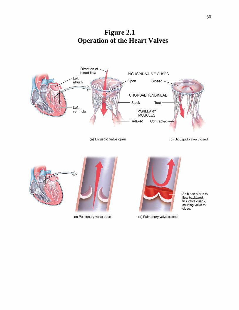
-Function -The AV valves permit unidirectional flow of blood from the atria into the ventricles. •When the ventricles are relaxed, the papillary muscles are also relaxed and the chordae tendineae are slack (loose) (Figure 2.1a, Derrickson). -Blood in the atria pushes the AV valves open and then moves into the ventricles. • When the ventricles contract, the papillary muscles also contract and pull on the chordae tendineae, which become taut (tight) (Figure 2.1b, Derrickson). -The chordae tendineae then pull on the cusps of the AV valves, thereby closing the valves and preventing backflow of blood from the ventricles into the atria. 2. semilunar valves -valves located at the junction between the ventricles and the arteries that leave the heart -2 types: a. pulmonary semilunar valve -located at the junction between the right ventricle and the pulmonary trunk -The pulmonary trunk is a large artery that branches to give rise to the smaller pulmonary arteries. b. aortic semilunar valve -located at the junction between the left ventricle and the aorta -Structure -Both semilunar valves contain 3 cusps, which are shaped like half moons (hence the name semilunar). -The cusps are attached to the inner walls of the pulmonary trunk and aorta and project into the lumen of these arteries. -Function -The semilunar valves permit unidirectional flow of blood from the ventricles into the pulmonary trunk and aorta. • During ventricular contraction, blood in the ventricles forces the semilunar valves to open and then moves into the pulmonary trunk and aorta (Figure 2.1c, Derrickson). • During ventricular relaxation, any blood that moves back towards the heart fills the pockets of the cusps of the semilunar valves (Figure 2.1d, Derrickson). -This causes the semilunar valves to come together and close, thereby preventing backflow of blood from the pulmonary trunk and aorta into the ventricles.

18
♦Function of the Heart -The heart functions as a pump that is responsible for the circulation of blood through the blood vessels of the body. ♦ Blood Flow Through the Heart and Body -Figures 20.4, 20.7, and 20.8 (Tortora) illustrate blood flow through the heart and the body. The right atrium receives deoxygenated blood (blood that has given up some of its O2 to body cells and picked up CO2 from body cells) through 3 veins: 1. superior vena cava -brings deoxygenated blood from blood vessels in the head, neck, upper limbs, and upper trunk to the right atrium of the heart 2. inferior vena cava -brings deoxygenated blood from blood vessels in the lower trunk and lower limbs to the right atrium of the heart 3. coronary sinus -brings deoxygenated blood from blood vessels in the heart wall to the right atrium of the heart The deoxygenated blood flows through the right atrium and continues into the right ventricle via the tricuspid valve. From the right ventricle, deoxygenated blood passes through the pulmonary semilunar valve into the pulmonary trunk. The pulmonary trunk divides into a right and left pulmonary artery, each of which carries deoxygenated blood to the alveoli of the lungs. In the alveoli, the blood becomes oxygenated as it picks up more O2 and releases some of its CO2 . The oxygenated blood flows through the pulmonary veins back to the heart, where it enters the left atrium. The oxygenated blood flows through the left atrium and continues into the left ventricle via the bicuspid (mitral) valve.
19
From the left ventricle, the oxygenated blood passes through the aortic semilunar valve into the aorta. The aorta branches to form smaller arteries that supply oxygenated blood to all of the organs of the body (except the alveoli of the lungs). -Note that the blood vessels of the body are classified into two groups: the pulmonary circulation and the systemic circulation. -The blood vessels that transport blood to and from the alveoli of the lungs form the pulmonary circulation. Examples: • pulmonary arteries • pulmonary veins -The blood vessels that transport blood to and from the rest of the organs of the body (except the alveoli of the lungs) form the systemic circulation. Examples: • aorta • inferior vena cava -In addition, the heart, like any other organ, has its own blood vessels. -This is due to the fact that the heart wall is thick; therefore, nutrients are not able to diffuse from the blood in the atria and ventricles to the cardiac muscle cells in the heart wall. -The blood vessels that supply the heart are collectively called the coronary (cardiac) circulation, which is part of the systemic circulation. -Examples: • coronary arteries • cardiac veins • coronary sinus

20
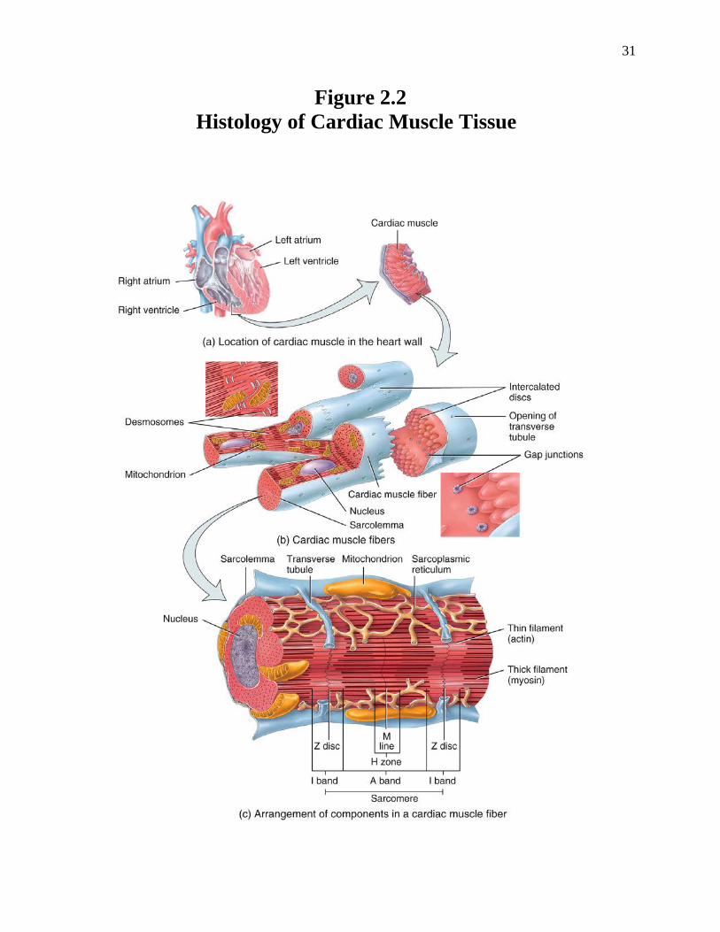
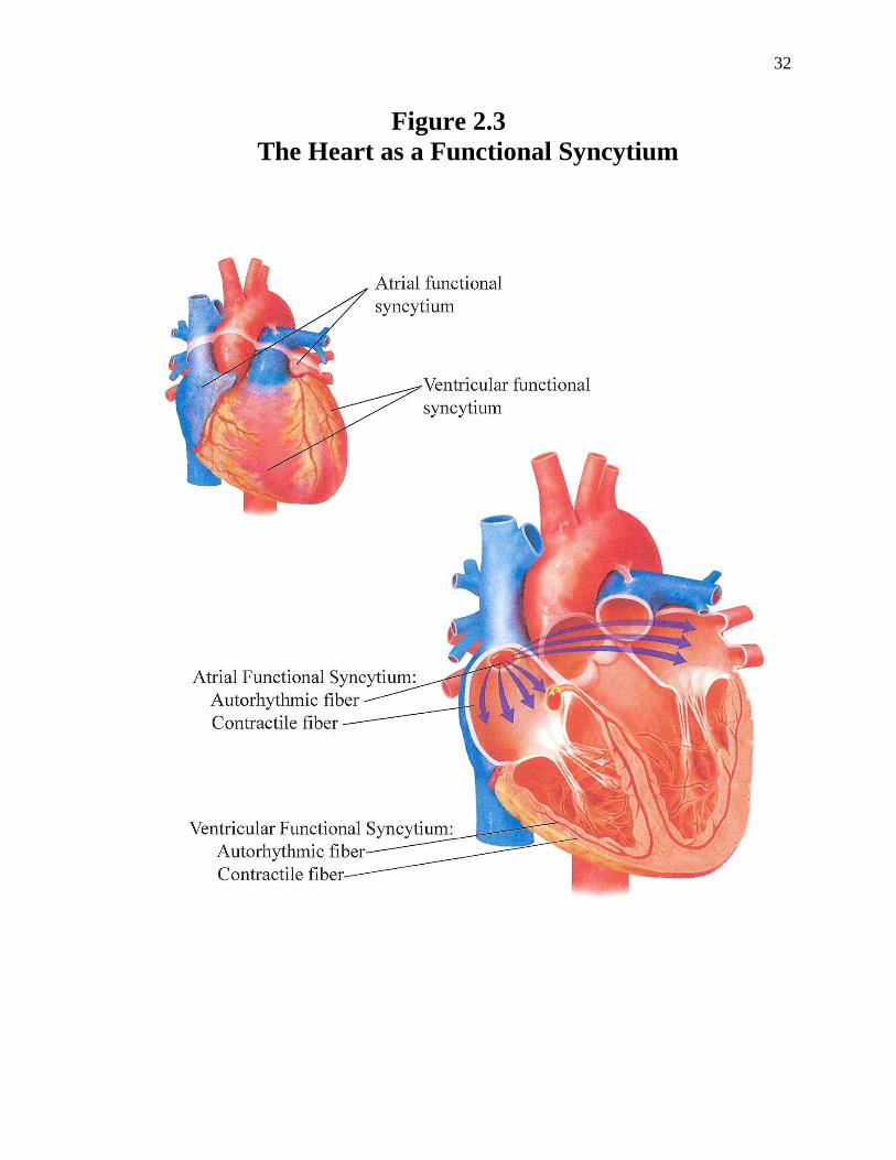
♦ Cardiac Muscle Tissue -The bulk of the heart is comprised of cardiac muscle tissue. -Like skeletal muscle fibers, cardiac muscle fibers are striated due to the presence of repeating sarcomeres consisting of thick and thin filaments that have a regular pattern of overlap (Figure 2.2, Derrickson). -The thick filaments contain myosin, and the thin filaments contain actin, troponin, and tropomyosin. -Cardiac muscle fibers contract due to myosin binding to actin and then causing the power stroke. -Unique to cardiac muscle fibers are intercalated discs, transverse thickening of the sarcolemma that connect the ends of cardiac muscle fibers to each other (Figure 2.2, Derrickson). -The intercalated discs contain desmosomes, which mechanically bind the cells together, and gap junctions, which electrically couple the cells together. ♦ Cardiac Muscle as a Functional Syncytium -Because cardiac muscle fibers are electrically coupled by gap junctions, when an action potential is generated in a mass of cardiac muscle fibers, it quickly spread to all the muscle fibers in that mass and then the muscle fibers contract together. -Such a mass of interconnected muscle fibers acts as a single, coordinated unit or functional syncytium. -The atria and the ventricles of the heart behave as two distinct functional syncytiums (Figure 2.3, Derrickson). -The atria and ventricles contract independently of each other, with the atria contracting before the ventricles. -This allows the ventricles to fill with blood from the atria before the ventricles eject blood out of the heart to the rest of the body. -Cardiac muscle, as a functional syncytium, consists of two types of muscle fibers: autorhythmic fibers and contractile fibers (Figure 2.3, Derrickson). 1. autorhythmic fibers -also known as pacemaker cells -account for only a very small number of cells in the functional syncytium and are usually grouped together. ■ Autorhythmic fibers include the components of the conduction system (SA node, AV node, bundle of His, bundle branches, and Purkinje fibers). - spontaneously generate action potentials - contain essentially no myofibrils and, therefore, are unable to contract 2. contractile fibers - constitute the great majority of cells in the functional syncytium ■ Contractile fibers include all cardiac muscle fibers in the atria and ventricles other than those of the conduction system. - have the necessary myofibrils to contract, but do not have the ability to initiate action potentials. - Instead, contractile fibers become excited and then contract together in response to action potentials conducted to them from autorhythmic fibers via gap junctions.
21
♦The Conduction System of the Heart -The conduction system causes the heart to undergo self-excitation (i.e. generate its own action potential) followed by coordinated contraction. -The built-in rhythm of the heart is known as autorhythmicity. -Components of the Conduction System -The conduction system consists of autorhythmic fibers that ensure that the chambers of the heart are excited and then contract in a coordinated manner (Figure 20.10a, Tortora): 1. sinoatrial (SA) node -located within the wall of the right atrium 2. atrioventricular (AV) node -located in the interatrial septum 3. bundle of His -also called the atrioventricular bundle -located in the interventricular septum 4. right and left bundle branches -located in the interventricular septum 5. Purkinje fibers -also called conduction myofibers -located within the walls of the ventricles -Recall that the components of the conduction system are autorhythmic fibers that lack actin and myosin and, therefore, do not contract. -Instead, the autorhythmic fibers are capable of initiating an action potential and and conducting (conveying) the action potential to one another and to the contractile fibers of the heart (those muscle fibers in the atria and ventricles that can contract). -Action potentials are able to be conducted throughout the heart by the conduction system because gap junctions connect the components of the conduction system to each other and to the contractile muscle fibers in the atria and ventricles. -Mechanism of the Conduction System The SA node generates an action potential. The action potential spreads from the SA node through the atria, causing the contractile muscle fibers of the atria to become excited and then to contract as a unit. Consequently, blood moves from the atria to the ventricles through the AV valves. Meanwhile, the action potential continues to move from the atria to the AV node.
22
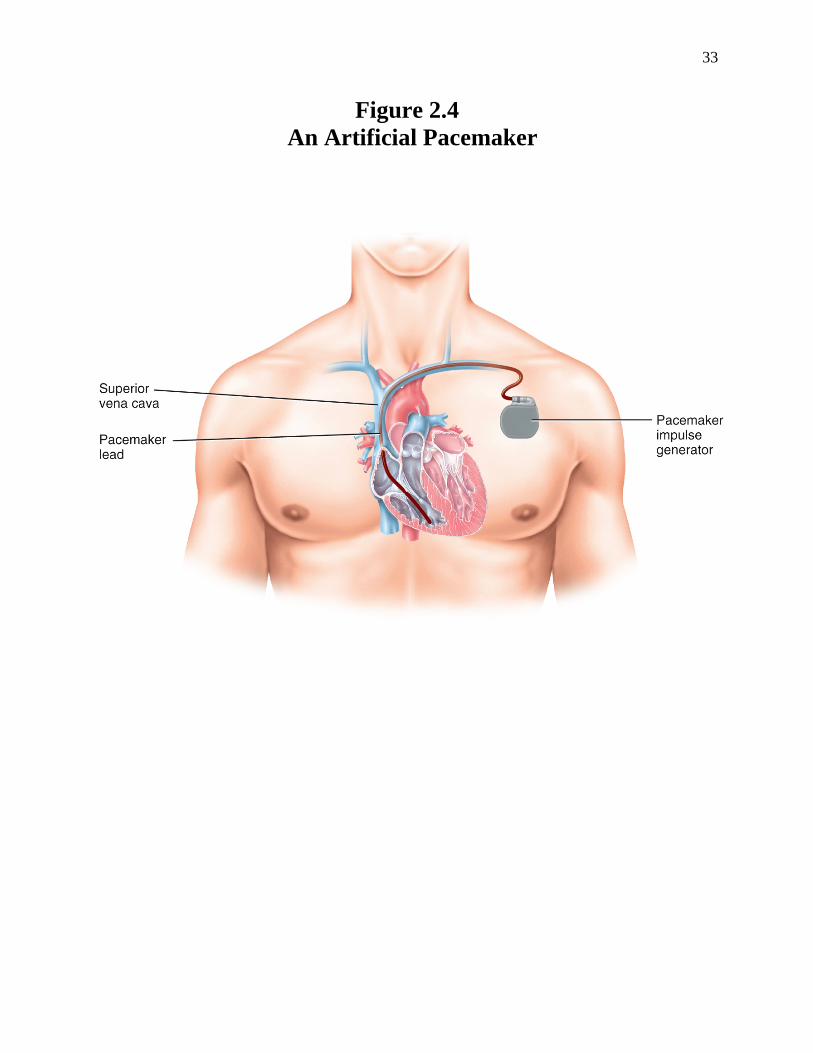
From the AV node, the action potential sequentially proceeds through the bundle of His, the right and left bundle branches, and the Purkinje fibers. From the Purkinje fibers, the action potential spreads through the ventricles, causing the contractile muscle fibers of the ventricles to become excited and then to contract as a unit. Consequently, blood moves through the semilunar valves into the pulmonary trunk and aorta. -Note that the atria contract before the ventricles because the conduction system brings the action potential to the atria before it brings it to the ventricles. -This allows the ventricles to fill up with blood before ejecting the blood into the pulmonary and systemic circulations. -The SA Node as the Heart’s Pacemaker -SA node cells do not have a stable resting membrane potential; instead, they repeatedly depolarize to threshold spontaneously. -This spontaneous depolarization is known as the pacemaker potential. -When the pacemaker potential reaches threshold, it triggers an action potential (Figure 20.10b). -The SA node generates action potentials before the other components of the conduction system have a chance to produce any action potentials of their own; hence, the SA node functions as the natural pacemaker of the heart. -Although the SA node produces these action potentials by itself, it is usually regulated (sped up or slowed down) by the autonomic nerves that supply the heart. -Under normal resting conditions, parasympathetic nerves slow down the SA node to the point that the SA node releases 75 action potentials per minute. –This causes the heart to beat 75 times per minute; each beat represents atrial contraction followed by ventricular contraction. –Thus, under normal resting conditions, the heart rate is 75 beats per minute. -Ectopic Pacemakers -If the SA node stops functioning normally, another component of the conduction system (usually the AV node) starts generating action potentials to keep the heart contracting. –The term ectopic pacemaker is used to refer to any site outside the SA node that becomes the pacemaker of the heart. –Usually, the heart rate is slower with an ectopic pacemaker. –Therefore, it may be necessary for an artificial pacemaker to be surgically implanted (including the batteries) underneath the skin with wires running to the atria and/or ventricles; such a device sends electrical activity to stimulate the heart to contract (Figure 2.4, Derrickson).

23
♦Action Potentials in a Contractile Cardiac Muscle Fiber -Unlike autorhythmic fibers, contractile cardiac muscle fibers have a stable resting membrane potential that is close to ̶ 90 mV. -When a contractile fiber is depolarized to threshold by an action potential initiated by an autorhythmic fiber of the conduction system, the contractile fiber produces its own action potential. -An action potential in a contractile cardiac muscle fiber has 3 main phases: rapid depolarization, plateau, and repolarization (Figure 20.11, Tortora). ♦Extrinsic Innervation of the Heart -Both divisions of the autonomic nervous system (ANS) supply nerves to the heart (Figure 2.5, Derrickson). -Parasympathetic Nervous System -Parasympathetic nerves, via the Vagus (X) nerves, innervate the SA node of the heart. -The neurotransmitter released by these nerves is acetylcholine. -Stimulation of the parasympathetic nerves to the heart has one major effect: • decrease in heart rate -This occurs because the Vagus nerves slow down the release of action potentials from the SA node. -Under normal resting conditions, the parasympathetic nervous system is dominant and the Vagus (X) nerves cause the heart rate to be around 75 beats/minute. -A person is said to exhibit bradycardia (slow heart rate) if the heart rate falls below 60 beats/min; this is often due to overactive parasympathetic (Vagal) stimulation of heart -Sympathetic Nervous System -Sympathetic nerves, via the cardiac accelerator nerves, innervate both the SA node and the ventricular myocardium of the heart. -The neurotransmitter released by these nerves is norepinephrine. -Stimulation of the sympathetic nerves to the heart causes two major effects: • increased heart rate -This occurs because the cardiac accelerator nerves speed up the release of action potentials from the SA node. -A person is said to exhibit tachycardia (rapid heart rate) if the heart rate is above 100 beats/minute; this is often caused by exercise or other conditions that cause stress on the body. • increased ventricular contraction -In addition, under sympathetic conditions, the adrenal glands release epinephrine and norepinephrine into the blood. -From the blood, these substances act as hormones that stimulate the heart in the same way as the cardiac accelerator nerves.

24
♦Electrocardiogram - An electrocardiogram (ECG) is a recording of the electrical activity (action potentials) that passes through the heart. ⇒The term elektrokardiogram (EKG) can also be used; kardia is German for heart. -The process of generating an electrocardiogram is known as electrocardiography (Figure 2.6, Derrickson). During electrocardiography, electrodes are placed on the skin of the limbs (limb leads) or on the skin of the chest (chest leads). From the body’s surface, these electrodes are able to detect the action potentials that pass through the heart as the heart beats (contracts). -Each standard limb lead consists of two electrodes (one designated as the positive electrode and the other as the negative electrode) that are attached to the surface of the body. ● In limb lead I, the positive electrode is connected to the left arm and the negative electrode to the right arm. ●In limb lead II, the positive electrode is connected to the left leg and the negative electrode to the right arm. ● In limb lead III, the positive electrode is connected to the left leg and the negative electrode to the left arm. -Each limb lead detects the electrical potential difference between the positive and negative electrodes and provides a separate electrocardiogram (ECG) recording. -The three standard limb leads form an imaginary triangle known as Einthoven’s triangle that extends between the arms and left leg to surround the heart. -Note that the right leg is not part of the triangle, but instead serves as the ground. -A typical ECG consists of 3 waves (Figure 20.12, Tortora): 1. P wave -represents atrial depolarization -This means that the atria become excited due to the movement of an action potential from the SA node through the atrial muscle fibers. -Shortly after the P wave appears, the atria contract. 2. QRS complex -represents ventricular depolarization -This means that the ventricles become excited due to the movement of the action potential through the ventricular muscle fibers. -Shortly after the QRS complex appears, the ventricles contract. 3. T wave -represents ventricular repolarization -This means that the ventricles revert back to their original, unexcited state as the action potential moves out of the ventricular muscle fibers. -Shortly after the T wave appears, the ventricles relax.
25
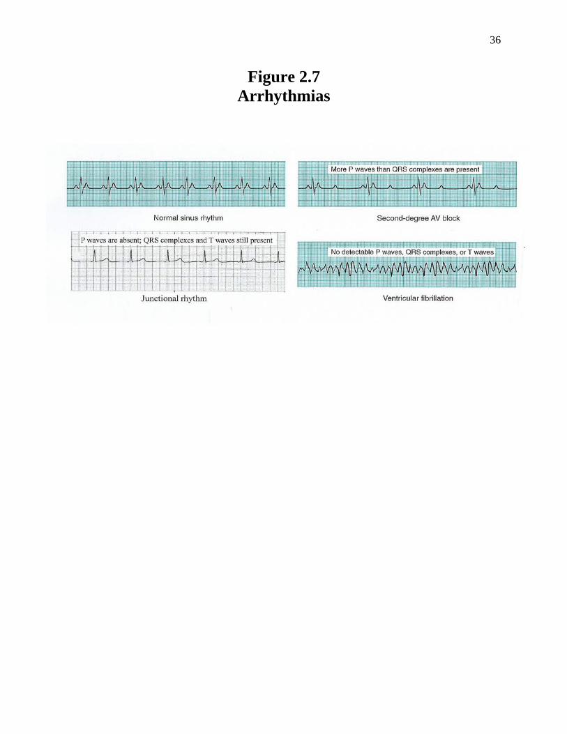
-The ECG can be used to determine if there is a problem with the heart. Examples (Figure 2.7, Derrickson): • SA node is nonfunctional (junctional rhythm) ECG pattern: P waves are absent; the AV node takes over as the pacemaker and sets the heart rate at 40 to 60 beats/min. • AV node is partially blocked or damaged (2nd degree heart block) ECG pattern: P waves outnumber QRS waves due to the fact that some of the action potentials are not conducted through the AV node. • ventricles contract too quickly and out of sequence (ventricular fibrillation) ECG pattern: Distorted QRS waves that are numerous and outnumber the P waves; fibrillating ventricles do not pump blood effectively and must be fixed by defibrillation using paddles (electrodes) that reset the SA node as the pacemaker. ♦Cardiac Cycle -The cardiac cycle refers to the events that occur in the heart with each heartbeat; it is associated with two heart sounds: “lubb” and “dupp.” -During the cardiac cycle, the muscle fibers of the atria and the ventricles undergo diastole (relaxation) and systole (contraction). -Phases -The cardiac cycle is divided into 5 phases: passive ventricular filling, atrial contraction, isovolumetric ventricular contraction, ventricular ejection, and isovolumetric ventricular relaxation (Figure 2.8, Derrickson). 1. passive ventricular filling During this phase, both the atria and ventricles are in diastole (relaxed); in addition, both the AV valves and semilunar valves are initially closed. Blood flows into the atria from the vena cavae and pulmonary veins. The blood then forces the AV valves to open as it moves into the ventricles. About 80% of ventricular filling (about 105 mL) occurs during this phase; the remaining 20% (about 25 mL) of ventricular filling occurs during the atrial contraction phase of the cardiac cycle. Toward the end of the passive ventricular filling phase, an action potential arises in the SA node and then spreads throughout the atria, causing the atria to depolarize. Atrial depolarization is indicated by the P wave on the ECG.
26
2. atrial contraction Atrial depolarization causes atrial systole (contraction). While the atria are in systole, the ventricles remain in diastole. Contraction of the atria pushes an additional 25 mL of blood into the ventricles. Thus, each ventricle now contains about 130 mL of blood. This volume will remain in the ventricles until the end of the ventricle’s relaxation period (diastole) and is, therefore, known as the end-diastolic volume (EDV). Toward the end of the atrial contraction phase, the action potential that had been generated in the SA node now sequentially moves through the AV node, bundle of His, right and left bundle branches, Purkinje fibers, and then into the ventricles. The QRS complex appears on the ECG, marking the onset of ventricular depolarization. 3. isovolumetric ventricular contraction Ventricular depolarization causes ventricular systole (contraction). While the ventricles are in systole, the atria are back in diastole. The AV valves close in response to the contraction of the ventricles in order to prevent backflow of blood into the atria. The closure of the AV valves causes the first heart sound: “lubb.” For a brief moment, both the AV and SL valves are closed. During this interval, ventricular muscle fibers are contracting and exerting force but are not yet shortening. Thus, the muscle contraction is isometric (same length). Moreover, because all four valves are closed, ventricular volume remains the same (isovolumic). For these two reasons, this phase is known as isovolumetric ventricular contraction (iso- = same).
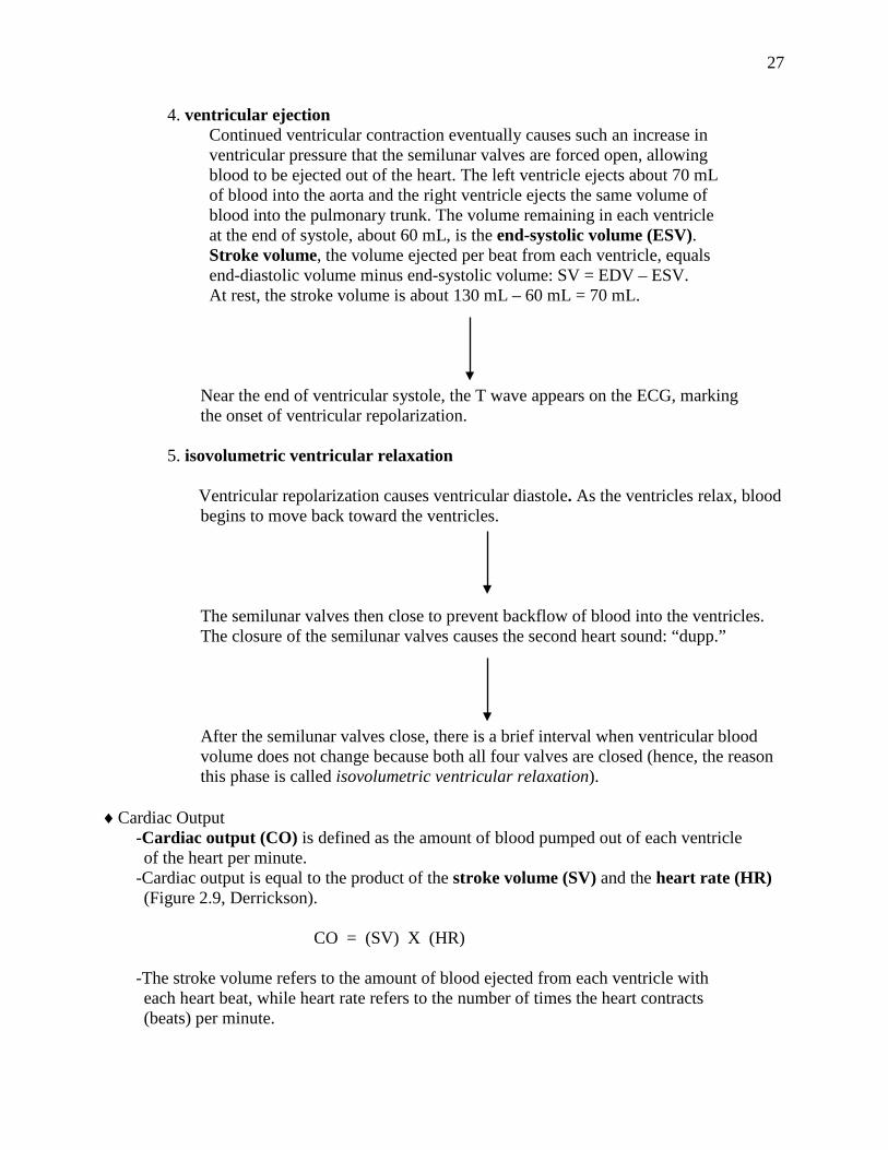
27
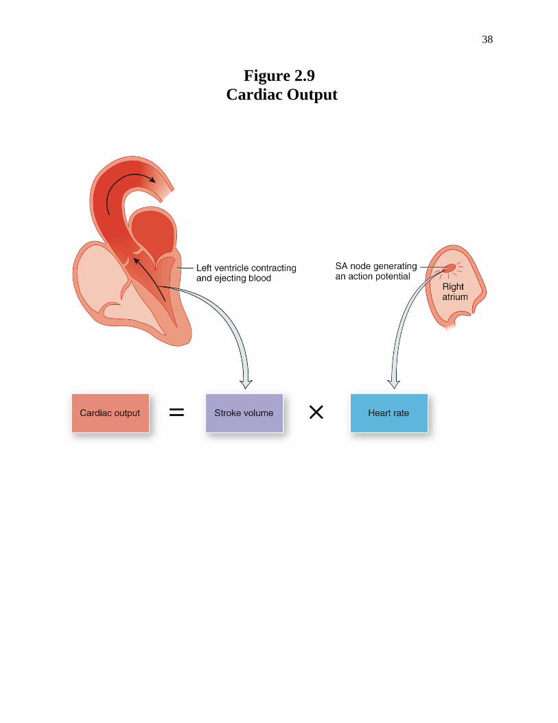
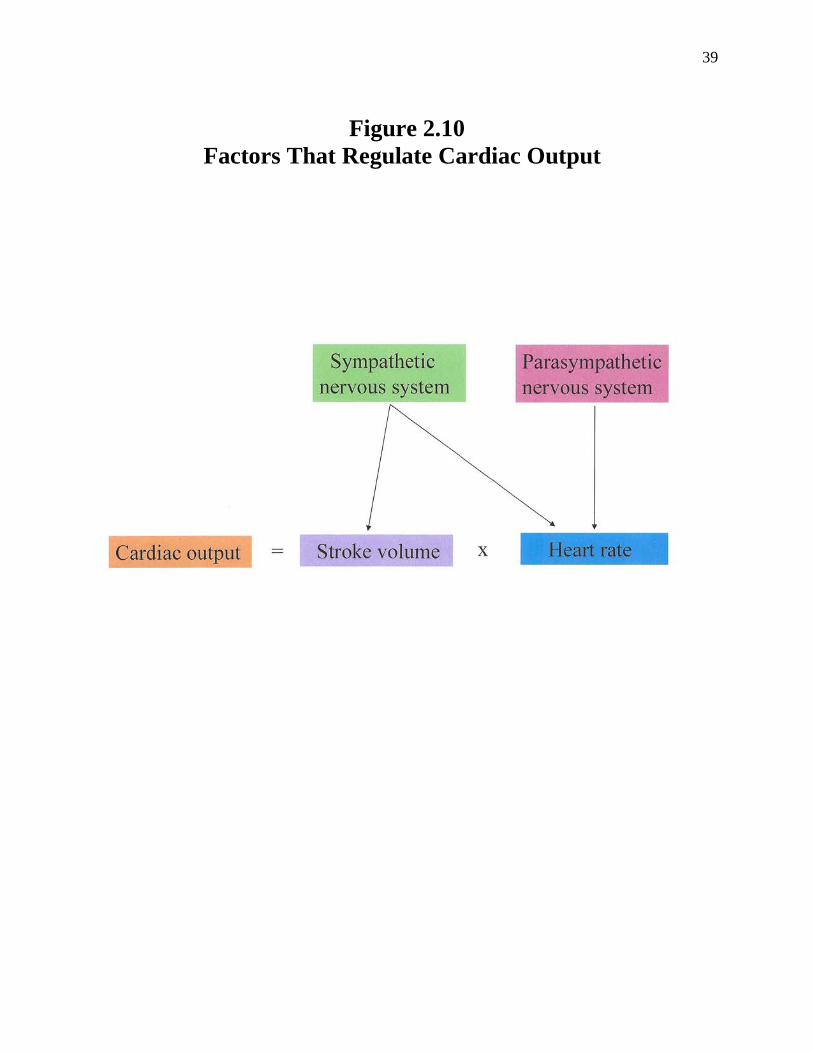
4. ventricular ejection Continued ventricular contraction eventually causes such an increase in ventricular pressure that the semilunar valves are forced open, allowing blood to be ejected out of the heart. The left ventricle ejects about 70 mL of blood into the aorta and the right ventricle ejects the same volume of blood into the pulmonary trunk. The volume remaining in each ventricle at the end of systole, about 60 mL, is the end-systolic volume (ESV). Stroke volume, the volume ejected per beat from each ventricle, equals end-diastolic volume minus end-systolic volume: SV = EDV – ESV. At rest, the stroke volume is about 130 mL – 60 mL = 70 mL. Near the end of ventricular systole, the T wave appears on the ECG, marking the onset of ventricular repolarization. 5. isovolumetric ventricular relaxation Ventricular repolarization causes ventricular diastole. As the ventricles relax, blood begins to move back toward the ventricles. The semilunar valves then close to prevent backflow of blood into the ventricles. The closure of the semilunar valves causes the second heart sound: “dupp.” After the semilunar valves close, there is a brief interval when ventricular blood volume does not change because both all four valves are closed (hence, the reason this phase is called isovolumetric ventricular relaxation). ♦Cardiac Output -Cardiac output (CO) is defined as the amount of blood pumped out of each ventricle of the heart per minute. -Cardiac output is equal to the product of the stroke volume (SV) and the heart rate (HR) (Figure 2.9, Derrickson). CO = (SV) X (HR) -The stroke volume refers to the amount of blood ejected from each ventricle with each heart beat, while heart rate refers to the number of times the heart contracts (beats) per minute.
28
-Under normal resting conditions, SV = 70 ml/beat and the HR = 75 beats/min; therefore, the normal CO equals 5250 ml/min: CO = (70 ml/beat) X (75 beats/min) = 5250 ml/min = 5.25 L/min -Notice that the cardiac output is close to the total blood volume of 5 L, which indicates that blood moves throughout the entire circulatory system of the body once every minute!!!!!!!!! -Cardiac output varies depending on how active the heart is. -Recall that parasympathetic nerves to the heart decrease heart rate. • A decrease in heart rate decreases cardiac output. -Also recall that sympathetic nerves to the heart increase heart rate and increase ventricular contraction. • An increase in heart rate increases cardiac output. • In addition, an increase in ventricular contraction causes an increase in stroke volume, which results in an increase in cardiac output. -Under maximal sympathetic stimulation, cardiac output can increase to as much as 25 L due to the increase in stroke volume and heart rate !!!!!!!! -Figure 2.10 (Derrickson) summarizes the factors that regulate cardiac output and its determinants. ♦Clinical Applications and Disorders -Look up the following clinical applications and disorders in Tortora: 1. cardiopulmonary resuscitation p. 698 2. pericarditis and cardiac tamponade p. 698 3. heart valve disorders p. 706 4. myocardial ischemia and infarction p. 709 5. regeneration of heart cells p. 709 6. artificial pacemakers p. 711

29
7. heart murmurs p. 719 8. coronary artery disease p. 727-730 9. congenital heart defects p. 730-731 10. arrhythmias p. 731-733 (read the first two paragraphs and also the section on heart block and ventricular fibrillation) 11. congestive heart failure p. 733 12. cardiac arrest p. 733 13. palpitation p. 734 14. paroxysmal tachycardia p. 734
30
Figure 2.1 Operation of the Heart Valves
31
Figure 2.2 Histology of Cardiac Muscle Tissue
32
Figure 2.3 The Heart as a Functional Syncytium
33
Figure 2.4 An Artificial Pacemaker

34
Figure 2.5 Extrinisic Innervation of the Heart

35
Figure 2.6 Electrocardiography
36
Figure 2.7 Arrhythmias

37
Figure 2.8 Cardiac Cycle
38
Figure 2.9 Cardiac Output
39
Figure 2.10
Factors That Regulate Cardiac Output