Heart Development II: Aortic Arches - Columbia University ... · Heart Development II Kimara L....
46
Human Embryology: Heart Development II Kimara L. Targoff, M.D. Division of Pediatric Cardiology, Columbia University Medical Center Developmental Genetics Program, Skirball Institute, NYU School of Medicine
Transcript of Heart Development II: Aortic Arches - Columbia University ... · Heart Development II Kimara L....

Human Embryology:Heart Development II
Kimara L. Targoff, M.D.Division of Pediatric Cardiology, Columbia University Medical Center
Developmental Genetics Program, Skirball Institute, NYU School of Medicine
Human Vascular Development
• Overview
• Aortic Arch Development
• Arterial Vascular Development
• Venous System Development
• Lymphatic Development
• Transition from Fetal to Post-Natal Circulation

Development of the Arterial and Venous Systems

Cranial Ends of the Dorsal Aortae Form a
Dorsoventral Loop: The First Aortic Arch

Aortic Arches Arise in a Craniocaudal Sequence
Surrounding the Pharynx
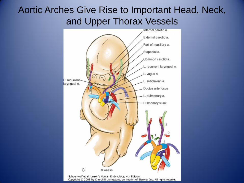
Aortic Arches Give Rise to Important Head, Neck,
and Upper Thorax Vessels
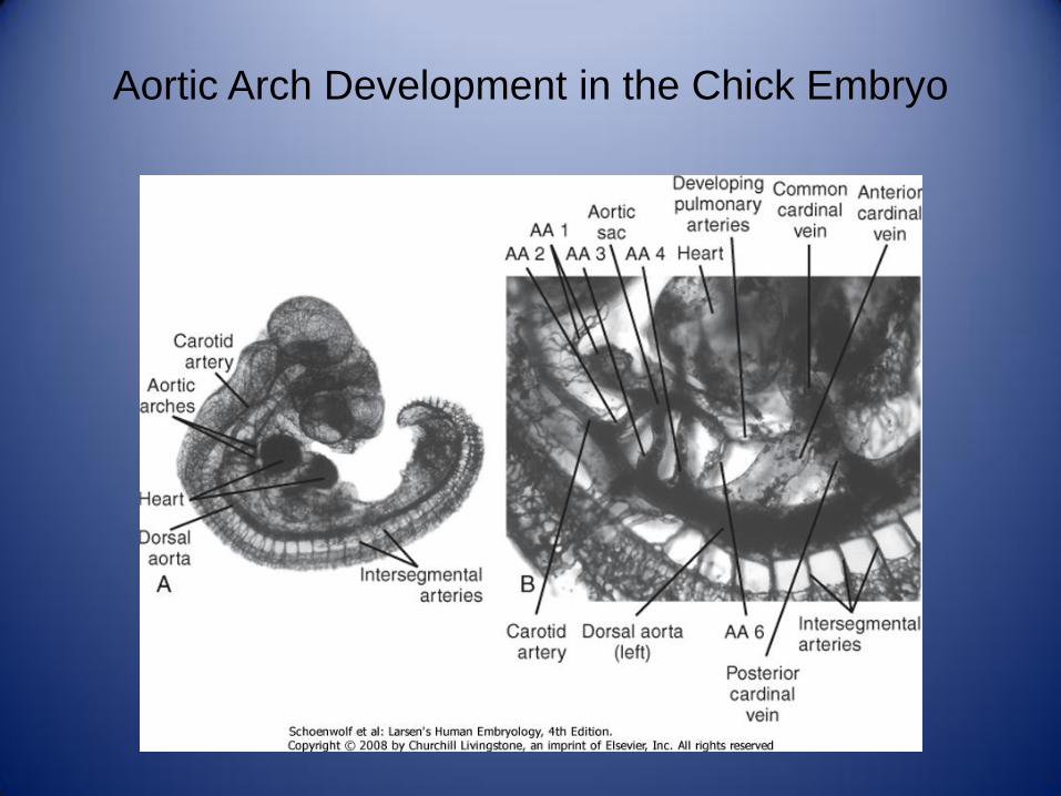
Aortic Arch Development in the Chick Embryo
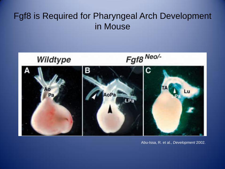
Fgf8 is Required for Pharyngeal Arch Development
in Mouse
Abu-Issa, R. et al., Development 2002.

Cardiovascular and Thymic Defects in Tbx1
Hypomorphic Mutant Neonates
Hu, T. et al., Development 2004.
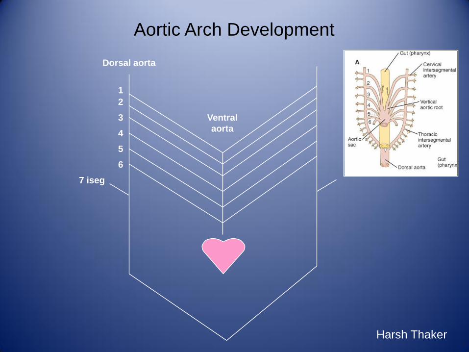
Aortic Arch Development
1
2
3
6
4
5
7 iseg
Ventral
aorta
Dorsal aorta
Harsh Thaker

Aortic Arch Development
1
2
3
6
4
5
7 iseg
Ventral
aorta
Dorsal aorta
Harsh Thaker

6
33
44
7 iseg7 iseg
Aortic sac
Truncus arteriosus
6
Aortic Arch and Derivatives
Harsh Thaker

6
33
44
7 iseg7 iseg
Harsh Thaker
Aortic Arch and Derivatives

6
33
4
47 iseg
7 iseg
Aortic Arch and Derivatives
Harsh Thaker
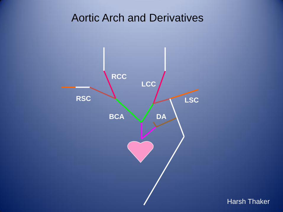
LCCRCC
RSC
BCA
LSC
DA
Aortic Arch and Derivatives
Harsh Thaker

LCCRCC
RSC
BCA
LSC
DA
Recurrent Laryngeal Nerves
Harsh Thaker

Defects in Normal Regression of the Arterial System
Lead to Vascular Anomalies
• Double Aortic Arch
– Failure of the right dorsal aorta to regress
• Aberrant Right Subclavian Artery
– Regression of the right fourth arch
– 1% of the general population
– 40% of patients with Trisomy 21 and CHD
• Right Aortic Arch
– Retention of the right dorsal aorta segment
– 13-35% of patients with TOF
– 8% of patients with TGA

Failure of Regression of the Right Dorsal Aorta
Leads to a Double Aortic Arch
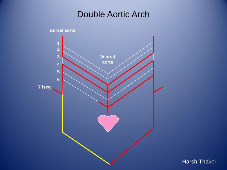
Double Aortic Arch
1
2
3
6
4
5
7 iseg
Ventral
aorta
Dorsal aorta
Harsh Thaker

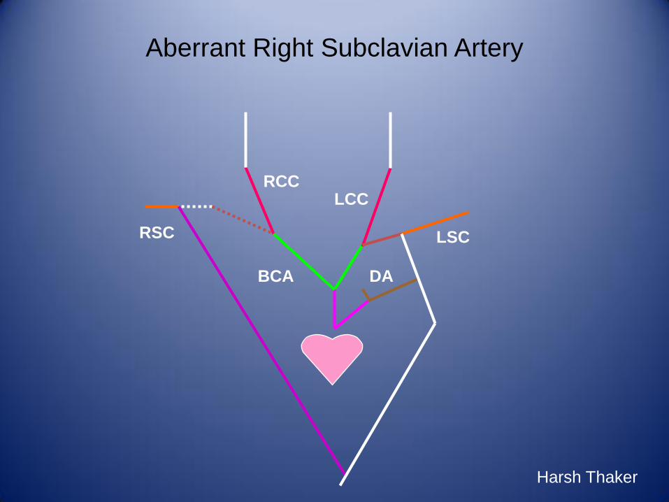
Regression of the Right Fourth Arch Results in an
Aberrant Right Subclavian Artery
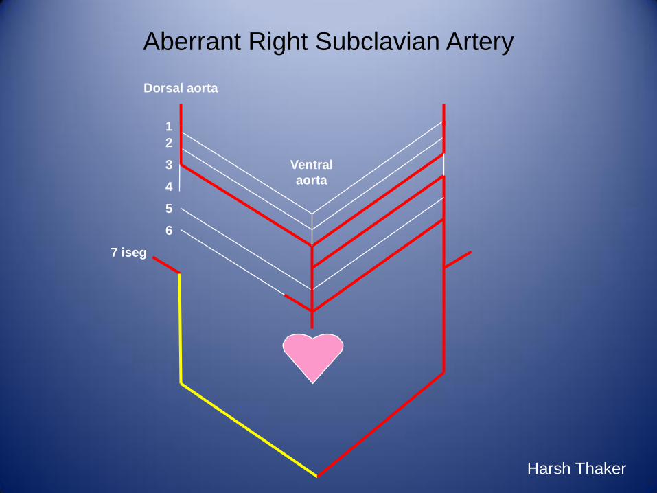
Aberrant Right Subclavian Artery
1
2
3
6
4
5
7 iseg
Ventral
aorta
Dorsal aorta
Harsh Thaker
LCCRCC
RSC
BCA
LSC
DA
Harsh Thaker
Aberrant Right Subclavian Artery

Retention of the Right Dorsal Aortic Segment
Yields a Right Aortic Arch

Right Aortic Arch
1
2
3
6
4
5
7 iseg
Ventral
aorta
Dorsal aorta
Harsh Thaker

Right Aortic Arch: Mirror Image Branching versus
Aberrant Left Subclavian Artery

Vascular Rings May Cause Compression of the
Trachea and the Esophagus
• Double Aortic Arch
– Failure of the right dorsal aorta to regress
• Right Aortic Arch
– Ductus arteriosus is directed towards the right
– If the ductus, or later, the ligamentum arteriosum, passes behind
the esophagus, constriction may occur

Double Aortic Arch Presenting with Dysphagia in a
31-Year-Old Woman

Aortic Arch Anomalies Can Cause Significant Clinically
Compromise in the Neonatal Period
• Interrupted Aortic Arch
– Obliteration of the right and left fourth aortic arches
• Coarctation of the Aorta
– Constriction of the aorta in the region of the ductus arteriosus
– 0.3% of live births
– Most common cardiac anomaly in Turner’s Syndrome

Obliteration of the Right and Left Fourth Aortic
Arches Leads to Interruption of the Aorta

Constriction of the Aorta in the Region of the
Ductus Arteriosus Produces Coarctation
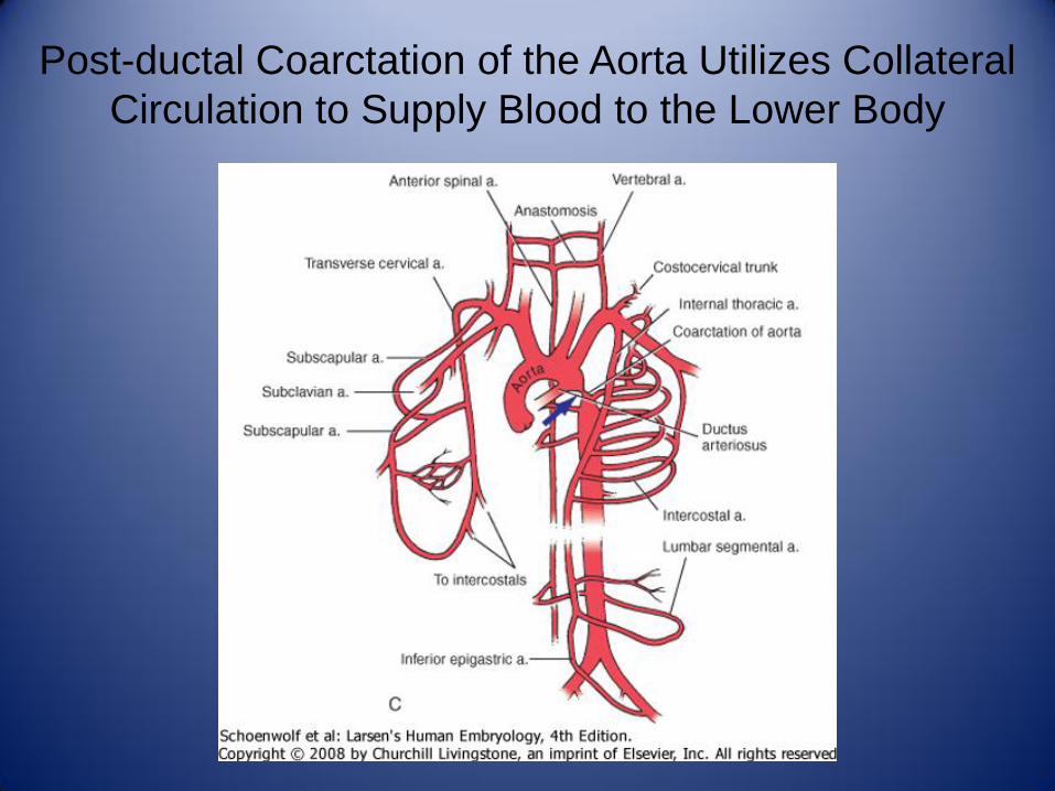
Post-ductal Coarctation of the Aorta Utilizes Collateral
Circulation to Supply Blood to the Lower Body
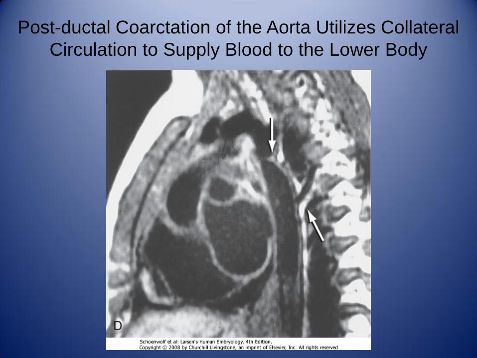
Post-ductal Coarctation of the Aorta Utilizes Collateral
Circulation to Supply Blood to the Lower Body

Vitelline Arteries Give Rise to the Arterial Supply of
the Gastrointestinal Tract

Lateral Branches of the Descending Aorta
Highlight Developmental Histories of Each Organ

Umbilical
Vitelline
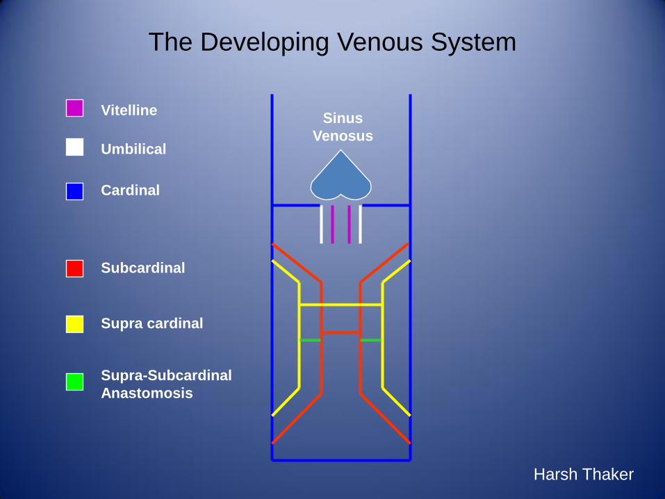
The Developing Venous System
Sinus
Venosus
Cardinal
Harsh Thaker
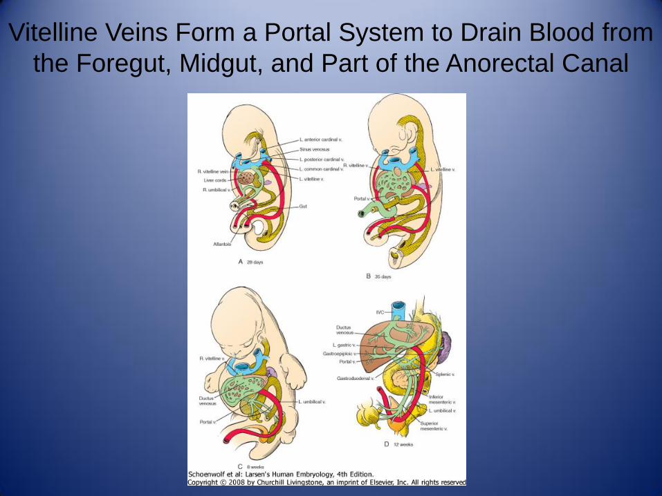
Vitelline Veins Form a Portal System to Drain Blood from
the Foregut, Midgut, and Part of the Anorectal Canal
Umbilical
Vitelline
The Developing Venous System
Sinus
Venosus
Cardinal
Harsh Thaker
Subcardinal
Supra cardinal
Supra-Subcardinal
Anastomosis

The Systemic Venous System Develops from Four
Bilaterally Symmetric Cardinal Veins

Following Remodeling of the Subcardinal System, the
Supracardinal Veins Sprout

Remodeling of Abdominal Venous System Occurs
through Obliteration of the Left Supracardinal Vein
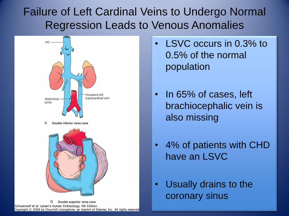
Failure of Left Cardinal Veins to Undergo Normal
Regression Leads to Venous Anomalies
• LSVC occurs in 0.3% to
0.5% of the normal
population
• In 65% of cases, left
brachiocephalic vein is
also missing
• 4% of patients with CHD
have an LSVC
• Usually drains to the
coronary sinus
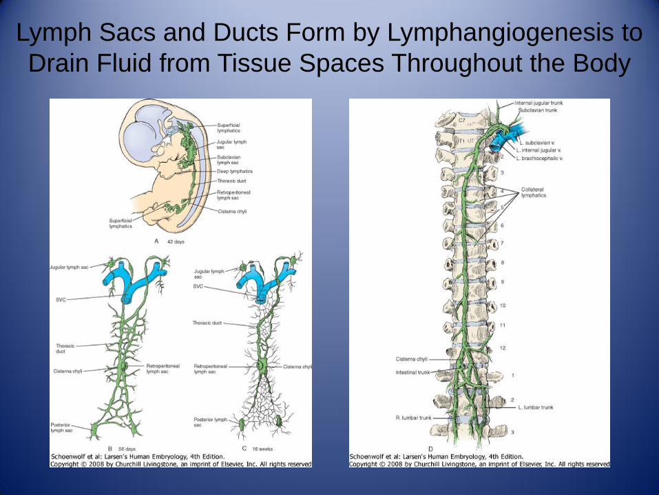
Lymph Sacs and Ducts Form by Lymphangiogenesis to
Drain Fluid from Tissue Spaces Throughout the Body

Cystic Hygromas Develop in Turner’s Syndrome
Patients Secondary to Blockage of Lymphatic Ducts
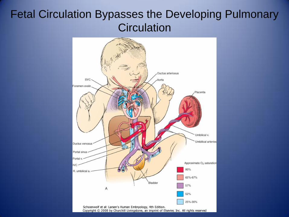
Fetal Circulation Bypasses the Developing Pulmonary
Circulation
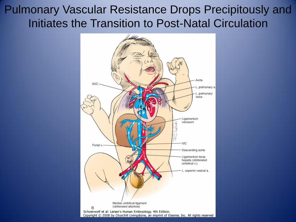
Pulmonary Vascular Resistance Drops Precipitously and
Initiates the Transition to Post-Natal Circulation

Normal Closure of the Ductus Arteriosus Occurs during
the Transition to Neonatal Circulation in Series
• Prostaglandins maintain a patent ductus arteriosus
• Indomethacin is used to induce ductal closure
• Physiologic closure occurs by 2 days in 82% of patients