Heamodynamics of pacing Dual Chamber - csnlc.nhs.uk · Abnormal Physiology Disease affecting HR, SV...
42
HEAMODYNAMICS OF PACING NASPE Training 2005 Lauren Butler – Lancashire & South Cumbria Cardiac Network
Transcript of Heamodynamics of pacing Dual Chamber - csnlc.nhs.uk · Abnormal Physiology Disease affecting HR, SV...
HEAMODYNAMICS OF PACING
NASPE Training2005Lauren Butler – Lancashire & South Cumbria Cardiac Network
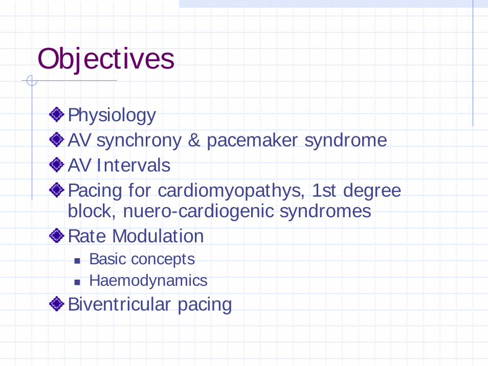
Objectives
PhysiologyAV synchrony & pacemaker syndromeAV IntervalsPacing for cardiomyopathys, 1st degree block, nuero-cardiogenic syndromesRate Modulation
Basic conceptsHaemodynamics
Biventricular pacing

Physiology
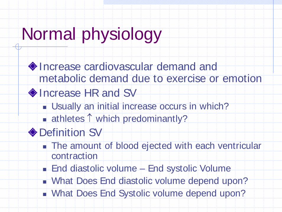
Normal physiology
Increase cardiovascular demand and metabolic demand due to exercise or emotionIncrease HR and SV
Usually an initial increase occurs in which?athletes ↑ which predominantly?
Definition SVThe amount of blood ejected with each ventricular contractionEnd diastolic volume – End systolic VolumeWhat Does End diastolic volume depend upon?What Does End Systolic volume depend upon?
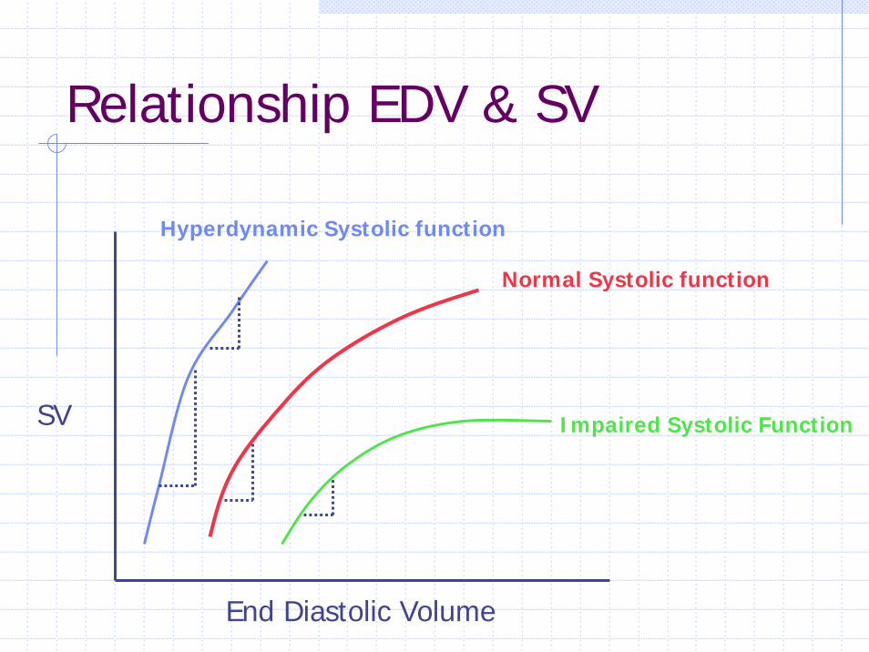
Relationship EDV & SV
SV
End Diastolic Volume
Hyperdynamic Systolic function
Normal Systolic function
Impaired Systolic Function
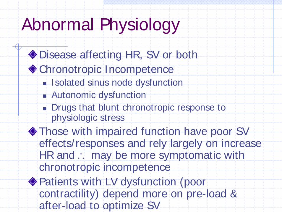
Abnormal Physiology
Disease affecting HR, SV or bothChronotropic Incompetence
Isolated sinus node dysfunctionAutonomic dysfunctionDrugs that blunt chronotropic response to physiologic stress
Those with impaired function have poor SV effects/responses and rely largely on increase HR and ∴ may be more symptomatic with chronotropic incompetencePatients with LV dysfunction (poor contractility) depend more on pre-load & after-load to optimize SV

How complicated is it?LV dysfunction
Metabolic abnormalities, chronic acidosis, hypoxia, hypercarbia (affect contractility)Autonomic dysfunction (affects HR & SV)Concomitant renal failure, diabetes, hypertension (affect pre-load, after-load, autonomic function , contractility)Drugs for treatment dysfunction, AF, CAD (suppress HR) Lets not forget associated conduction abnormalities!
AV synchrony & Pacemaker syndrome
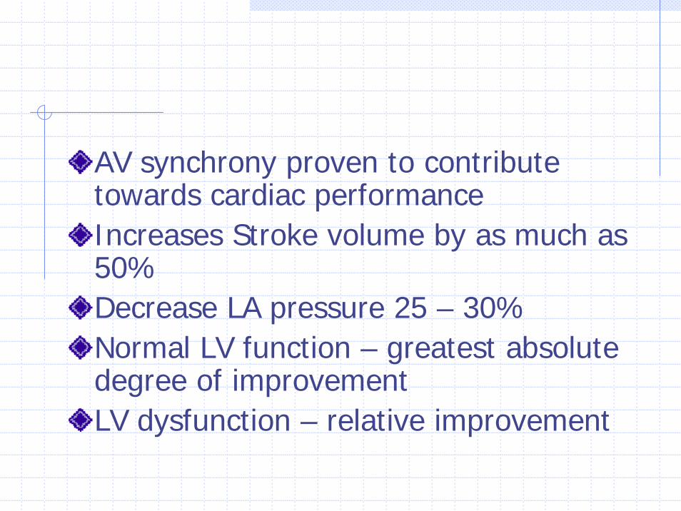
AV synchrony proven to contribute towards cardiac performanceIncreases Stroke volume by as much as 50%Decrease LA pressure 25 – 30%Normal LV function – greatest absolute degree of improvementLV dysfunction – relative improvement

AV synchrony (BP & CO)
In some patients, loss AV synchrony leads to a drop in BP & occasionally causes syncopeAssess supine & standing(symptoms exacerbated standing due to reduction venous return and ∴ reduction end diastolic volumeLoss atrial kick (pacing population 15 – 25% cardiac output)VA conduction and all associated with it!
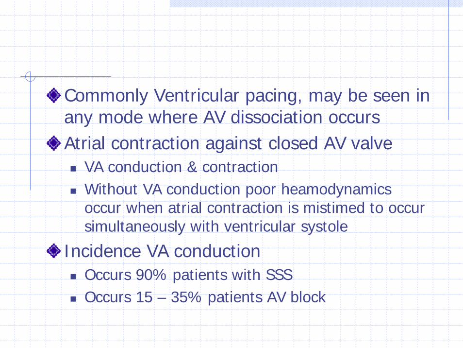
Commonly Ventricular pacing, may be seen in any mode where AV dissociation occursAtrial contraction against closed AV valve
VA conduction & contractionWithout VA conduction poor heamodynamicsoccur when atrial contraction is mistimed to occur simultaneously with ventricular systole
Incidence VA conductionOccurs 90% patients with SSSOccurs 15 – 35% patients AV block
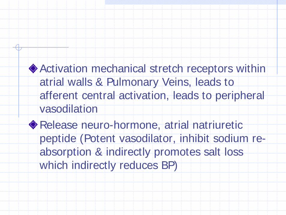
Activation mechanical stretch receptors withinatrial walls & Pulmonary Veins, leads to afferent central activation, leads to peripheralvasodilationRelease neuro-hormone, atrial natriureticpeptide (Potent vasodilator, inhibit sodium re-absorption & indirectly promotes salt loss which indirectly reduces BP)
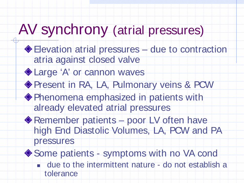
AV synchrony (atrial pressures)Elevation atrial pressures – due to contraction atria against closed valveLarge ‘A’ or cannon wavesPresent in RA, LA, Pulmonary veins & PCWPhenomena emphasized in patients with already elevated atrial pressuresRemember patients – poor LV often have high End Diastolic Volumes, LA, PCW and PA pressuresSome patients - symptoms with no VA cond
due to the intermittent nature - do not establish a tolerance
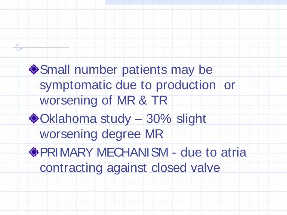
Small number patients may be symptomatic due to production or worsening of MR & TROklahoma study – 30% slight worsening degree MRPRIMARY MECHANISM - due to atria contracting against closed valve
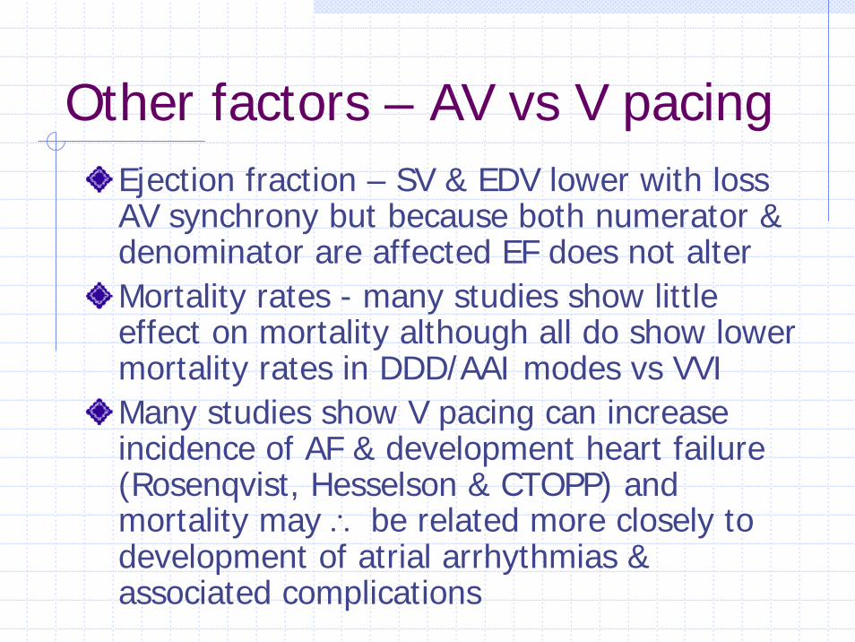
Other factors – AV vs V pacingEjection fraction – SV & EDV lower with loss AV synchrony but because both numerator & denominator are affected EF does not alter Mortality rates - many studies show little effect on mortality although all do show lower mortality rates in DDD/AAI modes vs VVIMany studies show V pacing can increase incidence of AF & development heart failure (Rosenqvist, Hesselson & CTOPP) and mortality may ∴ be related more closely to development of atrial arrhythmias & associated complications
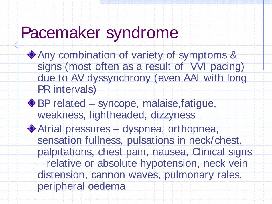
Pacemaker syndromeAny combination of variety of symptoms & signs (most often as a result of VVI pacing) due to AV dyssynchrony (even AAI with long PR intervals)BP related – syncope, malaise,fatigue, weakness, lightheaded, dizzynessAtrial pressures – dyspnea, orthopnea, sensation fullness, pulsations in neck/chest, palpitations, chest pain, nausea, Clinical signs – relative or absolute hypotension, neck vein distension, cannon waves, pulmonary rales, peripheral oedema
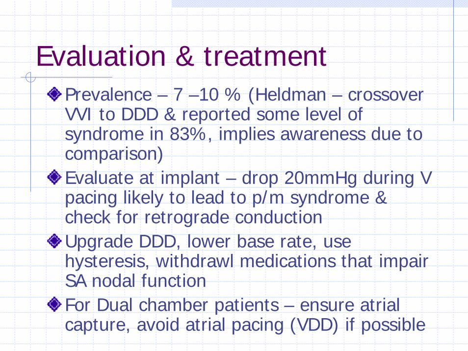
Evaluation & treatmentPrevalence – 7 –10 % (Heldman – crossover VVI to DDD & reported some level of syndrome in 83%, implies awareness due to comparison)Evaluate at implant – drop 20mmHg during V pacing likely to lead to p/m syndrome & check for retrograde conductionUpgrade DDD, lower base rate, use hysteresis, withdrawl medications that impair SA nodal functionFor Dual chamber patients – ensure atrialcapture, avoid atrial pacing (VDD) if possible

AV Intervals
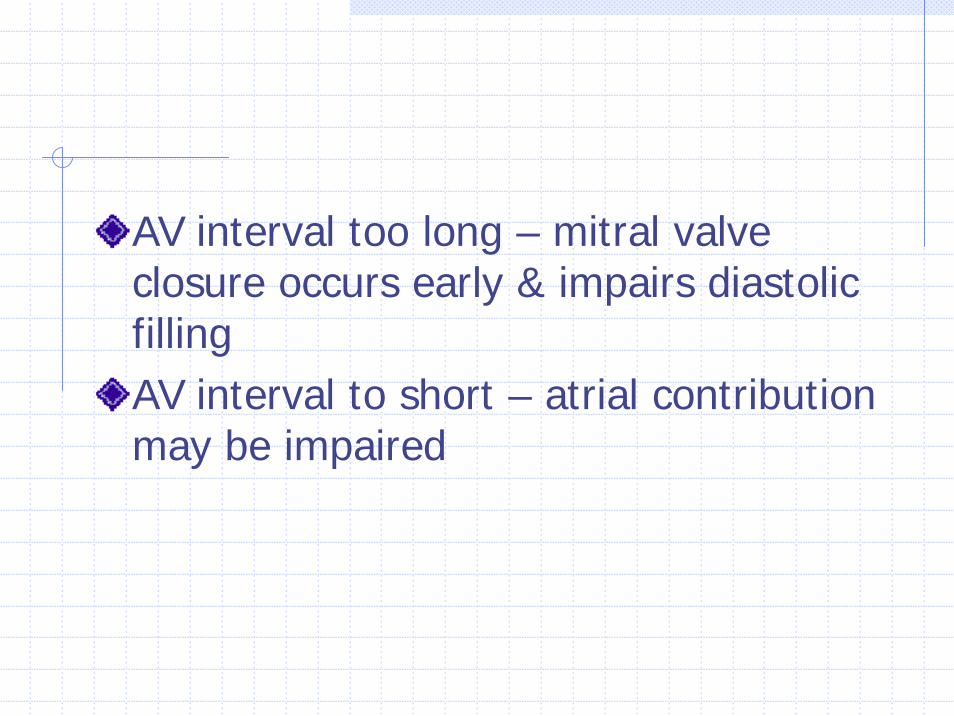
AV interval too long – mitral valve closure occurs early & impairs diastolic fillingAV interval to short – atrial contribution may be impaired
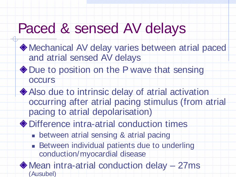
Paced & sensed AV delaysMechanical AV delay varies between atrial paced and atrial sensed AV delaysDue to position on the P wave that sensing occursAlso due to intrinsic delay of atrial activation occurring after atrial pacing stimulus (from atrial pacing to atrial depolarisation)Difference intra-atrial conduction times
between atrial sensing & atrial pacingBetween individual patients due to underling conduction/myocardial disease
Mean intra-atrial conduction delay – 27ms (Ausubel)
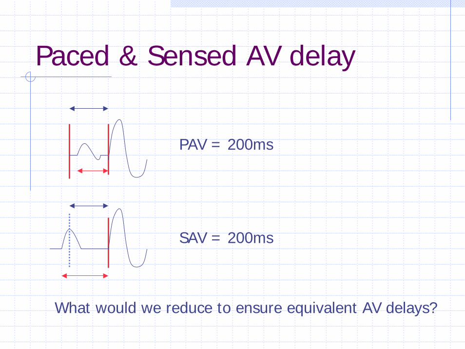
Paced & Sensed AV delay
PAV = 200ms
SAV = 200ms
What would we reduce to ensure equivalent AV delays?
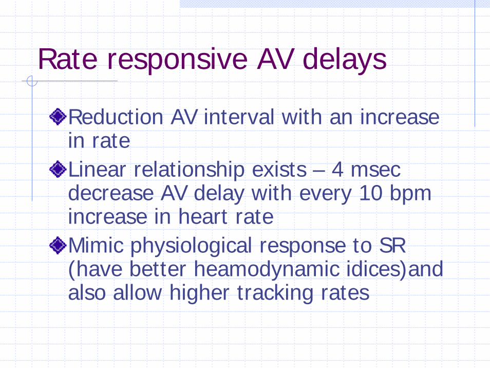
Rate responsive AV delays
Reduction AV interval with an increase in rateLinear relationship exists – 4 msec decrease AV delay with every 10 bpm increase in heart rateMimic physiological response to SR (have better heamodynamic idices)and also allow higher tracking rates
Heamodynamic effects
Cardiomyopathy1st degree blockNeuro-cardiogenic syncope
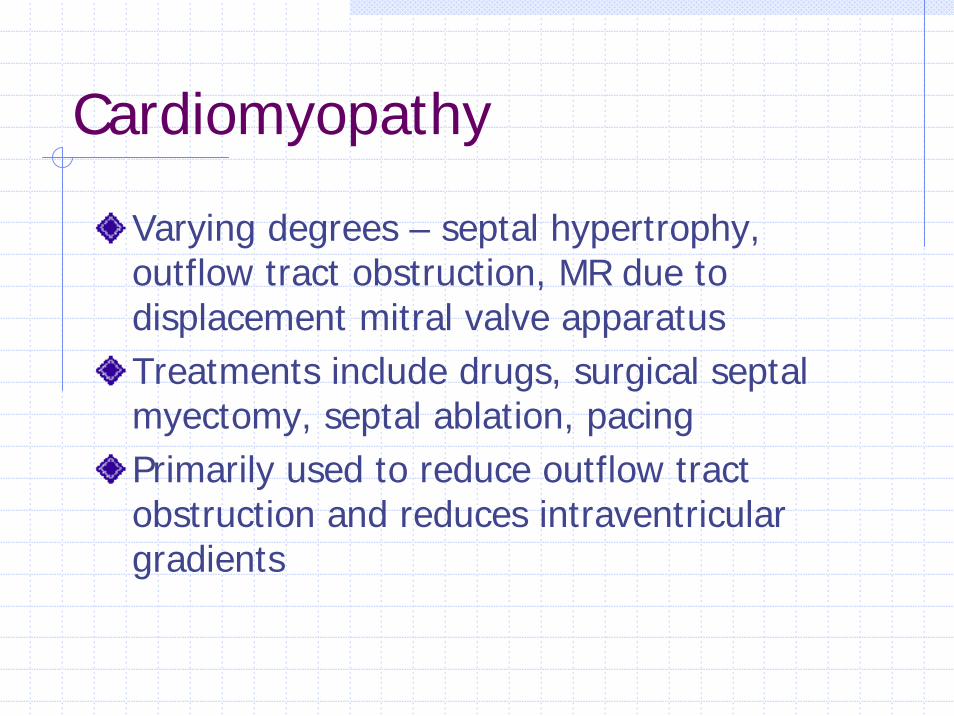
Cardiomyopathy
Varying degrees – septal hypertrophy, outflow tract obstruction, MR due to displacement mitral valve apparatusTreatments include drugs, surgical septalmyectomy, septal ablation, pacingPrimarily used to reduce outflow tract obstruction and reduces intraventricular gradients
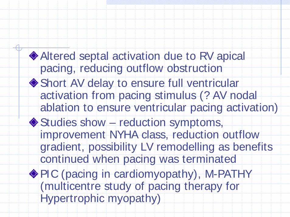
Altered septal activation due to RV apical pacing, reducing outflow obstruction Short AV delay to ensure full ventricular activation from pacing stimulus (? AV nodal ablation to ensure ventricular pacing activation)Studies show – reduction symptoms, improvement NYHA class, reduction outflow gradient, possibility LV remodelling as benefits continued when pacing was terminated PIC (pacing in cardiomyopathy), M-PATHY (multicentre study of pacing therapy for Hypertrophic myopathy)
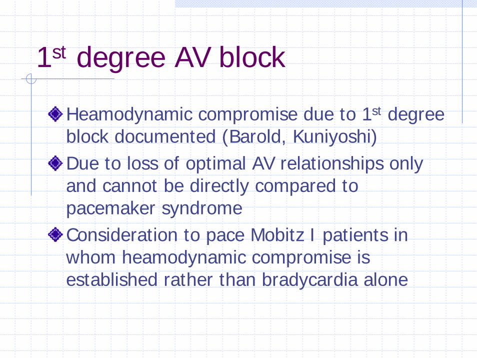
1st degree AV block
Heamodynamic compromise due to 1st degree block documented (Barold, Kuniyoshi)Due to loss of optimal AV relationships only and cannot be directly compared to pacemaker syndromeConsideration to pace Mobitz I patients in whom heamodynamic compromise is established rather than bradycardia alone
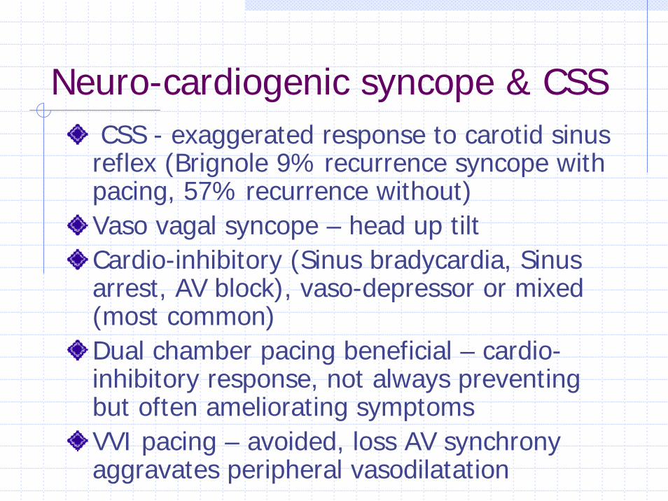
Neuro-cardiogenic syncope & CSSCSS - exaggerated response to carotid sinus
reflex (Brignole 9% recurrence syncope with pacing, 57% recurrence without)Vaso vagal syncope – head up tiltCardio-inhibitory (Sinus bradycardia, Sinus arrest, AV block), vaso-depressor or mixed (most common)Dual chamber pacing beneficial – cardio-inhibitory response, not always preventing but often ameliorating symptoms VVI pacing – avoided, loss AV synchrony aggravates peripheral vasodilatation

Rate drop therapies
VPS –1 (North American Vasovagalpacemaker study) – compared pacing with therapy (17% syncopal incidence) to 59% without any pacing therapyRemember – DDI with hysteresis, does not rate smooth down to allow intrinsic conduction to resume
Rate Modulation
Basic conceptsHeamodynamics
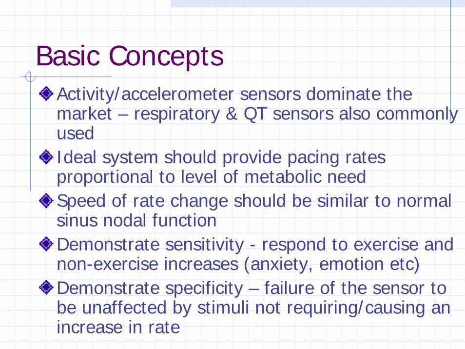
Basic ConceptsActivity/accelerometer sensors dominate the market – respiratory & QT sensors also commonly usedIdeal system should provide pacing rates proportional to level of metabolic needSpeed of rate change should be similar to normal sinus nodal function Demonstrate sensitivity - respond to exercise and non-exercise increases (anxiety, emotion etc)Demonstrate specificity – failure of the sensor to be unaffected by stimuli not requiring/causing an increase in rate
Technical considerations
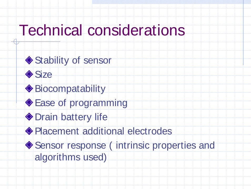
Stability of sensorSizeBiocompatabilityEase of programmingDrain battery lifePlacement additional electrodesSensor response ( intrinsic properties and algorithms used)
Accelerometers & activity sensors
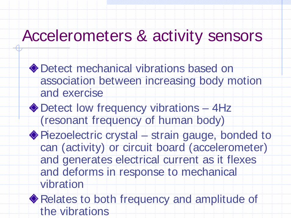
Detect mechanical vibrations based on association between increasing body motion and exerciseDetect low frequency vibrations – 4Hz (resonant frequency of human body)Piezoelectric crystal – strain gauge, bonded to can (activity) or circuit board (accelerometer) and generates electrical current as it flexes and deforms in response to mechanical vibration Relates to both frequency and amplitude of the vibrations
Activity sensors
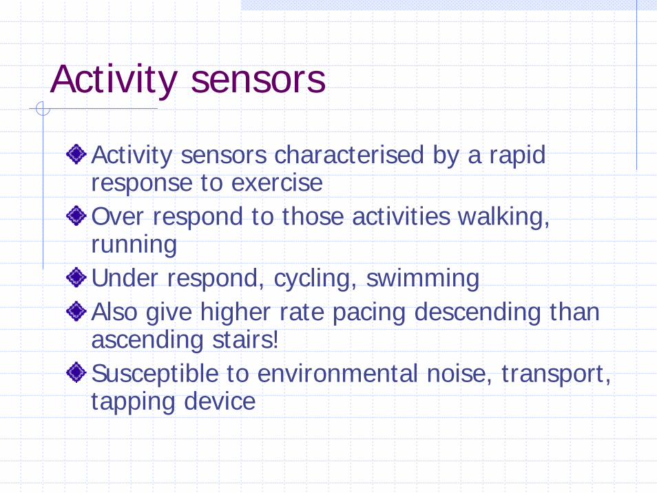
Activity sensors characterised by a rapid response to exerciseOver respond to those activities walking, runningUnder respond, cycling, swimmingAlso give higher rate pacing descending than ascending stairs!Susceptible to environmental noise, transport, tapping device
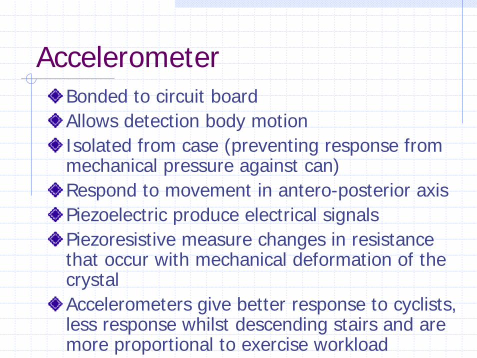
AccelerometerBonded to circuit boardAllows detection body motionIsolated from case (preventing response from mechanical pressure against can)Respond to movement in antero-posterior axisPiezoelectric produce electrical signals Piezoresistive measure changes in resistance that occur with mechanical deformation of the crystal Accelerometers give better response to cyclists, less response whilst descending stairs and are more proportional to exercise workload
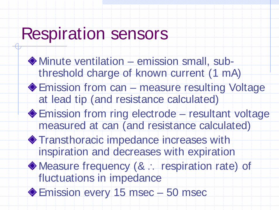
Respiration sensorsMinute ventilation – emission small, sub-threshold charge of known current (1 mA) Emission from can – measure resulting Voltage at lead tip (and resistance calculated)Emission from ring electrode – resultant voltage measured at can (and resistance calculated) Transthoracic impedance increases with inspiration and decreases with expiration Measure frequency (& ∴ respiration rate) of fluctuations in impedanceEmission every 15 msec – 50 msec
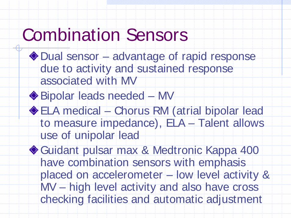
Combination SensorsDual sensor – advantage of rapid response due to activity and sustained response associated with MVBipolar leads needed – MVELA medical – Chorus RM (atrial bipolar lead to measure impedance), ELA – Talent allows use of unipolar leadGuidant pulsar max & Medtronic Kappa 400 have combination sensors with emphasis placed on accelerometer – low level activity & MV – high level activity and also have cross checking facilities and automatic adjustment
QT intervalAutonomic activity and heart rate affect the stimulus - T interval (affected by emotion)Shortens at increased pacing ratesQT interval varies between individuals but is consistent for each individual at restAffected by drugs and electrolytesMeasured - stimulus to apex T wave in the IEGMEarly devices – T wave undersensing (improved with T wave filters) and large polarisation impulses also interfered with measurement (lower polarisation with improved electrode design)
Other sensorsTemperature – thermistor in RA portion lead, slow response to exercisePre-ejection interval – shortens as exercise increases (pacing stimulus to onset ventricular contraction)Stroke volume – placement of impedance catheter, alters pacing rate to keep SV constantRV Pressure increase – transducer incorporated into distal portion lead
Other sensors
Wall motion and small changes of contractility – associated with metabolic changesMixed Venous Oxygen saturation –hemoreflectance oximetry placed in RV portion leadPaced Depolarisation Integral – measures ventricular depolarisation gradient (vector)Peak Endocardial acceleration (PEA) –incorporates an accelerometer in tip lead, correlates to changes in sympathetic tone (early signal to rate drop episode)
Heamodynamics
chronotropic competence most important contributor to cardiac output – Improve HR and exercise capacity (every 40% increase pacing rate – 10% increase exercise capacity)Important at higher levels of activity, AV synchrony at lower levelsDiagnosis chronotropic incompetence
70 –85% predicted maximum (difficult due to limited exercise capacity many patients)Wilkoff chronotropic assessment exercise protocol (CAEP) (2 minute stages gradual increase speed and incline)
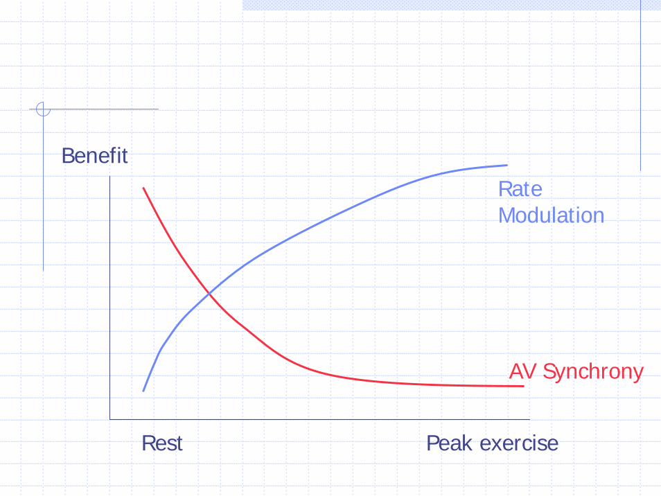
Benefit
Rest Peak exercise
AV Synchrony
Rate Modulation
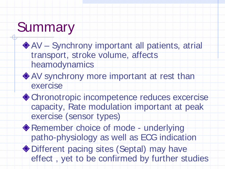
SummaryAV – Synchrony important all patients, atrial transport, stroke volume, affects heamodynamicsAV synchrony more important at rest than exerciseChronotropic incompetence reduces excercisecapacity, Rate modulation important at peak exercise (sensor types)Remember choice of mode - underlying patho-physiology as well as ECG indicationDifferent pacing sites (Septal) may have effect , yet to be confirmed by further studies