Head of College Scholars List Scheme Summer Studentship Report … · Head of College Scholars List...
10
Head of College Scholars List Scheme Summer Studentship Report Form This report should be completed by the student with his/her project supervisor. It should summarise the work undertaken during the project and, once completed, should be sent by email to: [email protected] within four weeks of the end of the studentship. 1. Student Surname: Bolton Forename: Victoria Lena E-mail address: [email protected] 2. Supervisor: Surname: Patel Forename: Arvind E-mail address: [email protected] 3. Research Project Report 3.1 Project Title (maximum 20 words): Identification of epitopes of a hepatits C virus neutralizing antibody (CBH-7) with a view to vaccine development. 3.2 Project Lay Summary (copied from application): Hepatitis C virus (HCV) causes liver cirrhosis and cancer. Expensive treatments are available but there is still no vaccine. A few infected individuals do clear infection without treatment, this is associated with the early appearance of broadly neutralising antibodies (bnAb) targeting the viral protein E2. One vaccine approach is to harness natural immunity against
Transcript of Head of College Scholars List Scheme Summer Studentship Report … · Head of College Scholars List...

Head of College Scholars List Scheme
Summer Studentship
Report Form
This report should be completed by the student with his/her project supervisor. It should summarise the work undertaken during the project and, once completed, should be sent by email to: [email protected] within four weeks of the end of the studentship.
1. Student
Surname: Bolton Forename: Victoria Lena
E-mail address: [email protected]
2. Supervisor:
Surname: Patel Forename: Arvind
E-mail address: [email protected]
3. Research Project Report
3.1 Project Title (maximum 20 words):
Identification of epitopes of a hepatits C virus neutralizing antibody (CBH-7) with a view to
vaccine development.
3.2 Project Lay Summary (copied from application):
Hepatitis C virus (HCV) causes liver cirrhosis and cancer. Expensive treatments are available
but there is still no vaccine. A few infected individuals do clear infection without treatment,
this is associated with the early appearance of broadly neutralising antibodies (bnAb)
targeting the viral protein E2. One vaccine approach is to harness natural immunity against
regions on E2 that elicit bnAb. The human bnAb CBH-7 binds to HCV E2 protein and
neutralizes HCV infection, however the binding site is not known. This project will use a
library of peptides spanning E2 to identify important regions for CBH-7 binding.
3.3 Start Date: 15/6/2015 Finish Date:7/8/2015
3.4 Original project aims and objectives (100 words max):
The binding site of bnAB CBH-7 binding of E2 is still unknown. The aim of this project
therefore was to identify the key epitope residues that define the binding site of bnAB CBH-7
on E2.
Key questions:
o What are key epitope residues in bnAB CBH-7 binding of E2?
3.5 Methodology: Summarise and include reference to training received in research
methods etc. (250 words max):
Preparation of E1E2 plasmid DNA: Competent E.coli were incubated with plasmid DNA on
ice for 30mins, heat-shocked at 42oC for 1 min and rested on ice before plating onto LB-Agar
plates containing Ampicillin. Plates were incubated overnight at 37C. LB-broth was
inoculated with a single colony and grown at 37C on a shaker overnight. Plasmid DNA was
extracted using the Qiagen spin miniprep kit as per the manufacturers’ protocol. DNA
concentration was quantified by measuring absorbance at 260nm.
Production of E1E2 lysate: HEK 293T cells were transfected with E1E2 plasmid DNA using
calcium phosphate transfection and incubated overnight at 37C. The transfection mix was
replaced with fresh media and incubated for 24h. Cells were lysed for 20min, lysate was
cleared by centrifugation (13000g, 10mins, 4C).
GNA capture ELISA assay: Immulon II enzyme immunoassay plates were coated with
Galanthus nivalis (GNA) lectin. Plates were blocked with 1% skimmed milk powder in PBS
containing 0.1% Tween (PBS-T). E1E2 lysate was incubated at RT for 2h. Plates were washed
in PBS-T (x3), incubated with an E2 antibody for 1h, washed and incubated in secondary
HRP-conjugated antibody for 1h. The plate was washed in PBS-T (x6) then TMB substrate
was added, the reaction was developed for 20min then stopped with 0.5M H2SO4.
Absorbance was measured on a platereader at 450nm.
Peptide library screen: The standard GNA ELISA assay was adapted as follows. H77
genotype 1a E1E2 lysate was used. Human bnAB CBH-7 at 0.1 ug/ml in the presence of 10
ug/ml peptide in 100ul of PBST was bound for 1h at RT. Bound antibody was detected with
human HRP-antibody Promega W 4031.
3.6 Results: Summarise key findings (300 words max). Please include any relevant tables or
images as an appendix to this report:
Peptide library screen:
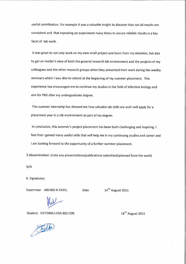
A library of peptides spanning HCV E2 were analysed in a competition E1E2 ELISA assay, with
the aim of identifying the E2 region containing the CBH-7 epitope. Genotype 1 H77 E1E2
lysate bound plates were incubated with Hmab CBH-7 in the presence of E2 peptide.
Competition for binding between Hmab CBH-7 and a peptide would be reflected in a lower
absorbance value. No inhibition was observed with the samples containing CBH-7 and
peptide. The positive control , Mab V3.2 however, in the presence of peptide 25, showed
significant inhibition confirming the assay was working (see Figure 1). These results confirm
that CBH-7 binds to a conformational epitope as opposed to a linear epitope.
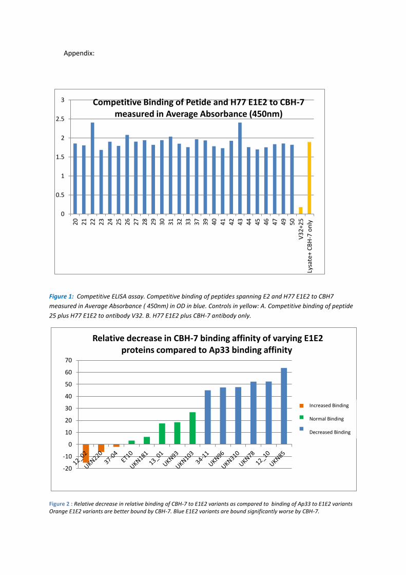
GNA capture ELISA essay:
An alternative strategy to identify residues that form part of the conformational epitope of
CBH-7 used a panel of genetically variable E1E2 proteins, cloned from clinical cohorts. The
expression levels of E2 were normalised using the linear anti-E2 Mab AP33 to a signal of 2
OD, to serve as a reference value. In parallel, Mab CBH-7 binding was measured. The panel
of E1E2 proteins were ranked according to the average relative binding affinity to CBH-7
then grouped into 3 groups: Increased binding, normal binding, and decreased binding
according to their relative binding affinity to CBH-7 compared to their binding affinity to
Ap33 (Figure 2).
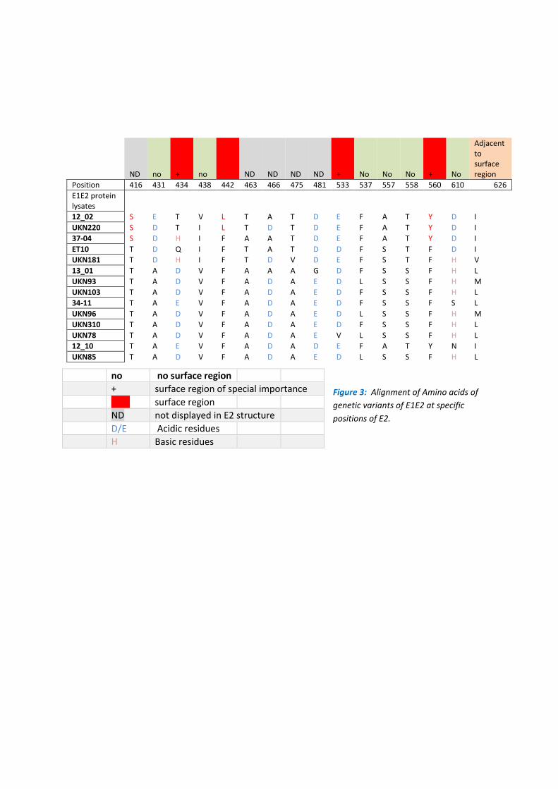
Alignment of amino acid sequences of each E1E2 protein according to their ranking revealed
patterns of amino acid grouping that correlate with the increase or decrease in binding
affinity of CBH-7 (Figure 3). Figure 3 shows E2 positions of special interest with known
surface amino acid residues e.g. 434, 442, 533, 560 highlighted in red. E1E2 protein variants
with decreased CBH-7 binding affinity demonstrate acidic amino acid residues at position
434 as opposed to non-acidic residues carried by E1E2 protein variants with increased
binding. At position 442 a change from phenylalanine to leucine was recorded for E1E2
variants 12.20 and UKN 220, that both showed an increase in CBH-7 binding affinity.
Moreover, the presence of a tyrosine at position 560 correlated mostly with an increased
binding affinity of E1E2 variants carrying this amino acid.
The position of these residues of interest within the E2 protein, were investigated with the
molecular modelling program Chimera 1.9 and are displayed in Figure 4. Residue 434 and
560 are closely associated with each other on the surface of the protein. Moreover residue
434 forms a close interface with epitope II.
Analysis of predicted CBH-7 glycosylation sites showed that N- glycosylation was reduced at
position 448 and 540 for lysates that were poorly bound by CBH-7 (Table 1).
3.7 Discussion (500 words max):
The aim of this project was to investigate and determine possible CBH-7 binding sites.
Earlier studies ( Keck et al. 2004) have suggested that CBH-7 binds to a conformational
epitope in Domain C, that differs spatially from other antibody binding domains A and B as
determined by competition studies. The peptide screen conducted in this project
investigated competition for CBH-7 between H77 E1E2 protein and linear peptides spanning
the E1E2 protein and found no inhibition providing further evidence that CBH-7 binds to a
conformational epitope.
GNA capture Elisa assay and downstream sequence analysis have identified positions 434 ,
442, 533 and 560 as putative CBH-7 interacting residues. Residue 533 falls within a region
of E2 involved in binding the receptor CD81. In addition, this residue is in close proximity to
residues of Domain A ( Keck et al. 2012), which has been suggested to be in proximity to
Domain C in previous competition studies ( Keck et al. 2004). Furthermore, residues 434 and
560 are in close proximity with epitope 2/ Domain B which includes a CD81 interaction site.
The same study ( Keck et al. 2004) have suggested that CBH-7 inhibited E2-CD81 interactions
suggesting that residues 533, 434 and 560 could be of further interest. Further experiments
including site-directed mutagenesis, mutating E1E2 variants of increased binding and
decreased binding by CBH-7 ( Figure 2) at residues 434 , 442, 533 and 560 to an alanine
could be conducted to investigate the effects on CBH-7 binding.In addition, other residues
of interest 416, 463, 466, 475, 481 could also be investigated by site- directed mutagenesis
to investigate their effect on CBH-7 binding. These residues are not included in the E2 core
protein structure, therefore it is not known for certain if these residues are surface-
accessible for antibody interaction . However these residues may be located on the surface
of the E2 protein and therefore may be putative CBH-7 interaction sites.

Analysis of the N-glycosylation pattern of E1E2 proteins, ordered according to ranking of
CBH-7 binding from highest to lowest revealed E2 residues 448 and 540 as of possible
interest. Both residues were more likely to be glycosylated in E1E2 variants that had reduced
CBH-7 binding affinity ( Table1). Interestingly these sequences were also less likely to have
glycosylation sites at two further residues within E1 (196 and 234). The effect of N-
glycosylation on CBH-7 binding could be investigated by knocking out glycosylation at
residues 448 and 540 from E1E2 variants e.g. 1202 and UKN 220 that were bound well by
CBH-7 by mutation of the asparagine residues. Finally, it would be interesting to create
chimeras of E1E2 variants that combine different E1 and E2 glycosylation sites of interest
e.g. 193, 234 and 448 and 540 to investigate this further.
4. Reflection by the student on the experience and value of the studentship (300 words max):
When I started the project I had little experience in working in a laboratory and related skills
and I was excited, but also nervous about my placement.
However through the work on my project , the support which I received from my friendly
and helpful colleagues and the direction of the supervising postdoc Vanessa Cowton in the
group, I soon felt integrated into the team.
During my placement I learned a number of techniques and was able to develop skills that
will be useful in my further studies and career, e.g ELISA assays, transfections and basic cell
culture techniques. The tasks I were given in my project were both interesting and
challenging and as my placement progressed my confidence in carrying them out increased.
Even when I made mistakes I was encouraged and assisted in solving the problems
encountered in these experiments , which gave me experience in both troubleshooting and
avoiding these mistakes. I could closely identify with the aim of the project and found
conducting my experiments led me both to learn a great deal and feel that I was making a
Appendix:
Figure 1: Competitive ELISA assay. Competitive binding of peptides spanning E2 and H77 E1E2 to CBH7
measured in Average Absorbance ( 450nm) in OD in blue. Controls in yellow: A. Competitive binding of peptide
25 plus H77 E1E2 to antibody V32. B. H77 E1E2 plus CBH-7 antibody only.
Figure 2 : Relative decrease in relative binding of CBH-7 to E1E2 variants as compared to binding of Ap33 to E1E2 variants Orange E1E2 variants are better bound by CBH-7. Blue E1E2 variants are bound significantly worse by CBH-7.
-20
-10
0
10
20
30
40
50
60
70
Relative decrease in CBH-7 binding affinity of varying E1E2 proteins compared to Ap33 binding affinity
37-04
UKN103
UKN85
Increased Binding Normal Binding Decreased Binding
0
0.5
1
1.5
2
2.5
3
20
21
22
23
24
25
26
27
28
29
30
31
32
33
37
39
40
41
42
43
44
45
46
47
49
50
V3
2+2
5
Lysa
te+
CB
H-7
on
ly
Competitive Binding of Petide and H77 E1E2 to CBH-7 measured in Average Absorbance (450nm)
ND no + no ND ND ND ND + No No No + No
Adjacent to surface region
Position 416 431 434 438 442 463 466 475 481 533 537 557 558 560 610 626
E1E2 protein lysates
12_02 S E T V L T A T D E F A T Y D I
UKN220 S D T I L T D T D E F A T Y D I
37-04 S D H I F A A T D E F A T Y D I
ET10 T D Q I F T A T D D F S T F D I
UKN181 T D H I F T D V D E F S T F H V
13_01 T A D V F A A A G D F S S F H L
UKN93 T A D V F A D A E D L S S F H M
UKN103 T A D V F A D A E D F S S F H L
34-11 T A E V F A D A E D F S S F S L
UKN96 T A D V F A D A E D L S S F H M
UKN310 T A D V F A D A E D F S S F H L
UKN78 T A D V F A D A E V L S S F H L
12_10 T A E V F A D A D E F A T Y N I
UKN85 T A D V F A D A E D L S S F H L
Figure 3: Alignment of Amino acids of
genetic variants of E1E2 at specific
positions of E2.
no no surface region
+ surface region of special importance
Red surface region
ND not displayed in E2 structure
D/E Acidic residues
H Basic residues
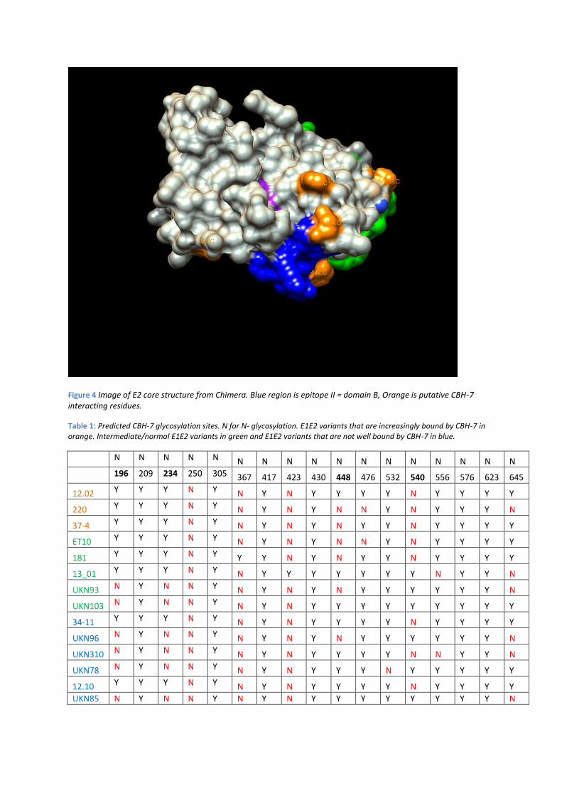
Figure 4 Image of E2 core structure from Chimera. Blue region is epitope II = domain B, Orange is putative CBH-7 interacting residues.
Table 1: Predicted CBH-7 glycosylation sites. N for N- glycosylation. E1E2 variants that are increasingly bound by CBH-7 in orange. Intermediate/normal E1E2 variants in green and E1E2 variants that are not well bound by CBH-7 in blue.
N N N N N N N N N N N N N N N N N
196 209 234 250 305 367 417 423 430 448 476 532 540 556 576 623 645
12.02 Y Y Y N Y N Y N Y Y Y Y N Y Y Y Y
220 Y Y Y N Y N Y N Y N N Y N Y Y Y N
37-4 Y Y Y N Y N Y N Y N Y Y N Y Y Y Y
ET10 Y Y Y N Y N Y N Y N N Y N Y Y Y Y
181 Y Y Y N Y Y Y N Y N Y Y N Y Y Y Y
13_01 Y Y Y N Y N Y Y Y Y Y Y Y N Y Y N
UKN93 N Y N N Y N Y N Y N Y Y Y Y Y Y N
UKN103 N Y N N Y N Y N Y Y Y Y Y Y Y Y Y
34-11 Y Y Y N Y N Y N Y Y Y Y N Y Y Y Y
UKN96 N Y N N Y N Y N Y N Y Y Y Y Y Y N
UKN310 N Y N N Y N Y N Y Y Y Y N N Y Y N
UKN78 N Y N N Y N Y N Y Y Y N Y Y Y Y Y
12.10 Y Y Y N Y N Y N Y Y Y Y N Y Y Y Y
UKN85 N Y N N Y N Y N Y Y Y Y Y Y Y Y N