Head and Neck Cancer - ASTRO · and postoperative head and neck cancer radiotherapy treatment....
133
Head and Neck Cancer Alexander Lin University of Pennsylvania
-
Upload
hoangkhanh -
Category
Documents
-
view
217 -
download
0
Transcript of Head and Neck Cancer - ASTRO · and postoperative head and neck cancer radiotherapy treatment....

Head and Neck Cancer
Alexander Lin
University of Pennsylvania

Disclosures
• Employer: University of Pennsylvania
• Grant support: National Institutes of Health
• R01-CA174976-01
• U01-DE022939-01
• Speaker: Ion Beam Applications
• Consultant: Elekta

Learning Objectives
• Predict disease outcomes and survival for the major categories/subsites of head and neck cancer.
• Review and discuss new, high-impact, clinical evidence published in the past year that may impact treatment paradigms for head and neck cancer therapy.
• Determine the best clinical and technical approaches for definitive and postoperative head and neck cancer radiotherapy treatment.

Basic Anatomy: Neck levels
Som et al. AJR 2000

Basic Anatomy: Level Ia (submental)
• Below mylohyoid muscle
• Above caudal edge of hyoid bone
• Between the anterior bellies of the digastric muscle

Basic Anatomy: Level Ia (submental)
• Below mylohyoid muscle
• Above caudal edge of hyoid bone
• Between the anterior bellies of the digastric muscle

Basic Anatomy: Level Ia (submental)
• At risk from cancers of the:• Oral cavity

Basic Anatomy: Level Ib (submandibular)
• Below mylohyoid muscle
• Above caudal edge of hyoid bone
• Posterolateral to the anterior belly of the digastic muscles

Basic Anatomy: Level Ib (submandibular)
• Below mylohyoid muscle
• Above caudal edge of hyoid bone
• Posterolateral to the anterior belly of the digastic muscles

Basic Anatomy: Level Ib (submandibular)
• At risk from cancers of the:• Oral cavity

Basic Anatomy: Level II, Superior internal jugular (deep cervical) chain
• Extends from skull base superiorly to caudal edge of hyoid bone inferiorly
• Medial to the sternocleidomastoid
• Anterior border: posterior border of the submandibular gland
• Posterior border: posterior border of the sternocleidomastoid
• Posterior edge of jugular vein separates IIa from IIb
Basic Anatomy: Level II, Superior internal jugular (deep cervical) chain
• At risk from cancers of the:• Nasopharynx
• Oral cavity
• Oropharynx
• Larynx/Hypopharynx

Basic Anatomy: Level III, Mid internal jugular (deep cervical) chain
• Extends from caudal edge of hyoid bone superiorly to caudal edge of cricoid cartilage inferiorly
• Medial to the sternocleidomastoid
• Anterior and posterior borders parallel the sternocleidomastoid

Basic Anatomy: Level III, Mid internal jugular (deep cervical) chain
• Extends from caudal edge of hyoid bone superiorly to caudal edge of cricoid cartilage inferiorly
• Medial to the sternocleidomastoid
• Anterior and posterior borders parallel the sternocleidomastoid

Basic Anatomy: Level III, Mid internal jugular (deep cervical) chain
• At risk from cancers of the:• Nasopharynx
• Oral cavity
• Oropharynx
• Larynx/Hypopharynx

Basic Anatomy: Level IV, Low internal jugular (deep cervical) chain
• Extends from caudal edge of cricoid cartilage superiorly to the level of the clavicle inferiorly
• Medial to the sternocleidomastoid
• Anterior and posterior borders parallel the sternocleidomastoid

Basic Anatomy: Level IV, Low internal jugular (deep cervical) chain
• Extends from caudal edge of cricoid cartilage superiorly to the level of the clavicle inferiorly
• Medial to the sternocleidomastoid
• Anterior and posterior borders parallel the sternocleidomastoid

Basic Anatomy: Level IV, Low internal jugular (deep cervical) chain
• At risk from cancers of the:• Nasopharynx
• Oropharynx
• Larynx/Hypopharynx

Basic Anatomy: Level V, Posterior triangle
• Posterior to the posterior edge of the SCM
• Anterior to the trapezius
• Level Va: posterior to levels II and III
• Level Vb: posterior to level IV

Basic Anatomy: Level V, Posterior triangle
• Posterior to the posterior edge of the SCM
• Anterior to the trapezius
• Level Va: posterior to levels II and III
• Level Vb: posterior to level IV

Basic Anatomy: Level V, Posterior triangle
• At risk from cancers of the:• Nasopharynx
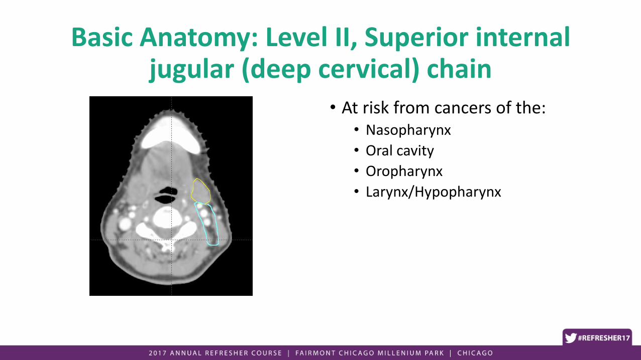
Basic Anatomy: Level VI, Prelaryngeal/Pretracheal
• Extends from caudal edge of hyoid superiorly to the manubrium of the sternum inferiorly
• Between the anterior edges of the sternocleidomastoid
• Anterior to levels III and IV
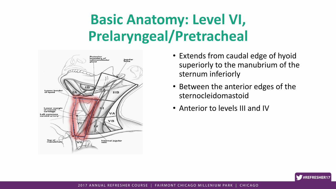
Basic Anatomy: Level VI, Prelaryngeal/Pretracheal
• Extends from caudal edge of hyoid superiorly to the manubrium of the sternum inferiorly
• Between the anterior edges of the sternocleidomastoid
• Anterior to levels III and IV

Basic Anatomy: Level VI, Prelaryngeal/Pretracheal
• At risk from cancers of the:• Thyroid
• Larynx• Primary subglottic location or
extension into subglottis
• Extension through thyroid cartilage
• Hypopharynx• Post-cricoid location

Basic Anatomy: Retropharyngeal
• Extends from base of skull superiorly to the cranial edge of the hyoid bone inferiorly
• Located medial to the carotid artery

Basic Anatomy: Retropharyngeal
• At risk from cancers of the:• Nasopharynx
• Oropharynx• Posterior tonsillar pillar
• Soft palate
• Posterior pharyngeal wall
• Hypopharynx• Posterior pharyngeal wall

Principles of RT and Combined Modality
• Concurrent chemotherapy
• Alternatives to standard chemotherapy• Induction chemotherapy
• Cetuximab
• Altered Fractionation
• Principles of RT technique/target delineation
The role of concurrent chemotherapy
For advanced stage (III, IVa, IVb)

• Meta-analysis of randomized trials from 1965-2000 comparing RT alone vs CRT.• Timing: Induction, Concurrent, Adjuvant• # agents: Mono- or polychemotherapy
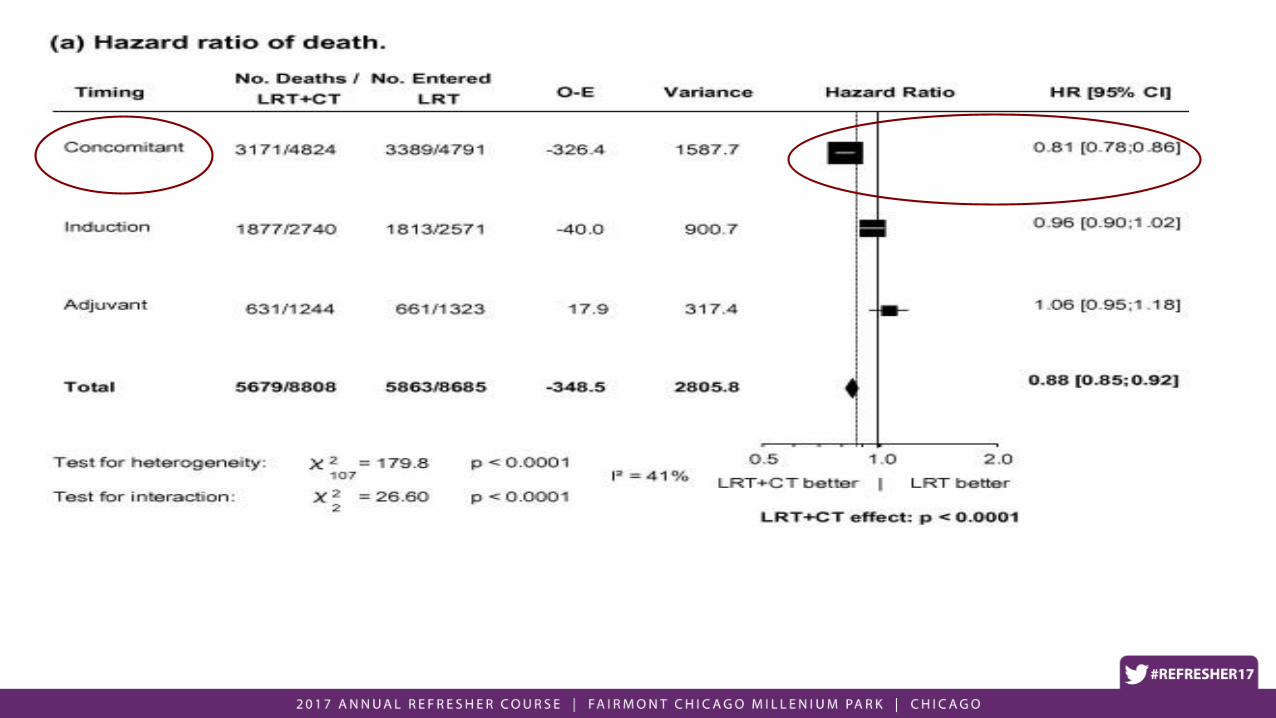

Suggests that main benefit of chemotherapy for HNC is from radiosensitization


• Concurrent, mono-agent platinum-based chemotherapy is the optimal approach for patients undergoing concurrent chemoradiation.• Superior to induction and adjuvant
Why the enthusiasm for induction chemotherapy?


Why Consider Induction Chemotherapy
• Pros:• Salvage subclinical M1 disease OS benefit?
• Assessment of response
• Reduce dose/volume of RT?
• Cons:• Prolongs treatment time/cost
• Increases toxicity
• No clinical benefit


• Study terminated early due to poor accrual (145 enrolled)
• Median f/u 49 mos
• 3-yr OS: (73% ICT vs 78% CRT, NS)
• Febrile neutropenia (23% ICT vs 1% CRT)
Haddad, Lancet Oncol. 2013; 14: 257.


• 285 Accrued (out of a planned 400)
• Adverse events more common with ICT• 47 vs. 28%, p = 0.002
• No differences in OS, DFFS, or RFS
Cohen JCO 2014; 32: 2735
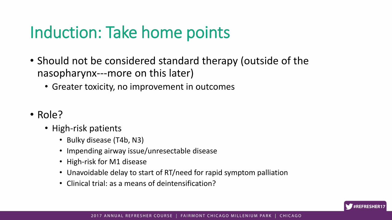
Induction: Take home points
• Should not be considered standard therapy (outside of the nasopharynx---more on this later)• Greater toxicity, no improvement in outcomes
• Role?• High-risk patients
• Bulky disease (T4b, N3)
• Impending airway issue/unresectable disease
• High-risk for M1 disease
• Unavoidable delay to start of RT/need for rapid symptom palliation
• Clinical trial: as a means of deintensification?


• Stage III, IV, OPX, hypopharynx, larynx
• Randomization• RT alone• RT + Cetuximab
• Cetuximab• 400 mg/m2 loading dose• 250 mg/m2 weekly
• RT• Once daily: 70 Gy, 35 fx• Twice daily: 1.2 Gy bid, 60-64 fx (72-76.8 Gy)• Concomitant Boost (72 Gy in 42 fx, 1.8 Gy daily x 3.6 wks, then 1.8 Gy AM
dose, 1.5 Gy PM dose for last 2.5 wks)


• RT + Cetuximab superior to RT alone wrt LRC and OS for LA-HNSCC
• Valid therapeutic option, for pts CI to receive platinum-based chemotherapy
• Alternative to platinum?• Efficacy
• Toxicity
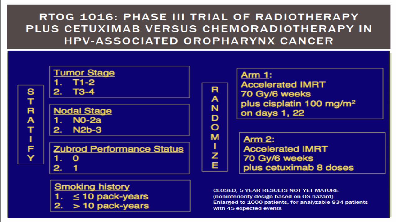
• Pending results (RTOG 1016)

Altered Fractionation RT

RTOG 90-03
• Locally advanced HNC, RT alone
• 4 arms• Standard (SFX): 2 Gy daily to 70 Gy (7 weeks)
• Hyperfractionation (HFX): 1.2 Gy bid to 81.6 Gy (7 weeks)
• Split course (AFX-S): 1.6 Gy bid to 67.2 Gy, with 2 week rest after 38.4 Gy (6 weeks)
• Concomitant boost (AFX-C): 1.8 Gy daily, with 1.5 Gy boost as 2nd daily trt for last 12 days, 72 Gy (6 weeks)

•HFX improved OS compared to SFX
•7 vs 6 wk trt• 6 wk trt: trend to increased grade 3-5 toxicity
• Worst toxicity per patient, by trt •AFX-C trended worse than SFX
Beitler IJROBP 2014; 89: 13
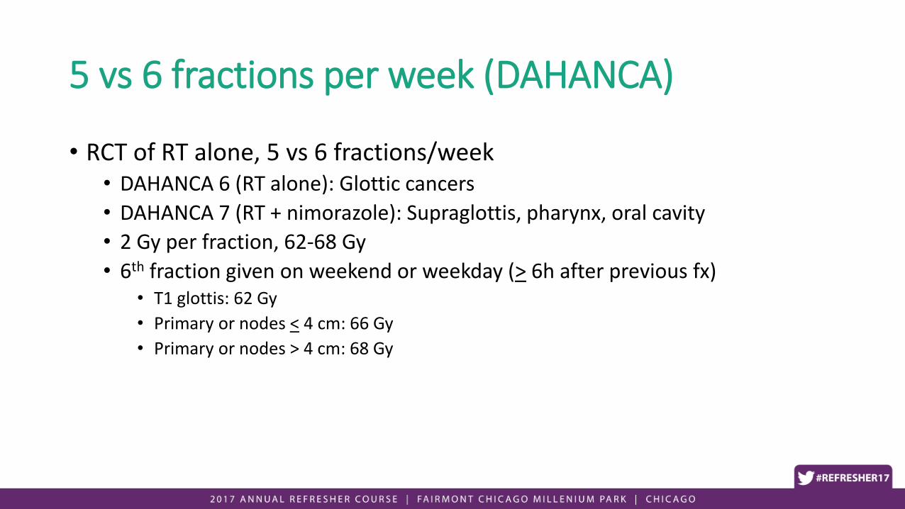
5 vs 6 fractions per week (DAHANCA)
• RCT of RT alone, 5 vs 6 fractions/week• DAHANCA 6 (RT alone): Glottic cancers
• DAHANCA 7 (RT + nimorazole): Supraglottis, pharynx, oral cavity
• 2 Gy per fraction, 62-68 Gy
• 6th fraction given on weekend or weekday (> 6h after previous fx)• T1 glottis: 62 Gy
• Primary or nodes < 4 cm: 66 Gy
• Primary or nodes > 4 cm: 68 Gy

Overgaard Lancet 2003; 362: 933

Overgaard Lancet 2003; 362: 933

Overgaard Lancet 2003; 362: 933

Altered Fractionation: Summary
• Improves disease outcomes when compared to standard fractionation when treating with RT alone for advanced stage HNC
• For pts who cannot receive chemotherapy, consider:• Hyperfractionation (RTOG 9003): 1.2 Gy bid to 81.6 Gy (7 weeks)
• 6 fractions per week, 2 Gy per fraction (DAHANCA)

Principles of RT technique/target delineation
• Simulation• Head extended• Supine• Arms down• IV contrast• 5-pt mask • Thin cut (2-3 mm)
• Technique: IMRT (except for early stage glottic cancer)
• Target delineation (elective nodes)• Primary echelon
• Location/drainage of primary • Lateralized (ipsilateral) vs. midline
(bilateral)
• Secondary echelon• At risk if primary echelon contains
bulky or high-volume disease

Doses/margins• Gross disease (70 Gy)
• GTV + CTV (0.5 – 1 cm) + PTV (3-5 mm)
• High-risk CTV elective regions (60 Gy)• Elective region around primary site (subclinical disease)• Primary echelon or involved nodal regions
• Low-risk CTV elective regions (50 Gy)• 2nd echelon regions
Daily IGRT, 3 mm PTV expansion

Clinical sections
• Nasopharynx
• Oral cavity
• Oropharynx
• Larynx/Hypopharynx

Clinical sections
• Nasopharynx
• Oral cavity
• Oropharynx
• Larynx/Hypopharynx
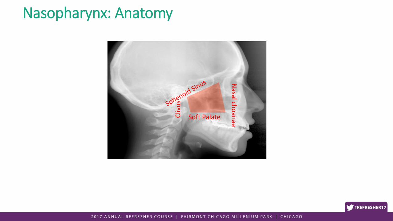
Soft Palate
Nasal ch
oan
ae
Cliv
us
Nasopharynx: Anatomy

Treatment Approach
• Stage I• RT alone (10 yr LC and DSS > 90%)
• Stage II-IVb• Concurrent chemoradiation + adjuvant chemotherapy
• Stage IVc (M1 disease)• Chemotherapy
• RT for symptom palliation

Concurrent Chemoradiation
Al-Sarraf, J Clin Oncol 1998; 16: 1310

• LRF: 14 vs 41%
• DM: 13 vs 35%
• 3-y PFS: 69 vs 24%
• 3-y OS: 76 vs 46%
Toxicity: 63% completed CRT, 55% completed adjuvant chemo
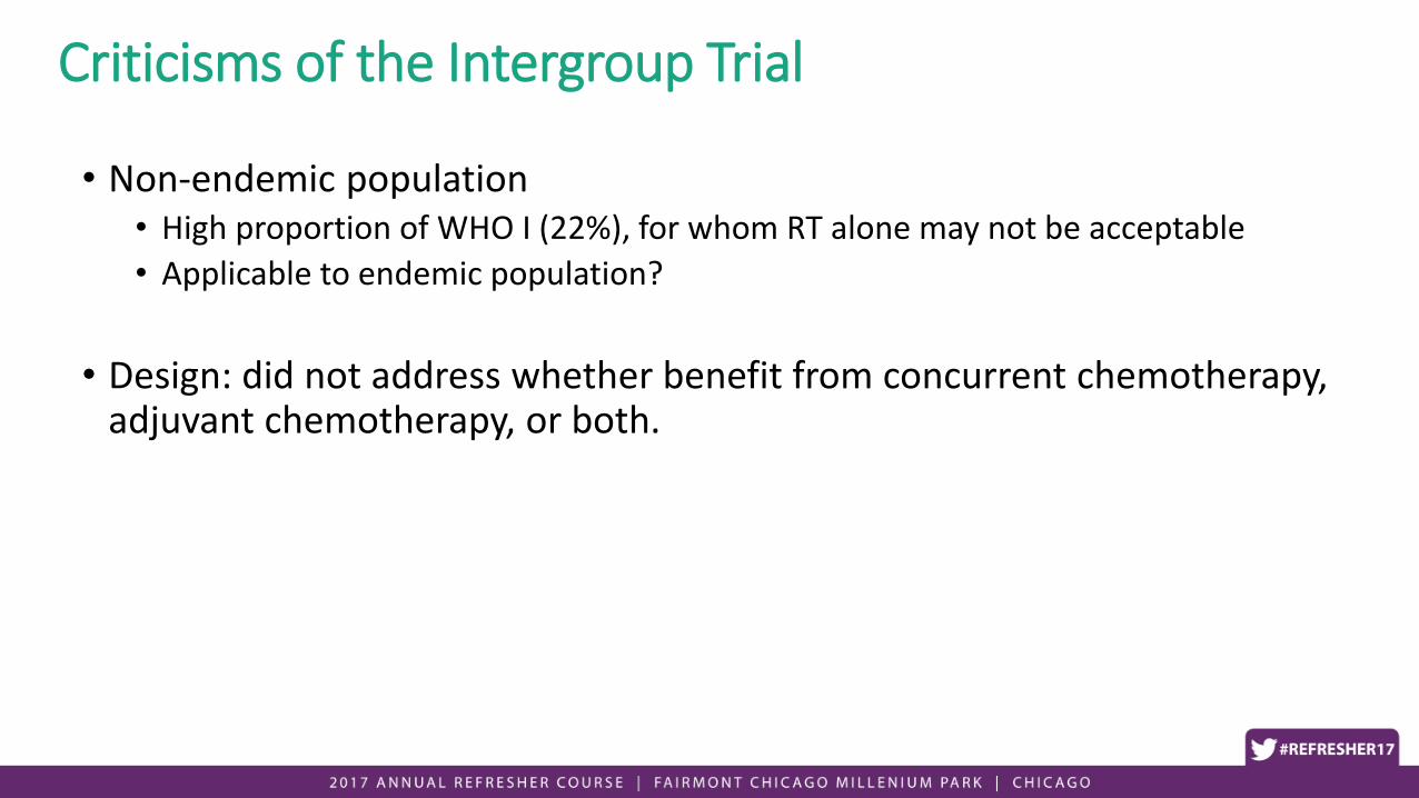
Criticisms of the Intergroup Trial
• Non-endemic population• High proportion of WHO I (22%), for whom RT alone may not be acceptable
• Applicable to endemic population?
• Design: did not address whether benefit from concurrent chemotherapy, adjuvant chemotherapy, or both.

AJCC 1988 Stage N Treatment OS LC PFS/FFS
Lin 2003JCO 21:631
III/IV 284 70-74Gy, 2D
CDDP, 5FU (LD)
54%
72%, 5 yr
73%
89%
53%
72%
Chan, 2005JNCI 97:536
II-IVB 350 66Gy, 2D
CDDP weekly
59%
70%, 5yr
NS 52%
60%
Kwong, 2004JCO 22:2643
II-IVB 219 62.5-68Gy, 2D
Con: UFT; Adj CDDP, 5FU/VBM
77%
87%, 3 yr
72%
80%
58%
69%
Lee 2005JCO 23:6966
III/IVB
N2-3
348 >66Gy, >50% 3D
Con: CDDP; Adj CDDP, 5FU
78%
78%, 3 yr
82%
92%
61%
70%
Wee, 2004JCO 23:487
III-IVB 221 70Gy, 2D
Con: CDDP; Adj CDDP, 5FU
77%
85%, 2 yr
NS 62%
76%
Chen, 2011
JNCI 103:1761
Chinese Stage
II (T2N0, T1-
2N1)
230 70Gy, 2D
Con: CDDP
86%
95%, 5yr
NS 78%
88%

Meta-analysis
• 8 trials, 1753 pts
• CRT vs RT alone
• CRT: 6% OS benefit at 5y
• Survival benefit most pronounced for WHO Type I
• No impact on OS from induction or adjuvant chemotherapy
Baujat IJROBP 2006; 64: 47

Adjuvant chemo: RCT (Guangzhou)
• Stage III/IV• Excluded T3-T4N0
• CRT vs CRT + adjuvant• Concurrent: weekly cisplatin (40 mg/m2)
• Adjuvant: Cisplatin + 5-FU x 3
Chen Lancet Oncol 2012; 13: 163

Chen Lancet Oncol 2012; 13: 163

Criticisms
• Design: not appropriately powered for non-inferiority
• Higher number of failures in the CRT arm (trend)
• >50% did not complete concurrent chemotherapy
• ~20% of pts randomized to receive adjuvant chemo did not receive it
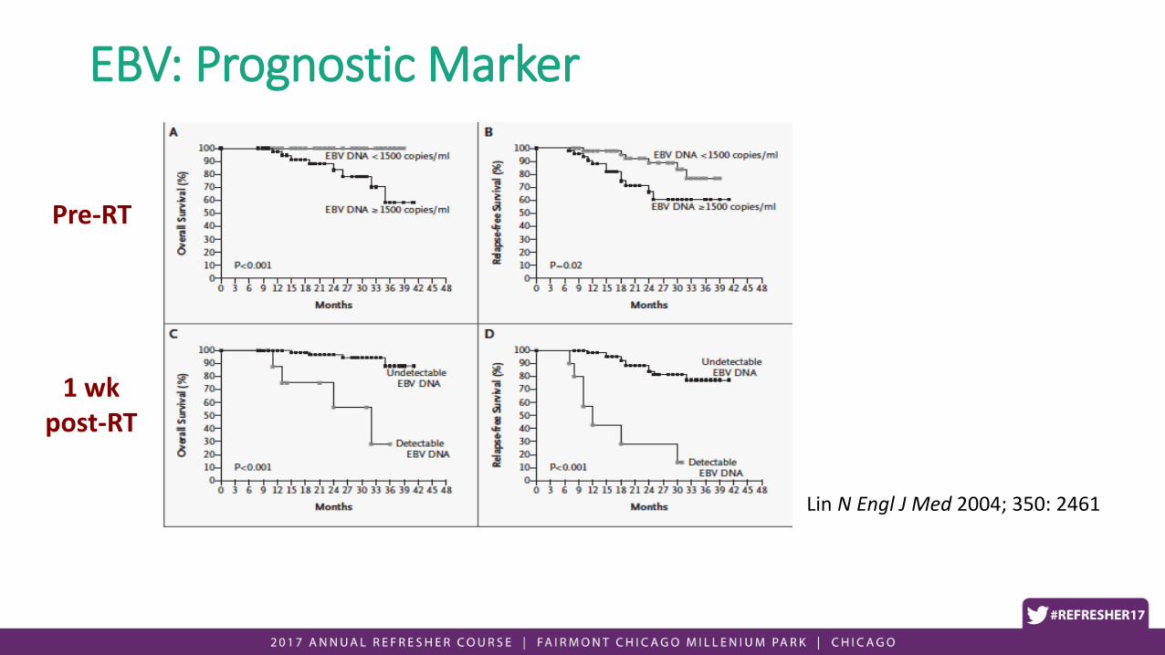
EBV: Prognostic Marker
Lin N Engl J Med 2004; 350: 2461
Pre-RT
1 wk post-RT


Insert slides on induction trial

Induction chemotherapy plus concurrent chemoradiotherapy versus concurrent chemoradiotherapy alone in
locoregionally advanced nasopharyngeal carcinoma: a phase 3, multicentre, randomised controlled trial
Prof Ying Sun, MD, Wen-Fei Li, MD, Prof Nian-Yong Chen, MD, Prof Ning Zhang, MD, Prof Guo-Qing Hu, MD, Prof Fang-Yun Xie, MD, Prof Yan Sun, MD, Prof Xiao-Zhong Chen, MD,
Prof Jin-Gao Li, MD, Prof Xiao-Dong Zhu, MD, Prof Chao-Su Hu, MD, Prof Xiang-Ying Xu, MD, Yuan-Yuan Chen, MD, Prof Wei-Han Hu, MD, Prof Ling Guo, MD, Prof Hao-Yuan Mo,
MD, Lei Chen, MD, Yan-Ping Mao, MD, Rui Sun, MD, Ping Ai, MD, Shao-Bo Liang, MD, Guo-Xian Long, MD, Bao-Min Zheng, MD, Xing-Lai Feng, MD, Xiao-Chang Gong, MD, Ling Li,
MD, Chun-Ying Shen, MD, Jian-Yu Xu, MD, Ying Guo, PhD, Prof Yu-Ming Chen, PhD, Fan Zhang, MD, Li Lin, MD, Ling-Long Tang, MD, Prof Meng-Zhong Liu, MD, Dr Prof Jun Ma,
MD
The Lancet Oncology
Volume 17, Issue 11, Pages 1509-1520 (November 2016) DOI: 10.1016/S1470-2045(16)30410-7
Copyright © 2016 Elsevier Ltd Terms and Conditions
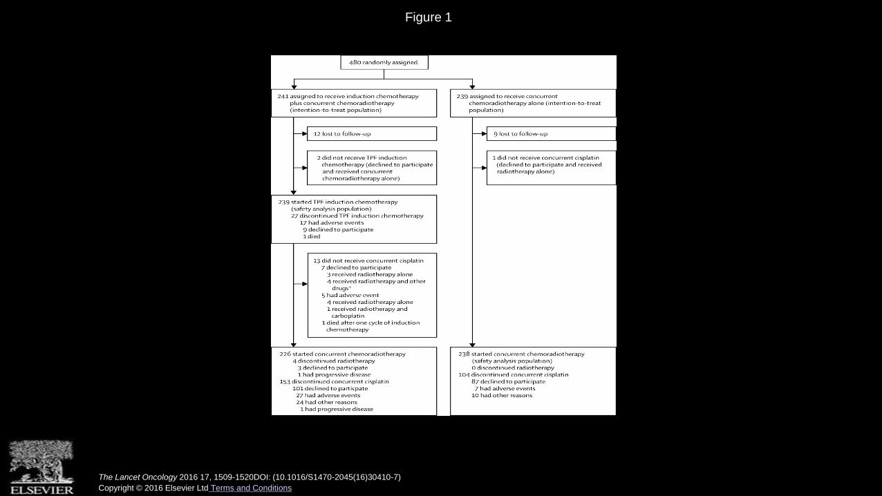
Figure 1
The Lancet Oncology 2016 17, 1509-1520DOI: (10.1016/S1470-2045(16)30410-7)
Copyright © 2016 Elsevier Ltd Terms and Conditions

Figure 2
The Lancet Oncology 2016 17, 1509-1520DOI: (10.1016/S1470-2045(16)30410-7)
Copyright © 2016 Elsevier Ltd Terms and Conditions

RT Treatment planning
• IMRT• LC > 90%
• Gross disease (primary + nodes): ~70 Gy
• High-risk CTV (bilateral RP, II-V, subclinical nasopharynx): 59-63 Gy
• Low-risk CTV: 56-59 Gy

Elective nasopharynx CTV
• Entire nasopharynx• Ant: posterior 1/3 of nasal cavity/maxillary sinuses (or greater
if anterior extension)• Post: anterior 1/2 of clivus (entire clivus if involved)• Sup: Inferior 1/2 of sphenoid (entire if T3/4, including
cavernous sinus)• Inf: Palate (or greater to ensure adequate inferior margin
below GTV)- Skull base (rotundum, ovale, lacerum)- Pterygoid- Parapharyngeal space

Pterygopalatine fossa Foramen ovale
Clivus

Foramen Lacerum

Foramen rotundum
Nasopharynx: Summary
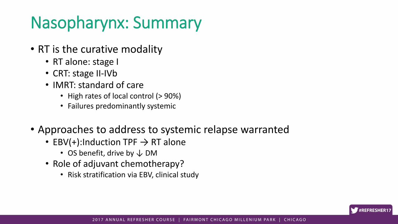
• RT is the curative modality• RT alone: stage I • CRT: stage II-IVb• IMRT: standard of care
• High rates of local control (> 90%)• Failures predominantly systemic
• Approaches to address to systemic relapse warranted• EBV(+):Induction TPF → RT alone
• OS benefit, drive by ↓ DM
• Role of adjuvant chemotherapy?• Risk stratification via EBV, clinical study

Clinical sections
• Nasopharynx
• Oral cavity
• Oropharynx
• Larynx/Hypopharynx

Oral cavity: Anatomy
• Lips
• Oral Tongue (ant 2/3)
• Floor of Mouth
• Retromolar Trigone
• Buccal Mucosa
• Hard Palate
• Gingiva/alveolar ridge

Treatment Approach• Initial surgery whenever possible
• Definitive RT: surgically unresectable or medically inoperable
• Postop RT• Early stage (I/II)
• Positive/close margins
• LVI or PNI
• Advanced stage• Concurrent chemotherapy if (+) margin or ECE

Role of therapeutic neck dissection
• RCT: • 596 pts, lateralized
T1/2 oral cavity SCCA
• Upfront (elective) vs salvage (therapeutic) neck dissection
D’Cruz et al. NEJM 2015
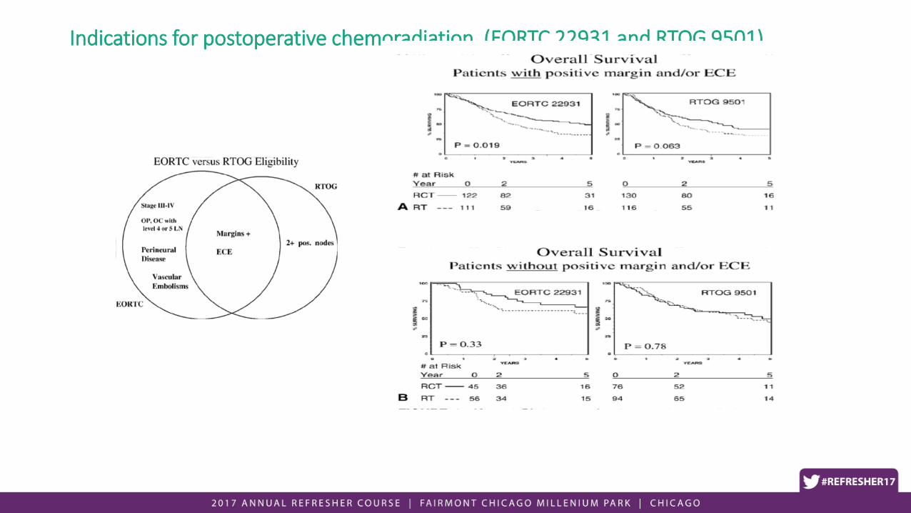
Indications for postoperative chemoradiation (EORTC 22931 and RTOG 9501)

• Retrospective NCDB analysis of ~11,000 pts, stage III-IVb SCCA of the HN, treated with surgery + adjuvant RT or CRT.
• Excluding pts with ECE or positive surgical margins.
• Patterns of care: 47% pts received adjuvant CRT.


Oral cavity: Summary
• Initial surgery, whenever possible• Upfront, elective neck dissection
• Postop RT • Early stage: intermediate risk factors (PNI/LVI)
• Advanced stage: all patients• Postop CRT for (+) margins and ECE
• Intermediate risk factors in absence of (+) margins or ECE: RT alone vs CRT


Clinical sections
• Nasopharynx
• Oral cavity
• Oropharynx
• Larynx/Hypopharynx


Palatine tonsil (if present)Palatoglossal arch
Palatopharyngealarch
Tonsillar Fossa
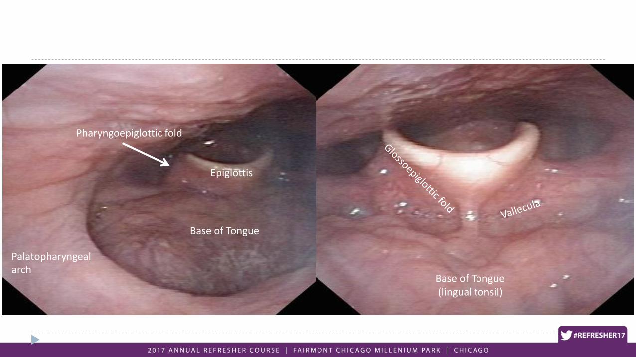
Epiglottis
Palatopharyngealarch
Base of Tongue
Base of Tongue(lingual tonsil)
Pharyngoepiglottic fold

Glossotonsillar sulcus

Soft Palate
BOT
Tonsil

Vallecula
Epiglottis

Treatment approach
• Early stage: single modality• RT alone vs. surgery
• RT• Unilateral neck: well-lateralized primary (tonsil)• Bilateral neck: central lesion (palate, BOT)
• Surgery• New, less invasive approaches: Transoral Robotic/Laser• Best for well-lateralized lesion (Tonsil, well-lateralized BOT)
• Advanced stage: combined modality• Organ preservation (CRT)• Surgery + adjuvant RT (+/- chemo)

Oropharyngeal SCCA • Traditionally associated with
smoking/drinking• Increasing incidence of
tumors associated with HPV (~ 85%)
• Males account for at least 80% of cases, generally younger
• Patients present with prominent neck adenopathy and relatively small primary tumors Chaturvedi, JCO 2011
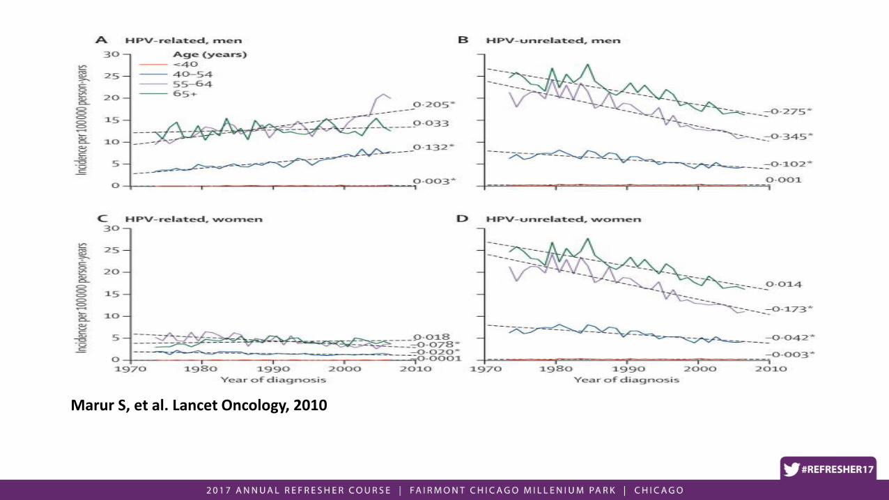
Marur S, et al. Lancet Oncology, 2010

Classification of the Study Patients into Risk-of-Death Categories and Kaplan-Meier Estimates of Overall
Survival According to Those Categories
Ang KK et al. N Engl J Med 2010;363:24-35
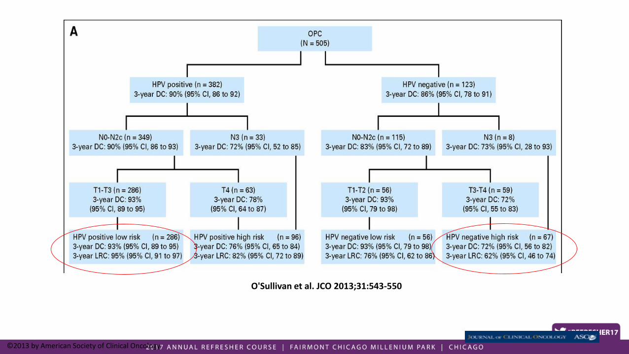
O'Sullivan et al. JCO 2013;31:543-550
©2013 by American Society of Clinical Oncology

Rationale for toxicity mitigation based on HPV status• Disease outcomes excellent
• Treatment is morbid (acute and chronic)
• Approaches:• Chemotherapy: alternative agents (cetuximab) or omit
• RT• Dose
• Volume
• Surgery: pathologic data to risk stratify

ECOG 3311


ECOG 1308: Phase II trial of IC followed by cetuximab with low orstandard dose IMRT in pts with HPV-associated resectable oropharyngealSCCA
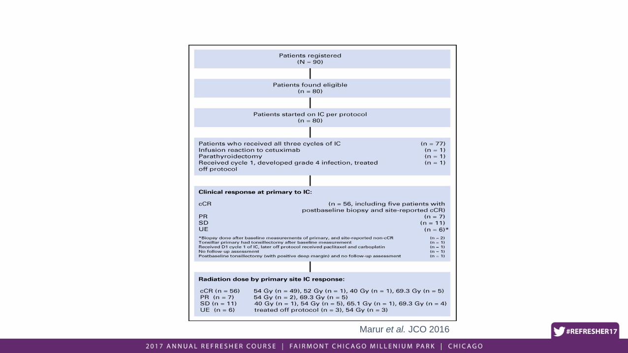
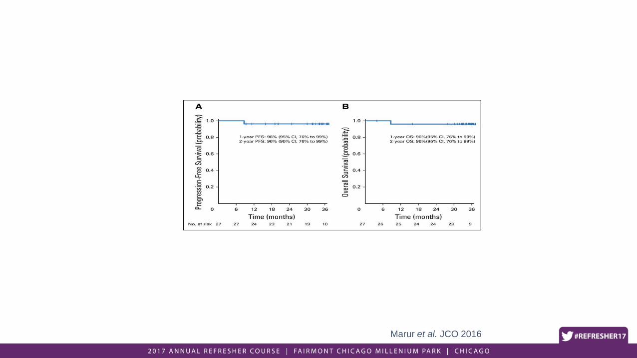
Marur et al. JCO 2016
Marur et al. JCO 2016

• Benefits:
• Pts receiving low dose RT had improved 12 mo dysphagia.
• Excellent results (96% 2yr PFS and OS)
• Concerns:
• Of 80 pts, only 46 eligible for dose reduction to both primary site and nodes (70% achieved primary site cCR, 58% nodal cCR)
• Toxicity:
• 3 pts could not finish induction
• 3 pts could not complete RT
• 21%: changed from cis-carboplatin during IC
• 28%: dose modication of cetuximab during IC or RT
• Pts who had T4 disease, N2c disease, and > 10 p-y tobacco did worse (2 yr PFS of 71% vs 96%)
• If pts clinical characteristics are more or equally predictive of outcome as response to IC, question whether IC is needed to select pts for de-intensification

Development and validation of a staging system for HPV-related oropharyngeal cancer by the International
Collaboration on Oropharyngeal cancer Network for Staging (ICON-S): a multicentre cohort study
Prof Brian O'Sullivan, MD, Shao Hui Huang, MD, Jie Su, MSc, Prof Adam S Garden, MD, Prof Erich M Sturgis, MD, Kristina Dahlstrom, PhD, Prof Nancy Lee, MD, Nadeem Riaz, MD,
Xin Pei, PhD, Shlomo A Koyfman, MD, Prof David Adelstein, MD, Prof Brian B Burkey, MD, Jeppe Friborg, MD, Claus A Kristensen, MD, Anita B Gothelf, MD, Frank Hoebers, MD,
Bernd Kremer, MD, Prof Ernst-Jan Speel, PhD, Daniel W Bowles, MD, Prof David Raben, MD, Sana D Karam, MD, Eugene Yu, MD, Wei Xu, PhD
The Lancet Oncology
Volume 17, Issue 4, Pages 440-451 (April 2016) DOI: 10.1016/S1470-2045(15)00560-4
Copyright © 2016 Elsevier Ltd Terms and Conditions

Figure 4
The Lancet Oncology 2016 17, 440-451DOI: (10.1016/S1470-2045(15)00560-4)
Copyright © 2016 Elsevier Ltd Terms and Conditions

Figure 3
The Lancet Oncology 2016 17, 440-451DOI: (10.1016/S1470-2045(15)00560-4)
Copyright © 2016 Elsevier Ltd Terms and Conditions

Oropharynx: Summary
• Early stage disease: single modality (RT or surgery alone)
• Advanced stage: Combined modality approach
• Demographics changing (HPV)• Improved disease outcomes
• Methods for toxicity mitigation are warranted for low-risk patients• Await results from clinical trials

Clinical sections
• Nasopharynx
• Oral cavity
• Oropharynx
• Larynx/Hypopharynx

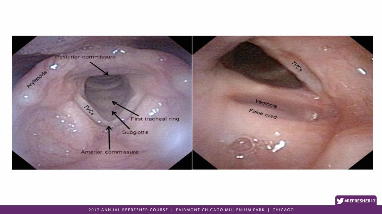
Anatomy• Supraglottis
– Epiglottis
– Arytenoids
– AE folds
– False cords
– Ventricles
• Glottis
– True vocal cords
• Subglottis
– 5mm below glottis to bottom of cricoid

Base of Tongue(lingual tonsil)
Epiglottis
Vallecula
Vallecula
Epiglottis
Hyoid

Aryepiglottic fold
Epiglottis
Arytenoids
Pyriform sinuses
Pyriform sinuses
AE fold
Arytenoids

Pyriform sinusesAE fold
Post-cricoid area
Posterior Pharyngeal Wall

Arytenoids Cricoid
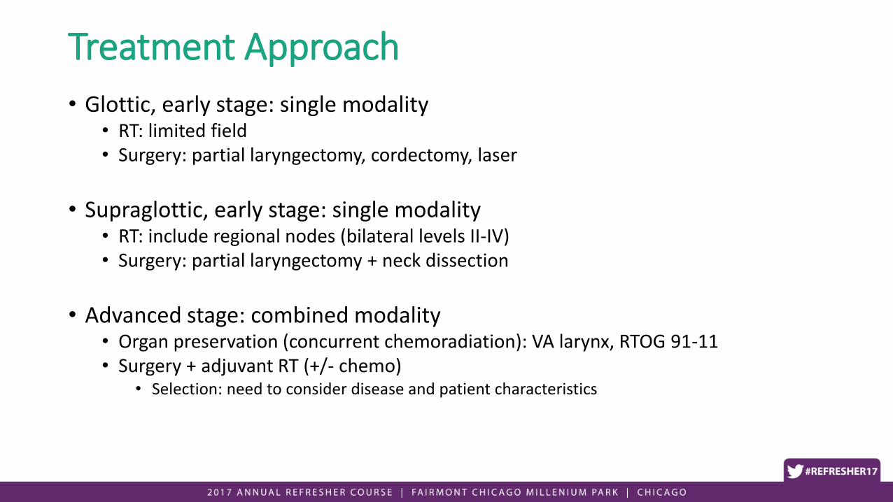
Treatment Approach
• Glottic, early stage: single modality• RT: limited field• Surgery: partial laryngectomy, cordectomy, laser
• Supraglottic, early stage: single modality• RT: include regional nodes (bilateral levels II-IV)• Surgery: partial laryngectomy + neck dissection
• Advanced stage: combined modality• Organ preservation (concurrent chemoradiation): VA larynx, RTOG 91-11• Surgery + adjuvant RT (+/- chemo)
• Selection: need to consider disease and patient characteristics

RT technique: T1 glottis• Superior: thyroid notch• Inferior: bottom of cricoid• Anterior: flash skin• Posterior: anterior to vertebral
body
• Field arrangements• Opposed laterals
• Risk: shooting through shoulders, underdosing the target
• Alternatives: obliques (superior or anterior)

Dose and fractionation: T1 glottis (Yamazaki et al. IJROBP 2006)
• Prospective, Randomized Trial, 1993-2001
• 180 pts with T1N0 Glottic Cancers
• Randomized to
A) 2.00 Gy/fraction
1) 60 Gy in 30 fractions (<2/3 VC)
2) 66 Gy in 33 fractions (>2/3 VC)
B) 2.25 Gy/fraction
1) 56.25 Gy in 25 fractions (<2/3 VC)
2) 63 Gy in 28 fractions (>2/3 VC)
Conclusion: Use 225 cGy per fraction to 63 Gy for T1 Glottic Ca
•No significant increase in acute or chronic toxicity

VA Larynx Study (NEJM 1991)
• Randomized, Prospective Phase 3, 1985-1989. • 332 patients, Stage III or IV (excluding T1N1) laryngeal cancer
• Arm 1) Total laryngectomy + Postop RT
• Arm 2) Induction Chemo + Definitive RT• Induction Chemo: Cisplatin 100mg/m2 + 5FU 1000mg/m2 Q3W x 3c
• Clinical Evaluation after cycle 2
• If PR (54%) or CR (31%) → proceed with cycle 3 → RT
• If < PR (15%) → TL + PORT

• Larynx Preservation Rate: 64%
• No difference in 2-yr OS
• Patterns of Failure: • Higher LF with ChemoRT• Higher DM with TL+PORT
2-yr LF DM OS
TL+PORT 2% 17% 68%
ChemoRT 12% 11% 68%
Established larynx preservation as a viable option
T4: 56% required salvage laryngectomy (excluded from RTOG 91-11)

RTOG 91-11 (Forastiere et al. NEJM 2003)
• Rationale: What about concurrent chemoRT instead of induction chemo?
• RTOG 91-11: Randomized, Prospective Phase 3, 1992-2000• 547 patients, Stage III/IV Laryngeal Cancer requiring TL
• Excluded large volume T4 (>1cm into BOT or penetration through thyroid cartilage)
• only 10% of pts ended up being T4
• Arm 1) Induction Chemo RT (same as VA Larynx study)
• Arm 2) Concurrent ChemoRT (cisplatin 100mg/m2 Q3wk)
• Arm 3) RT alone
• Primary endpoint: larynx preservation
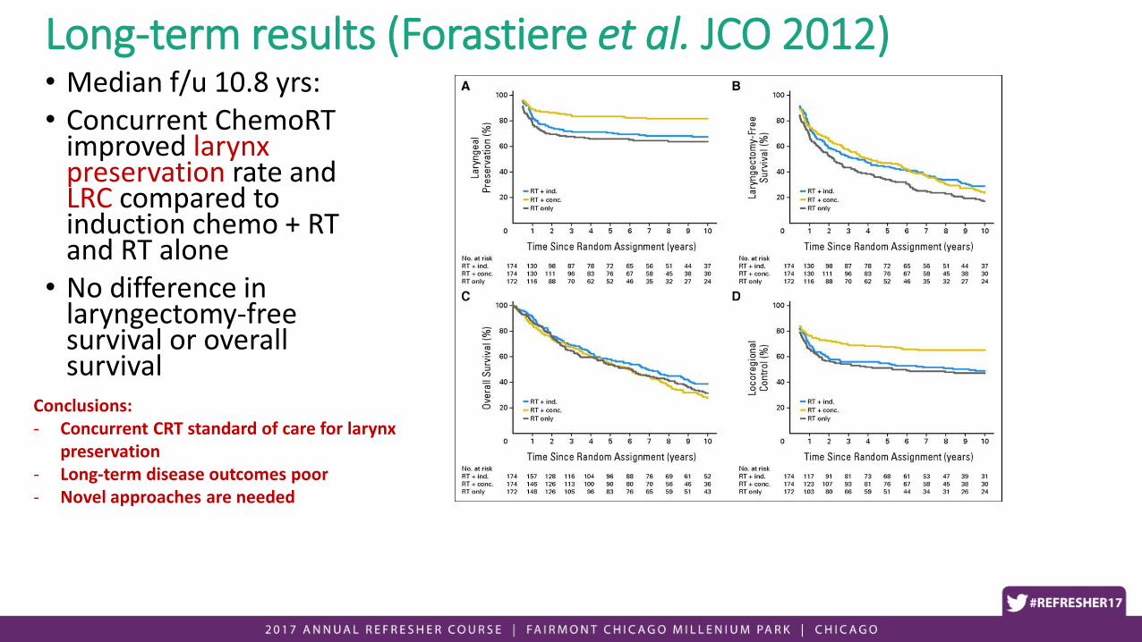
Long-term results (Forastiere et al. JCO 2012)• Median f/u 10.8 yrs: • Concurrent ChemoRT
improved larynx preservation rate and LRC compared to induction chemo + RT and RT alone
• No difference in laryngectomy-free survival or overall survival
Conclusions:- Concurrent CRT standard of care for larynx
preservation- Long-term disease outcomes poor- Novel approaches are needed

Larynx: Summary
• Early stage disease• Unimodality therapy: RT or surgery
• Disease limited to glottis: use > 2 Gy daily (2.25 Gy)
• Advanced-stage disease• Organ preservation vs surgery + PORT
• Patient selection: consider disease extent and function

Learning objectives
• HNC is a complex disease site where trt decisions/modalities often depend on multiple factors (site, stage, epidemiology).
• RT is a well-established modality of tx (definitive/postop)
• Knowledge of anatomy (visual inspection, CT) and patterns of spread are critical to proper trt/target delineation.
• Nasopharynx: CRT is standard (except for stage I)• IMRT: very high LC
• EBV(+)• Consider IC (TPF) → RT
• OS benefit via ↓DM vs. upfront CRT

Learning Objectives
• Oral cavity: surgery is initial trt of choice• RT used in adjuvant setting
• Oropharynx:• HPV has changed disease outcomes (disparate outcomes HPV (+) vs (-) despite
similar staging)
• Future directions: improving therapeutic ratio via toxicity mitigation (clinical trials)
• Larynx: need to improve outcomes• Organ preservation for advanced stage is a valid, standard option
• Consider patient, organ, and disease factors

Questions