HE Printed in U.S.A. Identification of Domain-Domain Docking
10
Identification of Domain-Domain Docking Sites within Clostridium symbiosum Pyruvate Phosphate Dikinase by Amino Acid Replacement* □ S Received for publication, July 12, 2000, and in revised form, September 5, 2000 Published, JBC Papers in Press, September 19, 2000, DOI 10.1074/jbc.M006149200 Min Wei‡, Zhong Li§, Dongmei Ye‡, Osnat Herzberg§¶, and Debra Dunaway-Mariano‡¶ From the ‡Department of Chemistry, University of New Mexico, Albuquerque, New Mexico 87131 and the §Center for Advanced Research in Biotechnology, Rockville, Maryland 20850 Potential domain-domain docking residues, identified from the x-ray structure of the Clostridium symbiosum apoPPDK, were replaced by site-directed mutagenesis. The steady-state and transient kinetic properties of the mutant enzymes were determined as a way of evaluating docking efficiency. PPDK mutants, in which one of two stringently conserved docking residues located on the N-terminal domain (Arg 219 and Glu 271 ) was substituted, displayed largely unimpeded catalysis of the phos- phoenolpyruvate partial reaction at the C-terminal do- main, but significantly impaired catalysis (>10 4 ) of the ATP pyrophosphorylation of His 455 at the N-terminal domain. In contrast, alanine mutants of two potential docking residues located on the N-terminal domain (Ser 262 and Lys 149 ), which are not conserved among the PPDKs, exhibited essentially normal catalytic turnover. Arg 219 and Glu 271 were thus proposed to play an impor- tant role in guiding the central domain and, hence, the catalytic His 455 into position for catalysis. Substitution of central domain residues Glu 434 /Glu 437 and Thr 453 , the respective docking partners of Arg 219 and Glu 271 , re- sulted in mutants impaired in catalysis at the ATP ac- tive site. The x-ray crystal structure of the apo-T453A PPDK mutant was determined to test for possible mis- alignment of residues at the N-terminal domain-central domain interface that might result from loss of the Thr 453 -Glu 271 binding interaction. With the exception of the mutation site, the structure of T453A PPDK was found to be identical to that of the wild-type enzyme. It is hypothesized that the two Glu 271 interfacial binding sites that remain in the T453A PPDK mutant, Thr 453 backbone NH and Met 452 backbone NH, are sufficient to stabilize the native conformation as observed in the crystalline state but may be less effective in populating the reactive conformation in solution. Pyruvate phosphate dikinase (PPDK) 1 catalyzes the inter- conversion of ATP, P i , and pyruvate with AMP, PP i , and PEP (1) using two separate active sites linked by a mobile domain containing the phosphoryl group carrier His 455 (2). At the first active site, to which ATP, P i and Mg(II) are bound, His 455 attacks the b-P of ATP, forming a pyrophosphorylhistidine enzyme intermediate (E-PP) and AMP (Scheme 1). The P i li- gand then reacts with the terminal phosphoryl group of E-PP to form a phosphorylhistidine intermediate (E-P) and PP i . The phosphorylhistidine of E-P then moves to the second active site where pyruvate, Mg(II), and a monovalent cation (K 1 or NH 4 1 ) are bound. Upon transfer of the phosphoryl group to pyruvate to form the final product PEP, the His 455 residue returns to the first active site to initiate a new catalytic cycle. The x-ray crystal structure (2) of the three-domain, 96-kDa subunit of the apo-PPDK homodimer from Clostridium symbio- sum is represented at the top of Fig. 1. A Mg(II) cofactor and the ATP and P i substrate ligands have been modeled into the structure at the N-terminal “ATP grasp” domain (residues 2– 430). The locations of the ligand binding sites (comprising active site 1) within the N-terminal domain are supported by the results from earlier affinity labeling and mutagenesis stud- ies (4, 5). As His 455 is located in close proximity to active site 1, the PPDK crystal structure, with some local adjustments, cor- responds to a conformer competent to catalyze the ATP/P i partial reaction (Scheme 1). Henceforth, this conformer will be referred to as “conformer 1.” Affinity labeling and mutagenesis studies have shown that the pyruvate ligand binds at a second active site, located on the a/b barrel C-terminal domain (residues 534 – 874) (6, 7). A model of an enzyme conformer (illustrated at the bottom of Fig. 1; henceforth referred to as “conformer 2”) judged to be effective in catalysis of the pyruvate partial reaction, was generated from the original structure by transferring the central domain (residues 390 –504) from its binding site at the concave surface of the N-terminal domain to the concave surface of the C- terminal domain (2). Mg(II) and PEP were modeled into active site 2 of conformer 2 (2) as pictured in Fig. 1 (bottom). In this model, the PEP phosphoryl group is aligned for nucleophilic attack by the catalytic His 455 residue. We anticipate that the rotation about the two interdomain linkers (residues 341–389 and 505–533), as required by the conformer 1 7 conformer 2 model of Fig. 1, occurs freely in solution, thus allowing the central domain to “swivel” between the two active sites. Full catalytic turnover on the enzyme will * This work was supported by National Institutes of Health Grant GM 36260 (to D. D. M.) and National Science Foundation Grant DMB9019340 (to O. H.). The costs of publication of this article were defrayed in part by the payment of page charges. This article must therefore be hereby marked “advertisement” in accordance with 18 U.S.C. Section 1734 solely to indicate this fact. □ S The on-line version of this article (available at http://www.jbc.org) contains Supplementary Figure 1, which shows the alignment of 17 known PPDK sequences generated using CLUSTALW program sup- ported by the GCG Wisconsin package. The atomic coordinates and structure factors (code 1ggo) have been deposited in the Protein Data Bank, Research Collaboratory for Struc- tural Bioinformatics, Rutgers University, New Brunswick, NJ (http:// www.rcsb.org/). ¶ To whom correspondence should be addressed Tel.: 505-277-3383; Fax: 505-277-2609; E-mail: [email protected]. 1 The abbreviations used are: PPDK, pyruvate phosphate dikinase; AMPPNP, adenylimidodiphosphate; PNP, imidodiphosphate; PEP, phosphoenolpyruvate; NADH, dihydronicotinamide adenine dinucle- otide; HPLC, high performance liquid chromatography. THE JOURNAL OF BIOLOGICAL CHEMISTRY Vol. 275, No. 52, Issue of December 29, pp. 41156 –41165, 2000 © 2000 by The American Society for Biochemistry and Molecular Biology, Inc. Printed in U.S.A. This paper is available on line at http://www.jbc.org 41156
Transcript of HE Printed in U.S.A. Identification of Domain-Domain Docking

Identification of Domain-Domain Docking Sites withinClostridium symbiosum Pyruvate Phosphate Dikinase byAmino Acid Replacement*□S
Received for publication, July 12, 2000, and in revised form, September 5, 2000Published, JBC Papers in Press, September 19, 2000, DOI 10.1074/jbc.M006149200
Min Wei‡, Zhong Li§, Dongmei Ye‡, Osnat Herzberg§¶, and Debra Dunaway-Mariano‡¶
From the ‡Department of Chemistry, University of New Mexico, Albuquerque, New Mexico 87131 and the §Center forAdvanced Research in Biotechnology, Rockville, Maryland 20850
Potential domain-domain docking residues, identifiedfrom the x-ray structure of the Clostridium symbiosumapoPPDK, were replaced by site-directed mutagenesis.The steady-state and transient kinetic properties of themutant enzymes were determined as a way of evaluatingdocking efficiency. PPDK mutants, in which one of twostringently conserved docking residues located on theN-terminal domain (Arg219 and Glu271) was substituted,displayed largely unimpeded catalysis of the phos-phoenolpyruvate partial reaction at the C-terminal do-main, but significantly impaired catalysis (>104) of theATP pyrophosphorylation of His455 at the N-terminaldomain. In contrast, alanine mutants of two potentialdocking residues located on the N-terminal domain(Ser262 and Lys149), which are not conserved among thePPDKs, exhibited essentially normal catalytic turnover.Arg219 and Glu271 were thus proposed to play an impor-tant role in guiding the central domain and, hence, thecatalytic His455 into position for catalysis. Substitutionof central domain residues Glu434/Glu437 and Thr453, therespective docking partners of Arg219 and Glu271, re-sulted in mutants impaired in catalysis at the ATP ac-tive site. The x-ray crystal structure of the apo-T453APPDK mutant was determined to test for possible mis-alignment of residues at the N-terminal domain-centraldomain interface that might result from loss of theThr453-Glu271 binding interaction. With the exception ofthe mutation site, the structure of T453A PPDK wasfound to be identical to that of the wild-type enzyme. Itis hypothesized that the two Glu271 interfacial bindingsites that remain in the T453A PPDK mutant, Thr453
backbone NH and Met452 backbone NH, are sufficient tostabilize the native conformation as observed in thecrystalline state but may be less effective in populatingthe reactive conformation in solution.
Pyruvate phosphate dikinase (PPDK)1 catalyzes the inter-conversion of ATP, Pi, and pyruvate with AMP, PPi, and PEP(1) using two separate active sites linked by a mobile domaincontaining the phosphoryl group carrier His455 (2). At the firstactive site, to which ATP, Pi and Mg(II) are bound, His455
attacks the b-P of ATP, forming a pyrophosphorylhistidineenzyme intermediate (E-PP) and AMP (Scheme 1). The Pi li-gand then reacts with the terminal phosphoryl group of E-PP toform a phosphorylhistidine intermediate (E-P) and PPi. Thephosphorylhistidine of E-P then moves to the second active sitewhere pyruvate, Mg(II), and a monovalent cation (K1 or NH4
1)are bound. Upon transfer of the phosphoryl group to pyruvateto form the final product PEP, the His455 residue returns to thefirst active site to initiate a new catalytic cycle.
The x-ray crystal structure (2) of the three-domain, 96-kDasubunit of the apo-PPDK homodimer from Clostridium symbio-sum is represented at the top of Fig. 1. A Mg(II) cofactor and theATP and Pi substrate ligands have been modeled into thestructure at the N-terminal “ATP grasp” domain (residues2–430). The locations of the ligand binding sites (comprisingactive site 1) within the N-terminal domain are supported bythe results from earlier affinity labeling and mutagenesis stud-ies (4, 5). As His455 is located in close proximity to active site 1,the PPDK crystal structure, with some local adjustments, cor-responds to a conformer competent to catalyze the ATP/Pi
partial reaction (Scheme 1). Henceforth, this conformer will bereferred to as “conformer 1.”
Affinity labeling and mutagenesis studies have shown thatthe pyruvate ligand binds at a second active site, located on thea/b barrel C-terminal domain (residues 534–874) (6, 7). Amodel of an enzyme conformer (illustrated at the bottom of Fig.1; henceforth referred to as “conformer 2”) judged to be effectivein catalysis of the pyruvate partial reaction, was generatedfrom the original structure by transferring the central domain(residues 390–504) from its binding site at the concave surfaceof the N-terminal domain to the concave surface of the C-terminal domain (2). Mg(II) and PEP were modeled into activesite 2 of conformer 2 (2) as pictured in Fig. 1 (bottom). In thismodel, the PEP phosphoryl group is aligned for nucleophilicattack by the catalytic His455 residue.
We anticipate that the rotation about the two interdomainlinkers (residues 341–389 and 505–533), as required by theconformer 1 7 conformer 2 model of Fig. 1, occurs freely insolution, thus allowing the central domain to “swivel” betweenthe two active sites. Full catalytic turnover on the enzyme will
* This work was supported by National Institutes of Health GrantGM 36260 (to D. D. M.) and National Science Foundation GrantDMB9019340 (to O. H.). The costs of publication of this article weredefrayed in part by the payment of page charges. This article musttherefore be hereby marked “advertisement” in accordance with 18U.S.C. Section 1734 solely to indicate this fact.
□S The on-line version of this article (available at http://www.jbc.org)contains Supplementary Figure 1, which shows the alignment of 17known PPDK sequences generated using CLUSTALW program sup-ported by the GCG Wisconsin package.
The atomic coordinates and structure factors (code 1ggo) have beendeposited in the Protein Data Bank, Research Collaboratory for Struc-tural Bioinformatics, Rutgers University, New Brunswick, NJ (http://www.rcsb.org/).
¶ To whom correspondence should be addressed Tel.: 505-277-3383;Fax: 505-277-2609; E-mail: [email protected].
1 The abbreviations used are: PPDK, pyruvate phosphate dikinase;AMPPNP, adenylimidodiphosphate; PNP, imidodiphosphate; PEP,phosphoenolpyruvate; NADH, dihydronicotinamide adenine dinucle-otide; HPLC, high performance liquid chromatography.
THE JOURNAL OF BIOLOGICAL CHEMISTRY Vol. 275, No. 52, Issue of December 29, pp. 41156–41165, 2000© 2000 by The American Society for Biochemistry and Molecular Biology, Inc. Printed in U.S.A.
This paper is available on line at http://www.jbc.org41156

thus require that ATP, Pi, and Mg(II) first bind to active site 1of conformer 2 (see Scheme 2). The central domain must thendissociate from the C-terminal domain and bind with the N-terminal domain to form conformer 1. Despite the fact that therotation about the linkers is intrinsically fast (ns time scale),the central domain must remain docked long enough for catal-
ysis at active site 1 to be completed (millisecond time scale(Ref. 3)), but not so long as to impede the ensuing reaction ofHis(P)455 with pyruvate at active site 2. The transition fromconformation 1 to conformation 2 allows dissociation of theAMP and PPi product ligands from active site 1 as well pre-pares the enzyme for catalysis at active site 2, provided thatthis active site was “pre-loaded” with pyruvate and the Mg(II)/NH4
1 cofactors while the enzyme was in conformation 1. Fol-lowing catalytic turnover at active site 2 in conformer 2, thecentral domain must dissociate from the C-terminal domain toallow release of the PEP product from conformer 1. If active site1 contains ATP and Pi, the next catalytic cycle can directlyfollow from this conformer.
The separate site catalysis of PPDK thus requires the tran-sient formation of precisely oriented domain-domain com-plexes. An alignment made of the 17 known PPDK sequences(see Fig. 1 in supplemental materials, available in the on-lineversion of the journal) shows that a majority of the residuesdefining the proposed binding surfaces on the N- and C-termi-nal domains are stringently conserved as are many of thesurface residues surrounding the catalytic His455 on the centraldomain (see Fig. 2). In contrast, the surface residues on theopposite, solvent-exposed faces of the three domains are vari-able. The conservation of residues at the respective domain-domain interfaces of the observed and modeled conformers hasbeen necessary throughout evolution for productive domaindocking and accurate targeting of the catalytic His455.
Through inspection of the domain-domain interface region ofthe x-ray structure of apo-PPDK and of the alignment made ofthe PPDK sequences, two stringently conserved residues of theN-terminal domain (namely Arg219 and Glu271), whose sidechains are positioned for favorable polar interaction with sidechain and/or backbone atoms of the central domain, were iden-tified. As is shown in Fig. 3, the guanidinium group of Arg219 iswithin interaction distance of the carboxylate groups of thecentral domain residues Glu434 (Glu or Asp conserved) andGlu437 (not conserved). The carboxylate group of Glu271 iswithin interaction distance of the hydroxyl group of the centraldomain Thr453 and the backbone amide NHs of Thr453 andMet452. In addition, N-terminal domain residues Ser262 andLys149, which are not conserved among all of the PPDK se-quences, are also positioned for favorable interaction with thecentral domain. The hydroxyl group of Ser262 is within H-bonding distance of the backbone C5O of central domain res-idue Ala398. The side-chain ammonium group of Lys149 is lo-cated 4.4 Å from the central domain Glu430 carboxylate, whichis too far for significant interaction. However, if the side chainof the Lys149 is free to rotate from the conformation observed inthe crystal, it can move within 3.5 Å of the carboxylate, closeenough for favorable interaction to occur.
Since productive domain-domain binding is an integral com-ponent of PPDK catalysis (Scheme 2), an examination of theroles of Arg219, Glu271, Ser262, and Lys149 in domain dockingcan be made by replacing these residues with amino acids thatcan not interact favorably and evaluating the catalytic efficien-cies of the corresponding mutant enzymes. In the text thatfollows, we report on the steady-state and transient kineticproperties of the C. symbiosum PPDK mutants R219A, R219E,E434A/E437A, E271A, T453A, S262A, S262W, and K149A. Therate constants obtained for the mutants in catalysis of the fullreaction and the E 1 ATP 3 E-PPzAMP, E 1 ATP 1 Pi 3E-PzAMPzPPi, and E 1 PEP 3 E-Pzpyruvate partial reactionsare compared with those determined in an earlier kinetic anal-ysis of the wild-type PPDK (8). The results suggest that theproductive docking of the central domain is controlled, in part,through the interaction of N-terminal domain residues Arg219
ATP/Pi Partial Reaction at Active Site 1 on N-terminal Domain:
(1) E 1 ATP 1 Pi 3 E ? ATP ? Pi 3 E-PP ? AMP ? Pi
(2) E-PP ? AMP ? Pi 3 E-P ? AMP ? PPi 3 EP 1 AMP 1 PPi
Pyruvate Partial Reaction at Active Site 2 on C-terminal Domain:
(3) E-P 1 pyruvate 3 E-P ? pyruvate 3 E-P ? PEP 3 E 1 PEP
SCHEME 1. The ATP/Pi and pyruvate partial reactions of pyru-vate phosphate catalysis (1, 3).
FIG. 1. MOLSCRIPT stereodiagrams of the C. symbiosumPPDK monomer generated from the x-ray coordinates of theapoPPDK structure reported in Ref. 2. Catalysis of the E 1 ATP 1Pi 3 E-P 1 AMP 1 PPi takes place at the N-terminal domain (green)active site in conformer 1 (top structure; ATP and Pi ligands are blue,and Mg(II) is magenta), while catalysis of the E-P 1 pyruvate partialreaction takes place at the C-terminal domain (blue) active site inconformer 2 (bottom structure; PEP ligand is red, and Mg(II) is magen-ta). Movement the catalytic His455 residue (black) between the twoactive sites is perceived to occur through the interconversion of con-formers 1 and 2 via the swivel of the central domain (yellow) about itslinkers (pink).
PPDK Domain Docking 41157

and Glu271 with central domain residues Glu434/Glu437 andThr453/Met452, respectively. The N-terminal domain Ser262 andLys149 residues, on the other hand, do not appear to play a vitalrole in orienting or stabilizing the domain-domain complex.
EXPERIMENTAL PROCEDURES
Preparation of C. symbiosum PPDK Mutants—Mutant genes wereprepared from the plasmid pACYC184-D12 (9) using polymerase chainreaction techniques analogous to those described in (4). Mutagenicprimers, 18–22 base pairs in length, were synthesized by Life Technol-ogies, Inc. BglII and BstXI restriction sites were employed in the con-struction of the Lys149, Arg219, Ser262, and Glu271 mutants, whereasBstXI and KpnI sites were used for the Glu434/Glu437 and Thr453 mu-tants. The sequences of the isolated mutant genes were determined atthe Center for Agricultural Biotechnology at the University of Mary-land. Wild-type and mutant PPDK genes were expressed in Escherichiacoli JM101 cells, and the protein products were purified to homogeneity(judged by SDS-polyacrylamide gel electrophoresis analysis) in yields of20–25 mg/g cell as described in Ref. 4. The chromatographic propertiesof the mutant proteins as well as their stability to storage in bufferedsolutions at 4 oC were observed to be very similar to the chromato-graphic properties and stability of the wild-type enzyme.
Steady-state Kinetic Analysis—The spectrophotometric assay de-scribed in Ref. 10 was used to monitor the initial velocity of the PPDK-
catalyzed reaction of AMP, PPi, and PEP to ATP, Pi, and pyruvate.Initial velocities were measured as a function of the concentration of thevaried substrate (in a range of 0.5–10-fold Km) at fixed, saturatingconcentrations of cosubstrates (0.5 mM AMP, 0.5 mM PEP, 1 mM PPi)and metal ion cofactors (5 mM MgCl2 and 40 mM NH4Cl) in 20 mM
imidazole (pH 6.8, 25 °C). The initial velocity data were analyzed usingEquation 1 and the computer programs of Cleland (11). The kcat wascalculated from vmax/[E].
v0 5 vmax [S]/(Km 1 @S]) (Eq. 1)
v0 is the initial velocity, [E] is the enzyme concentration, [S] is thesubstrate concentration, vmax is the maximum velocity, and Km is theMichaelis constant. The Ki values for the competitive inhibitors AMP-PNP and PNP were determined from initial velocity data obtained atvarying AMP or PPi concentrations and fixed, saturating concentra-tions of cosubstrates and cofactors. The initial velocity data were ana-lyzed using Equation 2, where Ki is the inhibition constant and [I] theinhibitor concentration.
v0 5 vmax [S]/[Km~1 1 @I#/Ki! 1 @S]] (Eq. 2)
Rapid Quench Analysis of PPDK-catalyzed Single-turnover Reactionsof [32P]PEP—[32P]PEP was prepared from [b-32P]ATP according to theprocedure described in Carroll et al. (3). [b-32P]ATP (specific activity .
FIG. 2. Front and back views of the space filling models of the PPDK conformers 1 and 2 of Fig. 1 are shown to illustrate theconservation of residues at the domain-domain interface regions. The dark green, dark blue, and gold residues are stringently conservedamong the 17 known PPDK sequences, while the pastel blue, pastel green, and yellow residues are variable.
FIG. 3. A stereoview of the Arg219-Glu434/Glu437 and Glu271-Met452/Thr453 docking sites (a) and the Ser262-Ala398 and Lys149-Glu430
interactions at the interface of the N-terminal and central domains (b). The N-terminal domain interface is shown in green, the centraldomain interface in yellow, and the domain linkers in pink. The ATP ligand is blue, the Pi ligand red, the Mg(II) ion is magenta, and the catalyticHis455 is black.
PPDK Domain Docking41158
1 Ci/mmol) was custom-synthesized by PerkinElmer Life Sciences. Thesingle-turnover reactions of 40 mM wild-type or mutant PPDK, 5 mM
[32P]PEP, 2.5 mM MgCl2, and 10 mM NH4Cl in 50 mM K1Hepes (pH 7.0,25 °C) were carried out in a rapid quench apparatus from KinTekInstruments. Reactions were initiated by mixing 32 ml of bufferedenzyme/cofactors with 32 ml of buffered substrate and then terminatedat varying conversion with 182 ml of 0.6 M HCl. The protein wasprecipitated from quenched solutions by vigorous mixing with CCl4 andseparated by centrifugation. The protein pellet (dissolved in 500 ml ofboiling 10 N H2SO4) and supernatant were analyzed for 32P by scintil-lation counting. The percentage of conversion was deduced from theratio of the radioactivity in the pellet to the total radioactivity in thepellet and supernatant. The concentration of [b-32P]E-P was calculatedfrom 40 mM X% conversion and plotted as a function of reaction time.The resulting time course was fit to a single exponential equation
(Equation 3) using the KaleidaGraph computer program to yield thereported rate constants.
@B#t 5 @B#f 3 ~1 2 exp~kobs 3 t!! (Eq. 3)
[B]t is the product concentration at time t, [B]f is the product concen-tration at equilibrium, and kobs is the observed rate constant for thereaction.
Rapid Quench Analysis of PPDK-catalyzed Single-turnover Reactionsof [14C]ATP in the Absence or Presence of Pi—[14C]ATP was synthesizedfrom [14C]AMP (PerkinElmer Life Sciences) as follows. A 1-ml reactioncontaining 0.08 mM [14C]AMP (50 mCi), 50 mM PPi, 0.5 mM PEP, 5 mM
MgCl2, 40 mM NH4Cl, 6 units of PPDK in 50 mM K1Hepes (pH 7.0) wascarried out at 25 °C for 1 h. The reaction mixture was then chromato-graphed on a 9-ml DEAE-Sepharose column using a linear gradient(0.25–1.1 M) of triethylamine bicarbonate (pH 7) as eluant. The frac-tions containing [14C]ATP were pooled and concentrated in vacuo (ro-tary evaporation) at room temperature. Excess triethylamine bicarbon-ate was removed by repeated water dilution followed by evaporation.The resulting [14C]ATP was stored frozen in 50 mM K1Hepes buffer (pH7.0) at 230 °C.
Single-turnover reactions of 40 mM wild-type or mutant PPDK, 10 mM
[14C]ATP, 2.5 mM CoCl2, and 10 mM NH4Cl in 50 mM K1Hepes (pH 7.0,25 °C) were carried out in the presence or absence of 11 mM Pi using arapid quench device as described previously. The protein was removedfrom the acid quenched samples by centrifugation through a 500-mlfilter (cut-off molecular mass of 30 kDa) purchased from Pall GelmanInc. The unconsumed [14C]ATP and [14C]AMP product were separatedby HPLC using a Beckman ultrasphere C18 reversed-phase analyticalcolumn and 25 mM KH2PO4, 2.5% triethylamine, and 5% methanol asthe mobile phase. The fractions containing ATP and AMP were ana-lyzed for 14C content by liquid scintillation counting. The time course fora single-turnover reaction was constructed, and the kobs was calculated,in the manner described in the previous section.
X-ray Crystallographic Analysis of T453A PPDK—The crystalliza-tion conditions used for the C. symbiosum T453A PPDK mutant werethe same as those used for the wild-type enzyme (2). Single crystals
E1 ¢O¡ E2
8 8E1-P ¢O¡ E2-P
E2 binds & releases ATP, Pi pair or AMP, PPi pair but reacts only withbound PEP
E2-P binds & releases ATP, Pi pair or AMP, PPi pair but reacts onlywith bound pyruvate
E1 binds & releases pyruvate or PEP but reacts only with bound ATP,Pi pair
E1-P binds & releases pyruvate or PEP but reacts only with boundAMP, PPi pair
SCHEME 2. An illustration of the free and phosphorylatedforms of PPDK conformer 1 (E1, E1-P) and conformer 2 (E2, E2-P)(see Fig. 1) with the ligands that these enzyme forms bind andconvert to products.
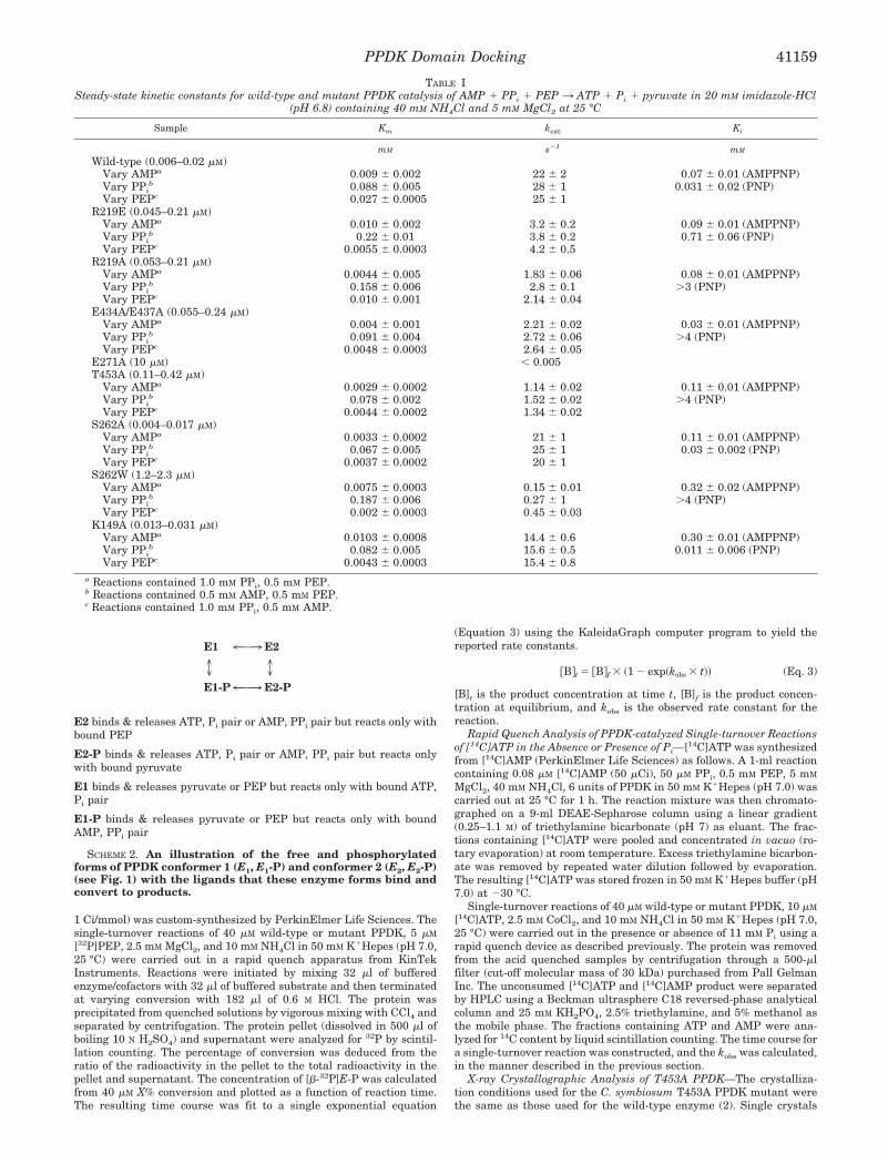
TABLE ISteady-state kinetic constants for wild-type and mutant PPDK catalysis of AMP 1 PPi 1 PEP 3 ATP 1 Pi 1 pyruvate in 20 mM imidazole-HCl
(pH 6.8) containing 40 mM NH4Cl and 5 mM MgCl2 at 25 °C
Sample Km kcat Ki
mM s21 mM
Wild-type (0.006–0.02 mM)Vary AMPa 0.009 6 0.002 22 6 2 0.07 6 0.01 (AMPPNP)Vary PPi
b 0.088 6 0.005 28 6 1 0.031 6 0.02 (PNP)Vary PEPc 0.027 6 0.0005 25 6 1
R219E (0.045–0.21 mM)Vary AMPa 0.010 6 0.002 3.2 6 0.2 0.09 6 0.01 (AMPPNP)Vary PPi
b 0.22 6 0.01 3.8 6 0.2 0.71 6 0.06 (PNP)Vary PEPc 0.0055 6 0.0003 4.2 6 0.5
R219A (0.053–0.21 mM)Vary AMPa 0.0044 6 0.005 1.83 6 0.06 0.08 6 0.01 (AMPPNP)Vary PPi
b 0.158 6 0.006 2.8 6 0.1 .3 (PNP)Vary PEPc 0.010 6 0.001 2.14 6 0.04
E434A/E437A (0.055–0.24 mM)Vary AMPa 0.004 6 0.001 2.21 6 0.02 0.03 6 0.01 (AMPPNP)Vary PPi
b 0.091 6 0.004 2.72 6 0.06 .4 (PNP)Vary PEPc 0.0048 6 0.0003 2.64 6 0.05
E271A (10 mM) , 0.005T453A (0.11–0.42 mM)
Vary AMPa 0.0029 6 0.0002 1.14 6 0.02 0.11 6 0.01 (AMPPNP)Vary PPi
b 0.078 6 0.002 1.52 6 0.02 .4 (PNP)Vary PEPc 0.0044 6 0.0002 1.34 6 0.02
S262A (0.004–0.017 mM)Vary AMPa 0.0033 6 0.0002 21 6 1 0.11 6 0.01 (AMPPNP)Vary PPi
b 0.067 6 0.005 25 6 1 0.03 6 0.002 (PNP)Vary PEPc 0.0037 6 0.0002 20 6 1
S262W (1.2–2.3 mM)Vary AMPa 0.0075 6 0.0003 0.15 6 0.01 0.32 6 0.02 (AMPPNP)Vary PPi
b 0.187 6 0.006 0.27 6 1 .4 (PNP)Vary PEPc 0.002 6 0.0003 0.45 6 0.03
K149A (0.013–0.031 mM)Vary AMPa 0.0103 6 0.0008 14.4 6 0.6 0.30 6 0.01 (AMPPNP)Vary PPi
b 0.082 6 0.005 15.6 6 0.5 0.011 6 0.006 (PNP)Vary PEPc 0.0043 6 0.0003 15.4 6 0.8
a Reactions contained 1.0 mM PPi, 0.5 mM PEP.b Reactions contained 0.5 mM AMP, 0.5 mM PEP.c Reactions contained 1.0 mM PPi, 0.5 mM AMP.
PPDK Domain Docking 41159

were obtained at 30 °C by vapor diffusion in hanging drops. Proteindrops were equilibrated against reservoir solutions containing 50–55%saturated ammonium sulfate and 100 mM Hepes buffer (pH 7.0). Thedrops consisted of protein solution (10 mg/ml, 20 mM imidazole buffer(pH 6.5), 100 mM KCl, 0.1 mM EDTA, and 1 mM mercaptoethanol),diluted by an equal volume of reservoir solution. The crystals belong tospace group P2, and within the accuracy of the data the unit celldimensions are the same as those of the wild-type protein crystals(Table II). The crystal was transferred to a mother liquor solutioncontaining 20% glycerol, and then flash-frozen using an Oxford cryo-system. X-ray diffraction data were collected on a Siemans area detectormounted on a three-circle goniostat, with a monochromated CuKa x-raysupplied by a Rigaku Rotaflex RU200BH rotating anode generator.Data were processed with the XENGEN package (12). Data collectionstatistics are provided in Table VI. Structure refinement was performedwith the program CNS (13). The simulated annealing slow-coolingprotocol at 3000 K was followed by positional and temperature factorrefinement cycles. Data up to 2.65 Å for which F $ 2s(F) were included.Adjustments to the model were made on a Silicon Graphics INDIGO IIcomputer graphics workstation using the program TURBO-FRODO(14).
RESULTS AND DISCUSSION
Steady-state Kinetic Properties of PPDK Mutants—ThePPDK mutants were first screened by measuring kcat and Km
values for substrates and Ki values for competitive inhibitors.The steady-state kinetic constants were determined for theAMP 1 PPi 1 PEP 3 ATP 1 Pi 1 pyruvate reaction (withMg21 and NH4
1 serving as cofactors) using a continuous spec-trophotometric assay to monitor pyruvate formation. PPDKadheres to an ordered, nonclassical bi (ATP, Pi) bi (PPi, AMP)uni (pyruvate) uni (PEP) kinetic mechanism (10). For conven-
ience, the Km (and Vm) for each substrate was evaluated frominitial velocity data measured with the concentrations of thetwo co-substrates held constant at saturating levels, while theconcentration of the third substrate was varied at ;0.5–10-foldthe Km value. The Ki values of the ATP analog AMPPNP(versus AMP) and PPi analog PNP (versus PPi) were deter-mined from the initial velocity data measured in the absenceand presence of the inhibitor.
The Ki, Km, and kcat values obtained for wild-type PPDK andthe PPDK mutants at pH 6.8 and 25 °C are listed in Table I.Except for S262A and K149A, each of the mutants displaysignificantly reduced catalytic efficiency compared with that ofthe wild-type PPDK. The kcat is deceased ;10-fold for theR219A, R219E, and E434A/E437A mutants, ;20-fold forT453A, ;100-fold for S262W, and .5000-fold for E271A. Aswill be described in the section that follows, the reduction inkcat derives from a reduction in the rate of the nucleotidepartial reaction.
The abilities of the mutant enzymes to bind ATP and PPi arereflected by the Ki values measured for the inert analogs AMP-PNP and PNP. AMPPNP competes with AMP for the samebinding site on conformer 1 of the free and phosphorylatedforms of the enzyme (see Scheme 2). Likewise, PNP competeswith PPi for the same site on the AMP complex of the free orphosphorylated enzyme in conformation 1. An increase in theKi value may signify that the structure or electronic environ-ment of the binding site has been altered by the mutation.Alternatively, if the ligand binding site is changed by domain-domain association, a mutation that affects domain binding canaffect the Ki of the inhibitor. Thus, an increase in the Ki valuesmay also indicate impaired domain-domain docking.
Based on the AMPPNP Ki values listed Table I, it is evidentthat the ATP binding affinity is not altered in the R219E,R219A, E343A/E347A, S262A, and T453A mutants, despite thefact that all but the S262A mutant has a significantly lowerturnover rate. On the other hand, the AMPPNP Ki valuesmeasured for the S262W and K149A mutants are ;4-foldlarger than that measured for wild-type PPDK. The exactcause of reduced ATP binding in these mutants is presentlyunknown; however, since the Ser262 and Lys149 residues do notappear to function as docking residues (i.e., the kcat values ofthe Ala mutants are normal), we have not pursued the issuefurther.
FIG. 4. The time courses for [32P]E-P formation in the single-turnover reaction of 40 mM wild-type or mutant PPDK, 5 mM [32P]PEP,2.5 mM MgCl2, and 10 mM NH4Cl in 50 mM K1Hepes (pH 7.0, 25 °C). A, wild-type (●), R219A (E), R219E (M), and E434A/E437A (Œ). B,wild-type (●), E271A (E), S262W (M), and T453A (Œ). The curves were generated by computer fitting the data to a first order rate equation usingthe computer program KaleidaGraph. The kobs values calculated from these curves are reported in Table II.
TABLE IIThe single-turnover rate constants measured wild-type, R219E,
R219A, E434A/E437A, E271A, T453A, and S262W catalysis of the E1 [32P]PEP 3 [32P]E-P z pyruvate partial reaction
Single-turnover reactions contained 40 mM enzyme, 5 mM [32P]PEP,2.5 mM MgCl2, and 10 mM NH4Cl in 50 mM K1Hepes (pH 7.0, 25 °C).See “Experimental Procedures” for details.
Enzyme kobs
s21
Wild-type 43 6 4R219E 38 6 4R219A 38 6 4E271A 49 6 4E434A/E437A 52 6 6T453A 17 6 1
PPDK Domain Docking41160

The PPi binding affinity was found to be severely weakenedin the R219E, R219A, E434A/E437A, S262W, and T453APPDK mutants, as indicated by the comparatively large PNPKi values observed for the mutant enzymes (;20–100-foldlarger than that of the wild-type PPDK). Only the K149A andS262A mutants, both of which display normal kcat values (andthus, unimpaired domain-domain docking), were unaffected.The PPi binding site lies outside of the regions mutated and,therefore, may not be directly effected by the amino acid re-placements made. On the other hand, PPi binds at the entranceof active site 1, which becomes desolvated by the region of thecentral domain surrounding (and including) the His(P)455. Alikely scenario is that the reduced PNP binding affinity ob-served with the mutant enzymes is a reflection of altered do-main-domain binding.2
Catalysis at the PEP/Pyruvate Active Site—The EzPEP 9E-Pzpyruvate partial reaction is catalyzed by PPDK in confor-mation 2 (Fig. 1; Scheme 2). Thus, mutations made at theN-terminal domain are not expected to inhibit catalysis at theC-terminal domain. This was tested by measuring the kobs forsingle-turnover reactions of R219A, R219E, E271A, and S262Wmutant PPDK 1 [32P]PEP 3 [32P]E-Pzpyruvate using rapidquench techniques. The time courses for these reactions areshown in Fig. 4, whereas the kobs values calculated from therate data are reported in Table II. All four mutants displayednormal catalysis of the EzPEP7 E-Pzpyruvate partial reaction.The fact that the kobs values for the four mutants are notnoticeably higher than that measured for the wild-type enzymesuggests that the population of conformer 2 has not been sig-nificantly increased and/or that the rate of conformer equili-bration is not limiting.
2 In contrast to the ATP binding site, the Pi binding site is located atthe entrance to the active site crevice where Arg337, and possiblyMg(II), are the probable binding residues (4) D. Ye, M. Wei, M. V.McGuire, K. Huang, G. Kapadia, O. Herzberg, and D. Dunaway-Mari-ano, unpublished data). The affinity of Pi for this site is quite low (Kd ;6 mM) and the binding rate, slow (kon 5 0.04 mM21 s21; koff ;200 s21) (8).Since ATP binds beneath the Pi, substrate binding is ordered, with ATPfirst and Pi second (10). When the central domain binds to the N-terminal domain, the active site is closed and the dissociation of the twosubstrate ligands is prevented. The Pi ligand is covered by the centraldomain region surrounding the catalytic His455 (see Fig. 1). Whether or
not direct binding interaction occurs between the bound Pi ligand andthe central domain, cannot be discerned on the basis of the data onhand. Nevertheless, it is reasonable to expect that the domain dockingproduces significant alterations in the Pi environment. The PPi liganddisplays a higher binding affinity for the E-PzAMP complex (Kd 5 50mM) than does Pi for the enzyme-ATP complex (8, 10). PPi binding (kon5 2 mM21 s21; koff 5 100 s21 (Ref. 8)) is followed by phosphohistidine 455binding. It is likely that the environment of the PPi ligand is altered bydomain-domain association and that the PPi binding constant is ef-fected by mutations that impair the domain-domain docking reaction.
FIG. 5. The time courses for[14C]AMP formation in the single-turnover reactions of 40 mM (●) or 80mM (E) enzyme, 10 mM [14C]ATP, 5 mMMgCl2, and 10 mM NH4Cl in 50 mMK1Hepes (pH 7.0, 25 °C) catalyzed bywild-type, R219E, R219A, E434A/E437A, T453A, and S262A PPDK. Theplots are generated by fitting the timepoint data to a first order rate equation.
PPDK Domain Docking 41161

The E434A/E437A and T453A mutations modify the centraldomain. Since the central domain must interface with theC-terminal domain in order to catalyze the EzPEP 7E-Pzpyruvate partial reaction, it was anticipated that the cen-tral domain mutants would display reduced catalytic efficiencyat both active sites. However, the time course measured for theE 1 [32P]PEP 3 [32P]E-Pzpyruvate single-turnover catalyzedby the E434A/E437A mutant is essentially the same as thatcatalyzed by wild-type PPDK, whereas that measured for theT453A mutant reflects ;2-fold reduction in kobs and ;30%reduction in the position of the reaction equilibrium ([E-P][pyruvate] [E-Pzpyruvate]/[E] [PEP] [EzPEP], where for wild-type PPDK the Kd values for E-Pzpyruvate and EzPEP com-plexes are 75 and 170 mM, respectively, and the internal Keq 5[E-Pzpyruvate]/[EzPEP] 5 1 (Ref. 8)) (Fig. 4; Table II). TheGlu434 and Glu437 residues of the central domain thus do notappear to function as docking sites in the association of thecentral and C-terminal domains. On the other hand, the Thr453
residue appears to play a minor role in promoting catalysis atthe C-terminal active site.
Catalysis at the ATP/Pi Active Site—Catalysis at the N-
terminal domain consists of two reaction steps: the pyrophos-phorylation of His455 by ATP, followed by the transfer of aphosphoryl group from the pyrophosphorylated His455 to phos-phate (see Scheme 1). Both reaction steps require activation ofthe enzyme by a divalent metal ion (Mg(II) is the physiologicalcofactor, but Co(II) and Mn(II) are also effective activators) (1).
The reaction of His455 with ATP does not require Pi and,thus, can be studied in isolation. The single-turnover reactionof excess PPDK with [14C]ATP to produce E-PPz[14C]AMP ismonitored using a rapid quench instrument in conjunction withHPLC techniques to separate the [14C]AMP released from thequenched enzyme (8). Previous studies of the wild-type PPDKhave shown that the amount of AMP formed under the condi-tions of the single-turnover reaction is governed by the level ofenzyme-bound versus unbound ATP and by the [E-PPzAMP]/[EzATP] equilibrium (8). Substitution of Co(II) for Mg(II) ascofactor in the reaction increases both ATP binding to theenzyme and the internal equilibrium, so that the amount ofE-PPzAMP produced from reaction of 40 mM PPDK with 10 mM
ATP increases from ;5% for the Mg(II)-activated enzyme to;25% for the Co(II)-activated enzyme. For this reason, the
FIG. 6. The time courses for [14C]AMP formation in the single-turnover reactions of 5 mM [14C]ATP, 11 mM Pi, 5 mM MgCl2, and 10mM NH4Cl in 50 mM K1Hepes (pH 7.0, 25 °C) catalyzed by 20 mM enzyme wild-type, R219E, R219A, E434A/E437A, E271A, T453A, S262A,and S262W PPDK. The plots are generated by fitting the time point data to a first order rate equation.
PPDK Domain Docking41162

abilities of the PPDK mutants to catalyze the E 1 [14C]ATP3E-PPz[14C]AMP partial reaction were evaluated using Co(II)rather than Mg(II) as cofactor.
On the other hand, Mg(II) is the superior choice for kineticanalysis of the single-turnover reaction of E (20 mM) 1 ATP (5mM) 1 Pi (11 mM) 3 E-PzAMPzPPi. For this reaction, it isadvantageous to minimize the level of AMP formed in theinitial EzATP Pi 3 E-PPzAMPzPi step so that the total amountof AMP formed will coincide with the amount of AMP producedas the result of the second partial reaction: E-PPzAMPzPi 3E-PzAMPzPPi, which, by mass action effect, drives the firstreaction forward. For wild-type PPDK, the kobs for the first stepis ; 300 s21 while the kobs for the second step is ;6 s21 (4).
If catalysis of the first step is impaired in the PPDK mutant,this will be reflected by the time course for [14C]AMP formationin the EzCo(II) 1 [14C]ATP3 E-PPz [ 14C]AMPzCo(II) reaction,as well as by the time course for [14C]AMP formation in theEzMg(II) 1 [14C]ATP 1 Pi 3 E-Pz[14C]AMPzPPizzMg(II) reac-tion. If, on the other hand, the first step is not severely inhib-ited, then the degree to which the second step is inhibited canbe discerned from the time course for [14C]AMP formation inthe EzMg(II) 1 [14C]ATP 1 Pi 3 E-PPz[14C]AMPzPPizMg(II)reaction (see for example, Ref. 4).
The time courses for [14C]AMP formation in the EzCo(II) 1
[14C]ATP3 E-PPz[14C]AMPzCo(II) reaction and in the EzMg(II)1 [14C]ATP 1 Pi 3 E-Pz[14C]AMPzPPizMg(II) reaction cata-lyzed by wild-type and mutant PPDK are shown in Figs. 5 and6, whereas the observed rate constants calculated from the ratedata are listed in Tables III and IV. S262A mutant catalyzesthe EzCo(II) 1 [14C]ATP 3 E-PPz [ 14C]AMPzCo(II) reaction atthe normal (wild-type) rate, while the rates observed for theR219E, R219A, E434A/E437A, E271A, T453A, and S262W mu-tants are drastically lower (104 to .105). The kobs was re-measured using double the concentration of mutant or wild-type PPDK to determine whether EzCo(II)z [ 14C]ATP complexformation or catalytic turnover is rate-limiting in the single-turnover reaction. The results, reported in Table V, indicatethat catalysis and not ATP binding is rate-limiting for each ofthese enzymes.
The kobs values measured for the EzMg(II) 1 [14C]ATP 1 Pi
3 E-Pz[14C]AMPzPPizMg(II) reaction catalyzed by the R219E,R219A, E434A/E437A, E271A, T453A, and S262W mutants(Table III) mirror (within error limits) the kobs values measuredfor the EzCo(II) 1 [14C]ATP3 E-PPz [ 14C]AMPzCo(II) reaction(Table IV). Thus, the first (E-PPzAMP forming) step is rate-limiting. The kobs values, therefore, are less than or equal to therate constants for the second step catalyzed in the mutants.
Altered Active Site Versus Altered Domain-Domain Dockingin PPDK Mutants—The kinetic properties measured for theR219E, R219A, E434A/E437A, E271A, T453A, and S262WPPDK mutants show that the amino replacements made in-hibit catalysis of the E 1 ATP 3 E-PPzAMP partial reaction.Because catalysis of this partial reaction depends both on theintegrity of the N-terminal domain active site and on the accu-rate placement of the His455 residue into the active site, im-paired catalysis in these mutants can derive from an alterationin the active site environment and/or an alteration in thedomain-domain docking process. Here, we argue that the N-terminal domain active sites in the PPDK mutants3 are intactand that impaired catalysis derives from altered domain-do-main docking.
First, we note that the mutation sites are located at thesurface of the N-terminal or central domains. We do not antic-ipate that the amino acid substitutions will alter the domainfold (as is demonstrated for the T453A mutant by x-ray crys-tallographic analysis; see below), and in this way inhibit catal-ysis of the E 1 ATP 3 E-PPzAMP partial reaction. Substitu-tions of Arg219, Glu271, Lys149, and Glu434/Glu437 involvechanges in the electrostatic properties of the domain surface,not in but near the active site. In previous (unrelated) studies,analogous electrostatic perturbations have been generated atthe N-terminal domain surface in close vicinity of the activesite, without alteration of catalytic activity (in the Lys163,
3 The exception may be the S262W mutant. This mutant displays amodest reduction in ATP binding affinity and large reduction in cata-lytic efficiency. We suspect that the large Trp side chain causes confor-mational changes effecting both the ATP binding site and the domain-domain binding site.
TABLE IIISingle-turnover rate constants for wild-type, R219E, R219A, E434A/E437A, E271A, T453A, S262A, and S262W PPDK catalysis of E 1
[14C]ATP 3 E-PP z [14C]AMPReaction contains 40 mM enzyme, 10 mM [14C]ATP, 2.5 mM Co21, 10
mM NH41, and 50 mM K1Hepes (pH 7.0, 25 °C).
Enzyme kobs
s21
Wild-type ;300 6 70a
R219E 0.03 6 0.03b
R219A 0.050 6 0.004b
E434A/E437A 0.08 6 0.05b
E271A ,0.005c
T453A 0.046 6 0.003S262A 200 6 20S262W #0.005c
a Average results of two reaction trials carried out using separatelyprepared enzymes. The marked error was the difference between thekobs values obtained from the two trials.
b The wild-type PPDK catalyzed ATP reaction reached more than60% conversion within the dead time of the rapid quench instrument;therefore, the wild-type kobs was only an estimated value.
c Mutant E271A- and S262W-catalyzed ATP turnover reactions weretoo slow to be accurately quantitated due to considerable decompositionof nucleotides at the active sites of these mutants when incubation timeexceeded 300 s. The lower limit of measurable rate constant is 0.005s21.
TABLE IVSingle-turnover rate constants for wild-type, R219E, R219A, E434A/E437A, E271A, T453A, S262A, and S262W PPDK catalysis of E 1
[14C]ATP 1 Pi 3 E-P 1 [14C]AMP 1 PPi
Single-turnover reaction contains 20 mM enzyme, 5 mM [14C]ATP, 11mM Pi, 5 mM Mg21, 10 mM NH4
1, and 50 mM K1Hepes (pH 7.0, 25 °C).
Enzyme kobs for trial 1 kobs for trial 2 kobs
s21 s21 s21
Wild-type 6.2 6 0.6 5.7 6 0.3 6.0 6 0.6a
R219A 0.015 6 0.001 0.015 6 0.001 0.015 6 0.001a
R219E 0.014 6 0.001 0.028 6 0.002 0.02 6 0.01a
E434A/E437A 0.13 6 0.01 0.21 6 0.01 0.17 6 0.08a
E271A 0.00029 6 0.00003T453A 0.071 6 0.005S262A 4.5 6 0.6S262W 0.0069 6 0.0003
a Average results of two reaction trials carried out using separatelyprepared enzymes. The marked error was the difference between thekobs values obtained from the two trials.
TABLE VRatios of kobs values measured for single-turnover reactions of E 1
[14C]ATP 3 E-P z [14C]AMP catalyzed by 40 mM and 80 mM wild-type,R219A, R219E, E434A/E437A, or T453A PPDK
Reaction contains 40 or 80 mM enzyme, 10 mM [14C]ATP, 2.5 mM Co21,10 mM NH4
1 and 50 mM K1Hepes (pH 7.0, 25 °C).
Enzymes WT R219A R219E DEDA T435A
Ratio trial 1a 1.3 1.15 1.26 0.96 0.80Ratio trial 2a 1.03 0.84 0.92
a Ratio of the kobs for the reaction containing 80 mM enzyme over thekobs for the reaction containing 40 mM enzyme.
PPDK Domain Docking 41163
Lys177, Glu168, Glu170, and Glu175 PPDK mutants examined)(5). Although it can be argued that these sites are not located atthe domain-domain interface, we point out that two of thedomain-domain interface residues examined in the presentstudy, Ser262 and Lys149 have been replaced by an Ala residuewithout loss of catalytic activity.
Second, we note that mutation of Arg219 to Glu has the sameimpact on catalysis at active site 1 that mutation of Arg219 toAla has. Thus, the loss of binding interaction between Arg219
and the Glu434/Glu437 residues is what is critical to catalysisand not the type of charge alteration made at position 219.Moreover, which partner of the paired docking residues, Arg219
or Glu434/Glu437, is replaced in the mutagenesis experimentdoes not effect the outcome; the catalytic properties of theGlu434/Glu437 mutant and the Arg219 mutant are similar.
Finally, we note that disruption of the Glu271-Thr453/Met452
docking site has more impact on catalysis than does disruptionof the Arg219-Glu434/Glu437 docking site. Unlike the Arg219-Glu434/Glu437 docking site, the Glu271-Thr453/Met452 dockingsite is close to the His455 residue (see Fig. 3). The His455 sidechain is positioned on the N terminus of a short helix, just downstream from the Glu271 docking residues Thr453 and Met452.The backbone amide NHs of both of these residues as well asthe side chain of the Thr453 residue bind with the Glu271 car-boxylate group, thereby drawing the His455 into the active siteregion. Consequently, the Glu271-Thr453/Met452 docking sitemight play a more dominant role in the productive domain-domain binding than the Arg219-Glu434/Glu437 site, which isfurther removed from the His455. Alternatively, the differentialeffects of mutating Arg219 and Glu271 may be attributed to thedifference in the number of interdomain electrostatic interac-tions lost. Specifically, the present data indicate that the re-moval of a single interaction, regardless of the docking siteinvolved, results in a ;104-fold reduction in kobs. This may bea simple coincidence, or it may suggest that the contributions ofthe electrostatic interactions are additive, in which case onecan rationalize the greater loss in activity for the E271A PPDKmutant in terms of the loss of three rather than one interaction.
Misalignment versus Decreased Residence Time by ImpairedDomain-Domain Docking—Domain movement in PPDK occursvia bond rotation within the solvated linkers (Fig. 1). Domain-
domain association occurs through the diffusion of the twotethered protein bodies through restricted solvent space. Thebinding equilibrium is controlled by the attraction between thetwo interfaced surfaces. Protein interfaces observed in proteindimers, protease-protein inhibitor complexes, and antibody-protein antigen complexes (where in permanent association isdesired) range from 700 to 4900 Å2 (16). In PPDK, for which thebinding of between the two domain surfaces is intended to betransient, the accessible area buried in conformation 1 (Fig. 1)is ;700 Å2 (2).
Hydrophobic interactions deriving from the desolvation ofnonpolar residues are often present at protein-protein inter-faces (17, 18). Inspection of the domain-domain interfacewithin the PPDK structure reveals two matched sets of hydro-phobic regions which may contribute to the intrinsic bindingenergy. The electrostatic interaction between Arg219 andGlu434/Glu437 and between Glu271 and Thr453/Met452(NH) mayalso contribute to the domain-domain binding energy as well asprovide specificity to the domain-domain interactions. A steer-ing mechanism, based on the pairing of specific residues at thedomain-domain interface, could reduce the number of collisionsrequired to achieve productive binding of the catalytic His455 inthe N-terminal domain active site.
Proof that the electrostatic interaction between Arg219 andGlu434/Glu437 and between Glu271 and Thr453/Met452(NH) isused for domain-domain steering is not easily obtained. Crys-
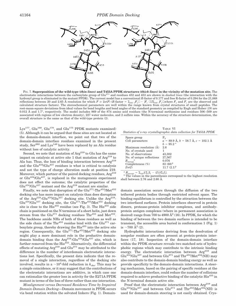
FIG. 7. Superposition of the wild-type (thin lines) and T453A PPDK structures (thick lines) in the vicinity of the mutation site. Theelectrostatic interactions between the carboxylate group of Glu271 and residues 452 and 453 are shown in dashed lines (the interaction with thehydroxyl group is eliminated in the mutant PPDK). The current model has a conventional R-factor of 0.177 and free R-factor of 0.294 for the 21,068reflections between 20 and 2.65 Å resolution for which F $ 2s(F) (R-factor 5 Shkl uuFo 2 uFc /Shkl uFo,where Fo and Fc are the observed andcalculated structure factors). The stereochemical parameters are well within the range known from crystal structures of small peptides. Theroot-mean-square deviations from ideal values for bond lengths and bond angles of the standard geometry as compiled by Engh and Huber (19) are0.012 Å and 1.7°, respectively. The model includes 869 of the 874 amino acid residues (the N-terminal methionine and residues 506–509 areassociated with regions of low electron density), 227 water molecules, and 2 sulfate ions. Within the accuracy of the structure determination, theoverall structure is the same as that of the wild-type protein (2).
TABLE VIStatistics of x-ray crystallographic data collection for T453A PPDK
Space group P2Cell parameters a 5 89.9 Å, b 5 58.7 Å, c 5 102.1 Å
b 5 95.2 °Maximum resolution (Å) 2.6No. of crystals used 1No. of observations 43,302No. of unique reflections 27,567Rmerge
a 0.076Completeness (%) 83 (68)b
I/s(I) 11.7 (2.1)b
a Rmerge 5 Shkl[(SiuIl 2 ^I&u)/SiIi].b The values in the parentheses correspond to the highest resolution
shell between 2.76 and 2.60 Å.
PPDK Domain Docking41164
tallization experiments of these mutant enzymes, which follow-ing structure determination would reveal if the relative orien-tation of the two domains has been altered, have beenunsuccessful. Thus far, only the x-ray structure of the T453Amutant has been determined (see Fig. 7, Table VI, and “Exper-imental Procedures” for details). Within the accuracy of thestructure determination, the overall structure is the same asthat of the wild-type protein (2). The Ca atomic coordinates ofthe mutant and wild-type molecules superimpose with a root-mean-square deviation of 0.5 Å. The electron density map atthe mutation site is consistent with the replacement of a thre-onine by an alanine residue at position 453. The interactionbetween the side chain of Glu271 and the hydroxyl group ofThr453 is therefore eliminated, but the remaining two interac-tions with the two NH groups of residues 452 and 453 aremaintained (Fig. 7). In addition, the side chains of Met452 andLeu369 shift concertedly so that the Met452 side chain occupiesspace that was partially occupied in the wild-type protein bythe methyl group of Thr453, and the Leu396 side chain shiftstoward the position occupied by Met452 in the wild-type protein.These changes are compensatory and therefore are not ex-pected to impact significantly on the catalytic efficiency of theenzyme. Thus, although the structure of T453A PPDK providesevidence that the reduction in catalytic efficiency is not causedby alteration in active site conformation, it does not provideany indication of misalignment of the domain-domain surfaces.This static picture may or may not provide an accurate view ofthe dynamics between protein species in solution. The absenceof a particular conformer in the crystal does not mean that it isalso absent in solution.
CONCLUSIONS
The current model of PPDK catalysis involves the swivel-type movement of a central, phosphoryl carrier domain be-tween the ATP/Pi active site of the N-terminal domain and thepyruvate active site of the C-terminal domain. We hypothesize
that the stringently conserved residues at the respective do-main-domain interfaces play an active role in guiding the cat-alytic His455 of the central domain into the active sites andholding it there long enough for catalysis to be completed. Theloss of catalytic activity observed for the mutants examined inthe present study supports the proposed roles of the N-terminalresidues Arg219 and Glu271 in the productive binding of thecentral domain for catalysis of the ATP/Pi partial reaction.
REFERENCES
1. Wood, H. G., O’Brien, W. E., and Micheales, G. (1977) Adv. Enzymol. Relat.Areas Mol. Biol. 45, 85–155
2. Herzberg, O., Chen, C. C., Kapadia, G., McGuire, M., Carroll, L. J., Noh, S. J.,and Dunaway-Mariano, D. (1996) Proc. Natl. Acad. Sci. U. S. A. 93,2652–2657
3. Carroll, L. J., Mehl, A. F., and Dunaway-Mariano, D. (1989) J. Am. Chem. Soc.111, 5965–5967
4. McGuire, M., Huang, K., Kapadia, G., Herzberg, O., and Dunaway-Mariano, D.(1998) Biochemistry 37, 13463–13474
5. McGuire, M., Carroll, L. J., Yankie, L., Thrall, S. H., Dunaway-Mariano, D.,Herzberg, O., Jayaram, B., and Haley, B. H. (1996) Biochemistry 35,8544–8552
6. Xu, Y., Yankie, L., Shen, L., Jung, Y. S., Mariano, P. S., Dunaway-Mariano, D.,and Martin, B. M. (1995) Biochemistry 34, 2181–2187
7. Yankie, L., Xu, Y., and Dunaway-Mariano, D. (1995) Biochemistry 34,2188–2194
8. Mehl, A., Xu, Y., and Dunaway-Mariano, D. (1994) Biochemistry 33,1093–1102
9. Pocalyko, D. J., Carroll, L. J., Martin, B. M., Babbitt, P. C., and Dunaway-Mariano, D. (1990) Biochemistry 29, 10757–10765
10. Wang, H. C., Ciskanik, L., Dunaway-Mariano, D., von der Saal, W., andVillafranca, J. J. (1988) Biochemistry 27, 625–633
11. Cleland, W. W. (1979) Methods Enzymol. 63, 500–51312. Howard, A. J., Gilliland, G. L., Finzel, B. C., Poulos, T. L., Ohlendorf, D. H.,
and Salemme, F. R. (1987) J. Appl. Crystallogr. 20, 383–38713. Brunger, A. T. (1992) X-PLOR Version 3.1: A System for X-ray Crystallography
and NMR, Yale University, New Haven, CT14. Roussel, A., and Cambillu, C. (1989) TURBO FRODO; Silicon Graphics
Geometry Partner Directory, Silicon Graphics, Mountain View, CA15. Deleted in proof16. Stites, W. E. (1997) Chem. Rev. 97, 1233–125017. Privalov, P. L., and Gill, S. J. (1988) Adv. Protein Chem. 39, 191–23418. Spolar, R. S., Ha, J. H., and Record, M. T. (1989) Proc. Natl. Acad. Sci. U. S. A.
86, 8382–838519. Engh, R. A., and Huber, R. (1991) Acta Crystallogr. Sect. A 47, 392–400
PPDK Domain Docking 41165