Safeguarding the global heparin supply chain: Bovine Heparin ...
Harris, L. F. and Killard, A. (2012) Heparin monitoring: From bloodtube to microfluidic device. In: Piyathilake, D. E. and Liang, R., eds.(2012) Heparin, properties, uses and side effects. New York: NovaScience Publishers. ISBN 9781621004318 Available from: http://eprints.uwe.ac.uk/17673
We recommend you cite the published version.The publisher’s URL is:https://www.novapublishers.com/catalog/
Refereed: Yes
(no note)
Disclaimer
UWE has obtained warranties from all depositors as to their title in the materialdeposited and as to their right to deposit such material.
UWE makes no representation or warranties of commercial utility, title, or fit-ness for a particular purpose or any other warranty, express or implied in respectof any material deposited.
UWE makes no representation that the use of the materials will not infringeany patent, copyright, trademark or other property or proprietary rights.
UWE accepts no liability for any infringement of intellectual property rightsin any material deposited but will remove such material from public view pend-ing investigation in the event of an allegation of any such infringement.
PLEASE SCROLL DOWN FOR TEXT.
In: Heparin: Properties, Uses and Side Effects ISBN 978-1-62100-431-8
Editors: D. E. Piyathilake, Rh. Liang, pp. © 2011 Nova Science Publishers, Inc.
Chapter IV
Heparin Monitoring: From Blood Tube
to Microfluidic Device
Leanne F. Harrisa and Anthony J. Killard
a,b,*
aBiomedical Diagnostics Institute, National Centre for Sensor Research, Dublin City
University, Glasnevin, Dublin 9, Ireland bDepartment of Applied Sciences, University of the West of England, Frenchay Campus,
Coldharbour Lane, Bristol BS16 1QY, UK
Abstract
Heparin anticoagulant therapy has been pivotal in both the treatment and prophylaxis
of thrombotic disease for many decades. It remains standard practice to monitor
unfractionated heparin (UFH) therapy due to its unpredictable pharmacokinetics. The
advent of low molecular weight heparins (LMWHs) reduced the need for continuous
laboratory monitoring due to the improved dose-response relationships and
pharmacokinetics of these drugs. However, special patient cohorts exist where monitoring
becomes essential irrespective of the drug being administered.
The standard assays used for heparin (UFH and LMWH) monitoring include the
activated partial thromboplastin time (aPTT), activated clotting time (ACT), thrombin
time (TT), and the anti-Xa assay. Clot-based assays such as the aPTT, ACT, and TT,
comprise some of the more traditional assays that are employed in the haemostasis
laboratory. The anti-Xa assay is a chromogenic assay more commonly used for
monitoring patients on LMWH therapy.
Over the last few years, significant efforts have been made towards point of care
testing (POCT) which offers greater ease of use, convenience, efficiency, and faster
turnaround times than laboratory-based tests. POCT, as its name suggests describes
testing that can be performed near or beside the patient, be it in a primary care facility
* Corresponding Author: Prof. Anthony J. Killard. Department of Applied Sciences, University of the West of
England, Frenchay Campus, Coldharbour Lane, Bristol BS16 1QY, UK. Tel: + 00 44 1173282147; Fax: + 00
44 1173442904; E-mail: [email protected].
Leanne F. Harris and Anthony J. Killard 2
such as a doctor’s surgery, the operating theatre, the emergency room or even in the
home.
While many point of care (POC) coagulation assays are available on the market,
there is a certain degree of reticence among the medical community in their uptake, as
these technologies compete with conventional laboratory testing, accompanied by reports
of poor correlations between the two systems. The popularity of these devices remains
controversial as they can face major challenges in the areas of regulatory compliance,
quality control, and financial cost.
Many POC technologies are commercially available for coagulation tests. For
heparin monitoring in particular, the devices available, e.g., Hemochron® and i-STAT®,
can perform tests such as aPTT, ACT, and TT. While current POC devices for measuring
heparin are suitable for use in the hospital setting rather than in the home, the POC
technologies of the future will need to encompass all patient settings. The future of
coagulation testing could see a move away from the more traditional clot-based tests
towards more modern analytical technologies, with a knock-on effect of improved assay
variability, precision, and reliability. Such developments can only improve medical
outcomes associated with heparin testing.
Introduction
Cardiovascular and thrombotic disease are leading causes of morbidity and mortality
worldwide and are the most common causes of death in the Western world. The incidence of
cardiovascular disease varies significantly both geographically and ethnically [1]. Arterial
disease or arteriosclerosis, hardening of the artery walls, underlies the development of
cardiovascular disease which often manifests itself more commonly as coronary heart disease
(CHD) or stroke. While anticoagulants can be used to prevent the occurrence of a thrombotic
event, over- or under-dosing can lead to excessive bleeding or severe clotting respectively,
and hence the need to closely link treatment with monitoring. Monitoring of anticoagulants
such as heparin has long been established in the central haematology laboratory, with many
advances in testing over the years, including the shift towards point of care testing (POCT)
which could transform the future of anticoagulant monitoring. This chapter will review
heparins and the evolution of its associated anticoagulant monitoring from the central
laboratory through to the point of care (POC).
Unfractionated Heparin
Unfractionated heparin (UFH) has dominated the anticoagulant market for over half a
century in the treatment and prophylaxis of thrombotic disorders. It was first discovered by
Jay McLean and William Henry Howell in the early 20th
century, that heparin had
antithrombotic properties but exerted its effect indirectly on thrombin and factor Xa (FXa),
through its interaction with a plasma co-factor called antithrombin (AT), which was
subsequently discovered in the 1960s. Since then UFH has become one of the most widely
used parenteral drugs worldwide [2].
Heparin Monitoring 3
UFH is a heterogenous mixture of glycosaminoglycans with molecular weights ranging
from 3,000 to 30,000 Da and a mean molecular weight of 15,000 Da, with commercial
preparations usually ovine, bovine, or porcine in origin. This variability in molecular weight
often translates into a variable therapeutic effect as a result of the variation in metabolic
clearance and the inhibition of both FXa and thrombin. Heparin also has a tendency to bind to
other biological artefacts including plasma proteins, endothelial cells, and macrophages,
rendering the dose-response a little difficult to predict [3].
The anticoagulant entity of UFH relies on the presence of a specific pentasaccharide
sequence, often referred to as the AT-binding domain, which has a strong affinity for AT,
thus accelerating its activity. This region comprises just one third of the heparin molecule, yet
it is responsible for its entire therapeutic effect. Binding of the pentasaccharide sequence of
UFH to AT causes a conformational change in its reactive centre and this accelerates its
interaction with FXa and thrombin. The heparin/AT complex can also inactivate factors IXa,
XIa and XIIa. However, thrombin and FXa are most sensitive to AT action, with thrombin
approximately 10-fold more sensitive to inhibition than FXa. UFH has an anti-FXa/anti-IIa
ratio of 1:1, with a biological half-life of 30 minutes. Heparin is administered subcutaneously
or intravenously and initial dosing is weight-based, which is also dependent upon the
thrombotic condition manifesting in the patient. Several advantages of UFH include easy
reversal in patients at risk of severe bleeding using protamine sulfate, a short half life of 1-2
hours after intravenous injection, and plasma clearance is not dependent upon renal excretion
[4].
Low Molecular Weight Heparin
Despite the widespread use of UFH, it is being rapidly replaced by low molecular weight
heparin (LMWH) as the treatment for choice for many indications. LMWHs are shorter chain
lengths of UFH achieved by chemical or enzymatic depolymerisation of the longer heparin
chain length and were first developed in the 1980s [3]. They usually range in molecular
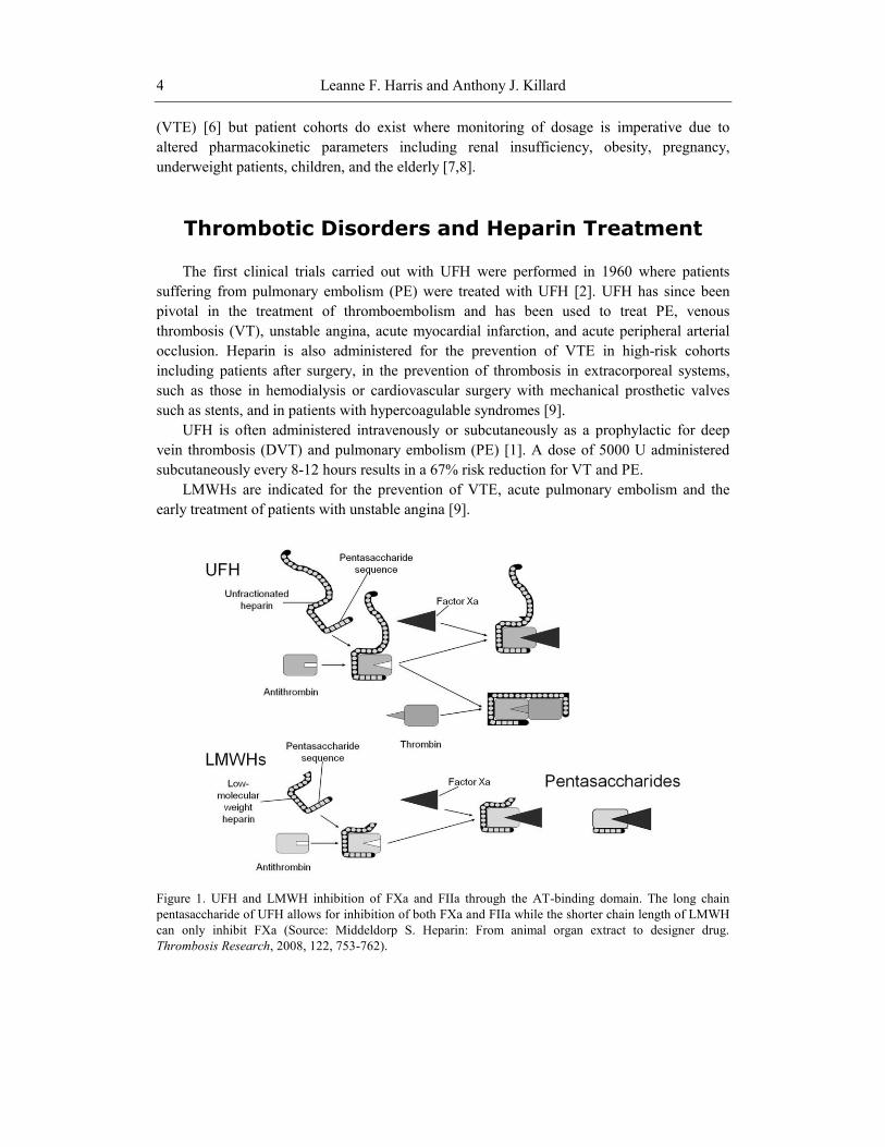
weight from 2,000 to 9,000 Da with a mean molecular weight of 4,500 Da. For thrombin
inactivation, a chain length of 18 saccharide units is essential for the formation of a ternary
heparin-AT-thrombin complex, which explains why there is less inhibitory action against
thrombin with LMWHs compared to UFH (Figure 1). LMWHs direct their inhibitory effect
primarily at FXa with less binding to plasma proteins, endothelial cells and platelets, resulting
in a more predictable dose-response relationship with improved pharmacokinetics.
Commercial preparations of LMWH usually have anti-FXa/anti-IIa ratios of between 2:1 and
4:1 as a result of their reduced ability to inhibit thrombin. The LMWH half-life is between 3
and 6 hours and administration is typically in fixed doses for thromboprophylaxis or in weight
adjusted doses for therapeutic effect. Associated with LMWH is a lower incidence of
heparin–induced thrombocytopenia (HIT) and heparin-induced osteoporosis [5].
While UFH has been used as an anticoagulant for many years, the advent of LMWH
brought with it greater bioavailability and a more predictable and a superior dose-response.
This improvement in predictability also implied a reduced need for laboratory monitoring in
clinically stable patients on LMWH therapy for the prevention of venous thromboembolism
Leanne F. Harris and Anthony J. Killard 4
(VTE) [6] but patient cohorts do exist where monitoring of dosage is imperative due to
altered pharmacokinetic parameters including renal insufficiency, obesity, pregnancy,
underweight patients, children, and the elderly [7,8].
Thrombotic Disorders and Heparin Treatment
The first clinical trials carried out with UFH were performed in 1960 where patients
suffering from pulmonary embolism (PE) were treated with UFH [2]. UFH has since been
pivotal in the treatment of thromboembolism and has been used to treat PE, venous
thrombosis (VT), unstable angina, acute myocardial infarction, and acute peripheral arterial
occlusion. Heparin is also administered for the prevention of VTE in high-risk cohorts
including patients after surgery, in the prevention of thrombosis in extracorporeal systems,
such as those in hemodialysis or cardiovascular surgery with mechanical prosthetic valves
such as stents, and in patients with hypercoagulable syndromes [9].
UFH is often administered intravenously or subcutaneously as a prophylactic for deep
vein thrombosis (DVT) and pulmonary embolism (PE) [1]. A dose of 5000 U administered
subcutaneously every 8-12 hours results in a 67% risk reduction for VT and PE.
LMWHs are indicated for the prevention of VTE, acute pulmonary embolism and the
early treatment of patients with unstable angina [9].
Figure 1. UFH and LMWH inhibition of FXa and FIIa through the AT-binding domain. The long chain
pentasaccharide of UFH allows for inhibition of both FXa and FIIa while the shorter chain length of LMWH
can only inhibit FXa (Source: Middeldorp S. Heparin: From animal organ extract to designer drug.
Thrombosis Research, 2008, 122, 753-762).
Heparin Monitoring 5
LMWH was assessed in the 1990s for the prevention of thrombosis in medium- and high-
risk surgical patients. Bedridden and accompanied by chronic debilitating diseases, the
susceptibility of these patients to adverse coagulation events was high. LMWH has impacted
the area of arterial thrombosis, whereby the subcutaneous administration of this anticoagulant
to patients with unstable angina was associated with a reduced rate of myocardial infarction,
emergency revascularisation, and death [5].
Heparin Monitoring
For the screening, diagnosis, and monitoring of haemostatic disorders, there is a wide
variety of tests at the disposal of the modern haematology laboratory that allow for accurate
patient diagnoses. The first tests developed for monitoring coagulation date back about 3000
years ago to China, where the length of time that blood flows from the skin after it has been
ruptured was recorded. It has been documented that this observation was subsequently made
by Hippocrates 2,300 years ago [10]. While the bleeding time was referred to in both the 16th
and 19th centuries, it was not until the 1900s when Duke and Ivy launched the standard
methods of bleeding time. It can be described as the time from incision on a patient’s forearm
until the time that bleeding ceases due to platelet plug formation [11].
The very first laboratory test of coagulation is often referred to as the manual ‘tilt tube’
clotting technique, where a blood sample is drawn by venipuncture and added to a glass test
tube which is tilted through 90° until a visible clot forms and the time recorded [12]. While
there are many automated tests of coagulation available, the manual ‘tilt tube’ technique is
still employed by some diagnostic laboratories as a result of sample to sample variability.
Coagulation monitoring assays have developed quite significantly since the introduction
of some of the first bleeding time tests, while retaining some of their basic assay principles.
The prothrombin time test (PT) was one such assay which progressed on from the classic ‘tilt
tube’ test and it remains one of the oldest tests of coagulation that is still performed. The PT
is used to monitor the extrinsic pathway, as the presence of tissue factor and phospholipids
allow it to mimic the effect of damaged tissue on the coagulation system. Internationally it is
used in the monitoring of vitamin K antagonists such as warfarin and not for heparin therapy.
The clotting tests outlined as follows were developed more specifically for heparin
monitoring.
Activated Partial Thromboplastin Time (aPTT)
The PT paved the way for the development of assays targeting the intrinsic pathway such
as the activated partial thromboplastin time (aPTT) developed in the 1960s by Proctor and
Rapaport [13]. Initially called the partial thromboplastin time (PTT) due to the presence of a
partial thromboplastin from rabbit brain, the aPTT was developed to include activators such
as kaolin, ellagic acid, and silica [14]. These are all negatively charged materials that
contribute to the surface-dependent activation of Hageman factor or factor XII. The aPTT is a
routine test used in the diagnosis and monitoring of heparin therapy which causes a
Leanne F. Harris and Anthony J. Killard 6
prolongation in the aPTT primarily due to thrombin inhibition [15]. The assay principle is
simple to follow in that the citrated plasma sample is incubated with an aPTT reagent, that
contains the surface activator of choice and phospholipids for 3 minutes at 37°C, which
allows for optimal activation of intrinsic pathway proteins. The recommended therapeutic
aPTT ratio range of 1.5-2.5 corresponds to a heparin level of 0.2-0.4 U/ml of blood or plasma
[9,16]. While there is widespread use of the aPTT internationally, the reagents and
instruments used in the determination of the aPTT have changed significantly over the last 25
years [3]. The existence of many companies specialising in the development and production
of haemostasis products translates into more than 300 laboratory methods currently in use
[17]. A distinct disadvantage of the aPTT is the loss of reproducibility across tests from
different manufacturers as well as lot-to-lot variation in test kits from the same manufacturer
[18]. The aPTT is also well-known for its variability in responsiveness as a result of
differences in reagent composition such as phospholipid or activator type, and
methodological differences such as the type of instrument/coagulometer used [15,19,20]. It is
recommended that the therapeutic range should be established for the particular reagent and
instrument being used in each laboratory [16]. The aPTT is not suitable for monitoring
LMWH therapy due to the inability of the short chain LMWH to bridge thrombin. LMWH
activity is primarily directed at FXa inhibition [15].
Activated Clotting Time (ACT) Test
Developed by Hattersley in 1966, the activated clotting time (ACT) test was the very first
bedside test to monitor coagulation during cardiopulmonary bypass (CPB) [21]. Hattersley
believed that the sensitivity of coagulation assays could be enhanced through the elimination
or shortening of the contact activation time. The initial assay involved drawing 1 ml of blood
by venipuncture into an evacuated tube which is replaced with a diatomite tube prewarmed to
37°C. The tube was inverted to mix and placed in a 37°C incubator. At one minute and at 5
second intervals thereafter, the tube was removed and tilted to spread the blood along its
length until the formation of the first visible clot could be detected [21]. When used for
monitoring high doses of heparin, the reference value for ACT ranges between 70 and 180
seconds. However, these values are subject to change based on the clinical indication and the
test method used [22,23]. While commonly used and widely accepted as a method for heparin
monitoring during CPB and PCI, it is also well known that the ACT does not correlate well
with the anti-Xa measures of heparin therapy [24]. The ACT is a global whole blood
coagulation test and as a result is affected by a number of different factors including platelets,
AT, hemodilution, and hypothermia [25]. In a study on the assessment of heparin
anticoagulation it was shown that due to the CPB-related effects of hemodilution and
hypothermia, the ACT was a poor test for heparin monitoring. It is also deemed an unsuitable
monitoring tool for heparin due to its poor correlation with anti-Xa assays, which are plasma
based tests and remain unaffected by the factors previously outlined [25,26].
Heparin Monitoring 7
Thrombin Time (TT) and High Dose Thrombin Time (HiTT)
The thrombin time test (TT) or the thrombin clotting time (TCT) as it is also known, is
primarily used in the hospital setting for monitoring critically ill patients as it allows for the
generation of rapid test results at the bedside [27]. The TT is one of the more procedurally
simple clotting assays to perform, measuring the rate of conversion of fibrinogen to fibrin,
after a plasma sample is incubated with an equal volume of thrombin. The time to clot
formation is thus recorded as the TT. It is sensitive to inhibitors such as heparin resulting in a
prolonged TT in patients on heparin therapy. One observation with the TT in heparin
monitoring was the lack of linearity with increasing heparin concentration resulting in assays
that cannot clot at high heparin doses [16]. Hence the high dose thrombin time (HiTT) was
developed which is sensitive to heparin doses beyond the upper limit of the therapeutic range.
It also overcomes some of the shortcomings of the ACT. It incorporates a large dose of
thrombin into a test tube to bind to a large proportion of the heparin-AT complexes that form
during heparinisation. The unbound heparin prolongs the time it takes for fibrin to form and is
measured as the HiTT, which decreases with CPB [24].
Protamine Sulfate Titration
Another assay that has been developed for measuring heparin dose is the protamine
sulfate titration assay. Protamine in excess prevents clot formation and a deficit of protamine
cannot neutralise heparin in the sample. However these assays are not widely available and
not routinely employed in the diagnostic laboratory [16,28].
Anti-Factor Xa Assays
With the advent of peptide substrates specific for coagulation proteins in the 1960s,
researchers became interested in alternative methods of coagulation testing, such as optical
measurement techniques, as opposed to the standard clot-based methodologies. It was at the
Karolinska Institute in Stockholm, that a small peptide - Bz-Phe-Val-Arg-OMe - was shown
to have a very high affinity for thrombin resulting in the synthesis of S-2160, the very first
chromogenic substrate for use in haemostasis research [29]. While the diagnostic laboratories
did not adapt well to the chromogenic versions of the PT and aPTT, the chromogenic
methods for AT and heparin did gain widespread acceptance. An assay with a colorimetric
endpoint is appealing as it is not affected by the many biological variables that interfere with
standard clot-based assays, such as the aPTT which can be prolonged by inherited or acquired
coagulation factor deficiencies such as factors XII, XI, IX, VIII, V and II [22,23]. Such
colorimetric tests are also in widespread use on central hospital laboratory analysers and so
can be readily adapted to operate on them.
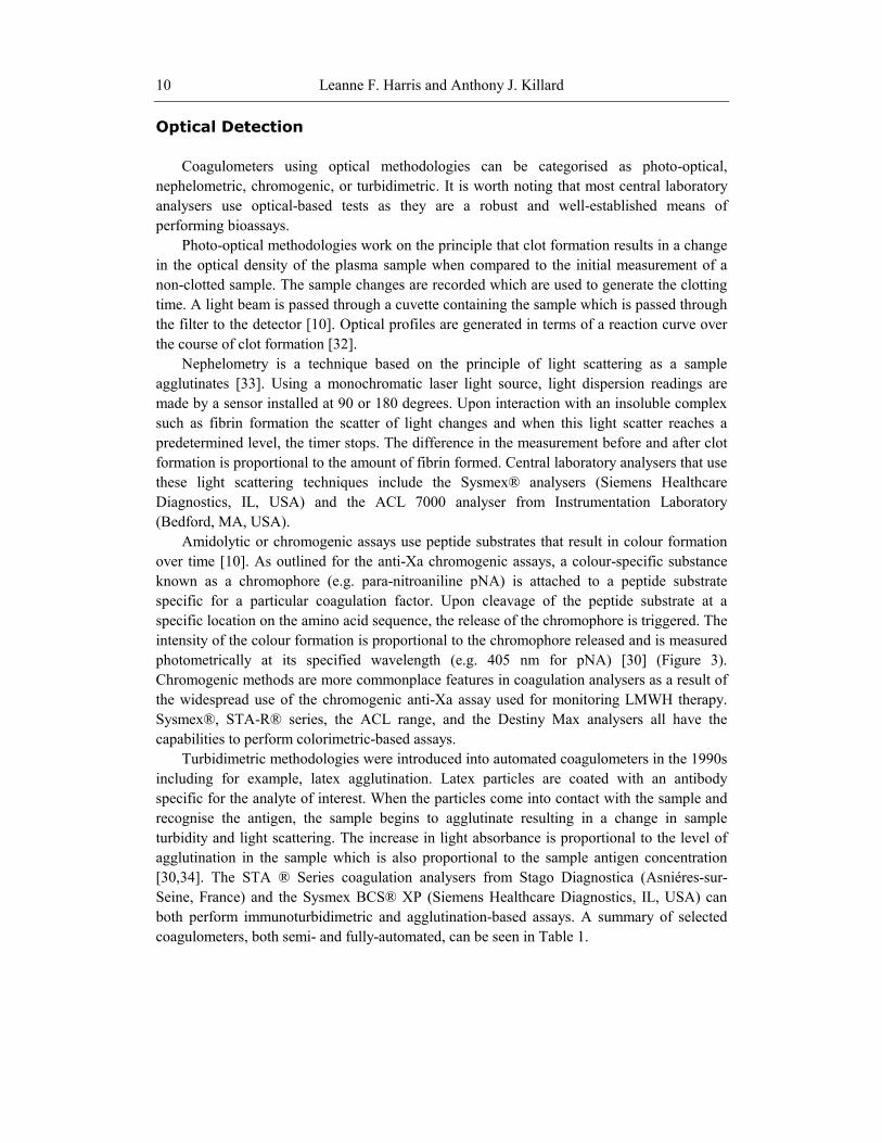
The anti-Xa assay is one such assay that is used to measure heparin therapy. The assay is
comprised of a FXa specific peptide substrate to which a chromophore is attached. Excess
FXa is added to the heparinised plasma sample which is incubated to allow the formation of
Leanne F. Harris and Anthony J. Killard 8
the heparin-AT-FXa complex. As FXa cleaves the substrate, the chromophore is released and
the resulting colour formation is recorded using a spectrophotometer. The rate of colour
formation is directly proportional to the free FXa in the sample and thus indirectly
proportional to the amount of heparin in the sample. Using standard curves of known heparin
concentration, the sample concentration can be extrapolated [22]. The anti-FXa assay is
currently the ‘gold standard’ method for monitoring patients on LMWH therapy.
Principles of Automated Laboratory Testing
for Heparin
The following section will be divided into methods of detection used in central laboratory
analysers and will incorporate more specific examples of the various types of technology
available commercially.
The early manual and visual tests of coagulation have long been replaced by central
diagnostic technologies which allow for fully automated, accurate, and precise high
throughput coagulation testing [10]. The 1970s saw the transition from tilt-tube testing to the
use of semi-automated equipment that used photometric and mechanical methods for the
detection of fibrin [30]. Fully automated coagulometers, using mechanical and optical
detection, are now available that allow for the automated performance of coagulation tests
including clot-based, chromogenic, and immunological assays. Often these analytical
methods can be performed simultaneously and the same assay can be carried out using two
different methods. Automation has led to improved reproducibility and standardisation,
increased user flexibility and faster sample processing times, in addition to reductions in
reagent and sample costs [31].
Mechanical Detection
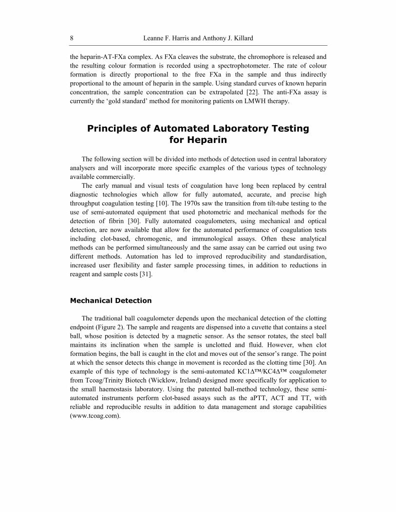
The traditional ball coagulometer depends upon the mechanical detection of the clotting
endpoint (Figure 2). The sample and reagents are dispensed into a cuvette that contains a steel
ball, whose position is detected by a magnetic sensor. As the sensor rotates, the steel ball
maintains its inclination when the sample is unclotted and fluid. However, when clot
formation begins, the ball is caught in the clot and moves out of the sensor’s range. The point
at which the sensor detects this change in movement is recorded as the clotting time [30]. An
example of this type of technology is the semi-automated KC1∆™/KC4∆™ coagulometer
from Tcoag/Trinity Biotech (Wicklow, Ireland) designed more specifically for application to
the small haemostasis laboratory. Using the patented ball-method technology, these semi-
automated instruments perform clot-based assays such as the aPTT, ACT and TT, with
reliable and reproducible results in addition to data management and storage capabilities
(www.tcoag.com).
Heparin Monitoring 9
Figure 2. Patented ball technology: mechanical detection of clot formation as used in the KC1∆™/KC4∆™
coagulometer. The movement of the metal ball is impeded by the formation of the clot which is returned as
the clotting time (Source: Tcoag KC4 Delta Semi aAutomated Coagulation Analyser, User Manual).
Mechanical clot detection using a steel rod as opposed to a steel ball is also a common
method of detection. This type of mechanical detection is used in Instrumentation
Laboratory’s (Bedford, MA, USA) extensive range of ACL coagulometers (ACL Elite Pro,
ACL AcuStar™, ACL™ Advance, ACL 700, ACL 200 and the CT analyser for the
execution of aPTT and TT clot-based assays (www.instrumentationlaboratory.com).
Helena Laboratories (Texas, USA) developed the Cascade M and Cascade M-4
coagulation analysers which are semi-automated and allow for low volume testing with
routine clotting assays in small haemostasis laboratories. Both the aPTT and TCT can be
measured using the Cascade system (www.helena.com). As it is a small semi-automated
benchtop analyser that can be used for most routine coagulation assays, it is often referred to
as a POC instrument.
The Destiny Max and Destiny Plus haemostasis analysers from Tcoag (Wicklow, Ireland)
are high throughput analysers that use micro-mechanical clot detection for aPTT and TT
monitoring of heparin (www.tcoag.com).
Other commercial coagulometers that perform clot-based assays using mechanical
detection methodologies, include the STA® Series from Diagnostica Stago and the Sysmex®
CA from Dade Behring (Siemens Healthcare Diagnostics, IL, USA). Modern coagulometers
now interface with computerised data processing units that allow for storage of calibration
curves and generated data, in addition to integrated quality control programmes that allow for
alignment with international standards [31]. With mechanical detection the sample can
comprise any form, including platelet poor plasma (PPP), platelet rich plasma (PRP), or
whole blood (WB).
Leanne F. Harris and Anthony J. Killard 10
Optical Detection
Coagulometers using optical methodologies can be categorised as photo-optical,
nephelometric, chromogenic, or turbidimetric. It is worth noting that most central laboratory
analysers use optical-based tests as they are a robust and well-established means of
performing bioassays.
Photo-optical methodologies work on the principle that clot formation results in a change
in the optical density of the plasma sample when compared to the initial measurement of a
non-clotted sample. The sample changes are recorded which are used to generate the clotting
time. A light beam is passed through a cuvette containing the sample which is passed through
the filter to the detector [10]. Optical profiles are generated in terms of a reaction curve over
the course of clot formation [32].
Nephelometry is a technique based on the principle of light scattering as a sample
agglutinates [33]. Using a monochromatic laser light source, light dispersion readings are
made by a sensor installed at 90 or 180 degrees. Upon interaction with an insoluble complex
such as fibrin formation the scatter of light changes and when this light scatter reaches a
predetermined level, the timer stops. The difference in the measurement before and after clot
formation is proportional to the amount of fibrin formed. Central laboratory analysers that use
these light scattering techniques include the Sysmex® analysers (Siemens Healthcare
Diagnostics, IL, USA) and the ACL 7000 analyser from Instrumentation Laboratory
(Bedford, MA, USA).
Amidolytic or chromogenic assays use peptide substrates that result in colour formation
over time [10]. As outlined for the anti-Xa chromogenic assays, a colour-specific substance
known as a chromophore (e.g. para-nitroaniline pNA) is attached to a peptide substrate
specific for a particular coagulation factor. Upon cleavage of the peptide substrate at a
specific location on the amino acid sequence, the release of the chromophore is triggered. The
intensity of the colour formation is proportional to the chromophore released and is measured
photometrically at its specified wavelength (e.g. 405 nm for pNA) [30] (Figure 3).
Chromogenic methods are more commonplace features in coagulation analysers as a result of
the widespread use of the chromogenic anti-Xa assay used for monitoring LMWH therapy.
Sysmex®, STA-R® series, the ACL range, and the Destiny Max analysers all have the
capabilities to perform colorimetric-based assays.
Turbidimetric methodologies were introduced into automated coagulometers in the 1990s
including for example, latex agglutination. Latex particles are coated with an antibody
specific for the analyte of interest. When the particles come into contact with the sample and
recognise the antigen, the sample begins to agglutinate resulting in a change in sample
turbidity and light scattering. The increase in light absorbance is proportional to the level of
agglutination in the sample which is also proportional to the sample antigen concentration
[30,34]. The STA ® Series coagulation analysers from Stago Diagnostica (Asniéres-sur-
Seine, France) and the Sysmex BCS® XP (Siemens Healthcare Diagnostics, IL, USA) can
both perform immunoturbidimetric and agglutination-based assays. A summary of selected
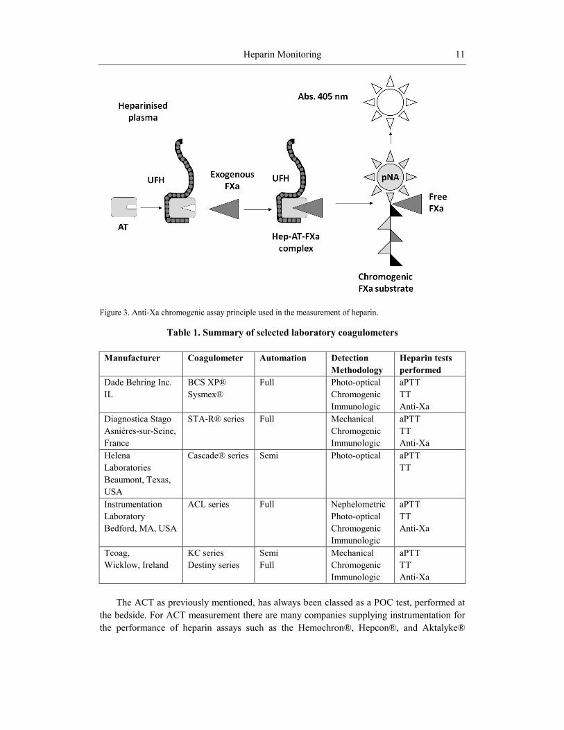
coagulometers, both semi- and fully-automated, can be seen in Table 1.
Heparin Monitoring 11
Figure 3. Anti-Xa chromogenic assay principle used in the measurement of heparin.
Table 1. Summary of selected laboratory coagulometers
Manufacturer Coagulometer Automation Detection
Methodology
Heparin tests
performed
Dade Behring Inc.
IL
BCS XP®
Sysmex®
Full Photo-optical
Chromogenic
Immunologic
aPTT
TT
Anti-Xa
Diagnostica Stago
Asniéres-sur-Seine,
France
STA-R® series Full Mechanical
Chromogenic
Immunologic
aPTT
TT
Anti-Xa
Helena
Laboratories
Beaumont, Texas,
USA
Cascade® series Semi Photo-optical
aPTT
TT
Instrumentation
Laboratory
Bedford, MA, USA
ACL series Full Nephelometric
Photo-optical
Chromogenic
Immunologic
aPTT
TT
Anti-Xa
Tcoag,
Wicklow, Ireland
KC series
Destiny series
Semi
Full
Mechanical
Chromogenic
Immunologic
aPTT
TT
Anti-Xa
The ACT as previously mentioned, has always been classed as a POC test, performed at
the bedside. For ACT measurement there are many companies supplying instrumentation for
the performance of heparin assays such as the Hemochron®, Hepcon®, and Aktalyke®
Leanne F. Harris and Anthony J. Killard 12
systems, which will be discussed in the next section on near-patient testing. However some
haemostasis companies such as Instrumentation Laboratory, manufacture reagents for
measuring clot-based ACT assays which can be used in many automated analysers.
Point of Care Testing
Often referred to as near patient testing or bedside testing, POCT is essentially a
technology that allows for the provision of a test, near to the patient, that can generate rapid
results, ultimately leading to improvements in healthcare [35]. POCT has been developed for
use in the home, the workplace, the doctor’s surgery, the pharmacy, as well as in the hospital
including the emergency room (ER) or operating theatre (OR) and the intensive care unit
(ICU). These tests are also suitable for use in mobile hospitals, ambulances, in military
settings, even space shuttle expeditions and also in the developing world where specialised
laboratories may not be available [36].
POCT offers many advantages over traditional diagnostics technologies including rapid
turnaround times, user-friendly technology which does not require trained personnel, smaller
sample volumes, and less invasive sampling techniques. A significant advantage of POCT is
improved patient compliance. The ability to routinely monitor and self-test resulting in the
attainment of knowledge about one’s medical status has been linked to health improvements.
It is thought that increased knowledge and education can improve adherence to treatment
[37]. In addition, wider economic benefits include reduced frequency of hospital visits and
length of hospital stay, improved drug treatment and patient management with the knock-on
effect of improved quality of life, reducing the economic burden [37]. Central laboratory
testing requires patient sampling, transportation and analysis which are all very time-
consuming and introduce a number of pre-analytical errors in measurement. With POCT,
immediate sample analysis can take place outside of the central laboratory, with a shorter time
to intervention, reduced pre-analytical errors and the potential to improve patient outcome. In
parallel, reductions in the burden of disease will automatically benefit the healthcare system
resulting in wider societal benefits.
Despite its many advantages, POCT essentially challenges conventional laboratory
testing and a major concern of the medical laboratory specialist in relation to patient self-
testing is the loss of control and quality that arises with POCT, such as the inaccurate
interpretation of results. Cost is another controversial area of debate with POC diagnostics,
but these issues also regularly arise with central laboratory diagnostics. While high initial
costs for new technologies are expected, it is predicted that the long term economic benefits
that arise from POCT will far outweigh the costs associated with central laboratory testing
[38,39]. While the adoption of POCT into the clinical arena has long been controversial, in
reality POCT has been reported to constitute 25% of total laboratory expenditure dollars
rendering it a critical component of laboratory medicine [40].
The main types of POC instruments include small benchtop analysers that are mostly
used in primary care settings and miniaturised handheld devices that were developed for use
directly by the patient, GP, or healthcare worker. Benchtop POC instruments were initially
developed based on central laboratory technologies but were miniaturised to allow adaptation

Heparin Monitoring 13
to smaller, more confined spaces such as the hospital ward, the doctor’s office, or outpatient
clinic [41]. These devices are less complex than standard laboratory instruments in that they
are simple to use, with a user-friendly interface, and the accompanying reagents are robust in
terms of storage and calibration. The portable handheld devices commonly associated with
POCT were initially developed for glucose, pregnancy, and urine testing. The first handheld
monitors for near patient testing were developed for measuring glucose in the 1980s [42].
Although developed primarily for hospital bedside use they have been used by diabetic
patients for self-monitoring of their glucose levels for many years. The handheld glucose
monitor progressed into a finger-stick capillary blood test which is now commonly used at the
bedside in hospital settings for adjusting insulin dosages with ease and rapidity.
Microfluidics in POCT
Microfluidics has been defined as the science and technology of systems that process or
manipulate small volumes of fluids using channels of micrometre dimensions [43].
Microfluidic devices can be assembled using a range of manufacturing techniques including
photolithography, hot-embossing, injection moulding, and laser/die cutting. Medical
diagnostics is a key application area for microfluidic technologies which has been maturing
over the last decade, incorporating more complex functions on-chip [44]. Technological
advances have greatly improved devices in terms of sample application, packaging,
interpretation of results, and disposable test cartridges. In comparison to traditional analysers,
the footprint of POC devices is significantly smaller, resulting in smaller sample and reagent
volumes which ultimately reduce cost [45].
POC Devices for Heparin Monitoring
While glucose and pregnancy testing are the current leaders in POCT, coagulation
monitoring is a close second due to the well-defined clinical need for anticoagulant
monitoring [41]. In the area of haemostasis testing, the PT test which is used for monitoring
warfarin is the only clotting test that is standardised across international laboratories,
correcting for differences in reagents. The International Normalised Ratio (INR) is used for
standardising PT tests at both the POC and in the central diagnostics laboratory. Other
haemostasis tests now available in POCT format include the ACT, aPTT and TT. The ACT,
developed as the first near-patient test during CPB is almost always used in POCT and not in
the laboratory setting [46]. While many of the initial POCT devices were developed for
monitoring ACT, these devices also have the capability to perform multiple clotting assays
due to clinical and technological advances, and also due to the fact that they are all based on
the measurement of clot formation [47,48]. Current POC devices for monitoring heparin use
established clot-based assays such as the aPTT and ACT. While the TT is used in the
laboratory setting for high dose heparin, few POC devices have this test available in their
repertoire of coagulation assays. The detection technologies used in central laboratory
analysers, such as optical, mechanical, and electrochemical techniques, have evolved to
Leanne F. Harris and Anthony J. Killard 14
enable the development of miniaturised POC devices, in parallel to advances in microfluidic
and microfabrication techniques. The following section will discuss detection technologies
currently used in POC devices that are available for heparin monitoring.
Optical Detection
Light Scattering
While optical detection in traditional coagulometers falls into four main categories
(photo-optical, nephelometric, chromogenic, and turbidimetric), the technologies that have
been adapted to POC devices differ according to the constraints of the device and are often
categorised under optical motion. Optical detection methodologies used in a miniaturised
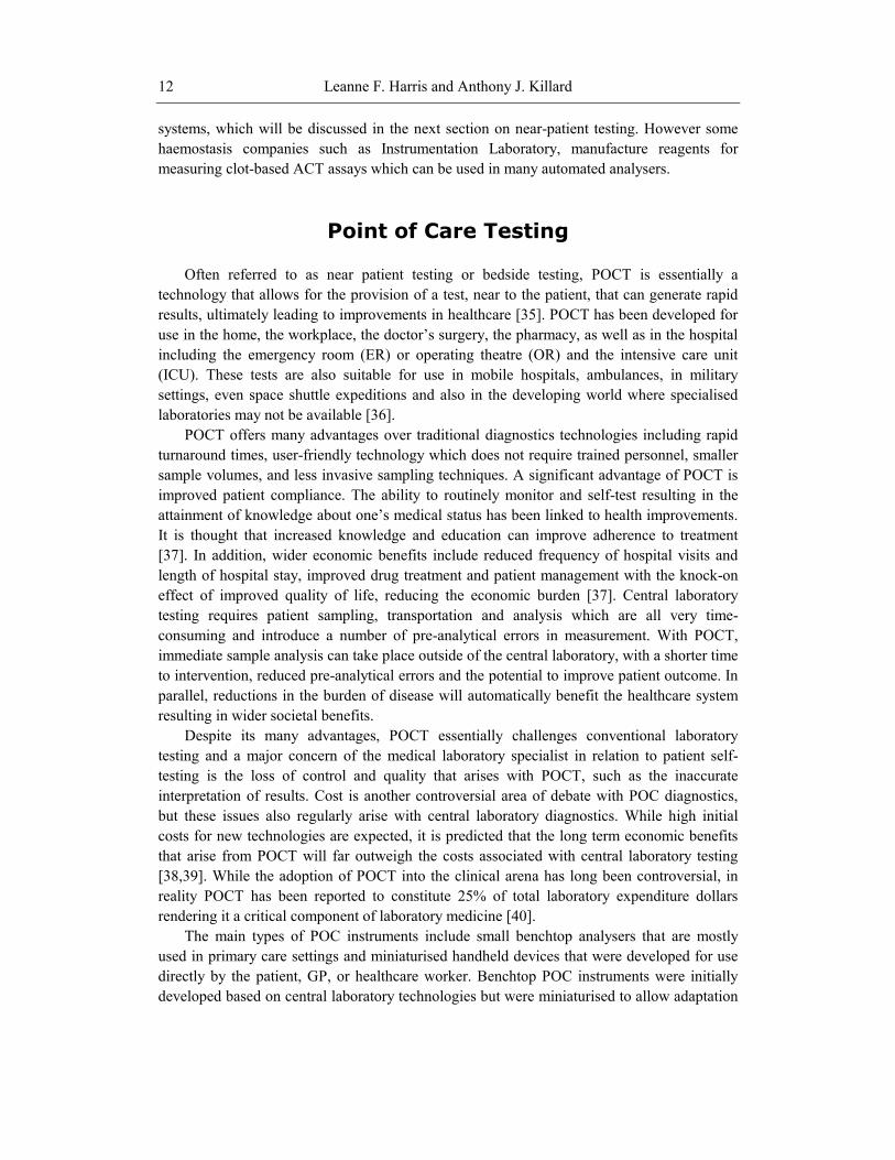
configuration often encompass the use of small optical detectors [45] (Figure 4).
Figure 4. Schematic of microfluidic cassette enclosed in a reader containing electronics and microoptical
detectors. The sample is applied to the sample port, undergoes immunochromatography in the nitrocellulose
capture areas, and a colour change is detected optically (Source: Pugia MJ; Price CP. Technology of
Handheld Devices for Point-of-Care Testing. In: Price CP, St John A, Hicks JM, editors. Point-of-Care
Testing. Washington, USA: American Association for Clinical Chemistry Inc.; 2004; 13-30).
As a clot develops it will occlude the light, blocking the detector which is then recorded as
the clotting time. One example of this technology is the Coagucheck® Plus/XS Pro (Roche
Diagnostics, IN, USA) which was developed developed for use with whole blood. The
disposable microfluidic cartridge contains a celite reagent to trigger clotting and as the sample
Heparin Monitoring 15
is applied and interacts with the activator the blood clots, and speckle detection technology is
used to measure the aPTT and ACT.
Optical-Mechanical
In some POC devices both optical and mechanical detection technologies are combined,
whereby a change in the physical movement of the sample as it clots, is detected optically.
The Hemochron® Whole Blood Coagulation System (ITC Nexus, NJ, USA) was developed
based on opto-mechanical detection technologies and can be divided into two systems, the
tube family and the cuvette family. The Hemochron® cuvette technologies comprise both the
Signature Elite™ and the Signature Plus™ whole blood microcoagulation systems. The
cuvette contains the activator (celite, silica or kaolin) for each specific test and as the sample
is applied and drawn along the channel mechanically, it interacts with the activator which
triggers coagulation. The blood sample is moved back and forth and as the clot forms the
movement is impeded which is monitored by two LED optical detectors aligned in the test
channel of the cuvette. A clot endpoint is reached when the speed of movement falls below a
predetermined rate and a clotting time is displayed on the instrument.
Another example of the cuvette technology is the GEM® analyser (Instrumentation
Laboratory, MA, USA) which is a portable whole blood coagulation system that can measure
aPTT, ACT, and ACT-LR. This small bench-top device uses cartridges that are preloaded
with activating reagents (kaolin, celite, silica), a small blood sample is applied and the change
in movement along the cuvette with clot formation is detected optically and reported as a
clotting time (www.instrumentationlaboratory.com).
The Hemochron® tube-based anticoagulation monitoring technology is the gold standard
for ACT testing and also uses opto-mechanical techniques for clotting time measurements
similar to the cuvette system. A magnet is located in the tube with the activating reagents,
usually celite or kaolin plus phospholipids. When the sample has been added to the tube, it is
placed in the instrument which triggers a timer. The principle of optical motion is used to
measure clotting time, whereby the time to clot formation is measured by the ability of a
magnet to move out of alignment stopping the timer [45].
Another technique that combines both mechanical and optical technologies is the
mechanical plunger-flag assembly that is inserted into and out of a blood sample, with the
time taken to move the plunger through the sample being optically detected. As the clot forms
the time it takes to drop the plunger into the sample changes and hits a pre-defined threshold
which is translated into a clotting time [48]. The Hepcon® HMS PLUS™ Haemostasis
Management System (Medtronic Inc, Minnesota, USA) is a POC device used for managing
individually targeted heparin levels and uses the mechanical plunger-flag method.
Paramagnetic-Optical
In addition to combined mechanical and optical detection techniques, optical detection
has also been incorporated into technologies that use magnetic iron particles to determine
clotting time. Paramagnetic iron oxide particles are present which move in response to the
oscillating magnetic field. As the sample is loaded and interacts with the dried activators,
coagulation begins causing a slowing down of particle movement. Using infrared detection
this technology can optically detect particle motion which senses changes in light
transmission [48]. The Rapidpoint® TAS from Bayer (NY, USA) is one such technology and
Leanne F. Harris and Anthony J. Killard 16
uses disposable test cards with test-specific reagents such as celite loaded onto the card and
can measure ACT, aPTT, and low heparin (LHMT).
Mechanical Detection
The most traditional method of clot detection involves the use of a sensor to detect the
movement of a ball in the bottom of the cuvette. As a clot forms in the cuvette the movement
of the ball is impeded and it becomes lodged at the edge of the cuvette. The Thrombotrack™
Solo from Axis-Shield (Norway) is a small bench-top instrument that uses this technology to
measure both TT and aPTT.
Electromechanical
Another principle of mechanical detection that also incorporates electronic and optical
mechnisms, is the plunger-flag technique where the rate of fall of a plunger into the blood
sample is recorded. As a clot forms, the fibrin mesh impedes the rate of fall through the
sample when compared to an unclotted sample. A photo-optical system can detect this change
in fall rate in each channel of the cartridge. The ACT PLUS™ (Medtronic Inc, Minnesota,
USA) is a microprocessor-controlled electromechanical instrument that can determine the
ACT in fresh and citrated whole blood samples using fibrin formation as the endpoint.
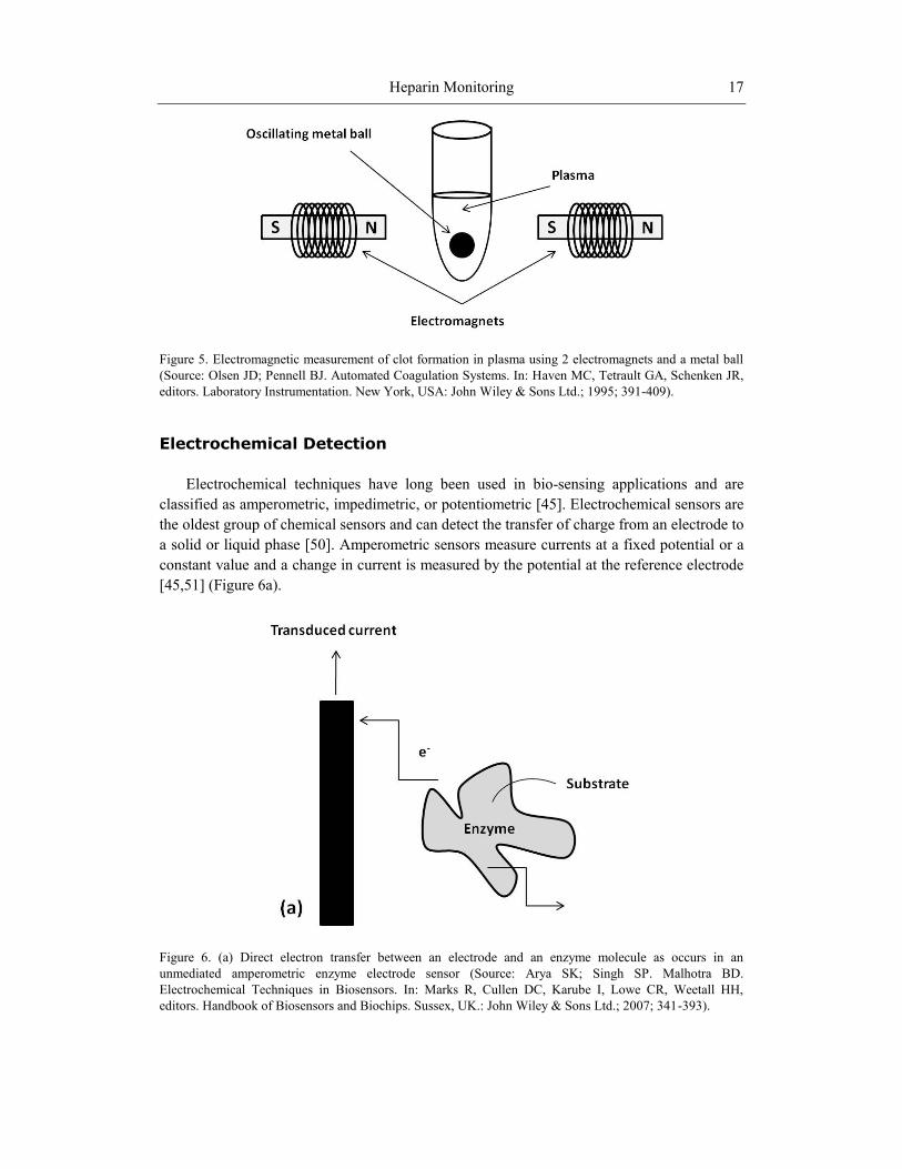
Electromagnetic
Electromagnetic clot detection uses a two-point detection mechanism, where two magnets
are positioned at 0° and 90°. As the magnet rotates through these positions, even early fibrin
formation can be detected [41,49]. In Figure 5 a plasma sample is mechanically rotated. A
metal ball is trapped by two magnets, but as clotting begins, the ball begins to rotate which is
recorded as the clotting time. The Actalyke® technology from Helena Laboratories (TX,
USA) uses this technique and is used primarily to measure the ACT. Heparinised samples
with an upper limit of 6 U/ml can be monitored with the Actalyke® MAX-ACT tubes.
Heparin Monitoring 17
Figure 5. Electromagnetic measurement of clot formation in plasma using 2 electromagnets and a metal ball
(Source: Olsen JD; Pennell BJ. Automated Coagulation Systems. In: Haven MC, Tetrault GA, Schenken JR,
editors. Laboratory Instrumentation. New York, USA: John Wiley & Sons Ltd.; 1995; 391-409).
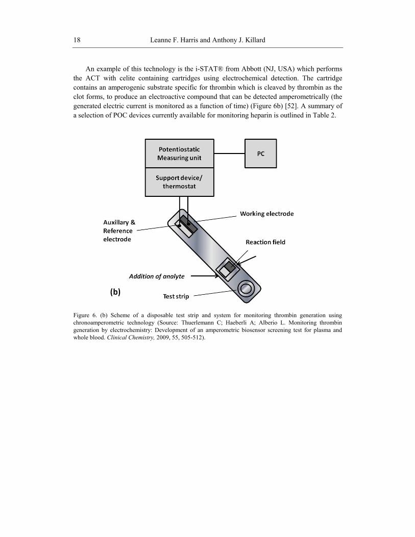
Electrochemical Detection
Electrochemical techniques have long been used in bio-sensing applications and are
classified as amperometric, impedimetric, or potentiometric [45]. Electrochemical sensors are
the oldest group of chemical sensors and can detect the transfer of charge from an electrode to
a solid or liquid phase [50]. Amperometric sensors measure currents at a fixed potential or a
constant value and a change in current is measured by the potential at the reference electrode
[45,51] (Figure 6a).
Figure 6. (a) Direct electron transfer between an electrode and an enzyme molecule as occurs in an
unmediated amperometric enzyme electrode sensor (Source: Arya SK; Singh SP. Malhotra BD.
Electrochemical Techniques in Biosensors. In: Marks R, Cullen DC, Karube I, Lowe CR, Weetall HH,
editors. Handbook of Biosensors and Biochips. Sussex, UK.: John Wiley & Sons Ltd.; 2007; 341-393).
Leanne F. Harris and Anthony J. Killard 18
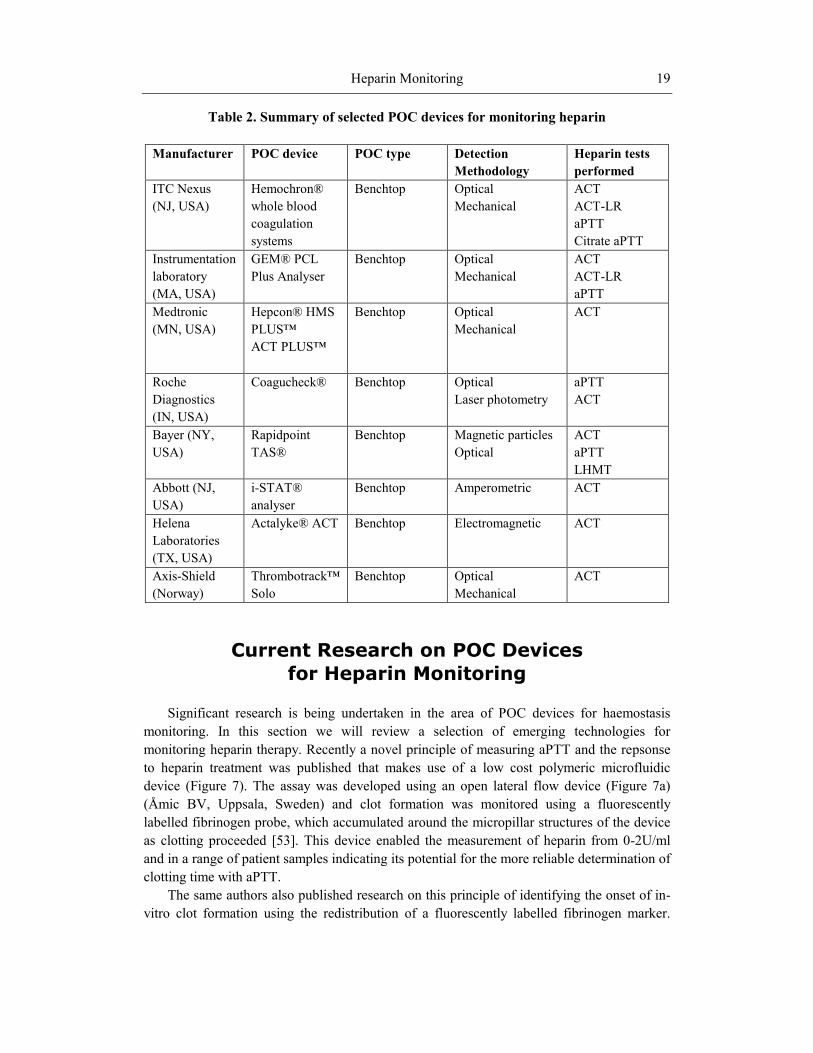
An example of this technology is the i-STAT® from Abbott (NJ, USA) which performs
the ACT with celite containing cartridges using electrochemical detection. The cartridge
contains an amperogenic substrate specific for thrombin which is cleaved by thrombin as the
clot forms, to produce an electroactive compound that can be detected amperometrically (the
generated electric current is monitored as a function of time) (Figure 6b) [52]. A summary of
a selection of POC devices currently available for monitoring heparin is outlined in Table 2.
Figure 6. (b) Scheme of a disposable test strip and system for monitoring thrombin generation using
chronoamperometric technology (Source: Thuerlemann C; Haeberli A; Alberio L. Monitoring thrombin
generation by electrochemistry: Development of an amperometric biosensor screening test for plasma and
whole blood. Clinical Chemistry, 2009, 55, 505-512).
Heparin Monitoring 19
Table 2. Summary of selected POC devices for monitoring heparin
Manufacturer POC device POC type Detection
Methodology
Heparin tests
performed
ITC Nexus
(NJ, USA)
Hemochron®
whole blood
coagulation
systems
Benchtop Optical
Mechanical
ACT
ACT-LR
aPTT
Citrate aPTT
Instrumentation
laboratory
(MA, USA)
GEM® PCL
Plus Analyser
Benchtop Optical
Mechanical
ACT
ACT-LR
aPTT
Medtronic
(MN, USA)
Hepcon® HMS
PLUS™
ACT PLUS™
Benchtop Optical
Mechanical
ACT
Roche
Diagnostics
(IN, USA)
Coagucheck® Benchtop Optical
Laser photometry
aPTT
ACT
Bayer (NY,
USA)
Rapidpoint
TAS®
Benchtop Magnetic particles
Optical
ACT
aPTT
LHMT
Abbott (NJ,
USA)
i-STAT®
analyser
Benchtop Amperometric ACT
Helena
Laboratories
(TX, USA)
Actalyke® ACT Benchtop Electromagnetic ACT
Axis-Shield
(Norway)
Thrombotrack™
Solo
Benchtop Optical
Mechanical
ACT
Current Research on POC Devices
for Heparin Monitoring
Significant research is being undertaken in the area of POC devices for haemostasis
monitoring. In this section we will review a selection of emerging technologies for
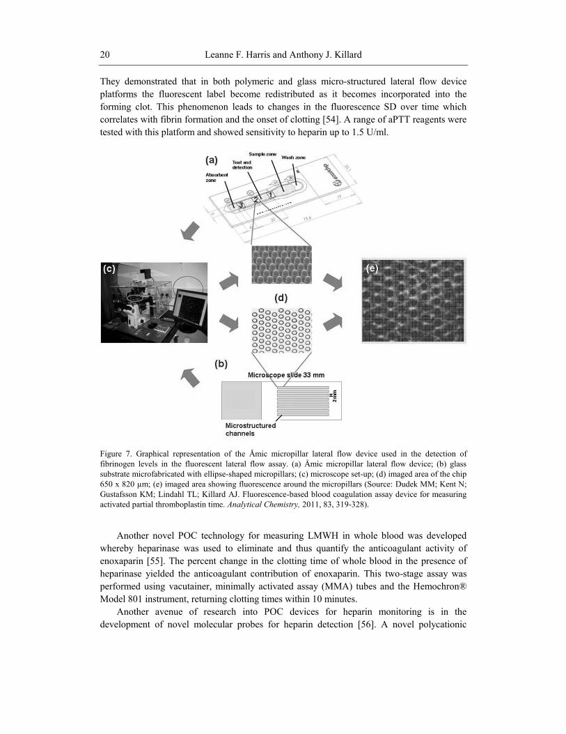
monitoring heparin therapy. Recently a novel principle of measuring aPTT and the repsonse
to heparin treatment was published that makes use of a low cost polymeric microfluidic
device (Figure 7). The assay was developed using an open lateral flow device (Figure 7a)
(Åmic BV, Uppsala, Sweden) and clot formation was monitored using a fluorescently
labelled fibrinogen probe, which accumulated around the micropillar structures of the device
as clotting proceeded [53]. This device enabled the measurement of heparin from 0-2U/ml
and in a range of patient samples indicating its potential for the more reliable determination of
clotting time with aPTT.
The same authors also published research on this principle of identifying the onset of in-
vitro clot formation using the redistribution of a fluorescently labelled fibrinogen marker.
Leanne F. Harris and Anthony J. Killard 20
They demonstrated that in both polymeric and glass micro-structured lateral flow device
platforms the fluorescent label become redistributed as it becomes incorporated into the
forming clot. This phenomenon leads to changes in the fluorescence SD over time which
correlates with fibrin formation and the onset of clotting [54]. A range of aPTT reagents were
tested with this platform and showed sensitivity to heparin up to 1.5 U/ml.
Figure 7. Graphical representation of the Åmic micropillar lateral flow device used in the detection of
fibrinogen levels in the fluorescent lateral flow assay. (a) Åmic micropillar lateral flow device; (b) glass
substrate microfabricated with ellipse-shaped micropillars; (c) microscope set-up; (d) imaged area of the chip
650 x 820 µm; (e) imaged area showing fluorescence around the micropillars (Source: Dudek MM; Kent N;
Gustafsson KM; Lindahl TL; Killard AJ. Fluorescence-based blood coagulation assay device for measuring
activated partial thromboplastin time. Analytical Chemistry, 2011, 83, 319-328).
Another novel POC technology for measuring LMWH in whole blood was developed
whereby heparinase was used to eliminate and thus quantify the anticoagulant activity of
enoxaparin [55]. The percent change in the clotting time of whole blood in the presence of
heparinase yielded the anticoagulant contribution of enoxaparin. This two-stage assay was
performed using vacutainer, minimally activated assay (MMA) tubes and the Hemochron®
Model 801 instrument, returning clotting times within 10 minutes.
Another avenue of research into POC devices for heparin monitoring is in the
development of novel molecular probes for heparin detection [56]. A novel polycationic
Heparin Monitoring 21
ruthenium compound 1 was investigated as a fluorescent probe for the direct, rapid, and
simple detection of LMWH in serum samples at 630 nm where sample autofluorescence is
low. In contrast to more conventional methods such as the ACT or anti-Xa assay which are
indirect measures of heparin, this new technology focuses on the direct detection of heparin
which would translate into less variability in measurement.
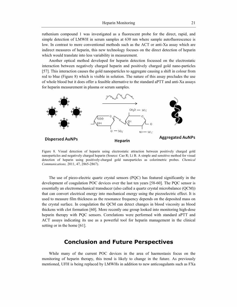
Another optical method developed for heparin detection focussed on the electrostatic
interaction between negatively charged heparin and positively charged gold nano-particles
[57]. This interaction causes the gold nanoparticles to aggregate causing a shift in colour from
red to blue (Figure 8) which is visible in solution. The nature of this assay precludes the use
of whole blood but it does offer a feasible alternative to the standard aPTT and anti-Xa assays
for heparin measurement in plasma or serum samples.
Figure 8. Visual detection of heparin using electrostatic attraction between positively charged gold
nanoparticles and negatively charged heparin (Source: Cao R; Li B. A simple and sensitive method for visual
detection of heparin using positively-charged gold nanoparticles as colorimetric probes. Chemical
Communications, 2011, 47, 2865-2867).
The use of piezo-electric quartz crystal sensors (PQC) has featured significantly in the
development of coagulation POC devices over the last ten years [58-60]. The PQC sensor is
essentially an electromechanical transducer (also called a quartz crystal microbalance (QCM))
that can convert electrical energy into mechanical energy using the piezoelectric effect. It is
used to measure film thickness as the resonance frequency depends on the deposited mass on
the crystal surface. In coagulation the QCM can detect changes in blood viscosity as blood
thickens with clot formation [60]. More recently one group looked into monitoring high-dose
heparin therapy with PQC sensors. Correlations were performed with standard aPTT and
ACT assays indicating its use as a powerful tool for heparin management in the clinical
setting or in the home [61].
Conclusion and Future Perspectives
While many of the current POC devices in the area of haemostasis focus on the
monitoring of heparin therapy, this trend is likely to change in the future. As previously
mentioned, UFH is being replaced by LMWHs in addition to new anticoagulants such as FXa
Leanne F. Harris and Anthony J. Killard 22
and thrombin inhibitors. This pattern will render POC tests such as the aPTT and ACT, which
are very specific for heparin, obsolete in the future. There remains the argument that LMWH
requires less monitoring than traditional anticoagulants such as warfarin and UFH, but there
are special patient cohorts where monitoring becomes imperative, so POC devices will be
targeted at such patient groups. Devices for monitoring FXa and thrombin may be developed
which will most likely incorporate some of the newer nanotechnologies (as outlined below),
as the test method will not rely on clot-based endpoints as do some of the more traditional
clotting assays.
While reticence among the medical community was a major factor in the slow rate of
uptake of POCT devices when they were first developed, there have been major
improvements in terms of device stability, robustness, and reliability contributing to wider
acceptance by healthcare professionals. Rapid technological advances are opening up many
avenues for POCT exploitation in the area of improved engineering, miniaturisation,
detection methodologies, and biomarker discovery. Haemostasis POCT may begin to adapt
more novel methods of detection that offer greater levels of sensitivity and specificity, and
nanotechnologies are at the forefront of cutting edge detection methodologies. The evolution
of nanoparticles, nanodots, and nanowires shows great potential for incorporation into
biodevices and biosensors. Nanostructures offer many advantages for miniaturised biodevices
such as controllable size-dependent properties, adaptable chemical composition, and
chemically and physically robust structures [62]. The ease with which materials such as
polyvalent nanoparticles, carbon nanotubes, and silicon nanowires can result in a change in
electrical conductance into a spectroscopic or electrical signal as a result of target binding,
makes them attractive options for incorporation into POC devices.
Other nanomaterials including nanoshells and noble-metal nanoparticles are easily
modified to recognise biomolecules such as proteins, elemental ions, and small molecules.
With the development of these strategies, excellent versatility and increased sensitivity can be
achieved due to the ease with which nanostructures can be manipulated. In addition, label-
free technologies are highly sought after, as they offer excellent sensitivity and selectivity
with a wide range of biomolecules such as the microsphere resonator biosensor for thrombin
measurement as developed by Zhu et al. in 2006 [63].
A significantly large pool of biomarkers for different coagulation diseases currently exists
that have the potential to be exploited by the POC market. The haemostaseologist mainly tests
the following coagulation markers: D-dimer, protein S, protein C, prothrombin fragment 1+2,
thrombin-AT complex and clotting factors such as FXIII [34]. Many of the assays conducted
to test for the above clotting proteins are immuno-based assays which take many forms.
Enzyme linked immunosorbent assays (ELISA) and latex agglutination immunoassay
technologies are the most popular applications, with technologies such as Luminescent
Oxygen Channeling Immunoassay (LOCI®) and chemiluminescence-based assays also
emerging into the haemostasis laboratory. One major drawback of the standard ELISA is that
it is often incompatible with the workflow requirements of the diagnostic laboratory due to its
time-consuming nature. Latex agglutination on the other hand is more convenient with a
shorter assay duration, however because it is reliant on low-light scattering it requires patient
samples with high levels of coagulation markers. Chemiluminescent technologies combine
the advantages of latex agglutination with extremely sensitive detection technologies and can
Heparin Monitoring 23
be used to detect a wide range of coagulation proteins that are not currently targeted by POC
devices. Further integration of immunoassays into the coagulation laboratory is desirable but
access to such analysers is often difficult as a result of associated high equipment costs. The
adaptation of these technologies to POCT is promising as many of the POC tests already
available for D-dimer testing are immunoassay-based. The Triage D-Dimer (Biosite
Diagnostics, CA, USA) is a fluorescent based immunoassay delivering results within 15
minutes. The MiniQuant (Trinity Biotech, Wicklow, Ireland) POC device uses latex
agglutination with a result generated in 4 minutes, and the Cardiac D-Dimer assay is an
antibody sandwich immunoassay reporting a result within 8 minutes [64]. If such
technologies exist for D-dimer quantification there is scope for the application of such
technologies to a wider panel of coagulation factors which will aid in anticoagulant
monitoring, cardiovascular disease diagnosis, and prevention.
While the principle of POCT is that they can be used in potentially any setting, they are
currently most commonly employed in hospitals, at the GP’s office, and in the case of self-
monitoring in the home, it tends to be confined to glucose, INR and pregnancy testing [65].
Future plans for POCT would include increased popularity in settings other than primary care
such as: ambulances and helicopters, prisons, pharmacies, nursing homes, and fitness centres.
The future of anticoagulation monitoring will depend upon the level of standardisation
that is implemented with the introduction of new POC devices, as it is essential in the
execution of reliable therapy management. The overall reliability and cost-benefit analysis of
these tests will ultimately determine their uptake. One school of thought is that POCT should
remain an adjunct to central laboratory testing as opposed to its replacement [39]. The trend
so far has been a steady rise in availability of both POCT and conventional laboratory testing
and it is expected that this trend will continue as the test menu for various conditions expands.
The combined ageing and growth of the global population will increase pressure on
diagnostic hospital laboratories, rendering POC tests an invaluable asset to both the
healthcare professional but more importantly to the patient.
References
[1] Summers, LKM, Marso, SP, Grant, PJ. Artherothrombosis, thrombolysis and anti-
platelets. In: Hoffbrand, AV, Catovsky, D, Tuddenham, EGD, editors. Postgraduate
Haematology. Oxford, UK: Blackwell Publishing; 2005; 945-964.
[2] Wardrop D; Keeling D. The story of the discovery of heparin and warfarin. British
Journal of Haematology, 2008, 141, 757-763.
[3] Hirsh J; Raschke R. Heparin and low-molecular-weight heparin - The Seventh ACCP
Conference on Antithrombotic and Thrombolytic Therapy. Chest, 2004, 126, 188S-
203S.
[4] Eikelboom JW; Hankey GJ. Low molecular weight heparins and heparinoids. Medical
Journal of Australia, 2002, 177, 379-383.
[5] Kakkar AK. Low- and ultra-low-molecular-weight heparins. Best Practice & Research
Clinical Haematology, 2004, 17, 77-87.
Leanne F. Harris and Anthony J. Killard 24
[6] Wood B; Fitzpatrick L. A review of the prevention and treatment of venous
thromboembolism. Formulary, 2010, 45, 91-100.
[7] Baglin T; Barrowcliffe TW; Cohen A; Greaves M. Guidelines on the use and
monitoring of heparin. British Journal of Haematology, 2006, 133, 19-34.
[8] Clark NP. Low-molecular-weight heparin use in the obese, elderly, and in renal
insufficiency. Thrombosis Research, 2008, 123, 58S-61S.
[9] Hirsh J; Warkentin TE; Shaughnessy SG; Anand SS; Halperin JL; Raschke R, et al.
Heparin and low-molecular-weight heparin mechanisms of action, pharmacokinetics,
dosing, monitoring, efficacy, and safety. Chest, 2001, 119, 64S-94S.
[10] Qari MH. High throughput coagulation analyzers review. Combinatorial Chemistry &
High Throughput Screening, 2005, 8, 353-360.
[11] Lippi G; Favaloro EJ; Favaloro EJ; Franchini M; Franchini M; Guidi GC, et al.
Milestones and perspectives in coagulation and hemostasis. Seminars in Thrombosis
and Haemostasis, 2009, 35, 9-22.
[12] Quick AJ; Stanleybrown M; Bancroft FW. A study of the coagulation defect in
hemophilia and in jaundice. American Journal of Medical Science, 1935, 190, 501-511.
[13] Proctor RR; Rapaport SI. Partial thromboplastin time with kaolin - a simple screening
test for first stage plasma clotting factor deficiencies. American Journal of Clinical
Pathology, 1961, 36, 212-219.
[14] White GC. The partial thromboplastin time: defining an era in coagulation. Journal of
Thrombosis and Haemostasis, 2003, 1, 2267-2270.
[15] Ng VL. Anticoagulation monitoring. Clinics in Laboratory Medicine, 2009, 29, 283-
304.
[16] Kitchen S. Problems in laboratory monitoring of heparin dosage. British Journal of
Haematology, 2000, 111, 397-406.
[17] Olson JD; Arkin CF; Brandt JT; Cunningham MT; Giles A; Koepke JA, et al.
Laboratory monitoring of unfractionated heparin therapy. Archives of Pathology &
Laboratory Medicine, 1998, 122, 782-798.
[18] Brill-Edwards P; Ginsberg JS; Johnston M; Hirsh J. Establishing a therapeutic range
for heparin therapy. Annals of Internal Medicine, 1993, 119, 104-109.
[19] Stevenson K; Easton A; Curry A; Thomson J; Poller L. The reliability of APTT
methods and the relationship to lipid-composition and ultrastructure. Thrombosis
research, 1986, 55, 250-258.
[20] Kitchen S; Jennings I; Woods TAL; Preston FE. Wide variability in the sensitivity of
APTT reagents for monitoring of heparin dosage. Journal of Clinical Pathology, 1996,
49, 10-14.
[21] Hattersley PG. Activated coagulation time of whole blood. Journal of the American
Medical Association, 1966, 196, 436-440.
[22] Bates SM; Weitz JI. Coagulation assays. Circulation, 2005, 112, e53-60.
[23] Kitchen S; Makris M. Laboratory tests of hemostasis. In: O'Shaughnessy D, Makris M,
Lillicrap D, editors. Practical Hemostasis and Thrombosis. Massachusetts, USA:
Blackwell Publishing Ltd.; 2006; 8-17.
Heparin Monitoring 25
[24] Shore-Lesserson L. Evidence based coagulation monitors: heparin monitoring,
thromboelastography, and platelet function. Seminars in Cardiothoracic and Vascular
Anesthesia, 2005, 9, 41-52.
[25] Fitch JC; Geary KL; Mirto GP; Byrne DW; Hines RL. Heparin management test versus
activated coagulation time during cardiovascular surgery: correlation with anti-Xa
activity. Journal of Cardiothoracic and Vascular Anesthesia, 1999, 13, 53-57.
[26] Flom-Halvorsen HI; Ovrum E; Abdelnoor M; Bjornsen S; Brosstad F. Assessment of
heparin anticoagulation: Comparison of two commercially available methods. Annals
of Thoracic Surgery, 1999, 67, 1012-1016.
[27] Waele JJ; Cauwenberghe S; Hoste E; Benoit D; Colardyn F. The use of the activated
clotting time for monitoring heparin therapy in critically ill patients. Intensive care
medicine, 2003, 29, 325-328.
[28] Smythe MA; Koerber JM. Heparin monitoring: The confusion continues.
Pharmacotherapy, 1999, 19, 1240-1242.
[29] Rosen S. Chromogenic methods in coagulation diagnostics. Hamostaseologie, 2005,
25, 259-266.
[30] Kitchen S; McCraw A; Echenagucia M. Diagnosis of Hemophilia and Other Bleeding
Disorders: A Laboratory Manual. Montréal, Québec, Canada: World Federation of
Hemophilia; 2010.
[31] Heins M; Reinauer H. Automation in coagulation testing. Journal of the International
Federation of Clinical Chemistry / IFCC, 1996, 8, 117-122.
[32] Milos M; Herak DC; Zadro R. Discrepancies between APTT results determined with
different evaluation modes on automated coagulation analyzers. International Journal
of Laboratory Hematology, 2010, 32, 33-39.
[33] Rodak BF; Fritsma GA; Doig K. Haematology: Clinical Principle and Applications.
3rd Edition. St. Louis, Missouri, USA: Saunders Elsevier; 2007.
[34] Kappel A; Ehm M. Immunoassays for diagnosis of coagulation disorders.
Hamostaseologie, 2010, 30, 194-201.
[35] Price CP; St John A. Hicks JM. Point-of-Care testing: What, Why, When and Where?
In: Price CP, St John A, Hicks JM, editors. Point-of-Care-Testing. Washington, USA:
American Association for Clinical Chemistry Inc.; 2004; 3-9.
[36] Von Lode P. Point-of-care immunotesting: Approaching the analytical performance of
central laboratory methods. Clinical biochemistry, 2005, 38, 591-606.
[37] Price CP. Point of care testing. British Medical Journal, 2001, 322, 1285-1288.
[38] Stief TW; Fareed J. Point of care: Diagnostics in hemostasis - the wrong direction?
Clinical and Applied Thrombosis/Hemostasis, 2003, 9, 191-195.
[39] Willmott C; Arrowsmith JE. Point-of-care testing. Surgery, 2010, 28, 159-160.
[40] Hammett-Stabler CA; Nichols JH. Point-of-care testing, a critical component of
laboratory medicine. Clinical Biochemistry, 2009, 42, 135.
[41] St John A. Benchtop Instruments for Point-of-Care Testing. In: Price CP, St. John A,
Hicks JM, editors. Point-of-Care Testing. Washington, USA: American Association for
Clinical Chemistry Inc.; 2004; 31-45.
[42] Lewandrowski K. Point-of-care testing: An overview and a look to the future. Clinics
in Laboratory Medicine, 2009, 29, 421-432.
Leanne F. Harris and Anthony J. Killard 26
[43] Whitesides GM. The origins and the future of microfluidics. Nature, 2006, 442, 368-
373.
[44] Rivet C; Lee H; Hirsch A; Hamilton S; Lu H. Microfluidics for medical diagnostics
and biosensors. Chemical Engineering Science, 2011, 66, 1490-1507.
[45] Pugia MJ; Price CP. Technology of Handheld Devices for Point-of-Care Testing. In:
Price CP, St John A, Hicks JM, editors. Point-of-Care Testing. Washington, USA:
American Association for Clinical Chemistry Inc.; 2004; 13-30.
[46] Rhee AJ; Kahn RA. Laboratory point-of-care monitoring in the operating room.
Current Opinion in Anesthesiology, 2010, 23, 741-748.
[47] Perry DJ; Fitzmaurice DA; Kitchen S; Mackie IJ; Mallett S. Point-of-care testing in
haemostasis. British Journal of Haematology, 2010, 150, 501-514.
[48] Prisco D; Paniccia R. Point-of-care testing of hemostasis in cardiac surgery.
Thrombosis Journal, 2003, 1, 1-10.
[49] Olsen JD; Pennell BJ. Automated Coagulation Systems. In: Haven MC, Tetrault GA,
Schenken JR, editors. Laboratory Instrumentation. New York, USA: John Wiley &
Sons Ltd.; 1995; 391-409.
[50] Janata J. Principles of Chemical Sensors. 2nd Edition. New York, USA: Springer;
2009.
[51] Arya SK; Singh SP. Malhotra BD. Electrochemical Techniques in Biosensors. In:
Marks R, Cullen DC, Karube I, Lowe CR, Weetall HH, editors. Handbook of
Biosensors and Biochips. Sussex, UK.: John Wiley & Sons Ltd.; 2007; 341-393.
[52] Thuerlemann C; Haeberli A; Alberio L. Monitoring thrombin generation by
electrochemistry: Development of an amperometric biosensor screening test for plasma
and whole blood. Clinical Chemistry, 2009, 55, 505-512.
[53] Dudek MM; Kent N; Gustafsson KM; Lindahl TL; Killard AJ. Fluorescence-based
blood coagulation assay device for measuring activated partial thromboplastin time.
Analytical Chemistry, 2011, 83, 319-328.
[54] Dudek MM; Kent NJ; Gu P; Fan ZH; Killard AJ. Development of a fluorescent method
for detecting the onset of coagulation in human plasma on microstructured lateral flow
platforms. Analyst, 2011, 136, 1816-1825.
[55] Inchiosa MA,Jr; Pothula S; Kubal K; Sanchala VT; Navarro I. Toward development of
a point-of-care assay of enoxaparin anticoagulant activity in whole blood. Journal of
Thrombosis and Thrombolysis, 2011, 32, 47-53.
[56] Szelke H; Harenberg J; Kraemer R. Detection and neutralisation of heparin by a
fluorescent ruthenium compound. Thrombosis and Haemostasis, 2009, 102, 859-864.
[57] Cao R; Li B. A simple and sensitive method for visual detection of heparin using
positively-charged gold nanoparticles as colorimetric probes. Chemical
Communications, 2011, 47, 2865-2867.
[58] Cheng TJ; Chang HC; Lin TM. A piezoelectric quartz crystal sensor for the
determination of coagulation time in plasma and whole blood. Biosensors and
Bioelectronics, 1998, 13, 147-156.
[59] Chang H; Cheng T; Wu T; Lin T. Determination of coagulation time in whole blood
containing anticoagulant by piezoelectric quartz crystal sensor. Sensors and Actuators
B: Chemical, 2000, 66, 296-298.
Heparin Monitoring 27
[60] Cheng T; Lin T; Wu T; Chang H. Determination of heparin levels in blood with
activated partial thromboplastin time by a piezoelectric quartz crystal sensor. Analytica
Chimica Acta, 2001, 432, 101-111.
[61] Lin TM; Cheng TJ; Wu TH; Chang HC. Comparing a piezoelectric quartz crystal with
an optical coagulometer in monitoring high-dose heparin therapy by determining whole
blood activated partial thromboplastin time and activated clotting time. Sensors and
Actuators B-Chemical, 2005, 109, 270-277.
[62] Giljohann DA; Mirkin CA; Mirkin CA. Drivers of biodiagnostic development. Nature,
2009, 462, 461-464.
[63] Zhu H; Suter JD; White IM; Fan X. Aptamer based microsphere biosensor for
thrombin detection. Sensors, 2006, 6, 785-795.
[64] Van Cott EM. Point-of-care testing in coagulation. Clinics in Laboratory Medicine,
2009, 29, 337-350.
[65] Luppa PB; Müller C; Schlichtiger A; Schlebusch H. Point-of-care testing (POCT):
Current techniques and future perspectives. Trends in Analytical Chemistry, 2011, 30,
887-898.