Harpellales Zygomycota Trichomycetes) associated with ... · Fungal Diversity Harpellales...
49
Fungal Diversity Harpellales (Zygomycota: Trichomycetes) associated with black flies (Diptera: Simuliidae): world review and synthesis of their gy and taxonomy ecolo P. Nelder 1,3* , Charles E. Beard 1 , Peter H. Adler 1 , Sam-Kyu Kim 1 , Mark 2 , Clemson 124 L of Biological Sciences, (Zygom ck flies (Diptera: Simuliidae): world review arthrop 36 spe lack flies are ideal organisms for the study of trichomycete trichom among the few symbiotes known to encompass the m. To synthe suggest areas requiring further Pennel mittium, streams, symbiosis Intro (Isopoda) and larval insects, including beetles (Coleoptera), true flies (Diptera), (Trich rdt, 2000; Lichtwardt et al., 2003). amil eriomycetaceae), 36 genera, and more than 170 species world richomycetes” is erived from the Greek word “tricho,” meaning hair, in reference to the hair- and John W. McCreadie 1 114 Long Hall, Department of Entomology, Soils, and Plant Sciences, Box 340315 University, Clemson, South Carolina 29634-0315, USA 2 ife Sciences Building, 307 University Blvd., Department University of South Alabama, Mobile, Alabama 36688-0002, USA Nelder, M.P., Beard, C.E., Adler, P.H., Kim, S.K. and McCreadie, J.W. (2006). Harpellales ycota: Trichomycetes) associated with bla and synthesis of their ecology and taxonomy. Fungal Diversity 22: 121-169. The Harpellales are obligate, symbiotic trichomycete fungi that colonize the digestive tracts of ods, including black flies (simuliids). Worldwide, black flies are hosts for 9 genera and cies of Harpellales. B ecology because they are taxonomically well known, allowing precise identifications of ycete hosts. Trichomycete fungi are entire spectrum of symbiotic relationships: commensalism, mutualism, and parasitis provide an enhanced framework for investigating trichomycete-host symbioses, we review and size the biology of Harpellales colonizing black flies, study, and present a key to the species of Harpellales that colonize black flies worldwide. Key words: aquatic insects, black flies, fungi, Harpella, identification key, Nematocera, la, S duction The Harpellales are obligate, fungal associates of the guts of isopods mayflies (Ephemeroptera), stoneflies (Plecoptera), and caddisflies optera) (Misra and Lichtwa Harpellales, the most species-rich order in the class Trichomycetes, contains 2 ies (i.e., Harpellaceae and Leg f wide (Lichtwardt et al., 2001a). The name “t d * Corresponding author: Mark Nelder; e-mail: [email protected] 121
Transcript of Harpellales Zygomycota Trichomycetes) associated with ... · Fungal Diversity Harpellales...

Fungal Diversity
Harpellales (Zygomycota: Trichomycetes) associated with black flies (Diptera: Simuliidae): world review and synthesis of their
gy and taxonomy ecolo
P. Nelder1,3*, Charles E. Beard1, Peter H. Adler1, Sam-Kyu Kim1, Mark2
, Clemson
124 L of Biological Sciences,
(Zygom ck flies (Diptera: Simuliidae): world review
arthrop36 spe lack flies are ideal organisms for the study of trichomycete
trichom among the few symbiotes known to encompass the m. To
synthe suggest areas requiring further
Pennel mittium, streams, symbiosis
Intro
(Isopoda) and larval insects, including beetles (Coleoptera), true flies (Diptera),
(Trich rdt, 2000; Lichtwardt et al., 2003).
amil eriomycetaceae), 36 genera, and more than 170 species world richomycetes” is
erived from the Greek word “tricho,” meaning hair, in reference to the hair-
and John W. McCreadie
1114 Long Hall, Department of Entomology, Soils, and Plant Sciences, Box 340315University, Clemson, South Carolina 29634-0315, USA 2 ife Sciences Building, 307 University Blvd., DepartmentUniversity of South Alabama, Mobile, Alabama 36688-0002, USA
Nelder, M.P., Beard, C.E., Adler, P.H., Kim, S.K. and McCreadie, J.W. (2006). Harpellales ycota: Trichomycetes) associated with bla
and synthesis of their ecology and taxonomy. Fungal Diversity 22: 121-169.
The Harpellales are obligate, symbiotic trichomycete fungi that colonize the digestive tracts of ods, including black flies (simuliids). Worldwide, black flies are hosts for 9 genera and cies of Harpellales. B
ecology because they are taxonomically well known, allowing precise identifications of ycete hosts. Trichomycete fungi are
entire spectrum of symbiotic relationships: commensalism, mutualism, and parasitisprovide an enhanced framework for investigating trichomycete-host symbioses, we review and
size the biology of Harpellales colonizing black flies, study, and present a key to the species of Harpellales that colonize black flies worldwide.
Key words: aquatic insects, black flies, fungi, Harpella, identification key, Nematocera, la, S
duction
The Harpellales are obligate, fungal associates of the guts of isopods
mayflies (Ephemeroptera), stoneflies (Plecoptera), and caddisflies optera) (Misra and Lichtwa
Harpellales, the most species-rich order in the class Trichomycetes, contains 2 ies (i.e., Harpellaceae and Legf
wide (Lichtwardt et al., 2001a). The name “td
*Corresponding author: Mark Nelder; e-mail: [email protected]
121

like appearance of thalli in the host gut, and “mycetes,” meaning fungi (Duboscq et al., 1948). Harpellales is probably derived from the Latin word “harpe” for a curved sword, a reference to certain trichomycetes (i.e., Harpella spp.) that have curved propagules.
Larval black flies provide a model host system for the study of arpellales because of their ease of collection, ubiquity, and status as one of e taxonomically best-known groups of aquatic insects at the species level
n black flies compared to other host groups. ymbioses in lotic habitats are poorly understood (e.g., Brönmark and
g midges (Diptera:
om black flies Nelder et al., 2005a). By the end of 2004, 1907 valid species of extant black
Trichomycetes were originally described by Leidy (1849) as r colorless algae (Confervaceae), from beetles and millipeds
iplopoda). The study of Harpellales of black flies began in France, with the descr
C. Williams. The increase also coincides with a period of expanding
Hth(Adler et al., 2004). These attributes have contributed to a rich investigative history of trichomycetes iSHansson, 2005). The trichomycete-simuliid relationship, therefore, can be particularly instructive. Species richness of Harpellales in larval black flies is high compared
ith that of other aquatic insects such as bitinwCeratopogonidae), mosquitoes (Diptera: Culicidae), mayflies (Ephemeroptera), and stoneflies (Plecoptera), and is exceeded only by larval midges (Diptera: Chironomidae). Thirty-six species of Harpellales are known fr(flies had been described worldwide (Crosskey and Howard, 1997; Adler, unpublished data) – about 53 species of black flies per species of simuliid-associated trichomycete. We review the existing knowledge of the trichomycete-simuliid symbiosis, synthesize the scattered literature, and suggest areas of the trichomycete-simuliid relationship needing further study. We also present a key to the described Harpellales in black flies worldwide. History of trichomycete study in black flies
“entophyta,” o(D
iption of Harpella melusinae Léger & Duboscq from larvae of Simulium ornatum Meigen (Léger and Duboscq, 1929). From 1929 to 1959, descriptions of Harpellales associated with black flies increased at the rate of approximately two species every 10 years (Fig. 1). Since 1959, descriptions of new species of Harpellales in black flies have appeared at the rate of six species every 10 years. The increased rate of descriptions since 1959 can be attributed partly to an interest in black flies as fungal hosts by workers such as Robert W. Lichtwardt, Jehanne-Françoise Manier, Odette Tuzet, and Marvin
122

Fungal Diversity
40
10
20
30
mm
ulat
ive
Num
ber o
fH
arpe
llale
s Spe
cies
01929-1939
1940-1949
1950-1959
1960-1969
1970-1979
1980-1989
1990-1999
2000-2005
Calendar Date
Cu
Fig. 1. Species accumulation curve for simuliid-associated Harpellales described from 1929 to present. The first species (Harpella melusinae Léger & Duboscq) associated with black flies
s in areas such as ), Central America (Lichtwardt,
2003). The absence of a plateau the
was described in 1929. knowledge of simuliid taxonomy and appreciation of black flies as vectors of disease agents. The rapid increase in descriptions of Harpellales since 1990 is ttributable to the study of trichomycetes in black fliea
Australia (e.g., Lichtwardt and Williams, 1992a997), and South America (e.g., Alencar et al.,1
in species accumulation curve suggests that more species remain to be discovered (Fig. 1). The higher classification of gut fungi has developed over about 150 years and, in light of recent advances in molecular biology, continues to be reworked (Table 1). Two independent groups of entophyta (Eccrindides and Amoebidium) were recognized in the 1800s. Léger and Duboscq (1929) added another group, the “Harpellacées,” which they believed were related to the Entomophthorales. Duboscq et al. (1948) combined these three groups of entophyta into the new fungal taxon Trichomycetes, which for about 50 years would include four orders: Amoebidiales, Asellariales, Eccrinales, and Harpellales (Benny, 2001). Some researchers (e.g., Lichtwardt, 1986), however, suggested that the Amoebidiales might not be fungal.
123

Table 1. Time line for investigations of trichomycetes associated with black flies (1850-present). Aff. = affinity. Desc. = described. Cl. = classified 1850 1860 1870 1880 1890 1900 1910 1920Affinities and higher taxa described
1849, 1850-Leidy described first trichomycetes. Aff. Confervaceae (algae). “Eccrinides” 1853-Robin. Aff. Saprolegniales
1861-Ciekowski. Aff. Algae and lower mushrooms
1882-Bütschli 1895-Hauptfleisch. Aff. Saprolegniales 1883-Balbiani.
Aff. “Sporozoaires” (Butschli, similar to gregarines)
1897-Schröter. Aff. Myxomycetes 1892-Perrier 1896-Delage et Hérouard 1899-Labbé 1899-Mesnil. Aff. “Sporozoaires”
1906-Chatton. Aff. Lower fungi near myxomycetes or chytrids, not algae
1914-Mercier. Aff. “Protophytes” 1917-Lichtenstein. Aff. “Protoascomycetes”
1929a-Léger & Duboscq. Desc. Eccrinales and Amoebidiales. 1929-Léger & Duboscq. Desc. & Cl. Phycomycetes Entomophthorales Harpellacées 1929-Poisson. Aff. Thallophytes
Genera described
1849-Leidy. Enterobrus (sic)
1861-Cienkowski. Amoebidium
1929-Léger &Duboscq. Harpella2 & Paramoebidium Other
events 1856-Lieberkühn 1858-Schenk. Independently discovered Amoebidium but did not name it (Lieberkühn, Aff. Sporozaires)
1895-Hauptfleischsuggested aff. with Saprolegniales, based on his discovery of Astreptonemia
1909-Chatton & Roubaud. Amoebidiales from black fly1
1First trichomycete in black flies (Chatton and Roubaud 1909). 2First fungal trichomycete in black flies (Léger and Dubosq 1929).

Table 1, continued. 1930 1940 1950 1960 1970 1980 1990 2000Affinities and higher taxa described
1932-Léger & Gauthier. Desc. Génistellacées
1948-Duboscq et al. Cl. Trichomycètes Eccrinides Amoebidiales Eccrinales Harpellides Harpellales Harpellales Genistellales
1950(1951)-Manier. Cl. Trichomyctes Eccrinides Amoebidiales Eccrinales Palavascides Palavasciales Harpellides
Génistellides Génistellales Spartiellales Asellariales 1955-Manier. Cl. Prototrichomyceta Amoebida Eutrichomyceta Eccrinida Harpellida
1960-Lichtwardt. Cl. Trichomycetes Amoebidiales Eccrinales Harpellales Asellariales Genistellales 1968- Manier & Lichtwardt. Cl. Trichomycetes Amoebidiales Eccrinales Asellariales Harpellales
1986-Lichtwardt. Cl. Zygomycota Trichomycetes Harpellales Asellariales Eccrinales Amoebidiales
2005-Cafaro. Cl.Mesomycetozoa Ichythyophonida Amoebidiales Eccrinales
Genera described
1932-Léger & Gauthier. Stachylina & Stipella 1936-Poisson. Smittium
1961-Tuzet, Rioux, & Manier. Rubetella (syn Smittium) 1963-Manier. Pennella (nom. nud.) 1968-Manier & Lichtwardt. Pennella (validated)
1972-Lichtwardt. Genistellospora & Simuliomyces
1997-Lichtwardt. Genistelloides
2005-New genus from Great Smoky Mountains National Park (White, Siri, & Lichtwardt, in press)
Other events
1948-Duboscq et al. Coined the name Trichomycete which grouped the various taxa of “entophyta”
1960-Whisler cultured Amoebidium 1963-Clark, Kellen, & Lindegren cultured Smittium
1972-Pouzar. Legeriomycetaceae replaces Genistellaceae of Léger & Gauthier 1932. Type genus had been used for a legume in 1773. Sanger. Serology studies
1987-Peterson and Lichtwardt. Serology studies.
1996-Grigg & Lichtwardt. Isozyme studies.
2000-Benny & O’Donnell. Confirmed Amoebidium is a protozoan in Ichthyosporea 2000-2003-PEET grant: molecular studies by Cafaro, Gottlieb, Lichtwardt, & White (University of Kansas)
Theses
1951-Lichtwardt (MS) 1954-Lichtwardt (PhD)
1961-Whisler (PhD) 1965-Farr (MA) 1966-Chapman (MA) 1969-Sangar (PhD)
1970- Coste-Mathiez (PhD) 1971-Galt (MS), Grizel (PhD), Williams (PhD) 1972-El-Buni (MA), Moss (PhD) 1973-Preisner (PhD) 1975-El-Buni (PhD), El-Sherif (PhD) 1976-Starr (MA) 1978-Hollingsworth (MS) 1979-Dang (MS)
1980-Horn (MS), Yeboah (MS) 1984-Peterson (PhD) 1988-Grigg (MS) 1989-Horn (PhD)
1992-Taylor (PhD) 1994-Grigg (Ph.D) 1996-Frost (BS Honors)
2002-Beard (PhD), White (PhD) 2003-Alencar (PhD), Cafaro (PhD), Nelder (MS) 2004-Valle (PhD) 2005-Kim (PhD)

In the late 1980s and the 1990s, serology and molecular techniques, spectively, demonstrated that Amoebidiales do not belong with the
ccrinales in a es. Within the
moebidiales, the genera Paramoebidium and Amoebidium include at least four species associated with black flies: Paramoebidium curvum Lichtwardt, Paramoebidium grande Lichtwardt, and Paramoebidium chattoni Léger & Duboscq (nomen nudum), which inhabit the hindgut, and Amoebidium colluviei Lichtwardt, which lives on the external cuticle. The Amoebidiales are now considered protists (Benny and O’Donnell, 2000; Ustinova et al., 2000; Benny and White, 2001; Reeves, 2003a) in a clade with the Eccrinales, leaving the class Trichomycetes with two fungal orders, the Asellariales and the Harpellales (Cafaro, 2005).
The symbiotes: Harpellales The generalized life cycle of a harpellid in a black fly host is presented in Fig. 2. Harpellales consist of a holdfast and a main thallus with terminal or subterminal propagules that can be asexual (trichospores) or sexual (zygospores). They colonize the guts of larval black flies, attaching to the peritrophic matrix of the midgut or the cuticle of the hindgut by means of a permanent holdfast produced by the basal cell(s) of the thallus (Lichtwardt, 1986; Moss, 1998). The structure of the holdfast varies among and within genera of Harpellales. The holdfast is tapered or rounded in Harpella species, often bifurcated and covered by a mucilaginous sheath in Pennella species, and typically small and sometimes limuloid in Smittium species. The hindgut cuticle of a black fly folds around the holdfast of species such as Genistellospora homothallica Lichtwardt (Mayfield and Lichtwardt, 1980). The thalli of Harpellales exhibit one of two growth forms: branched, typically with a thallus giving rise to lateral branches (Legeriomycetaceae) (Fig. 3) or unbranched (Harpellaceae). The ultrastructure of trichomycetes colonizing black flies has been treated by Reichle and Lichtwardt (1972), Moss and Lichtwardt (1976, 1977), Mayfield and Lichtwardt (1980), Horn (1989b), and Sato (2002). The production of reproductive propagules in higher fungi is related to physiological and abiotic factors. Hence, the biomass of higher fungi increases as nutrients become available and biomass is converted to reproductive propagules as nutrients diminish (Griffin, 1994). Trichomycetes might respond similarly. For example, Smittium spp. cultured in high-nutrient broth produce predominantly vegetative growth, whereas low-nutrient broth results in high
retrichomycetes. Cavalier-Smith (1998) placed Amoebidiales and Enew class, Enteromycetes, because they share Golgi dictyosomA
126

Fungal Diversity
Zygospores
Resting sporestage
Flypupa
Fly adult
Flylarva
Vegetative thallus
Trichospores
Trichomycete life cycle
Fly eggs or Fungal cysts
Fig. 2. Generalized life cycle of Harpellales, with a simuliid host. Bold line represents fungal life cycle; thin line represents fly life cycle. Question marks indicate hypothesized routes. production of trichospores (El-Buni and Lichtwardt, 1976a). Some workers have suggested that trichospore production increases prior to host molting (Lichtwardt et al., 2001a) and responds to host-moulting hormones. As black flies prepare to molt, they cease feeding (Hinton, 1958). Thus, nutrients available to the fungus might decrease at this time. Harpellales reproduce asexually by trichospores (El-Buni and Lichtwardt, 1976a,b) (Fig. 4), which are basipetal, monosporous sporangia (housing a single sporangiospore); they are produced terminally or laterally from generative cells on fertile branches (Lichtwardt, 1986). Although trichospores have been found in the shed exuviae of the host (Lichtwardt et al., 2003), they are observed most often in the arthropod gut where the fungus is growing. Tri
?
chospores often have various numbers of nonmotile, basal
?
appendages, depending on the genus (Fig. 4) (Moss and Lichtwardt, 1976). The appendages can be coiled or spiralled within the thallus (but outside the plasma membrane) before the trichospore is released (Fig. 5), and in Genistellospora homothallica they are directed downward along the inner wall (Moss and Lichtwardt, 1976). Appendages are believed to entangle with food consumed by the host (Lichtwardt, 1996). They also allow for direct entanglement with
127

Fig. 3. Pennella sp. attached to the hindgut cuticle of Simulium tribulatum. Bar = 100 µm. the microtrichia on the labral fans of black flies during filter feeding (Fig. 6). Once in the gut, appendages might snag on the thalli of existing fungi and other materials, keeping them in the appropriate microhabitat for a longer period of time. The Harpellales reproduce sexually by zygospores, which are either pointed at one terminus or, more commonly, biconical (Fig. 7) (Lichtwardt, 1986). The production of pointed zygospores is a distinguishing feature of the order Harpellales. The shape of the zygospore possibly is conducive to rapid extrusion in the host gut (Lichtwardt, 1996). Production of zygospores requires thallial conjugation in some species (e.g., Pennella) but not in others (e.g., Genistellospora). Zygospores are produced on zygosporophores, probably after karyogamy and, unlike other Zygomycota, after meiosis (Moss and Lichtwardt, 1977). Reproduction by means of zygospores is rare in many Harpellales and might have been essentially lost in some species (e.g., Smittium culisetae Lichtwardt). Zygospores are classified according to their orientation with the zygosporophore: perpendicular = type I, median attachment to zygosporophore (Fig. 8); oblique = type II, submedian attachment; parallel = type III, median attachment (Fig. 7); and polar = type IV, coaxial attachment (type IV zygospores are not found in black flies) (Lichtwardt, 1986).
128
Fungal Diversity
129
Figs. 4-6. Trichospopres of Harpellales. 4. Smittium brasiliense in axenic culture; a = appendage, c = collar. 5. Harpella sp. from the gut of Simulium innoxium; s = spiraled appendages. 6. Trichospore of Smittium culisetae captured by microtrichia of the labral fan of Simulium tribulatum; a = appendage, c = collar, m = microtrichia, pf = primary fan ray. Bars = 25 µm, except Fig. 5 = 30 µm.
Figs. 7-8. Zygospores of Harpellales. 7. Pennella sp. (immature stage) in hindgut of Simulium sp. 8. Simuliomyces microsporus in hindgut of Simulium tribulatum. z = zygospores, zs = zy .gosporophores. Bars = 30 µm
130
Fungal Diversity Fungal Diversity
131
Moss (1998) suggested that zygospores probably are not required for
rema
et al.,
fromthe gut.
er clim
ales
mam
Adler
colonization of other hosts. Lichtwardt et al. (2003), however, suggested that zygospores are transmitted from larva to larva. The latter scenario has not been tested and the role of zygospores in the life history of simuliid-gut fungi
ins unknown (Beard and Adler, 2003). Zygospores might be part of a parasitic stage of some Harpellales (Horn, 2001) or a resistant stage during unfavorable conditions in the life cycle of the Harpellales (Lichtwardt 2001a). These hypotheses, however, are difficult to test because zygospores have not been produced in axenic cultures of Harpellales and, in field collections, they are unpredictable.
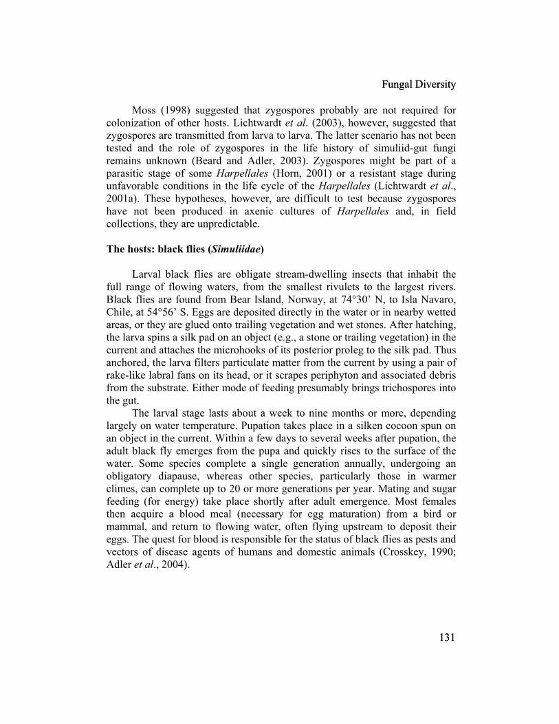
The hosts: black flies (Simuliidae)
Larval black flies are obligate stream-dwelling insects that inhabit the full range of flowing waters, from the smallest rivulets to the largest rivers. Black flies are found from Bear Island, Norway, at 74°30’ N, to Isla Navaro, Chile, at 54°56’ S. Eggs are deposited directly in the water or in nearby wetted areas, or they are glued onto trailing vegetation and wet stones. After hatching, the larva spins a silk pad on an object (e.g., a stone or trailing vegetation) in the current and attaches the microhooks of its posterior proleg to the silk pad. Thus anchored, the larva filters particulate matter from the current by using a pair ofrake-like labral fans on its head, or it scrapes periphyton and associated debris
the substrate. Either mode of feeding presumably brings trichospores into
The larval stage lasts about a week to nine months or more, depending largely on water temperature. Pupation takes place in a silken cocoon spun on an object in the current. Within a few days to several weeks after pupation, the adult black fly emerges from the pupa and quickly rises to the surface of the water. Some species complete a single generation annually, undergoing an obligatory diapause, whereas other species, particularly those in warm
es, can complete up to 20 or more generations per year. Mating and sugar feeding (for energy) take place shortly after adult emergence. Most femthen acquire a blood meal (necessary for egg maturation) from a bird or
mal, and return to flowing water, often flying upstream to deposit their eggs. The quest for blood is responsible for the status of black flies as pests and vectors of disease agents of humans and domestic animals (Crosskey, 1990;
et al., 2004).
131
Techniques for study of Harpellales Host rearing and collection Laboratory studies of the trichomycete-simuliid symbiosis have been facilitated by the relative ease of rearing larval black flies. Eggs of species such as Simulium vittatum Zetterstedt can be collected from the field by anchoring yellow strips of plastic (resembling trailing grasses) in the current and retrieving the strips a few days later. We have routinely obtained eggs of Simulium vittatum from a parasite-free colony at the University of Georgia, Athens, Georgia, USA (Gray and Noblet, 1999). Eggs can be placed in aerated ontainers of water at room temperature. When the larvae hatch, they can be
us-generation rearing of black flies is ossible but time intensive (Edman and Simmons, 1985a,b; Cupp and amb
ae are refrigerated on moist filter paper in a Petri dish until dissection.
etection of Harpellales in the host To assay black flies for trichomycetes, live larvae are slit ventromedially in a drop of water on a glass slide, using fine needles or a scalpel. The larva is
cfed pulverized fish food. ContinuopR erg, 1997; Gray and Noblet, 1999) Larval black flies in the field are best collected by hand, using forceps to remove them from in-stream objects such as trailing vegetation, artificial substrates (e.g., plastic), and stones (Adler et al., 2004). Although most larval instars can be colonized by trichomycetes, older instars generally contain more fungi, often with zygospores (Lichtwardt et al., 2003). Larvae in large rivers can be collected by boat or by anchoring monofilament fishing line or plastic tubing to a bank or bridge, allowing it to trail in the current, and removing it several days later. Accessible watercourses are sampled by hand collecting larvae from all available substrates while walking swaths from bank to bank. Live larvLarvae can be held for several days at 4°C for evacuation of food from the gut, facilitating fungal detection. Ecological data can be obtained at the time of collection by recording selected stream characteristics such as stream conductivity, discharge, pH, temperature, and width (McCreadie and Adler, 1998). These data, particularly if used in multivariate analyses, can be useful predictors of both host and symbiote distributions among streams (Beard, 2002; Beard et al., 2003; Nelder, 2003). Larval black flies can be identified using the illustrated keys of Yankovsky (2002) for the Palearctic Region, Adler et al. (2004) for the Nearctic Region, and Takaoka (2003) for the Indonesian portion of the Oriental Region. Keys to other areas of the world are listed by Crosskey and Howard (1997). D
132

Fungal Diversity
cut transversely just posterior to the thoracic proleg and a second cut is made mediately anterior to the anal proleg. (The larval host morphology is
004)). The midgut is grasped with forceps and
lactophenol containing1 part phenol
ndgut – midgut fungi cannot yet be cultured – is viewed under low agnification (ca. 40×), without a coverslip, and the mycelia removed with
placed the entire slide with fungus in a etri dish (100 mm × 15 mm) with sufficient broth to cover the slide.
imreviewed by Adler et al. (2pulled anteriorly from the body. The remainder of the gut is pulled posteriorly from the body. With the gut removed, the larval carcass is placed in acetic ethanol (1:3) or 80% ethanol for subsequent identification. The midgut musculature is stripped from the cellophane-like peritrophic matrix. The hindgut musculature requires more care to tease it from the cuticle. The peritrophic matrix and hindgut cuticle are then slide-mounted in a drop of water, a coverslip is applied, and the preparation is examined with phase-contrast microscopy. Morphological features of trichomycetes are measured using an ocular micrometer. A drop of lactophenol with cotton blue (a fixative and stain) is added to the edge of the coverslip and the preparation is sealed with clear nail polish. We use liquid (e.g., 20 ml for a final volume of 100 ml), 1 part lactic acid (20 ml), 2 parts glycerol (40 ml), 0.5 g cotton blue, and 1 part distilled water (20 ml). The solution is kept in a tightly closed glass bottle. Harpellales also can be recovered from larvae fixed in ethanol (> 70%) or acetic ethanol (Carnoy’s solution) (Adler et al., 1996; Nelder et al., 2004, 2005a). Larvae stored in acetic ethanol (1: 3) contain identifiable fungi after at least 15 years of storage at 4°C (Nelder, unpublished data). Trichomycetes are more difficult to remove from fixed larvae (e.g., in museum collections) than from live larvae (Lichtwardt et al., 2003) but are a valuable source of trichomycetes. The relative abundance of hyphae among hosts can be assessed by viewing the gut at 400× through a 10-mm × 10-mm ocular grid (McCreadie and Beard, 2003). The number of grid squares that contain one or more hyphae is counted and relative abundance expressed as the percentage of grid squares containing hyphae, relative to the total number of grid squares over the gut surface. Culturing of Harpellales Most modern culturing techniques are modifications of those presented by Lichtwardt (1964) and Lichtwardt et al. (2001a). Cultures can be derived from trichomycetes taken from live or freshly killed larvae. A slide with the larval himfine forceps. On occasion, we have P
133

Media preparation before isolation Standard culture dishes consist of 15-20 ml of 3.7 g/l Brain Heart
Infusion (BHI, Difco 237400), and 1.5-2% agar in a standard Petri dish (100 mm × 15 mm). We find a harder agar (1.75-2%) preferable to the standard bacterial formulation (1.5%) because it resists gouging if aggressively scraped with an inoculating loop. Although these pre-made dishes of media can be used for initial isolation,
amins are prepared in a similar an
acycline HCl can be used as an alternative antibiotic. A
0 µg iamine. The final solution is made by adding 1 ml of vitamin concentrate to
When a suitable mass of hyphae is found in a dissected larva, one tube
we alternatively use a broth method, which facilitates the addition of antibiotics. A small amount (ca. 100 ml) of concentrate of BHI broth is prepared at 15 times (i.e., 55.5 g/l) the final concentration. This concentrate is dispensed in single-use units by allocating 0.7 ml (sufficient for 10 ml final volume) to small cryovials (ca. 2 ml capacity) or microfuge tubes. These small vials are stored at about -80°C until needed. We do not store unused, diluted broth media for more than 24 hours. Standard culture dishes of agar can be sealed with Parafilm and stored for several months at room temperature. Single-use aliquots of antibiotics and vitm ner. Antibiotic concentration for the final solution is about 8,000 units/ml streptomycin and 4,000 units/ml penicillin G. Concentrated antibiotics are made up in a stock solution with 80,000 units/ml streptomycin and 40,000 units/ml penicillin G. (The exact amounts of powdered antibiotics will depend on the assay for the production batch). Aliquots (1 ml) of concentrated antibiotics are stored at -80ºC. When bacteria seem resistant to control by these antibiotics, tetrconcentration of 3 ppt in an overlay is beneficial. If vitamins are to be used in fungal isolations, a diluted vitamin solution, rather than water, is used to prepare the isolation medium. The vitamin concentrate, which is made using 5 mg biotin/ml water and 20 mg thiamine/ml water, is 100× the final concentration, yielding 50 µg/ml biotin and 20th99 ml of distilled water. each of BHI and antibiotics is defrosted and placed in a small beaker. The volume is brought up to 10 ml with water (or vitamin solution) and loaded into a 10-ml syringe. A sterile syringe filter (0.4 µm or smaller) is added to the syringe and the solution is sterile filtered into a sterile Petri dish, typically 35 or 60 mm × 15 mm; we dispense only enough solution to cover the bottom of the dish. The fungus is then placed in the dish.
134
Fungal Diversity
Maintenance
, oving the trichomycete to a BHI-agar plate and ear
t. We
n be archived at the United
Dishes should be examined daily for growth and for bacterial or fungal contaminants. If trichomycete growth is evident after 4-5 days, the hyphae can be moved aseptically to a BHI-agar plate or another broth dish without antibiotics. Antibiotics can slow the growth of freshly extracted trichomycetes. If competing fungi develop msm ing it around (without overlays) can provide isolated colonies. If bacteria develop, a transfer of the mycelial mass to a dish with fresh broth and antibiotics is beneficial. Once an axenic isolate is growing, it can be maintained by monthly transfers of mycelia, using an inoculating loop. The Petri dishes with trichomycetes are sealed with Parafilm. Backup cultures can be refrigerated for months in sealed dishes or culture tubes, and maintenance cultures can be kept at room temperature (ca. 25°C). For backup cultures, we use screw-cap tubes 20 mm × 125 mm, with 8 ml of media (BHI agar) solidified at a slanseldomly use a water overlay for maintenance cultures, although it is commonly used by other workers (e.g., Lichtwardt, 1986). However, cultures grow faster and produce more trichospores with water. We have had month-old cultures produce many trichospores within about 48 hours after a water overlay is added to a previously non-overlay dish. Trichomycetes in culture grow slowly relative to some common contaminants such as Penicillium and they need to be monitored. If a contaminant appears, the trichomycete can be transferred to another dish before it is overgrown. Trichomycetes isolated from black flies caStates Department of Agriculture-Agricultural Research Service Collection of Entomopathogenic Fungal Cultures, Ithaca, New York (http://www.ppru.cornell.edu/mycology/Insect_Mycology.htm). We usually ship specimens on slants in screw-cap vials, as in backup cultures. The host gut as habitat The vegetative stage of trichomycetes is restricted to the larval host gut and, therefore, depends on the host for protection. Hosts must select appropriate microhabitats. Larval black flies, for example, can relocate in inchworm fashion or on silk life lines to optimize the flow regime for feeding (Eymann, 1991; Poff and Ward, 1991) or to minimize exposure to ultraviolet light (Donahue and Schindler, 1998).
135

Gut s
trix of larval black flies is secreted by the anteriormost
colonized by trichomycetes, axim
at range in ze from colloids to particles up to 350 µm in diameter, including algae,
ngi (Wallace and Merritt, 1980; Adler et al., 2004). he food bolus typically contains sand grains, mica flecks, and similar
ched to the hindgut cuticle, they are oughed with the cuticular exuviae during larval molting. The thalli of
Smittium species convert most of their biomass to trichospores during host molting (McCreadie, unpublished data). Under laboratory conditions, thalli of
tructure
The gut of the larval black fly, roughly 3-15 mm long in mature larvae, provides the developmental milieu for Harpellales. It is divided into three sections: foregut, midgut, and hindgut (Crosskey, 1990). The cellophane-like envelope of the midgut (i.e., the peritrophic matrix, Figs. 9-10) and the cuticle-lined hindgut provide attachment sites for trichomycete holdfasts. No Harpellales have been found attached to the cuticular lining of the foregut of black flies. The peritrophic masection of the midgut (i.e., the cardia) and is termed a Type II peritrophic matrix. The matrix consists of proteins, proteoglycans, and chitin, and is permeable to dissolved materials (Peters, 1992). The peritrophic matrix acts as a mechanical barrier between the food and the gut epithelium (Lehane, 1997). The Type II peritrophic matrix moves posteriorly, eventually breaking up in the hindgut (Shao et al., 2001). Harpellales, such as Harpella spp., must attach and grow before the matrix travels to the end of the midgut. The Harpellales that colonize the midgut are unbranched and have relatively determinate growth, which might be an adaptation to the continuous posterior advancement of the matrix. The hindgut is a looped structure with three distinct regions: pylorus, colon, and rectum. The cuticular lining of the pylorus is often beset with posteriorly directed spines that might help move the peritrophic matrix posteriorly. Although the pylorus and rectum are m um trichomycete abundance occurs in the posterior colon (McCreadie and Beard, 2003) (Fig. 11). The fungi in the hindgut are generally branched and of indeterminate growth, limited only by the periodic moulting of the hindgut cuticle. The lumen of the larval gut is filled with ingested materials thsibacteria, detritus, and fuTinorganic matter that might act as roughage, pushing organic material through the gut (Colbo and Wotton, 1981). Although the inorganic matter might have abrasive properties, it evidently does not damage attached trichomycetes. Movement of food through the gut is temperature dependent, requiring about 20 minutes to 2 hours (Colbo and Wotton, 1981).
Because trichomycetes are attasl
136

Fungal Diversity
Figs. 9-10. Peritrophic matrix of the black fly midgut. 9. Harpella melusinae colonizing the peritrophic matrix of Cnephia ornithophilia. Bar = 200 µm. 10. Uncolonized peritrophic matrix of Simulium innoxium; the somewhat thickened anterior end is to the left. Bar = 300 µm.
137
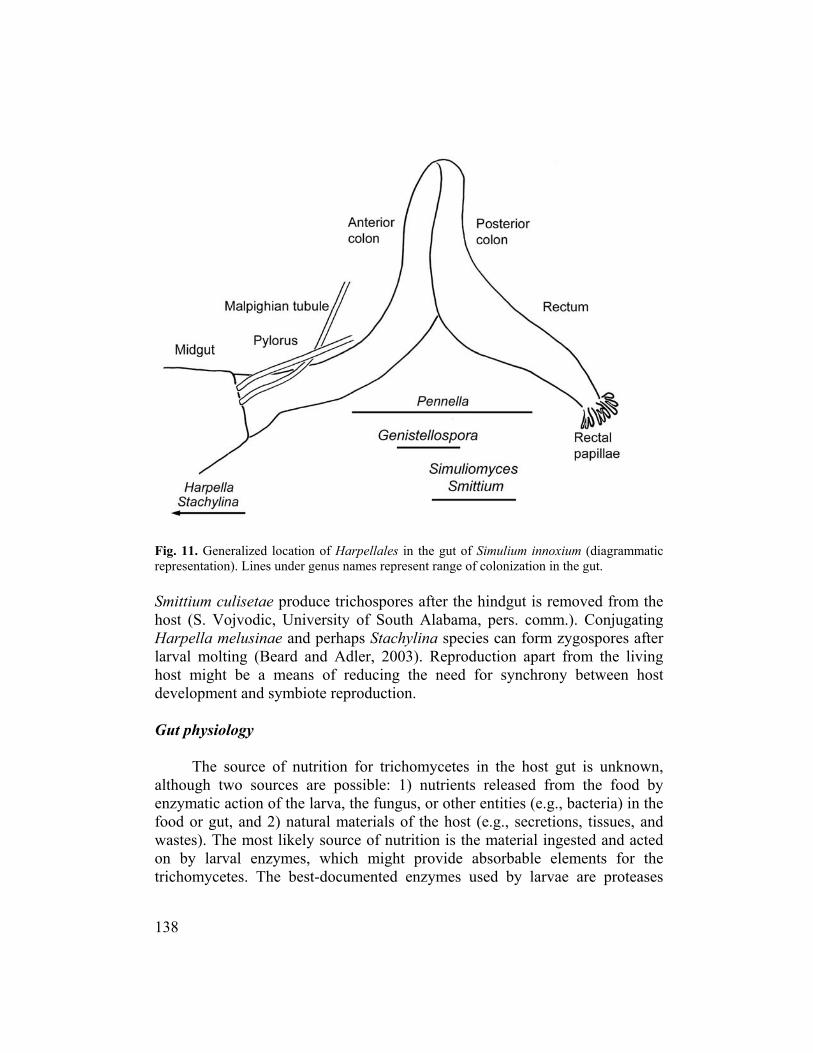
Fig. 11. ammatic represent
the
larval m the living host mdevelopm
the food by enzym
ichomycetes. The best-documented enzymes used by larvae are proteases
Generalized location of Harpellales in the gut of Simulium innoxium (diagration). Lines under genus names represent range of colonization in the gut.
Smittium culisetae produce trichospores after the hindgut is removed fromhost (S. Vojvodic, University of South Alabama, pers. comm.). Conjugating Harpella melusinae and perhaps Stachylina species can form zygospores after
olting (Beard and Adler, 2003). Reproduction apart fromight be a means of reducing the need for synchrony between host
ent and symbiote reproduction.
Gut physiology
The source of nutrition for trichomycetes in the host gut is unknown, although two sources are possible: 1) nutrients released from
atic action of the larva, the fungus, or other entities (e.g., bacteria) in the food or gut, and 2) natural materials of the host (e.g., secretions, tissues, and wastes). The most likely source of nutrition is the material ingested and acted on by larval enzymes, which might provide absorbable elements for the tr
138

Fungal Diversity
released in the midgut (Martin et al., 1985). The action of the high pH derived from carbonates (Boudko et al., 2001) might play a part in digestion by hydrolyzing materials and releasing nutrients. Bacteria inhabiting the gut (Malone and Nolan, 1978) also might aid release of nutrients. The enzymand chemicals secreted by the larva might be used by the fungus, possibly reducing the fitness of the host. If trichomycetes assist digestion by secreting enzymes, they might provide some benefit to host larvae by making mnutrients available, as demonstrated in mosquitoes (Horn, 1980). Starved larval black flies with trichomycetes have significantly higher survival than do trichomycete-free larvae (McCreadie et al., 2005). The basis of this murelationship is unknown, but might be nutritional. Gas exchange is probably not a limiting factor for Harpellalesblack flies live in running water that is usually high in dissolved oxygen. Aerobic bacteria inhabit the gut (Taylor et al., 1996), suggesting that oxygen is available to trichomycetes in the gut. Other gases, such as carbon dioxide and ammonia, probably diffuse into the water through the body of the host. Living in a host protects the trichomycetes from the vagaries of pH and osmotic changes in the host’s external environment. The larval miblack fly varies in pH from 8.2 anteriorly to 11.4 near the middle (Lacey and Federici, 1979). This high pH would be suboptimal for most fungi, but the Harpellaceae thrive in this environment. The available nutrients in the m
es
ore
tualistic
. Larval
dgut of a
dgut
udko et l., 2001). The hindgut probably has a near-neutral pH, based on data available
The relationship between Harpellales and their hosts generally has been mensalistic (Lichtwardt, 1996; Moss, 1998), although in
osquitoes, examples of parasitism (Williams and Lichtwardt, 1972; Sweeney,
ihave not been documented, but probably include carbonate complexes, which have been implicated in the maintenance of high pH in mosquitoes (Boafor mosquitoes (Bradley, 1985). Ecology of the symbiosis Symbiotic nature of the relationship considered comm1981) and mutualism (Horn and Lichtwardt, 1981) have been suggested. The symbiotic association between Smittium culisetae and black flies is dynamic: commensalistic when larvae are well fed, but mutualistic when larvae are starved (McCreadie et al., 2005). The basis of this mutualistic relationship is believed to be nutritional; hyphae in stressed larvae might produce sufficient vital nutrients to increase survival. In the female black fly, the symbiotic association can be parasitic, the trichomycetes replacing eggs with fungal cysts
139

(Undeen and Nolan, 1977; Taylor, 1992; Lichtwardt, 1996; White, 2002). The symbiosis between Harpellales and black flies thus represents one of the few examples of a relationship shifting among the three states of symbiosis (commensalism, mutualism, parasitism) in a single pair of associated species. Dispersal of Harpellales The propagules of the Harpellales, once released from the host, are subject to downstream displacement in flowing water. Upstream dispersal by the fungus is, therefore, essential. Dispersal of gut fungi by black flies is poorly understood and generally assumed to be via females flying upstream to deposit
, 1998),
in the United Kingdom (Moss and Descals, 986; Taylor, 1992; Rizzo and Pang, 2005a). Possible trichomycete cysts in
n recorded from Cameroon (Lewis, 1960a, 1965), uatemala (Garms, 1975), Ivory Coast (Walsh et al., 1981), Liberia (Lewis,
sina, 1963), Sudan (Lewis, 1953), and anganyika (Lewis, 1960b). Trichomycete infection of female black flies is mm
their eggs, thereby releasing cysts (i.e., chlamydospores of Taylor, 1992) into the water and maintaining the fungal population locally (Lichtwardt et al., 2003). In this manner, Harpellales are parasitic, although the role that ovarian cysts play in populations of black flies remains poorly known. Harpellales might also be dispersed on the feathers of birds (Williams, 1983; Misrathrough human activities (Lichtwardt, 1986, 1996), on alternative host species carrying the fungi in their guts or on their body surface (Lichtwardt, 1996), and on or in lotic organisms moving along the stream course. The production of ovarian cysts by species of Harpellales (Genistellospora, Harpella, Pennella, and Smittium) has been observed in black flies on the Avalon Peninsula of Newfoundland, Canada (Undeen and Nolan, 1977; Undeen, 1978; Yeboah, 1980; Yeboah et al., 1984); in New York State, USA (Tarrant, 1984; Labeyrie et al., 1996); in Liberia (Steinke and Garms, 1990; Taylor, 1992); and1female black flies have beeG1960a; Garms, 1973), Russia (ShipitTco on in some populations in Newfoundland, Canada (M.H. Colbo, Memorial University of Newfoundland, pers. comm.). However, our searches for ovarian cysts in female black flies in South Carolina, USA, have been unsuccessful, suggesting that it is not a universal means of dispersal. Spatial-temporal ecology and host preferences of Harpellales Few established laws exist in community ecology but the abundance, biodiversity, distribution, and species composition of organisms are almost universally recognized as uneven across any landscape and over time
140

Fungal Diversity
(Rosenzweig, 1995). Trichomycetes are no exception to this rule. Both the patterns of distribution and the causal factors of these patterns vary with scale. The scales of distribution considered here are the gut, the host, and the stream habitat. At the smallest scale of distribution (the gut), the distribution of hyphae
ithin the fore-, mid-, and hindgut. Members of the milies Harpellaceae and Legeriomycetaceae attach to the lining of the idgu
in species f Smittium differentially colonize particular species of black flies under
uggesting host references or differential survival in the different host species. Similarly, the
varies both across and wfam t or hindgut, respectively (Lichtwardt, 1986, 1996). Physiological differences in the midgut and hindgut are believed to be responsible for this difference in location of Harpellaceae and Legeriomycetaceae (e.g., Horn, 1989a). Although the restriction of trichomycetes at the family level to either the midgut or hindgut of arthropods is well documented, we know much less about the distribution and abundance of thalli in each region of the gut. For example, Simuliomyces microsporus Lichtwardt attaches to Paramoebidium chattoni, which in turn attaches to the anterior section of the hindgut in black flies (Beard and Adler, 2002). Smittium culisetae in mosquitoes is restricted to the rectum (Horn, 1989a, 1989b). In contrast, this species is most abundant in the posterior colon of black flies. The distribution of Harpellales in the gut, therefore, can be related to the host taxon. The next scale of spatial distribution is the host, which is exhibited (or measured) as differences in host specificity and preference. Genera of most Harpellales are specific to a single family of hosts. Apparent host specificity at some taxonomic levels of trichomycetes, however, might be the result of insufficient sampling of hosts. For example, the genus Stachylina, previously known only from chironomids, recently has been found in black flies (Labeyrie et al., 1996; Reeves et al., 2004). As more black flies are screened for trichomycetes, more genera of trichomycetes currently known only from other host families possibly could be found in black flies. Some species of Smittium can colonize several families of Diptera. Smittium culisetae, for example, colonizes larvae of the families Chironomidae, Culicidae, and Simuliidae. However, some species such as Smittium orthocladii Manier colonize only chironomid midges (Lichtwardt et al., 2001a). The wide host range of certain members of Smittium might reflect unresolved cryptic species or broader physiological tolerances. Certaocontrolled laboratory conditions (Nelder et al., 2005b), spprevalence of Harpella melusinae in the field is significantly higher in Simulium venustum Say than in Prosimulium magnum Dyar & Shannon in the same stream (Beard et al., 2003).
141

Relatively little is known about the distribution of trichomycetes within and among streams. Based on other symbiotic relationships (e.g., Sapp, 1994) several suppositions can be offered. First, symbiote distribution is influenced by both host and environment; hence, symbiotes will occur within a subset of
valence varies with season
Lictwardt (New Zealand), are apparently more restricted in distribution. Biogeographic information is constrained by
the sites occupied by their hosts. For example, Harpella melusinae readily colonizes Simulium vandalicum Dyar & Shannon, Simulium tuberosum (Lundström), and Simulium verecundum Stone & Jamnback, although not all sites where these hosts occur harbor Harpella melusinae (Beard et al., 2003). Because all trichomycetes have a “free-living” stage (or at least spend time outside the host), stream conditions have the potential to influence the distribution of this stage in much the same way that stream conditions influence the distributions of their hosts (Ross and Merritt, 1988; Adler and McCreadie, 1997; McCreadie and Adler, 1998). In larvae of Simulium tuberosum, Harpella melusinae is most prevalent in acidic streams with low conductivity, whereas in larvae of Simulium verecundum it is most prevalent in streams of lower velocity (Beard et al., 2003). In contrast, the prevalence of Harpella melusinae in black flies is not correlated with location of the hosts along streams in the Rocky Mountains of Colorado, USA (Lichtwardt and Williams, 1988). The temporal distribution of organisms can be examined on scales from the longevity of the organism to its evolutionary history. Other than several studies on the phenology of trichomycetes, little is known about the temporal ecology of these organisms. The monthly prevalence of Harpella melusinae and Stipella vigilans Léger & Gauthier shows little change over a year in some streams in North Wales (El-Sherif, 1975 in Taylor, 1992). In contrast, the prevalence of Harpella melusinae in Simulium ornatum in another English stream is low in February, March, and April (5-42%) but high (>75%) in most other months (Taylor et al., 1996). Trichomycete prein New York State (Labeyrie et al., 1996), and Harpella melusinae and Simuliomyces microsporus show significant seasonality in black flies in South Carolina (Beard and Adler, 2002). Harpella melusinae quickly spreads through a black fly population from an initial low inoculum to a maintained high prevalence (Lichtwardt and Williams, 1988; Taylor et al., 1996). Distribution and biogeography of Harpellales (Tables 2 and 3) Some species of Harpellales, such as Harpella melusinae and Simuliomyces microsporus, have a cosmospolitan distribution, whereas other species, such as Harpella leptosa Moss & Lichtwardt (western USA) and Pennella asymetrica Williams &
142

Table 2. Geographical distribution of Harpellales colonizing larval black flies worldwide. Harpellid fungus1 Distribution2 Reference Undescribed genus and species USA: NC White et al., in press Genistellospora guanacastensis
Lichtwardt, 1997 Costa Rica Lichtwardt, 1997
Argentina
Lichtwardt et al., 1999, 2000 Armenia Nelder et al., 2005a Chile Lichtwardt and Arenas, 1996 Costa Rica Lichtwardt, 1997, 2000 Spain Gibral and Santamaria, 1998 UK Moss and Descals, 1986; Taylor et al., 1995
Genistellospora homothallica Lichtwardt, 1972
USA: AL, AR, CA, CO, FL, KS, MO, NE, NY, OK, Puerto Rico, SC, TN, UT, VT, WY
Lichtwardt, 1972; Preisner, 1973; Moss and Lichtwardt, 1976, 1977; Mayfield and Lichtwardt, 1980; Grigg, 1988; Labeyrie et al., 1996; Slaymaker, 1998; Beard and Adler, 2000, 2002; White et al., 2000, Beard, 2002; Cafaro, 20023, 20033; White, 2002; Nelder, 2003; Reeves, 2003b; Adler et al., 2004; Kim and Adler, 2005
Genistellospora nubila Lichtwardt, 1997 Costa Rica Lichtwardt, 1997 Genistellospora tepidaria Lichtwardt,
1997 Costa Rica Lichtwardt, 1997
Genistellospora tropicalis Ríos-Velásquez, Alencar, Lichtwardt & Hamada, 2003
Brazil Alencar et al., 2003
Graminelloides biconica Lichtwardt, 1997
Costa Rica Lichtwardt, 1997
Harpella amazonica Ríos-Velásquez, Lichtwardt, Hamada & Alencar, 2003
Brazil Alencar et al., 2003
Harpella leptosa Moss & Lichtwardt, 1980
USA: AZ, MT, TX, UT Moss and Lichtwardt, 1980, Alencar et al., 2003, Adler et al., 2004 (Harpella near leptosa), Reeves and Adler, 2004

Table 2 continued. Geographical distribution of Harpellales colonizing larval black flies worldwide. Harpellid fungus1 Distribution2 Reference
Armenia Nelder et al., 2005a Australia Lichtwardt and Williams, 1990, 1992a, 1992b Canada
Brassard et al., 1971; Frost and Manier, 1971; Lichtwardt et al., 2001b; Adler et al., 2004, 2005; Kim and Adler, 2005
China
Adler et al., 1996
Harpella melusinae Léger & Duboscq, 1929
France Léger and Duboscq, 1929; Grenier, 1944; Manier, 1950; Tuzet and Manier, 1955 (= Harpella melusinae var. eyziesi nom. nud.); Manier, 1963, 1969/1970
Japan Lichtwardt, 1967; Lichtwardt et al., 1987 Malaysia Takaoka and Adler, 1997 New Zealand (Campbell Islands)
Crosby, 1974, 1980; Williams and Lichtwardt, 1990; Lichtwardt and Williams, 1992b
Norway White and Lichtwardt, 2004 UK Moss, 1970; El-Sherif, 1975 (in Taylor, 1992); Moss and Descals, 1986;
Taylor et al., 1995, 1996; Rizzo and Pang, 2005 Spain Gibral and Santamaria, 1998 Thailand Takaoka and Adler, 1997
USA: AK, AR, AZ, CA, CO, GA, KS, MN, MT, NC, NV, NY, SC, TN, UT, WY
Chapman, 1966; Lichtwardt, 1967, 1972, 1984; Williams and Lichtwardt, 1971; Reichle and Lichtwardt, 1972; Moss and Lichtwardt, 1977; Lichtwardt and Williams, 1988; Labeyrie et al., 1996; Slaymaker, 1998; Beard and Adler, 2000, 2002, 2003; Beard, 2002; Cafaro, 20023, 20033; White, 2002; Beard et al., 2003; Reeves, 2003b; Adler et al., 2004; Kim and Adler, 2005; White et al., 2006
Argentina Lichtwardt et al., 1999, 2000; White, 2002 Harpella meridianalis Lichtwardt & Arenas 1996 Chile Lichtwardt and Arenas, 1996
Argentina Lichtwardt et al., 2000 Costa Rica Lichtwardt, 1997
Harpella tica Lichtwardt, 1997
USA: Puerto Rico White et al., 2000, White, 2002

Table 2 continued. Geographical distribution of Harpellales colonizing larval black flies worldwide. Harpellid fungus1 Distribution2 Reference
Armenia Nelder et al., 2005a Japan
Lichtwardt et al., 1987, Sato, 2002 Spain Valle, 2004
Pennella angustispora Lichtwardt 1972
USA: CA, CO, UT, WY Lichtwardt, 1972; Mayfield and Lichtwardt, 1980; Cafaro, 20023, 20033 Norway White and Lichtwardt, 2004
Sweden Lichtwardt, 1984Pennella arctica Williams &
Lichtwardt, 1984 USA: MT Lichtwardt, 1984
Pennella asymetrica Williams & Lichtwardt, 1990
New Zealand Williams and Lichtwardt, 1990, Lichtwardt and Williams, 1992b
Pennella grassei Manier, 1968 France Tuzet and Manier, 1955; Manier, 1963, 1968, 1969/1970 Armenia Nelder et al., 2005a Canada Frost and Manier, 1971
Pennella hovassi Manier, 1968
France USA: SC
Manier, 1963, 1968; Peterson and Lichtwardt, 1987 Beard and Adler, 2002 (Pennella near hovassi)
Argentina Lichtwardt et al., 2000 Pennella montana Lichtwardt, 1997 Costa Rica Lichtwardt, 1997 Canada Lichtwardt et al., 2001b, White, 2002, Kim and Adler, 2005 Costa Rica Lichtwardt, 1997 UK Moss and Descals, 1986
Pennella simulii Williams & Lichtwardt, 1971
USA: SC, NC, TN, WY Williams and Lichtwardt, 1971, Beard and Adler, 2000, Reeves, 2003b, Kim and Adler, 2005, White et al., 2006
Argentina Lichtwardt et al., 1999, 2000 Armenia Nelder et al., 2005a Australia Lichtwardt and Williams, 1990; Lichtwardt and Williams, 1992a, 1992c Canada Lichtwardt et al., 2001b, Kim and Adler, 2005 Chile Lichtwardt and Arenas, 1996 Costa Rica Lichtwardt, 1997, 2000 France Manier, 1955 (= Stipella vigilans, misidentified)
Simuliomyces microsporus Lichtwardt, 1972
Norway White and Lichtwardt, 2004

Table 2 continued. Geographical distribution of Harpellales colonizing larval black flies worldwide. Harpellid fungus1 Distribution2 Reference
Spain Gibral and Santamaria, 1998 UK Ingold, 1967; Moss, 1970 (= Smittium sp. ?), Moss and Descals, 1986,
Taylor et al., 1995
USA: AL, AR, CA, CO, KS, MO, NC, NY, OK, SC, TN, UT, WY
Lichtwardt 1972, 1984; Lichtwardt and Williams 1988; Labeyrie et al. 1996; Beard and Adler, 2002; Cafaro, 20023, 20033; Nelder, 2003; Reeves, 2003b; Kim and Adler, 2005; White et al., 2006
Smittium aciculare Lichtwardt, 1990 Australia Lichtwardt and Williams, 1990 Brazil
Alencar et al. 2003 Smittium annulatum Lichtwardt, 1997 Costa Rica Lichtwardt, 1997, Gottlieb and Lichtwardt, 2001 Smittium brasiliense Alencar,
Lichtwardt, Ríos-Velásquez & Hamada, 2003
Brazil Alencar et al., 2003
Smittium colaradense Lichtwardt & Williams, 1987
USA: CO Williams and Lichtwardt, 1987, Lichtwardt and Williams, 1988
Australia Lichtwardt and Williams, 1990, 1992c Canada Preisner, 1973; El-Buni and Lichtwardt, 1976a,b; Starr et al., 1979; Horn,
1989a; Grigg, 1994; Gottlieb and Lichtwardt, 2001 Costa Rica Grigg and Lichtwardt, 1996
France Manier, 1969/1970New Zealand Williams and Lichtwardt, 1990
Smittium culicis Manier, 1970
USA: AR, KS, MO, NY, OK
Williams et al., 1982 (state not given); Horn, 1989a; Labeyrie et al., 1996; Cafaro, 20023, 20033
Costa Rica Lichtwardt, 1997 Smittium culicisoides Lichtwardt, 1997 Crozet Islands Reeves et al., 2004
Smittium culisetae Lichtwardt, 1964 USA: AR, KS, MO, NC, OK, SC, TN
Williams et al., 1982 (state not given); Horn, 1989a,b; Grigg, 1994; Grigg and Lichtwardt 1996; Beard and Adler, 2000, 2002; Gottlieb and Lichtwardt, 2001; Cafaro, 20023, 20033; White, 2002; McCreadie and Beard, 2003; Reeves, 2003b; McCreadie et al., 2005; Nelder et al., 2005b

Table 2 continued. Geographical distribution of Harpellales colonizing larval black flies worldwide. Harpellid fungus1 Distribution2 Reference
Costa Rica Lichtwardt, 1997, White, 2002, Gottlieb and Lichtwardt, 2001 Smittium dipterorum Lichtwardt, 1997 Spain Valle and Santamaria, 2004
Smittium imitatum Lichtwardt & Arenas, 1996
Chile Lichtwardt and Arenas, 1996, Gottlieb and Lichtwardt, 2001
Canada Kim and Adler, 2005 Smittium megazygosporum Manier & Coste, 1971 USA: SC Beard and Adler, 2000, 2002; Gottlieb and Lichtwardt, 2001; Nelder and
McCreadie, 2003; Nelder et al., 2005b Smittium morbosum Sweeney Armenia
Nelder et al., 2005a Smittium pennelliLichtwardt, 1984 USA: CO, MT Lichtwardt, 1984, Lichtwardt and Williams, 1988
Australia Lichtwardt and Williams, 1990 Argentina Lichtwardt et al., 2000 Chile Lichtwardt and Arenas, 1996 France Manier, 1963 Japan Preisner, 1973; El-Buni and Lichtwardt, 1976a,b; Moss and Lichtwardt,
1976; Starr et al., 1979; Lichtwardt et al., 1987; Horn, 1989a; Grigg, 1994; Grigg and Lichtwardt, 1996; Gottlieb and Lichtwardt, 2001
New Zealand Williams and Lichtwardt, 1990; Lichtwardt and Williams, 1992b Norway White and Lichtwardt, 2004
Smittium simulii Lichtwardt, 1964
Spain Gibral and Santamaria 1998, Valle and Santamaria, 2004 UK Moss in Crosskey, 1990 USA: AL, AR, CA, CO, KS, MO, NY, OK
Lichtwardt, 1964; Sanger et al., 1972; El-Buni and Lichtwardt, 1976a,b; Lichtwardt et al., 1987; Peterson and Lichtwardt 1987; Lichtwardt and Williams 1988; Horn 1989a; Grigg 1994; Grigg and Lichtwardt 1996; Horn and Lichtwardt 1996; Labeyrie et al. 1996; Gottlieb and Lichtwardt 2001; Cafaro 20023, 20033; Nelder 2003
Smittium tronadorium Lichtwardt, Ferrington & López Lastra
Armenia Nelder et al., 2005a
Stachylina litoralis Lichtwardt, White & Colbo
Crozet Islands Reeves et al., 2004

Table 2 continued. Geographical distribution of Harpellales colonizing larval black flies worldwide. Harpellid fungus1 Distribution2 Reference
Armenia Nelder et al., 2005a France Léger and Gauthier, 1932; Manier, 1950; Manier, 1955b (identified as
Simuliomyces sp.?); Tuzet and Manier, 1955; Chadefaud and Emberger, 1960; Manier 1963, 1969/1970
Spain Valle, 2004
Stipella vigilans Léger & Gauthier, 1932
UK Moss, 1970, El-Sherif, 1975 (in Taylor, 1992) 1Only those Harpellales with valid published names and identified to species are included. Published works mentioning isolates of Harpellales are included if they reference the isolation from a simuliid host. Dogma (1975) reported the presence of Enterobryus sp. (Trichomycetes: Eccrinales: Eccinaceae) from Simulium sp. larvae in the Philippines; however, the fungus is likely a member of the Harpellales, based on the presence of “diamond-shaped” zygospores, which are found only in the Harpellales. 2 Reports from USA indicated by states; all others reported by country. 3 The author did not specifically mention from which state(s) (AR, KS, MO, OK) the material was collected.

Fungal Diversity
Table 3. Zoogeographical distribution of Harpellales (arranged alphabetically) colonlarval black
izing flies; the Afrotropical Region was excluded because no published reports are
vailable for the region.
asiantic Neo-
tropicalO ale-
a Harpellales1 Austral- Nearc
ceanic Oriental P
arctic
Undescribed species2 + Genistellospora guanacastensis +3
Genistellospora homothallica + + + Genistellspora nubila +
+ + +
tispora + + la arctica + +
Pennella asymetrica + Pennella grassei + Pennella hovassi + + Pennella montana + Pennella simulii + + + Simuliomyces microsporus + + + + Smittium aciculare + + Smittium annulatum + Smittium brasiliense + + Smittium colaradense + Smittium culicis + + + + Smittium culicisoides + + Smittium culisetae + Smittium dipterorum + Smittium imitatum + Smittium megazygosporum + Smittium morbosum + Smittium pennelli + Smittium simulii + + + + Smittium tronadorium + Stachylina litoralis + Stipella vigilans + Total Species Richness 6 16 20 2 1 13
Genistellospora tepidaria + Genistellopsora tropicalis + Graminelloidae biconica + Harpella amazonica + Harpella leptosa + Harpella melusinae + Harpella meridianalis + Harpella tica + Pennella angusPennel
1 Only simuliid-associated Harpellales are included; Harpellales from other host taxa from other regions are not included, 2 White et al., in press, 3 + = present.
149
■
■
□
□
□
□
□ □
□
□
□
□
□ □
□
□
□ □
□
□
□
□
□
□
□
□ □
□□
□
□
□
■ □
■
■
■ □
□
■
■
■ ■
■ ■
■ ■
■ ■
■ ■
□ ■ ■
■
■
■
■
■
■ □
■
■ ■ ■
■ ■■ ■
□ ■ □
■
■ ■
■ □ ■
■ □
■
■
■ □ ■
■ □
■ ■ ■
■
■ ■
■ □ ■
■
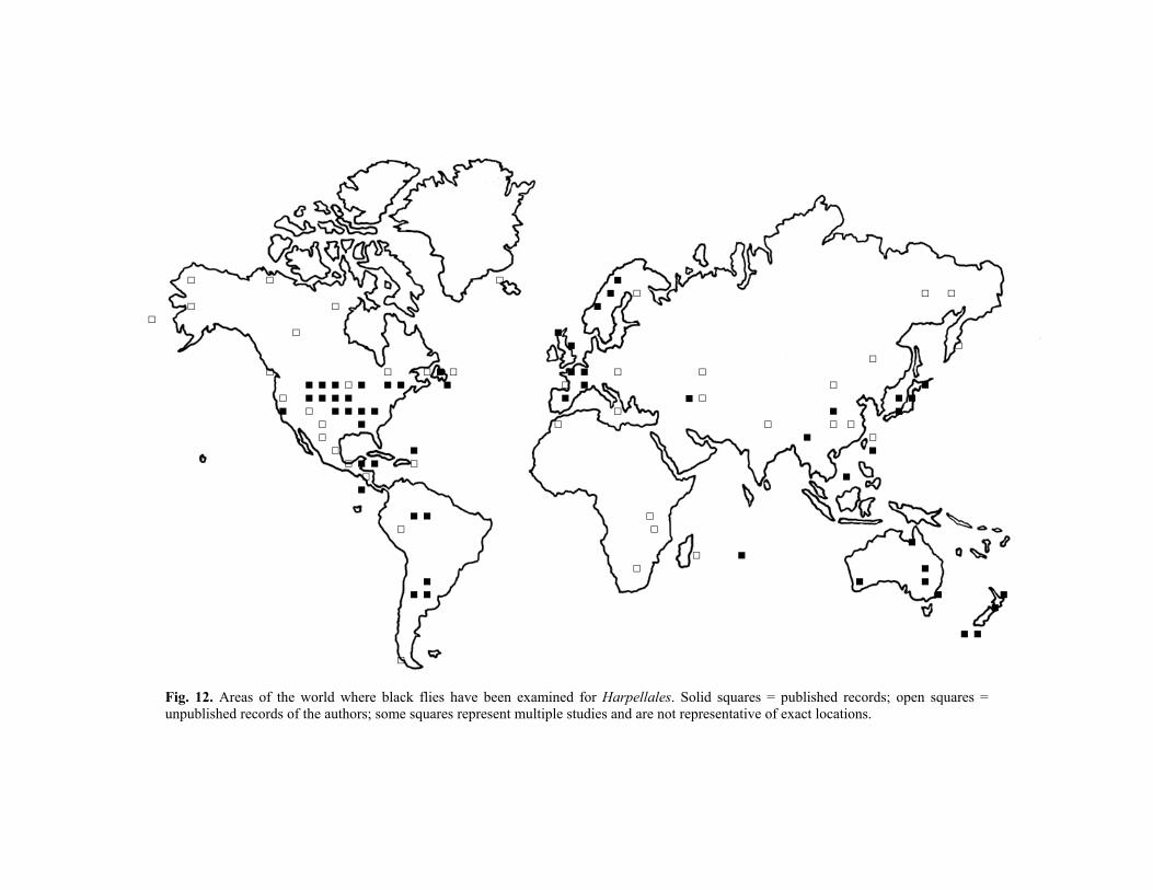
Fig. 12. Areas of the world where black flies have been examined for Harpellales. Solid squares = published records; open squares = unpublished records of the authors; some squares represent multiple studies and are not representative of exact locations.

Fungal Diversity
sampling efforts, which have been largely in the USA, Central America (Costa ica, Lichtwardt, 1997), South America (Argentina, Lichtwardt et al., 2000; razil, Alencar et al., 2003; Chile, Lichtwardt and Arenas, 1996), and western urope (England, Taylor et al., 1995; France, Manier, 1963) (Fig. 12). Large eographic areas, including Africa, Canada, many oceanic islands, and the riental and Palearctic Regions, remain poorly explored for Harpellales.
Areas of similar size with high species richness of Harpellales in black lies include Costa Rica (13 species, Lichtwardt, 1997); Alabama, USA (9 pecies, Nelder, 2003); and Armenia (8 species, Nelder et al., 2005a). Areas ith apparently low richness are Japan (3 species, Lichtwardt et al., 2001a) and orway (4 species, White and Lichtwardt, 2004). Although the Caucasus egion of Armenia is noted for high levels of endemic black flies, the gut
ymbiotes are typically widespread species (Nelder et al., 2005a). Australia nd New Zealand have high endemicity of black flies but low endemicity of arpellales (Lichtwardt and Williams, 1990; Williams and Lichtwardt, 1990). he lack of endemic gut symbiotes in larval black flies might reflect their lack f host specificity; an absence of host specificity would not constrain the eographic distribution of these symbiotes (Nelder et al., 2005a).
The distributions of widespread trichomycetes such as Harpella elusinae and Simuliomyces microsporus likely are limited only by the ispersal of adult black flies and the presence of suitable larval host habitats. owever, Simuliomyces microsporus is closely associated with other
ymbiotes (i.e., Paramoebidium spp., Pennella spp., and Genistellospora spp.) which they often attach their holdfast-like structures. Because some species
f Smittium can colonize several host families in various habitat types, their ispersal does not depend on a single host family; the geographic range of
RBEgO fswNRsaHTog mdHstoodthese species, therefore, is potentially greater (Nelder et al., 2005b). Taxonomy, classification, and evolutionary relationships Classification and phylogeny of Harpellales The class Trichomycetes is a member of a larger clade of Zygomycota that includes the Dimargaritales, Kickxellales, and possibly Zoopagales (Benny and White, 2001; Tanabe et al., 2004). The class Trichomycetes contains two fungal orders, the Asellariales and Harpellales (Benny and White, 2001; Cafaro, 2005). The Asellariales inhabit isopods (Isopoda) and springtails (Collembola) in terrestrial, freshwater, and intertidal habitats (Lichtwardt, 1986). They are not known from black flies or other aquatic insects, with the exception of a possible Asellariales record from one larva of Simulium ubiquitum Adler, Currie & Wood in Alabama (Nelder, 2003). The
151

order Harpellales comprises two families, the Harpellaceae and Legeriomycetaceae; however, molecular evidence suggests that these two families are not in separate clades (White, 2002). The Harpellaceae contain five genera, two of which (Harpella and rarely Stachylina) are found in black flies. The Legeriomycetaceae comprises 30 genera, seven of which (Genistellospora, Graminelloides, Pennella, Simuliomyces, Smittium, Stipella,
longer and ider in broth culture and in larval chironomids than in larvae of Simulium
t because species escriptions of Harpellales often rely on trichospore sizes from a single
dimensions of Harpella species, or example, change dramatically with the method of fixation (Kim, 2005). We
gge
and one undescribed genus (White et al., in press)) are found in black flies. The genera Genistellospora, Graminelloides, Harpella, Pennella, Simuliomyces, Stipella, and the undescribed genus are found exclusively in larval black flies, whereas Smittium and Stachylina are found in other host taxa. Morphological and molecular phylogenies within and among genera of the Harpellales are not congruent (Gottlieb and Lichtwardt, 2001; White, 2002). Phylogenetic relationships within the Harpellales have yet to be fully elucidated (Lichtwardt et al., 2003). The genus Smittium, for example, appears to be polyphyletic. More molecular work, such as that of White (2002), who used the gene sequences of 18S and 28S ribosomal DNA (rDNA) to infer a phylogeny of the Harpellales, is needed to resolve the evolutionary relationships within the Harpellales. Taxonomy of the Harpellales
Morphological characters used in trichomycete taxonomy include those of the trichospore, zygospore, generative cell, and holdfast, along with thallial growth form. Some characters routinely used to define species of Harpellales are subject to environmental influence. For example, the size of trichospores of species such as Smittium megazygosporum Manier & Coste varies with host and medium (Beard and Adler, 2000); trichospores are significantly winnoxium Comstock & Comstock. This finding is significandspecies, genus, or family of host. Trichosporefsu st that when new species are described, descriptions and, especially measurements, be made of both fresh and fixed (or stained) material. Preliminary efforts to distinguish species of Harpellales molecularly have used 5S rRNA (Walker, 1984), 18S rDNA (O’Donnell et al., 1998; Rizzo and Pang, 2005b), ITS and 18S rDNA (Gottlieb and Lichtwardt, 2001), and both 18S and 28S rDNA (White, 2002). Molecular techniques also offer promise for revealing hidden biodiversity (i.e., cryptic species) of Harpellales (White, 2002). Some species analyzed molecularly (e.g., Smittium culisetae) are composed of geographically and ecologically distinct populations,
152

Fungal Diversity
suggesting cryptic species (Peterson and Lichtwardt, 1987; Gottlieb and Lichtwardt, 2001). Harpella melusinae is probably a complex of cryptic species (Adler et al., 1996). All diagnostic and developmental stages of a fungus are seldom present in a single dissected host and identification of Harpellales by host association alone is poor practice (Lichtwardt, 1986). Therefore, trichomycetes from other hosts should be considered when identifying specimens from black flies. Axenic culturing is not possible for most species of Harpellales, making identification and description challenging. The investigator must rely on slide preparations, photomicrographs, and detailed notes and drawings of
A morphologically based dichotomous key to the Harpellales that orldwide is presented below. Moss (1981)
ublished the first key to simuliid trichomycetes, including only 9 of the 36 Harp
species,
representative fungal specimens. Slides treated with lactophenol and cotton blue are not permanent under most situations and do not provide long-term voucher or type specimens. Photomicrographs on high-quality (i.e., acid-free) paper and digital images on compact discs can be used in conjunction with semipermanent slides as a means of documenting specimens. Key to Harpellales colonizing larval black flies
colonize larval black flies wp
ellales species now known from black flies. The following key, compiled from original descriptions and the authors’ experience, follows the terminology of Lichtwardt (1986) and Lichtwardt et al. (2001a). The key pertains to fungal material from freshly killed larval black flies. It is useful only for sporulating thalli with mature trichospores (i.e., recently detached propagules or those near ready to detach) or zygospores. Thalli lacking trichospores are generally insufficient for species identification, even when thallial form or holdfast shape matches a described taxon. In these cases, identifications often can be made to the family or generic level. Only trichomycete species occurring naturally in larval black flies (i.e., not laboratory induced) are included in the key. In the laboratory, several species of Smittium isolated from non-simuliid hosts colonize and sporulate in larval black flies (Beard and Adler, 2003; Nelder et al., 2005b). All Smittiumtherefore, should be considered in making identifications. Other keys for identifying Harpellales are those of Lichtwardt (1973, 1986, 1997), Moss (1981), Lichtwardt and Arenas (1996), Lichtwardt et al. (1999), Misra and Lichtwardt (2000), Lichtwardt et al. (2001a), Valle and Santamaria (2004), and Ferrington et al. (2005).
153

Key to Harpellales 1. Thallus unbranched, attached
(F to peritrophic matrix or rarely to anterior of hindgut cuticle
. 14), w
immediately above holdfast ...................................................................................................... Harpella leptosa
Basal cell tapered gradually above holdfast (Fig. 15) .................................................................................. Harpella melusinae
de ..........................................................................................14 3. Trichospore greater than 3 µm wide..................................................................................15
amily Harpellaceae) .......................................................................................................2 1. Thallus branched, attached to hindgut cuticle, to other Harpellales (i.e., Pennella spp. or
Genistellospora spp.), or to Paramoebidium spp. (Family Legeriomycetaceae).................7 2. Trichospore cylindrical, often curled or coiled (Fig. 13), sometimes straight (Fig
ith 4 (rarely 2 or 3) appendages (Genus Harpella) ...........................................................3 2. Trichospore ovoid, straight, with 1 appendage (Genus Stachylina) .......Stachylina litoralis 3. Thallus twice as wide as trichospore ...............................................................Harpella tica 3. Thallus as wide as or narrower than trichospore .................................................................4 4. Basal cell tapered. Trichospore greater than or equal to 110 µm long ................................5 4. Basal cell rounded. Trichospore less than or equal to 100 µm long ....................................6 5. Thallus 4-6 µm wide. Trichospore 4.5 µm wide. Basal cell tapered
5. Thallus 6-10 µm wide. Trichospore 6-10 µm wide.
6. Trichospore 80-100 µm long by 4-8 µm wide. Holdfast wider than thallus.......................... .......................................................................................................... Harpella meridianalis 6. Trichospore 33-52 µm long by 3-4 µm wide. Holdfast narrower than thallus ...................... ............................................................................................................. Harpella amazonica 7. Trichospore with collar (Fig. 4) (Genus Smittium) ..............................................................8
. T7 richospore without collar (Fig. 16)..................................................................................21 8. Trichospore greater than 24 µm long...................................................................................9 8. Trichospore less than or equal to 24 µm long....................................................................12 9. Trichospore greater than or equal to 6 µm wide................................................................10 9. Trichospore less than or equal to 5 µm wide .....................................................................11 10. Holdfast and basal thallus region covered by mucilaginous sheath......... Smittium pennelli 10. Holdfast and basal thallus region not covered by mucilaginous sheath................................. ...........................................................................................................Smittium coloradense 11. Trichospore less than or equal to 30 µm long....................................................................12 11. Trichospore greater than or equal to 36 µm long......................Smittium megazygosporum 12. Trichospore with medial bulge, averaging more than 28 µm long .......... Smittium acicular 12. Trichospore without medial bulge, averaging less than 28 µm long Smittium tronadorium
3. Trichospore 3 µm or less wi11
154
Fungal Diversity
14. Holdfast limuloid. Trichospore 1.6 µm wide.......................................Smittium brasiliense 4. Holdfast not limuloid. Trichospore 2-3 µm wide ............................... Smittium dipterorum
15.
6. Trichospore widest proximal to midregion............................................. Smittium culisetae
7. Thalli with branching less compact. Widespread ................................ Smittium morbosum
8. Basal cell wider than thallus ..............................................................................................20
9. Generative cells 4-6 ....................................................................................Smittium culicis
20.
20. swelling ..................................................................................................... Smittium simulii
2. Trichospore ovoid or obpyriform, with thin appendages. Zygospore parallel to
zygosporophore. Known only from Europe ............................................... Stipella vigilans
3. Trichospore about 41 µm or more long .............................................................................25
Zygospore less than or equal to 80 µm long, 13.6 µm wide......................Pennella hovassi
5. Trichospore ovoid or obpyriform ......................................................................................26
6. Trichospore ovoid..............................................................................................................27
7. Holdfast pointed. Trichospore about 42 µm long by 14 µm wide ....... Pennella asymetrica
1
Collar less than or equal to 2.5 µm long............................................................................16 15. Collar 3 µm or more long ..................................................................................................18
116. Trichospore widest at midregion .......................................................................................17
17. Thalli with compact branching. Known from Chile ...............................Smittium imitatum 1 18. Basal cell as wide as or narrower than thallus ...................................................................19 1 19. Generative cells 1-4 ............................................................................Smittium culicisoides 1
Holdfast and basal region composed of 6 cells arranged in ring. Trichospore elongate, oval ..................................................................................................... Smittium annulatum Holdfast and basal region arched, wrench-like. Trichospore cylindrical with medial
21. Holdfast surrounded by mucilaginous sheath (Fig. 17) .....................................................22 21. Holdfast not surrounded by mucilaginous sheath..............................................................29 2
zygosporophore. Widespread (Genus Pennella)................................................................23 22. Trichospore cylindrical, with petiolate appendages. Zygospore perpendicular to
23. Trichospore less than 41 µm long......................................................................................24 2 24. Trichospore obpyriform (Fig. 18), less than 33 µm long, less than or equal to 7 µm wide.
24. Trichospore ovoid, greater than or equal to 28 µm long, 7 µm wide. Zygospore greater than or equal to 74 µm long, 16 µm wide...................................................Pennella simulii
225. Trichospore cylindrical (Fig. 19) .......................................................................................28 226. Trichospore obpyriform..............................................................................Pennella arctica 227. Holdfast bifurcated (Fig. 20). Trichospore about 60 µm long by 8-19 µm wide (Fig. 21).... ................................................................................................................ Pennella montana
155
28. Trichospore about 80 µm long, 4 µm wide.......................................Pennella angustispora 28. Trichospore less than or equal to 59 µm long, greater than 4.5 µm wide.. Pennella grassei 29. Holdfast C shaped, with refractive material (Fig. 22). Trichospore ovoid or obpyriform
(Genus Genistellospora)....................................................................................................30 29. Holdfast pointed or not C shaped; if C shaped, without refractive material. Trichospore
0. Trichospore about 38 µm or less long ...............................................................................31
1. Zygospore 50-70 µm long, less than or equal to 15 µm wide............................................32
2. Trichospore greater than or equal to 9 µm wide.......................... Genistellospora tepidaria
3. Trichospore greater than 50 µm long. Thallus less than 250 µm long...................................
33.
4. Trichospore typically with terminal cap or fine filaments.....................................................
35.
rela
taxo
e the estimation of basic community parameters such as species richness,
biconical or cylindrical ......................................................................................................34 330. Trichospore greater than or equal to 39 µm long...............................................................33 331. Zygospore about 100 µm long, 20 µm wide (Fig. 23).......... Genistellospora homothallica 332. Trichospore less than or equal to 8 µm wide ...............................Genistellospora tropicalis 3 ...........................................................................................Genistellospora guanacastensis
Trichospore less than 41 µm long. Thallus greater than 400 µm long................................... ......................................................................................................... Genistellospora nubila
3 ..................................................... Undescribed genus and species (White et al., in press) 34. Trichospore without terminal cap or fine filaments...........................................................35 35. Trichospore biconical. Thallus attached to hindgut cuticle .......... Graminelloides biconica
Trichospore cylindrical (Fig. 24). Thallus typically attached to Paramoebidium spp. (Fig. 25), Genistellospora spp., Pennella spp. (rarely hindgut cuticle).......................................... .................................................................................................. Simuliomyces microsporus
Future directions and questions
Since the discovery of trichomycetes in black flies in 1929, this tionship has fascinated scientists and led to a better understanding of
symbioses in aquatic systems. Most studies of trichomycetes have focused on nomy, culturing, host records, life cycles, and geographic distributions.
Little is known of the community structure of trichomycetes in black flies or other hosts. A goal of future studies of trichomycete ecology, therefore, should bdiversity, dominance, species turnover, and rank abundance distributions. Characterizing ecological patterns can illuminate the underlying mechanisms responsible for these patterns. Future research also should consider how these parameters vary across both temporal and spatial scales. For example, what is the relationship between local trichomycete species richness (e.g., in a stream)
156

Fungal Diversity
Figs. 13-15. Species of Harpella in larval black flies. 13. Harpella melusinae in Simulium tuberosum. Bar = 30 µm. 14. Harpella sp. in Simulium innoxium; st = straight trichospores. Bar = 100 µm. 15. Harpella melusinae in Simulium innoxium; hf = holdfasts. Bar = 100 µm.
157
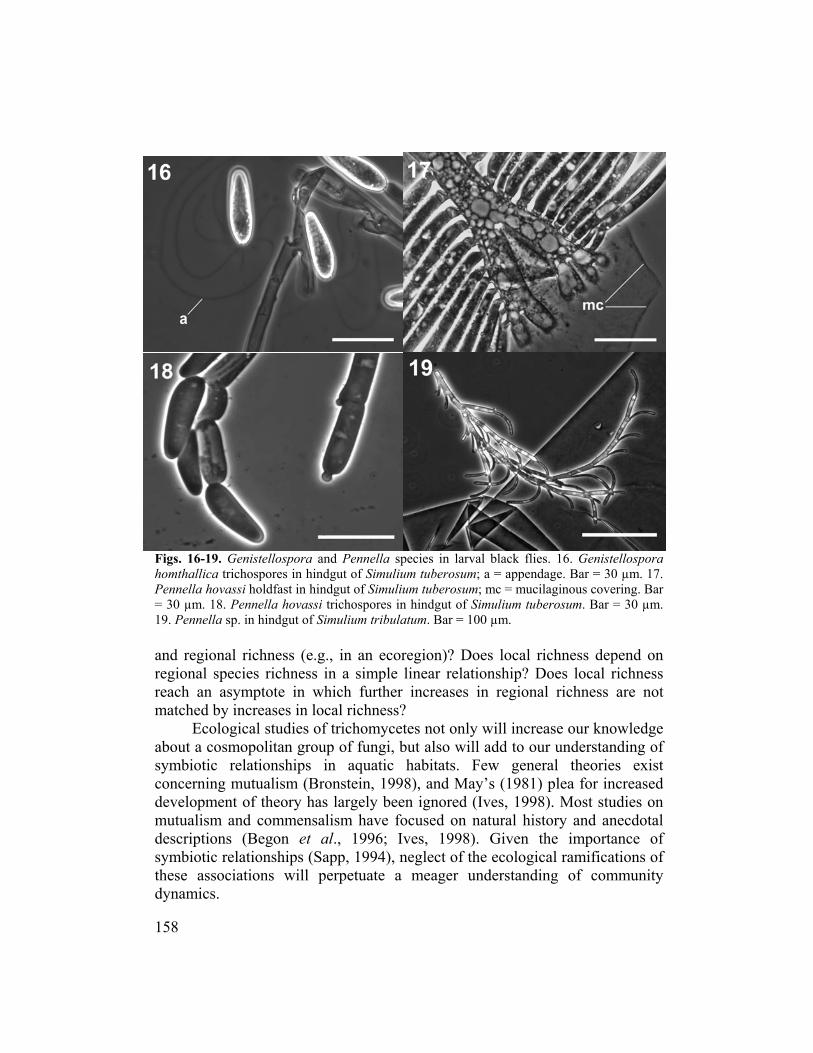
Figs. 16-19. Geni Genistellospora hom allica tric ar = 30 µm. 17. Pennella hovassi nous covering. Bar = 30 µm. 18. . Bar = 30 µm. 19. ennella sp. i
Does local richness depend on Does local richness
reach an asymma hed by i
about a cosmsymbconcerning mdevelopm
utualism
stellospora and Pennella species in larval black flies. 16. hospores in hindgut of Simulium tuberosum; a = appendage. B holdfast in hindgut of Simulium tuberosum; mc = mucilagi
th
P
tc
m
Pennella hovassi trichospores in hindgut of Simulium tuberosumn hindgut of Simulium tribulatum. Bar = 100 µm.
and regional richness (e.g., in an ecoregion)?regional species richness in a simple linear relationship?
ptote in which further increases in regional richness are not ncreases in local richness?
Ecological studies of trichomycetes not only will increase our knowledge opolitan group of fungi, but also will add to our understanding of
iotic relationships in aquatic habitats. Few general theories exist utualism (Bronstein, 1998), and May’s (1981) plea for increased
ent of theory has largely been ignored (Ives, 1998). Most studies on and commensalism have focused on natural history and anecdotal
descriptions (Begon et al., 1996; Ives, 1998). Given the importance of symbiotic relationships (Sapp, 1994), neglect of the ecological ramifications of these associations will perpetuate a meager understanding of community dynamics.
158
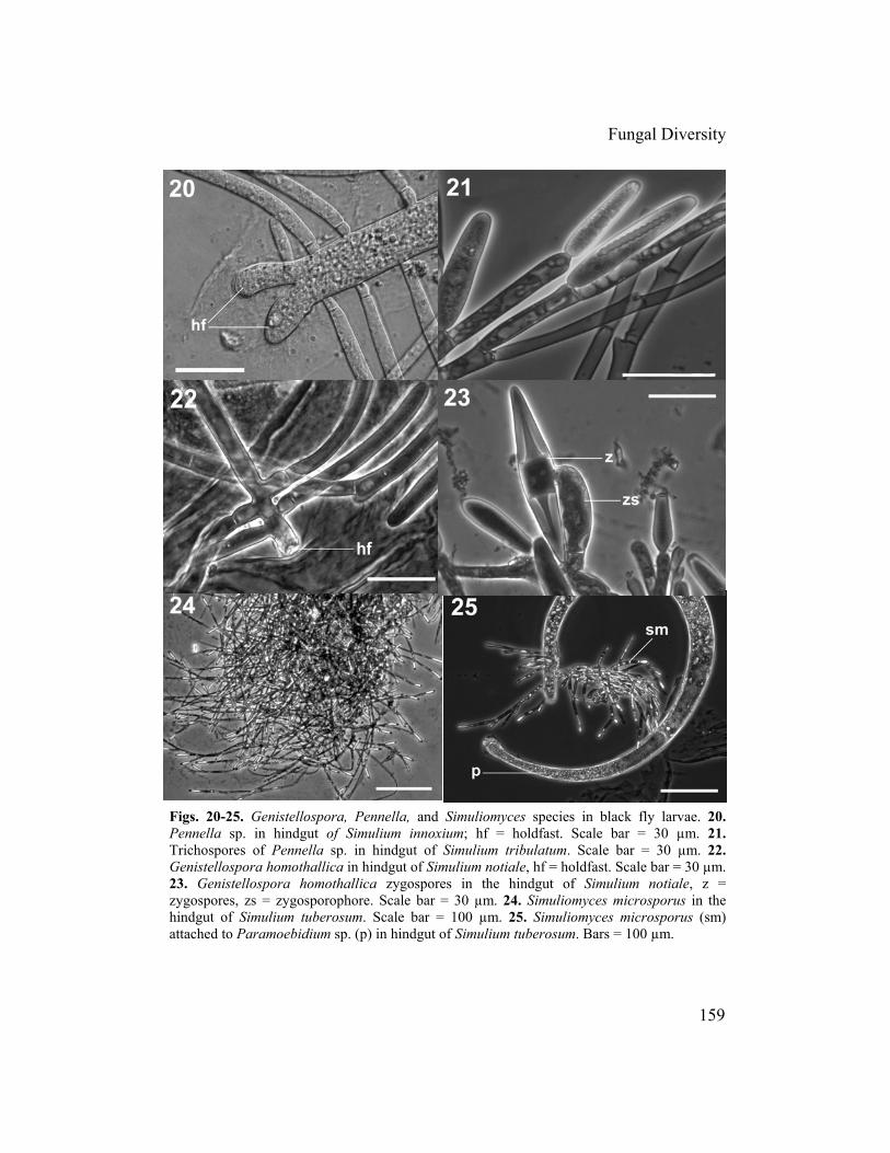
Fungal Diversity
Figs. 20-25. Genistellospora, Pennella, and Simuliomyces species in black fly larvae. 20. Pennella sp. in hindgut of Simulium innoxium; hf = holdfast. Scale bar = 30 µm. 21. Trichospores of Pennella sp. in hindgut of Simulium tribulatum. Scale bar = 30 µm. 22. Genistellospora homothallica in hindgut of Simulium notiale, hf = holdfast. Scale bar = 30 µm. 23. Genistellospora homothallica zygospores in the hindgut of Simulium notiale, z = zygospores, zs = zygosporophore. Scale bar = 30 µm. 24. Simuliomyces microsporus in the hindgut of Simulium tuberosum. Scale bar = 100 µm. 25. Simuliomyces microsporus (sm) attached to Paramoebidium sp. (p) in hindgut of Simulium tuberosum. Bars = 100 µm.
159
Reasons for the production of zygosvarian cysts are produced rema
some Harpellales and not in others? Wygospore production?sistant stage of the Harpellales
roduction of ovarian cysts might explaiopulations of cyst-infected black flies ractical perspective, an increase in ouetermine if they might be used as a estiferous black flies.
An understanding of trichomycete
pores and the mechanism by which in unknown. Why are zygospores produced in
hat are the biological cues that induce Does the zygospore represent an environmentally
life cycle? A deeper understanding of the n why some areas of the world have
while others apparently do not. From a r knowledge of ovarian cysts will help
possible means of biological control of
fitness, including spore production,
o
zrepppdp relies partly on a better understanding
e host gut). Although the physiology e same cannot be said for black flies. W
ssential eleme fungi or vice-versa.
A more complete picture of the biodiith black flies is needed. Global arpellalesnexplored. Parallel ecological work inst of the ecological patterns and hypo
of the physiology of their habitat (i.e., of the mosquito gut is well understood,
e do not fully understand the ents (e.g., nutritional) that are provided to the simuliid host by
versity of the gut mycota associated prospecting for simuliid-associated
should pay dividends. Areas such as Africa and Asia are virtually these areas of the world will provide a theses that are based largely on North
ththeth wHuteAmerican simuliid-trichomycete relationships.
Because the species is the ecologicalecies in any investigation is critical.
f cryptic (sibling) species of Harpellalesxpanding field of molecular biology is likiscovering and resolving complexes ofecies. Similarly, phylogenetic studies of the
enefit from molecular investigations, tegrated with other approaches, inclu
cknowledgements
unit of study, the detection of all New methods for detecting the presence
, therefore, are needed. The rapidly ely to afford multiple approaches for
morphologically similar trichomycete Harpellales will continue to
particularly as molecular studies are ding classical morphological studies.
spoedspbin A
This research was financed by National Science Foundation grants DEB-9629456 awarded to PHA and DEB-0075269 awarded to JWM, PHA, CEB, and T. McInnis. We thank M.H. Colbo and S. Vojvodic for sharing their unpublished data. We also thank G.R. Carner, R.W. Lichtwardt, A.G. Wheeler, and an anonymous reviewer for critiquing an earlier version of the manuscript. This is Technical Contribution No. 5169 of the Clemson University Experiment Station.
160

Fungal Diversity
References Adler, P.H. and McCreadie, J.W. (1997). The hidden ecology of black flies: sibling species and
ecological scale. American Entomologist 43: 153-161. Adler, P.H., Wang, Z. and Beard, C.E. (1996). First records of natural enemies from Chinese
blackflies (Diptera: Simuliidae). Medical Entomology and Zoology 47: 291-292. Adler, P.H., Currie, D.C. and Wood, D.M. (2004). The Black Flies (Simuliidae) of North
America. Cornell University Press, Ithaca, NY. Adler, P.H., Giberson, D.J. and Purcell, L.A. (2005). Insular black flies (Diptera: Simuliidae)
of North America: tests of colonization hypotheses. Journal of Biogeography 32: 211-220.
Alencar, Y.B., Ríos-Velásquez, C.M., Lichtwardt, R.W. and Hamada, N. (2003).
(2002). Seasonality of trichomycetes in larval black flies from
cology: Individuals, Populations and
.R. (2001). In situ analysis of pH gradients in mosquito larvae using non-invasive, self-
sitive micro electrodes. Journal of Experimental Biology 204: 691-
ss, Oxford, UK. ronstein, J.L. (1998). The contribution of ant-plant protection studies to our understanding of
mutualism. Biotropica 30: 150-161.
Trichomycetes (Zygomycota) in the digestive tract of arthropods in Amazonas, Brazil. Memórias do Instituto Oswaldo Cruz 98: 799-810.
Beard, C.E. (2002). Colonization of blackflies (Diptera: Simuliidae) by trichomycete fungi (Zygomycota) in South Carolina, USA. Ph.D. thesis, Clemson University, SC.
Beard, C.E. and Adler, P.H. (2000). Bionomics, axenic culture, and substrate-related variation in trichospores of Smittium megazygosporum. Mycologia 92: 296-300.
Beard, C.E. and Adler, P.H.South Carolina, USA. Mycologia 94: 200-209.
Beard, C.E. and Adler, P.H. (2003). Zygospores of selected trichomycetes in larval Diptera of the families Chironomidae and Simuliidae. Mycologia 95: 317-320.
Beard, C.E., McCreadie, J.W. and Adler, P.H. (2003). Prevalence of the trichomycete fungus Harpella melusinae (Harpellales: Harpellaceae) in larval black flies (Diptera: Simuliidae) across a heterogeneous environment. Mycologia 95: 577-583.
Begon, M., Harper, J.L. and Townsend, C.R. (1996). ECommunities. Blackwell Science, Oxford, UK.
Benny, G.L. (2001). Zygomycota: Trichomycetes. In: The Mycota. VII. Part A. Systematics and Evolution (eds. D.J. McLaughlin, E.G. McLaughlin and P.A. Lemke). Springer-Verlag, Berlin: 147-160.
Benny, G.L. and O'Donnell, K. (2000). Amoebidium parasiticum is a protozoan, not a Trichomycete. Mycologia 92: 1133-1137.
Benny, G.L. and White, M.M. (2001). The classification and phylogeny of Trichomycetes and Zygomycetes. In: Trichomycetes and Other Fungal Groups (eds. J.K. Misra and B.W. Horn). Science Publishers, Enfield, NH: 39-53.
Boudko, D.Y., Moroz, L.L., Linser, P.J., Trimarchi, J.R., Smith, P.J.S. and Harvey, W
referencing, pH sen699.
Bradley, T.J. (1985). The excretory system: structure and physiology. In: Comprehensive Insect Physiology, Biochemistry, and Pharmacology. Vol. 4 (eds. G.A. Kerkut and L.I. Gilbert). Pergamon Press, New York: 421-465.
Brassard, G.R., Frost, S., Laird, M., Olsen, O.A. and Steele, D.H. (1971). Studies of the spray zone of Churchill Falls, Labrador. Biological Conservation 4: 13-18.
Brönmark, C. and Hansson, L.A. (2005). The Biology of Lakes and Ponds. 2nd edn. Oxford University Pre
B
161

Cafaro, M.J. (2002). Species richness patterns in symbiotic gut fungi (Trichomycetes). Fungal Diversity 9: 47-56.
ed on rDNA sequences. Ph.D.
Cafaro early
Cavali of life. Biological Reviews 73: 203-
Chadeles. In: Traité de Botanique Systématique, Vol. 1, Sec. 5: Masson et Cie, Paris:
Chapm
adaires
Colbo, : Blackflies: the
Crossk .
story Museum, London.
. Chapman & Hall, London: 31-40.
Edmanae). Journal of Medical Entomology 22: 1-17.
572.
ia 68: 573-582.
nd South East England. Ph.D. thesis, University of London, UK.
Cafaro, M.J. (2003). Systematics of the Trichomycetes as an ecological group with emphasis on the phylogeny of Eccrinales and Asellariales basthesis, University of Kansas, Lawrence, KS. , M. (2005). Eccrinales (Trichomycetes) are not fungi, but a clade of protists at thedivergence of animals and fungi. Molecular Phylogenetics and Evolution 35: 21-34. er-Smith, T. (1998). A revised six-kingdom system266.
faud, M. and Emberger, L. (1960). Les Trichomycètes: Amoebidiales, Eccrinales et Harpella895-902. an, M.E. (1966). Isolation and experimental studies on some Trichomycetes. M.A. thesis, University of Kansas, Lawrence, KS.
Chatton, E. and Roubaud, E. (1909). Sur un Amoebidium du rectum des larves de Simulies (Simulium argyreatum Meig. et S. fasciatum Meig.). Comptes Rendus Hebdomdes Séances et Mémoires de la Société de Biologie 66: 701-703. M.H. and Wotton, R.S. (1981). Preimaginal black fly bionomics. InFuture for Biological Methods in Integrated Control (ed. M. Laird). Academic Press, New York: 209-225.
Crosby, T.K. (1974). Trichomycetes (Harpellales) of New Zealand Austrosimulium larvae (Diptera: Simuliidae). Journal of Natural History 8: 187-192.
Crosby, T.K. (1980). Invertebrate animal studies from Campbell Island. New Zealand Department of Land Survey Reserves Series 7: 99-100. ey, R.W. (1990). The Natural History of Blackflies. John Wiley & Sons, NY
Crosskey, R.W. and Howard, T. (1997). A New Taxonomic and Geographical Inventory of Blackflies (Diptera: Simuliidae). The Natural Hi
Cupp, E.W. and Ramberg, F.B. (1997). Care and maintenance of blackfly colonies. In: The Molecular Biology of Insect Disease Vectors: a Methods Manual (eds. J.M. Crampton, C.B. Beard and C. Louis)
Dogma, I.J., Jr. (1975). Of Philippine mycology and lower fungi. Kalikasan, Philippine Journal of Biology 4: 69-105.
Donahue, W.F. and Schindler, D.W. (1998). Diel emigration and colonization responses of black fly larvae (Diptera: Simuliidae) to ultraviolet radiation. Freshwater Biology 40: 357-365.
Duboscq, O., Léger, L. and Tuzet, O. (1948). Contribution à la connaissance des Eccrinides: les Trichomycètes. Archives de Zoologie Expérimentale et Générale 86: 29-144. , J.D. and Simmons, K.R. (1985a). Rearing and colonization of black flies (Diptera: Simuliid
Edman, J.D. and Simmons, K.R. (1985b). Simuliids (mainly Simulium decorum Walker). In: Handbook of Insect Rearing. Vol. II. (eds. P. Singh and R.F. Moore). Elsevier, Amsterdam, Netherlands: 145-152.
El-Buni, A.M. and Lichtwardt, R.W. (1976a). Asexual sporulation and mycelial growth in axenic cultures of Smittium spp. (Trichomycetes). Mycologia 68: 559-
El-Buni, A.M. and Lichtwardt, R.W. (1976b). Spore germination in axenic cultures of Smittium spp. (Trichomycetes). Mycolog
El-Sherif, H.K. (1975). Microsporidian and fungal infections of larval blackfly (Simuliidae) in rivers and streams of North Wales a
162

Fungal Diversity
Eymann, M. (1991). Dispersion patterns exhibited by larvae of the black flies Cnephia dacotensis and Simulium rostratum (Diptera: Simuliidae). Aquatic Insects 13: 99-106.
Frost, ycetes (Harpellales: Harpellaceae and
ium damnosum in the Bong Range, Liberia. Zeitschrift für Tropenmedizin und
Garmsin Guatemala, with reference to the transmission of Onchocerca volvulus.
Girbal tes (Fungi, Zygomycotina) comensals de
Gottlie and among species of
Gray, an, Animal, and Plant Pathogen Vectors (eds. K. Maramorosch
Grenie s Simulies. Bulletin de la
Griffin the Platte
Grigg, Ph.D. thesis,
Grigg,
y Magazine 94: 14-16.
KS.
ecies.
Horn, n host-mediated sporangiospore
Horn, ions of Trichomycetes to the insect gut: Smittium
lisetae (Trichomycetes). Mycologia
Ingold
Ferrington, L.C., Lichtwardt, R.W., Hayford, B. and Willams, M.C. (2005). Symbiotic Harpellales (Trichomycetes) in Tasmanian aquatic insects. Mycologia 97: 254-262. S. and Manier, J.-F. (1971). Notes on TrichomGenistellaceae) in larval blackflies (Diptera: Simuliidae) from Newfoundland. Canadian Journal of Zoology 49: 776-778.
Garms, R. (1973). Quantitative studies on the transmission of Onchocerca volvulus by SimulParasitologie 24: 358-372. , R. (1975). Observations on filarial infections and parous rates of anthropophilic blackflies Tropenmedizin und Parasitologie 26: 169-182.
, J. and Santamaria, S. (1998). Trichomycelarves de Simuliidae (Diptera) a la Peninsula Iberica. Acta Botanica de Barcelona 45: 49-56. b, A.M. and Lichtwardt, R.W. (2001). Molecular variation withinHarpellales. Mycologia 93: 66-81. E.W. and Noblet, R. (1999). Large scale laboratory rearing of black flies. In: Maintenance of Humand F. Mahmood). Oxford & IBH Publishing, New Delhi, India: 85-105. r, P. (1944). Remarques sur la biologie de quelques ennemis deSociété Entomologique de France 49: 130-133. , D.H. (1994). Fungal Physiology. 2nd edn. John Wiley, New York.
Grigg, R.D. (1988). Growth and distribution patterns of selected Trichomycetes onRiver floodplain of Central Nebraska. M.S. thesis, Kearney State College, Kearney, NE. R.D. (1994). Isozyme variation in selected cultured Trichomycetes.University of Kansas, Lawrence. KS. R.D. and Lichtwardt, R.W. (1996). Isozyme patterns in cultured Harpellales. Mycologia 88: 219-229.
Hinton, H.E. (1958). The pupa of the fly Simulium feeds and spins its own cocoon. Entomologist’s Monthl
Horn, B.W. (1980). Studies on the nutritional relationship of larval Aedes aegypti (Diptera: Culicidae) with Smittium culisetae (Trichomycetes). M.A. thesis, University of Kansas, Lawrence,
Horn, B.W. (1989a). Requirement for potassium and pH shift in host-mediated sporangiospore extrusion from trichospores of Smittium culisetae and other Smittium spMycological Research 93: 303-313.
B.W. (1989b). Physiological and ultrastructural studies oextrusion from trichospores of Smittium culisetae and other Smittium species. Ph.D. thesis, University of Kansas, Lawrence, KS. B.W. (2001). Physiological adaptatculisetae and its mosquito host. In: Trichomycetes and Other Fungal Groups (eds. J.K. Misra and B.W. Horn). Science Publishers, Enfield, NH: 99-116.
Horn, B.W. and Lichtwardt, R.W. (1981). Studies on the nutritional relationship of larval Aedes aegypti (Diptera: Culicidae) with Smittium cu73: 724-740. , C.T. (1967). Why not look for Harpellales. Bulletin of the British Mycological Society 1: 43-44.
163

Ives, A.R. (1998). Population ecology. In: Ecology (eds. S.I. Dodson, T. Allen, S.R. Carpenter, A.R. Ives, R. Jeanne, J.K. Kitchell, N. Langston and M.G. Turner). Oxford University
Kim, eir
Kim, S.-K. and Adler, P.H. (2005). Multiple symbiotes in larval black flies (Diptera:
Labey Lichtwardt, R.W. (1996). An investigation of Harpellales
Lacey, and midgut histopathology of Bacillus
Léger, L. and Duboscq, O. (1929). Harpella melusinae n. g. sp. entophyte eccriniforme
f the Academy of
Lewis,
Lewis, ium damnosum in the southern Cameroons and
Lewis, Observations on the Simulium neavei complex at Amani in Tanganyika.
Lewis, m population of the Kumba area in West
Lichtwal of Botany 51: 836-842.
Lichtw era and species of Harpellales (Trichomycetes)
Lichtwan). Academic Press, New York:
Lichtw
Lichtw 96). Trichomycetes and the arthropod gut. In: The Mycota, Animal and
Press, New York: 235-315. S.-K. (2005). Ecology and phylogeny of black flies (Diptera: Simuliidae) and thtrichomycete (Zygomycota: Trichomycetes) symbiotes. Ph.D. thesis, Clemson University, Clemson, SC.
Simuliidae): associations of trichomycete fungi with ichthyosporeans, mermithid nematodes, and microsporidia. Journal of Invertebrate Pathology 88: 247-253.
rie, E.S., Molloy, D.P. and (Trichomycetes) in New York State blackflies (Diptera: Simuliidae). Journal of Invertebrate Pathology 68: 293-298. L.A. and Federici, B.A. (1979). Pathogenesis thuringiensis in Simulium vittatum (Diptera: Simuliidae). Journal of Invertebrate Pathology 33: 171-182.
parasite des larves de Simulie. Comptes Rendus Hebdomadaires des Séances de l'Académie des Sciences Paris 188: 951-954.
Léger, L. and Gauthier, M. (1932). Endomycètes nouveaux des larves aquatiques d'Insectes. Comptes Rendus Hebdomadaires des Séances de l'Académie des Sciences Paris 194: 2262-2265.
Lehane, M.J. (1997). Peritrophic matrix structure and function. Annual Review of Entomology 42: 525-550.
Leidy, J. (1849). Enterobrus, a new genus of Confervaceae. Proceedings oNatural Sciences of Philadelphia 4: 225-233. D.J. (1953). Simulium damnosum and its relation to onchocerciasis in the Anglo-Egyptian Sudan. Bulletin of Entomological Research 43: 597-644. D.J. (1960a). Observations on SimulLiberia. Annals of Tropical Medicine and Parasitology 54: 208-223. D.J. (1960b).Bulletin of Entomological Research 51: 95-113. D.J. (1965). Features of the Simulium damnosuCameroon. Annals of Tropical Medicine and Parasitology 59: 365-374. ardt, R.W. (1964). Axenic culture of two new species of branched Trichomycetes. American Journ
Lichtwardt, R.W. (1967). Zygospores and spore appendages of Harpella (Trichomycetes) from larvae of Simuliidae. Mycologia 59: 482-491. ardt, R.W. (1972). Undescribed genfrom the guts of aquatic insects. Mycologia 64: 167-197. ardt, R.W. (1973). Trichomycetes. In: The Fungi: an Advanced Treatise. Vol. IVB. (eds. G.C. Ainsworth, F.K. Sparrow and A.S. Sussm237-243. ardt, R.W. (1984). Species of Harpellales living within the guts of aquatic Diptera larvae. Mycotaxon 19: 529-550.
Lichtwardt, R.W. (1986). The Trichomycetes: Fungal Associates of Arthropods. Springer-Verlag, New York. ardt, R.W. (19Human Relations (eds. D. Howard and D. Miller). Springer-Verlag, New York: 315-330.
164

Fungal Diversity
Lichtwardt, R.W. (1997). Costa Rican gut fungi (Trichomycetes) infecting lotic insect larvae. Revista de Biología Tropical 45: 1339-1383. ardt, R.W. (2000). Gut Lichtw fungi of invertebrates. In: Monteverde: Ecology and
Lichtw 1996). Trichomycetes in aquatic insects from southern Chile.
Lichtwstreams.
Lichtw8: 1057-1074.
Lichtw , M.C. (1992b). Smittium bullatum from a New Zealand midge
Lichtw stern Australian species of Smittium and
Lichtwical Society of Japan 28: 359-412.
quatic insect larvae. Mycologia 91: 1060-1082.
a. Mycologia 92: 332-340.
the Internet at
Lichtw n Newfoundland
Lichtw ). Freshwater trichomycetes and their
) from Newfoundland, Canada. Journal of
Manie cètes. Annales des Sciences Naturelles
Manie ycètes. Annales des Sciences
Manies Sciences Naturelles Botanique, Paris, Série 12 4: 737-750.
ris, Série 12 9: 93-108.
s Naturelles Botanique, Paris, Série 12 10: 469-471.
emistry and Physiology 82B: 37-39.
Conservation of a Tropical Cloud Forest. (eds. N.M. Nadkarni and N.T. Wheelwright). Oxford University Press, New York: 83-85. ardt, R.W. and Arenas, J. (Mycologia 88: 844-857. ardt, R.W. and Williams, M.C. (1988). Distribution and species diversity of trichomycete gut fungi in aquatic insect larvae in two Rocky Mountain Canadian Journal of Botany 66: 1259-1263. ardt, R.W. and Williams, M.C. (1990). Trichomycete gut fungi in Australian aquatic larvae. Canadian Journal of Botany 6
Lichtwardt, R.W. and Williams, M.C. (1992a). Tasmanian Trichomycete gut fungi in aquatic insect larvae. Mycologia 84: 384-391. ardt, R.W. and Williamslarva and new records of other trichomycete gut fungi. Canadian Journal of Botany 70: 1193-1195. ardt, R.W. and Williams, M.C. (1992c). Weother Trichomycetes in aquatic insect larvae. Mycologia 84: 392-398. ardt, R.W., Kobayasi, Y. and Indoh, H. (1987). Trichomycetes of Japan. Transactions of the Mycolog
Lichtwardt, R.W., Ferrington, L.C., Jr. and López Lastra, C. (1999). Trichomycetes in Argentinean a
Lichtwardt, R.W., López Lastra, C. and Mazzucchelli, M.G. (2000). Fungi living in the guts of larval aquatic insects in northwestern Argentin
Lichtwardt, R.W., Cafaro, M.J. and White, M.M. (2001a). The Trichomycetes: Fungal Associates of Arthropods, revised edition. Published onwww.nhm.ku.edu/~fungi. ardt, R.W., White, M.M. and Colbo, M.H. (2001b). Harpellales iaquatic insect larvae. Mycologia 93: 764-7873. ardt, R.W., White, M.M. and Cafaro, M.J. (2003arthropod hosts. In: Freshwater Mycology (eds. C.K.M. Tsui and K.D. Hyde). Fungal Diversity Research Series 10: 81-100.
Malone, K.M. and Nolan, R.A. (1978). Aerobic bacterial flora of the larval gut of the black fly Prosimulium mixtum (Diptera: SimuliidaeMedical Entomology 14: 641-645.
r, J.-F. (1950). Recherches sur les TrichomyBotanique 11: 53-162.
r, J.-F. (1955). Classification et nomenclature des TrichomNaturelles Zoologie, Série 11 17: 395-397.
r, J.-F. (1963). Trichomycètes de larves de Simulies (Harpellales du proctodeum). Annales de
Manier, J.-F. (1968). Validation de Trichomycètes par leur diagnose latine. Annales des Sciences Naturelles Botanique, Pa
Manier, J.-F. (1969-1970). Changement de nom pour Eccrinaflexilis Léger et Duboscq, 1906. Annales des Science
Martin, M.M., Kukor, J.J., Martin, J.S. and Merritt, R.W. (1985). The digestive enzymes of larvae of the black fly, Prosimulium fuscum (Diptera, Simuliidae). Comparative Bioch
165

May, R.M., ed. (1981). Theoretical Ecology – Principles and Applications. Sinauer Associates, Oxford.
Mayfield, S.D. and Lichtwardt, R.W. (1980). Comparative study of the holdfast structure in four Trichomycetes. Canadian Journal of Botany 58: 1074-1087. adie, J.W. and Adler, P.H. (1998). Scale,McCre time, space, and predictability: species
McCre icrodistribution of the trichomycete Smittium
McCre 5). Context-dependent symbiosis between
Moss, S.T. (1970). Trichomycetes inhabiting the digestive tract of Simulium equinum larvae.
Moss, with Simuliidae
Moss, bionts of Insecta. Botanical Journal of
Moss, age in the life cycle of
Moss, in
Moss, . (1977). Zygospores of the Harpellales: an ultrastructural
Moss, ptosa, a new species of Trichomycetes
Nelderal Alabama. M.S. thesis, University of South
Nelderae) and their effect on Trichomycete (Zygomycota)
omycetes) inhabiting larvae of Simulium
in the Caucasus of Armenia?
Nelder estigations of
distributions of preimaginal black flies (Diptera: Simuliidae). Oecologia 114: 79-92. adie, J.W. and Beard, C.E. (2003). The mculisetae in the hindgut of the black fly host Simulium vittatum. Mycologia 95: 998-1003. adie, J.W., Beard, C.E. and Adler, P.H. (200black flies (Diptera: Simuliidae) and trichomycete fungi (Harpellales: Legeriomycetaceae). Oikos 108: 362-370.
Misra, J.K. (1998). Trichomycetes - fungi associated with arthropods: review and world literature. Symbiosis 42: 179-220.
Misra, J.K. and Lichtwardt, R.W. (2000). Illustrated Genera of Trichomycetes: Fungal Symbionts of Insects and Other Arthropods. Science Publishers, Enfield, NH.
Transactions of the British Mycological Society 54: 1-13. S.T. (1981). Key to the genera and species of Trichomycetes associatedlarvae. Newsletter of the British Simuliid Group 5: 1-8. S.T. (1998). Harpellales (Trichomycetes); mycoScotland 50: 137-152. S.T. and Descals, E. (1986). A previously undescribed stHarpellales (Trichomycetes). Mycologia 78: 213-222. S.T. and Lichtwardt, R.W. (1976). Development of trichospores and their appendages Genistellospora homothallica and other Harpellales and fine-structural evidence for the sporangial nature of trichospores. Canadian Journal of Botany 54: 2346-2364. S.T. and Lichtwardt, R.Wstudy. Canadian Journal of Botany 55: 3099-3110. S.T. and Lichtwardt, R.W. (1980). Harpella lesubstantiated by electron microscopy. Canadian Journal of Botany 58: 1035-1044. , M.P. (2003). The ecology of Trichomycete (Zygomycota) fungi inhabiting black fly larvae (Diptera: Simuliidae) of coastAlabama, Mobile, AL. , M.P. and McCreadie, J.W. (2003). Bdelloid rotifers (Rotifera: Bdelloidea) inhabiting larval black flies (Diptera: Simuliidfungal abundance. Proceedings of the Entomological Society of Washington 105: 794-796.
Nelder, M.P., McCreadie, J.W., Coscaron, C. and Brockhouse, C.L. (2004). First report of a trichomycete fungus (Zygomycota: Trichochraceum sensu lato Walker (Diptera: Simuliidae) from the Galapagos Islands. Journal of Invertebrate Pathology 87: 39-44.
Nelder, M.P., Adler, P.H. and Kachvoryan, E.A. (2005a). Do gut symbiotes reflect the endemism of their host black flies (Diptera: Simuliidae) Journal of Biogeography 32: 1333-1341. , M.P., McCreadie, J.W. and Beard, C.E. (2005b). Laboratory invtrichomycete prevalence, abundance and fecundity in a Smittium-simuliid model. Mycologia 97: 338-345.
166

Fungal Diversity
O'Donnell, K., Cigelnick, E. and Benny, G.L. (1998). Phylogenetic relationships among the Harpellales and Kickxellales. Mycologia 90: 624-639.
Peterso within and between
uatic Sciences 48: 1926-1936.
ansas, Lawrence, KS.
omparative Parasitology 70: 78-79.
A. Ph.D.
Reeve uliidae) and their symbiotes
Reeve te symbiotes of Crozetia seguyi
Reichl , R.W. (1972). Fine structure of the Trichomycete, Harpella
Rizzo, the Harpellales (Trichomycetes,
Rizzo,
ated
Sangar ., Kirsch, J.A.W. and Lester, R.N. (1972). Immunological
Sapp,
Sato, H. (2002). Two ultrastructural aspects of the trichospore of Penella angustispora
Shao, sect Biochemistry and Physiology 47: 119-125.
Starr, A cChesney, J.D. and Baer, T.A. (1979). Sterols synthesized by cultured Trichomycetes. Archives of Microbiology 120: 185-189.
Peters, W. (1992). Peritrophic Membranes. Springer-Verlag, Berlin. n, S.W. and Lichtwardt, R.W. (1987). Antigenic variation populations of three genera of Harpellales (Trichomycetes). Transactions of the British Mycological Society 88: 189-197.
Poff, N.L. and Ward, J.V. (1991). Drift responses of benthic invertebrates to experimental streamflow variation in a hydrologically stable stream. Canadian Journal of Fisheries and Aq
Preisner, T.R. (1973). Studies on the morphology and ultrastructure of vegetative hyphae, trichospores, and trichospore development of Smittium spp. and Genistellospora homothallica. Ph.D. thesis, University of K
Reeves, W.K. (2003a). Emendation of the family name Amoebidiaceae (Choanozoa, Mesomycetozoa, Ichthyosporea). C
Reeves, W.K. (2003b). Biodiversity and behavior of selected endosymbiotes and hematophagous flies (Diptera) in the southern Appalachian Mountains, U.S.thesis, Clemson University, SC.
s, W.K. and Adler, P.H. (2004). Black flies (Diptera: Simfrom Guadalupe Mountains National Park, Texas. Studia Dipterologica 11: 3-7.
s, W.K., Beard, C.E. and Craig, D.A. (2004). Trichomyce(Diptera: Simuliidae) a primitive black fly. Journal of Invertebrate Pathology 87: 67-69. e, R.E. and Lichtwardtmelusinae, from black-fly guts. Archives für Mikrobiologie 81: 103-125. A.M. and Pang, K.-L. (2005a). A zygosporic stage of Zygomycota) associated with simuliid egg masses. Mycological Research 109: 266-270. A.M. and Pang, K.-L. (2005b). New primers for detection of Smittium spp. (Trichmycetes, Zygomycota) in insect hosts. Fungal Diversity 19 129-136.
Rosenzweig, M.L. (1995). Species Diversity in Space and Time. Cambridge University Press, Cambridge, UK.
Ross, D.H. and Merritt, R.W. (1988). Factors affecting larval black fly distribution and population dynamics. In: Black Flies: Ecology, Population Management and AnnotWorld List. Kim, K.C. and Merritt, R.W., eds. Pennsylvania State University. University Park, PA: 90-108. , V.K., Lichtwardt, R.Wstudies on the fungal genus Smittium (Trichomycetes). Mycologia 64: 342-358. J. (1994). Evolution by Association: a History of Symbiosis. Oxford University Press, New York.
(Harpellales): canals in the sporangiospore cell wall and appendage formation. Mycoscience 43: 33-36. L., Davenport, M. and Jacobs-Lorena, M. (2001). The peritrophic matrix of hematophagous insects. Archives of In
Shipitsina, N.K. (1963). Infestation of simuliids (Diptera) with their parasites and its effect upon ovarian functioning. Zoologichesky Zhurnal 42: 291-294. [In Russian; English summary].
Slaymaker, A. (1998). Diversity of trichomycete species and prevalence in aquatic hosts in two north-eastern Kansas streams. M.A. thesis, University of Kansas, Lawrence, KS. .M., Lichtwardt, R.W., M
167

Steinke, B. and Garms, R. (1990). Parasites from adult Simulium sanctipauli s. l. and S. yahense in Liberia. Zeitschrift für Angewandte Zöologie 77: 159-175.
omycetes) pathogenic to
Takaouoka, Japan.
and Hygiene 25: 17-27.
-449.
Taylor (1995). Scanning electron microscopy of the digestive
Undee d blackflies.
of the Society for Invertebrate Pathology, September 11-17, 1978,
Undee
Ustino .A.R. (2000). Hyaloraphidium curvatum is not a green
arcelona, Spain.
Walke omal RNA sequences from Zygomycotina and evolutionary
Wallac l
Walshramme in the Volta River Basin. In: Blackflies: the future for biological
White, les
Sweeney, A.W. (1981). An undescribed species of Smittium (Trichmosquito larvae in Australia. Transactions of the British Mycological Society 77: 55-60.
ka, H. (2003). The Black Flies (Diptera: Simuliidae) of Sulawesi, Maluku and Irian Jaya. Kyushu University Press, Fuk
Takaoka, H. and Adler, P.H. (1997). A new subgenus, Simulium (Daviesellum), and a new species, S. (D.) courtneyi, (Diptera: Simuliidae) from Thailand and peninsular Malaysia. Japanese Journal of Tropical Medicine
Tanabe, Y., Saikawa, M., Watanabe, M.M. and Sugiyama, J. (2004). Molecular phylogeny of Zygomycota based on EF-1α and RPB1 sequences: limitations and utility of alternative markers to rDNA. Molecular Phylogenetics and Evolution 30: 438
Tarrant, C.A. (1984). The vertical transmission of black fly (Diptera: Simuliidae) parasites. Ph.D. thesis, Cornell University, Ithaca, NY.
Taylor, M.R. (1992). Characterization of the microbial community within the digestive tracts of Simuliidae (Gut Flora, Harpellales). Ph.D. thesis, University of Portsmouth, UK. , M.R., Moss, S.T. and Ladle, M.tract of larval Simulium ornatum Meigen (Complex) (Diptera: Simuliidae) and its associated microbial flora. Canadian Journal of Zoology 73: 1640-1646.
Taylor, M.R., Moss, S.T. and Ladle, M. (1996). Temporal changes in the level of infestation of Simulium ornatum Meigen (Complex) (Simuliidae: Diptera) larvae by endosymbiotic fungus Harpella melusinae Lichtwardt (Harpellales: Trichomycetes). Hydrobiologia 328: 117-125.
Tuzet, O. and Manier, J.-F. (1955). Etude des Trichomycètes de l'intestin des larves de Simulium equinum Linné récoltés aux Eyzies (Dordogne). Annales des Sciences Naturelles Zoologie, Série 11 17: 55-62. n, A.H. (1978). Observations on the ovarian phycomycete of NewfoundlanProceedings of the International Colloquium on Invertebrate Pathology and XIth Annual Meeting Prague: 251-255. n, A.H. and Nolan, R.A. (1977). Ovarian infection and fungal spore oviposition in the blackfly Prosimulium mixtum. Journal of Invertebrate Pathology 30: 97-98. va, I, Krienitz, L. and Huss, Valga, but a lower fungus; Amoebidium parasiticum is not a fungus, but a member of the DRIPS. Protist 151: 253-262.
Valle, L.G. (2004). Tricomicets Ibèrics. Ph.D. dissertation, Universitat Autònoma de Barcelona, B
Valle, L.G. and Santamaria, S. (2004). The genus Smittium (Trichomycetes, Harpellales) in the Iberian Peninsula. Mycologia 96: 682-701. r, W.F. (1984). 5S ribosimplications. Systematic and Applied Microbiology 5: 448-456. e, J.B. and Merritt, R.W. (1980). Filter-feeding ecology of aquatic insects. AnnuaReview of Entomology 25: 103-132.
, J.F., Davies, J.B. and Cliff, B. (1981). World Health Organization Onchocerciasis Control Progmethods in integrated control (ed. M. Laird). Academic Press, New York: 209-226. M.M. (2002). Taxonomic and molecular systematic studies of the Harpella(Trichomycetes) toward understanding the diversity, evolution and dispersal of gut fungi. Ph.D. thesis, University of Kansas, Lawrence, KS.
168
Fungal Diversity
White, M.M. and Lichtwardt, R.W. (2004). Fungal symbionts (Harpellales) in Norwegian aquatic insect larvae. Mycologia 96: 891-910.
White, M.M., Cafaro, M.J. and Lichtwardt, R.W. (2000). Arthropod gut fungi from Puerto Rico and summary of tropical Trichomycetes worldwide. Caribbean Journal of Science 36: 210-220. M.M., Siri, A. and Lichtwardt, R.W. Fungal insectWhite, symbionts (Trichomycetes) in the
Willia
Willia vae by axenic
Yanko Simuliidae) of
Yeboa some Newfoundland blackflies. M.S. thesis, Memorial University of
Yeboartebrate Pathology 43: 363-373.
Great Smoky Mountains National Park and vicinity. Mycologia (in press). ms, M.C. (1983). Spore longevity of Smittium culisetae (Harpellales, Legeriomycetaceae). Mycologia 75: 171-174.
Williams, M.C. and Lichtwardt, R.W. (1971). A new Pennella (Trichomycetes) from Simulium larvae. Mycologia 63: 910-914.
ms, M.C. and Lichtwardt, R.W. (1972). Infection of Aedes aegypti larcultures of the fungal genus Smittium (Trichomycetes). American Journal of Botany 59: 189-193.
Williams, M.C. and Lichtwardt, R.W. (1987). Three new species of Smittium (Trichomycetes) with notes on range extensions. Mycologia 79: 832-838.
Williams, M.C. and Lichtwardt, R.W. (1990). Trichomycete gut fungi in New Zealand aquatic insect larvae. Canadian Journal of Botany 68: 1045-1056. vsky, A.V. (2002). A Key for the Identification of Blackflies (Diptera: Russia and Adjacent Countries (Former USSR). Handbook for the Identification of the Fauna of Russia 170. Zoological Institute, Saint Petersburg, Russia. [In Russian]. h, D.O. (1980). A survey of the prevalence and study of the effects of an ovarian phycomycete inNewfoundland. St. John's, Newfoundland. h, D.O., Undeen, A.H. and Colbo, M.H. (1984). Phycomycetes parasitizing the ovaries of blackflies (Simuliidae). Journal of Inve
(Received 13 February 2006; accepted 20 April 2006)
169