hapter 3 Article for publication in Pharmacognosy
45
41 Chapter 3 Article for publication in Pharmacognosy Chapter 3 is presented in the form of a research article which was accepted by the journal entitled “Pharmacognosy Magazine” for publication. The complete guide for authors for this journal is given in Appendix G and it advised that the manuscript be written in the Times New Roman font with double-spaced line spacing. Chapter 3 was therefore formatted according to the specifications in the guide for authors of this journal. Please note that this chapter differs from the rest of the thesis as it is written in U.S. English and not in U.K. English.
Transcript of hapter 3 Article for publication in Pharmacognosy

41
CCCChapter 3
Article for publication in Pharmacognosy
Chapter 3 is presented in the form of a research article which was accepted by the journal
entitled “Pharmacognosy Magazine” for publication. The complete guide for authors for this
journal is given in Appendix G and it advised that the manuscript be written in the Times New
Roman font with double-spaced line spacing. Chapter 3 was therefore formatted according
to the specifications in the guide for authors of this journal. Please note that this chapter
differs from the rest of the thesis as it is written in U.S. English and not in U.K. English.

42
In Vivo Skin Hydration And Anti-Erythema Effects Of Aloe Vera, Aloe Ferox
And Aloe Marlothii Gel Materials After Single And Multiple Applications
Lizelle T. Fox1, Jeanetta du Plessis1 (PhD), Minja Gerber1 (PhD), Sterna van Zyl2, Banie
Boneschans2 (PhD), Josias H. Hamman1* (PhD)
1Centre of Excellence for Pharmaceutical Sciences, Faculty of Health Sciences, North-West University, Private Bag X6001, Potchefstroom, 2520, South Africa
2Centre for Pharmaceutical and Biomedical Services, Faculty of Health Sciences, North-West University, Potchefstroom, 2520, South Africa
* Corresponding author:
JH Hamman (PhD), Centre of Excellence for Pharmaceutical Sciences,
North-West University, Private Bag X6001,
Potchefstroom, 2520,
South Africa. Tel.: +2718 299 4035; Fax: +2787 231 5432 E-mail address: [email protected]

43
ABSTRACT
Background: Although certain skin activities of Aloe vera extracts have been studied before,
less is known about the effects of other aloe species. Objective: To investigate the skin
hydrating and anti-erythema activity of gel materials of A. marlothii and A. ferox and comparing
it to that of A. vera in healthy human volunteers. Methods: Aqueous solutions (3% w/v) of the
polysaccharidic fraction of A. vera, A. marlothii and A. ferox leaf gel were applied to the volar
forearm skin of female subjects. The hydration effect of the aloe gel materials were measured by
utilizing a Corneometer® CM 825 and Visioscan® VC 98 after single and multiple applications as
well as by the Cutometer® dual MPA 580. The Mexameter® MX 18 was used to determine the
anti-erythema effects of the aloe material solutions on irritated skin areas. Results: The A. vera
and A. marlothii gel materials hydrated the skin after a single application, whereas the A. ferox
gel material showed dehydration effects compared to the placebo. After multiple applications all
the aloe materials exhibited dehydration effects on the skin. Mexameter® readings showed that
A. vera and A. ferox have anti-erythema activity similar to that of the positive control group (i.e.
hydrocortisone gel) after 6 days of treatment. Conclusion: The polysaccharide component of
the gel materials from selected aloe species has a dehydrating effect on the skin after multiple
applications. Both A. vera and A. ferox gel materials showed potential to reduce erythema on the
skin similar to that of hydrocortisone gel.
KEYWORDS: Aloe ferox; Aloe marlothii; Aloe vera; Anti-erythema, Herbs, Skin Hydration
44
INTRODUCTION
The general public is showing more interest in alternative medications of natural origin.[1] This
is not only due to their promising health benefits but also the potential they offer to rural-based
economies. Furthermore, numerous developing countries depend on traditional medicine as their
primary source of healthcare.[2] Some aloe species have a long history as traditional folk
remedies and are most commonly used to treat conditions such as constipation, arthritis, blood
pressure problems, burns, wounds, frostbite, diabetes, eczema, psoriasis and skin cancer . Other
biological activities of A. vera leaf gel include immunomodulatory, anti-inflammatory, anti-
oxidant, hepatoprotective, and skin hydrating effects.[2, 3, 4, 5, 6] Even though aloe plants have
been used for centuries to treat certain conditions, very little scientific data is available to support
the grounds for aloe’s therapeutic and medicinal properties.[7]
There are more than 360 species of the genus aloe known worldwide[8], of which 160 are
indigenous to South Africa.[6] Therapeutic uses of aloe are based almost exclusively on research
obtained for A. vera; therefore it is vital for scientists to investigate and determine the medicinal
uses and pharmaceutical applications of other aloe species.[2] The whole leaf extracts and pulp
(or gel) of A. vera are used in many commercial products such as cosmetics, lotions, sun screens,
shampoos and various other products due to their soothing, astringent and healing properties.[3; 9]
The main polysaccharide of A. vera gel, acetylated mannan (Acemannan), which consists of a
polydispersed β-1,4-linked mannan substituted with O-acetyl groups[10] is a proprietary substance
covered by many patents.[4] Commercially available CarrisynTM (Acemannan) by Carrington
Laboratories, Texas, is one among a range of products available.[4, 11]
45
There is a lot of controversy over the active ingredient(s) in A. vera, and several mechanisms of
action have been suggested.[7] Polysaccharides as well as miscellaneous bioactive constituents
have been identified from the leaves and roots of the A. vera plant.[12] Polysaccharides can
exhibit physiological as well as pharmacological activity, and therefore it can be assumed that
the mucilaginous gel of the aloe consisting mainly of polysaccharides holds the secret to the
medicinal properties of this family of plants.[7] However, it is believed that the phytoconstituents
in the aloe plant encourage healing in a concerted action rather than acting alone.[12]
It has for example been shown that the moisturizing properties of A. vera extracts may be due to
its polysaccharide-rich composition[5], which may be aided by traces of magnesium lactate.[13]
Inappropriate processing procedures are responsible for inadequate amounts of these
mucilaginous polysaccharides in many of the commercially available aloe products. Therefore,
it is crucial that the leaf of the aloe plant is processed in a manner ensuring that every bioactive
component is retained.[7] Freeze-dried A. vera extract was found to show a humectant
mechanism when improving skin moisture as it significantly increased the water content of the
stratum corneum (SC) although it did not alter the transepidermal water loss (TEWL). At a low
concentration, such as 0.10% w/v, the extract showed significant increases in SC water content
only after long-term application.[5]
Whole-leaf juice preparations of A. ferox and A. arborescens Miller showed progression of
wound closure in addition to facilitated healing of incisional wounds. The first step in wound
healing is anti-inflammation and this effect of the two aloe preparations is thought to play a
direct part in assisting the fast healing process. No signs of irritant contact have been
observed.[12]

46
The uses of substances such as aloe materials without the side-effects (e.g. dyspigmentation,
teleangiesctasia and skin atrophy) that are normally associated with long-term use of topical
corticosteroids to treat chronic inflammatory skin conditions are desirable. An in vivo study
tested the anti-inflammatory potential of a concentrated A. vera gel (97.5%) compared to 1%
hydrocortisone in placebo gel, and two commercial preparations containing 1% hydrocortisone
or 0.25% prednicarbate.[14] The A. vera gel did not show any anti-inflammatory effect after 24 h;
although, significant effect could be detected after 48 h. Onset of the effect was therefore
delayed, but was stronger than that of the 1% hydrocortisone in placebo gel although weaker
compared to the commercially available corticosteroids. The A. vera gel showed no side effects
and was well tolerated, although no conclusions could be made with regards to the sensitization
potential of A. vera gel due to the rarity of an allergy after a single application.[14]
Moisturizers enhance the skin’s ability to absorb moisture and also act as a barrier against
moisture loss. Quantification of water in the SC is a useful measurement that gives valuable
information pertaining to the barrier function and the biophysical properties of the skin.[15] The
retention of water and the hydration balance in the superficial skin layers ensures the skin’s
elasticity and flexibility[16] as dehydration of the skin causes a decrease in skin elasticity.[15]
In the present study, the hydration and anti-erythema effects of A. vera, A. ferox and A. marlothii
gel materials were evaluated in human subjects. The instruments used (i.e. Corneometer® CM
825, Visioscan® VC 98, Cutometer® dual MPA 580 and Mexameter® MX 18) are considered to
be non-invasive and therefore cause no harm or discomfort during the in vivo investigation of the
skin parameters, while accurately measure different aspects of the skin.[17]

47
MATERIALS AND METHODS
Plant material preparation
Organic solvent insoluble residues (or polysaccharidic fractions) were isolated from the leaf gel
materials of the three selected aloe species (i.e. A. vera, A. ferox and A. marlothii) as described
below.
Ethanol insoluble residues were separated from A. vera and A. marlothii gel materials according
to a method previously described.[18,19] The starting material for A. vera was dehydrated gel
powder (Daltonmax 700®) obtained from Improve USA, Inc. (Texas, United States of America).
The starting material for A. marlothii was obtained from natural populations near Koster in the
North West Province of South Africa for which a specimen voucher (PUC 11151) was deposited
at the Herbarium of the North-West University, South Africa. The traditional hand-filleting
method for processing of the A. marlothii leaves was used as it prevents contamination of the gel
with the yellow sap (latex/aloin).[20] The A. ferox 200:1 gel powder was obtained from Organic
Aloe (Pty) Ltd and consists of the methanol insoluble gel polysaccharidic fraction obtained from
the leaves of A. ferox natural populations near Albertinia in the Western Cape Province of South
Africa.
Nuclear magnetic resonance (1H-NMR) fingerprinting of aloe gel materials
Approximately 30 mg, 3 mg and 1 mg of the A. ferox, A. vera and A. marlothii precipitated, dried
gel materials were dissolved in 1.5 ml deuterium oxide (D2O; Merck, South Africa) respectively.
These solutions were filtered and a small quantity of 3-(trimethylsilyl) propionic acid-D4 sodium
salt (Merck, South Africa) was added. 1H-NMR spectra of the solutions were obtained in an

48
Avance III 600 Hz NMR spectrometer (Bruker, Germany). The resultant 1H-NMR spectra were
used to identify certain marker molecules (e.g. aloverose, glucose and malic acid) in the test
solutions, which are known to be present in aloe leave gel materials.
Aloe and hydrocortisone gel preparations for application to the skin
Each of the selected aloe gel materials (i.e. A. vera, A. ferox and A. marlothii) was dissolved in
ultrapure deionized water to obtain a 3% w/v solution with a gel structure. The composition of
the 1% w/v hydrocortisone gel used as the positive control during the erythema study is given in
Table 1.
To prepare the 1% w/v hydrocortisone gel, Carbopol Ultrez 20 was homogenized with distilled
water for approximately 30 min using a Heidolph® Diax 600 homogenizer (Heidolph, Germany)
at approximately 536 rpm. The polyethylene glycol was melted and hydrocortisone acetate was
slowly added together with ethanol. This mixture was slowly added to the homogenized
Carbopol Ultrez 20 dispersion in distilled water while continuously homogenizing the mixture.
Tri-ethanol amine was used to adjust the pH of the gel to approximately 6.8.
[Insert Table 1 here]
Clinical study protocol
This study was carried out according to the ethical principles of the Declaration of Helsinki and
was approved by the Ethics Committee of the North-West University, South Africa under the
title of “(In vivo) Cosmetic efficacy studies” (NWU-0097-10-A5).

49
Race, sex and age are considered to be important variables that can affect skin function and
biophysical measurements, which should be controlled or standardized and it has therefore been
suggested that studies should be designed within the same age range, ethnic group and sex.[21]
Consequently, volunteers were selected using strict inclusion/exclusion criteria. Female
volunteers between the ages of 20 and 40 years with a good health state and Fitzpatrick skin
types II and III (based on Mexameter® readings on untreated skin) were included in the study.
Exclusion criteria included a history of eczema, psoriasis within 6 months prior to study, allergic
skin reaction 30 days prior to the study, pregnant or lactating woman, having undergone
cosmetic surgery within previous 12 months, recent treatment with aloe containing products,
uncontrolled systemic disease, dermatological illnesses or conditions that may interfere with
neuromuscular function such as myasthenia gravis, treatment with topical or systemic drugs that
may influence the test results and recent history of intolerance to drugs and/or cosmetic products.
The study population included a total number of 59 subjects with 19, 23 and 17 volunteers that
participated in the short-term, long-term and erythema studies, respectively.
Prior to the study, informed consent was obtained from each volunteer. A washout period was
started 7 days prior to onset of the study. During this time and for the remainder of the study, the
volunteers were only allowed to wash with Dove® soap. The application of other skin products,
moisturizers, body powders and perfume on or near the test areas were prohibited during the
study. On the day of the measurements the use of alcohol, caffeine and vasoactive medications
was prohibited as they may alter skin microcirculation which can indirectly influence the skin
hydration profile.[16]
The volar forearm was chosen as the anatomical test site due to its relatively large available skin
surface area, its hairlessness and the fact that it contains only a small number of sebaceous

50
glands. It can also be used to assess the efficacy of facial products as it was found to be an
excellent representation of the facial skin.[15] The wrist and cubital fossa (anatomic occlusion
zone) were avoided.[16]
The temperature of the environment where measurements were made on the volunteers (i.e. the
Cosmetic Efficacy Laboratory, North-West University, Potchefstroom Campus, South Africa)
was controlled at 20 to 25°C and 50 ± 10% relative humidity.[16] Volunteers acclimatized in the
Cosmetic Efficacy Laboratory for at least 30 min prior to measurements to allow full skin
adaptation. In order to exclude the effect of circadian rhythms, measurements were performed
during the same time of day. Direct sun light and air flow was also avoided.[16]
Due to the differences in the hydration level of the SC between individuals, the baseline
hydration levels (before the application of the aloe gels to the skin) were measured to function as
an internal control.[17] One test field on the volar forearm was left untreated and measured at
every time point so that each volunteer served as her own control. The test materials were
applied with a glove-covered finger to avoid interference with sebum and sweat secretion.[16] All
the test materials were code named and neither the subjects nor the technical assistant knew the
content of the treatment groups and therefore a double-blind study was conducted.
Single application (short-term) and multiple applications (longer-term) hydration study
During the short-term and longer-term studies, the guidelines for the assessment of SC hydration
by The European Group for Efficacy Measurements on Cosmetics and Other Topical Products
(EEMCO) were followed.[22] The volar forearm skin of the dominant arm (short-term study) and
non-dominant arm (longer-term study) was divided into 5 sites of 6 cm2 each bordered with a
cosmetic pencil. Space was left open between the sites to prevent any cross-contamination. The
51
first three sites on the forearm were treated with 0.5 ml of a test material (i.e. 3% w/v A. vera, A.
marlothii, A. ferox gel solution) and the fourth site was treated with deionized ultrapure water
(placebo). The fifth site was left as ‘untreated skin’ (control).
A single application study (short-term study) was performed before the longer-term multiple
application study commenced[22] and to investigate the short-term hydration effects of the aloe
gel materials. A baseline reading (T0) was taken followed by measurements at 30 (T1), 90 (T2)
and 150 (T3) min after application of the aloe gel materials.[22]
During the long-term study, the aloe gel solutions were each applied twice daily (i.e. in the
morning and in the evening). A baseline reading (T0) was taken, followed by measurements
after 1 (T1), 2 (T2), 3 (T3) and 4 (T4) weeks after commencement of treatment. Measurements
were performed 12-20 h after the last treatment was applied in the evening prior to the day of
measurements.
The following instruments were used to measure the hydration effect of the test materials on the
skin: a Corneometer® CM 825 and Visioscan® VC 98 (Courage + Khazaka Electronic GmbH,
Germany) during the short- and long-term studies and a Cutometer® dual MPA 580 (Courage +
Khazaka Electronic GmbH, Germany) during the long-term study.
The Corneometer® operates at a low frequency (40-75 Hz) and measures the electrical
capacitance of the SC. Since water has the highest di-electrical constant in the skin, capacitance
values will increase with an increase in water content/skin hydration. The mean of three
measurements are displayed in arbitrary units ranging from 0-130.[16, 22, 23] The Corneometer®
only measures the moisture of the upper layers of the epidermis to an approximate depth of 10
µm due to the very small penetration depth of the electrical scatterfield.[15, 23]

52
The Visioscan® was used to analyze the skin topography. An image (6 x 8 mm) of the skin was
taken by a built in CCD-camera. The Visioscan® was connected to a computer by means of an
image digitalization unit which configures the image in 256 grey levels pixel by pixel, where
black was resembled by 0, and white by 255.[24]
Energy (NRJ), entropy (ENT) and homogeneity (HOM) texture parameters were used in this
study. The aforementioned parameters analyze differences in colors of neighbored pixels.
Energy is an indicator for the homogeneity of an image, entropy indicates the “mess” of an
image and homogeneity indicates the uniformity of an image. An increase in these parameter
values indicates an increase in skin hydration.[24]
In addition to the hydration measurements with the Corneometer®, the Cutometer® was used to
assess the skin’s viscoelastic properties, which indirectly relate to skin hydration.[21] Skin
viscoelasticity is the ability of the skin to return to its original position, after a certain delay, once
a force is removed.[25]
The principle by which the Cutometer® measures the elasticity of the upper skin layer is based on
the suction method. The skin was drawn into the aperture of the probe due to a negative pressure
that was created in the probe, and released again after a specific time. A non-contact optical
measuring system determined the penetration depth. During the measurement, the skin’s
resistance to the negative pressure (firmness) and its capability to return to its original position
(elasticity) were displayed as curves (penetration depth in mm/time). Measurement parameters
were calculated from these curves of which the R-parameters were used.[25] The curves
represented the viscoelastic properties of the skin and consisted of two phases, suction and

53
relaxation phase, which each consist of two parts. Fig. 1 illustrates a typical skin deformation
curve obtained by the Cutometer®.
[Insert Figure 1 here]
During the first part of the suction phase, the skin enters the probe straight and immediately. The
immediate elastic deformation/distension of the skin is expressed in Ue. The second part
represents the viscoelastic suction part, Uv, when the skin “creeps” into the probe (delayed
distension). Uf is the maximum penetration after suction time (final distension/skin
distensibility). The complete relaxation (Ua) can be divided into two parts: the immediate elastic
return/retraction (Ur) and the flat, viscoelastic part (Ua-Ur). The overall ability of the skin to
return to its original shape is shown by Uf – Ua. R (resilient distension) is the residual
deformation at the end of the measuring cycle.[25, 26]
During this study, Mode 1 was used, which is a measurement with constant negative pressure. A
probe with a 2 mm diameter aperture was used with 350 mBar pressure applied to suck the skin
into the probe. The measurement consisted of two cycles with 5 s suction followed by 5 s of
relaxation. The total skin deformation (R0 = Uf), the gross-elasticity (R2 = Ua/Uf), the
viscoelastic to elastic extension (R6 = Uv/Ue), the biological elasticity (R7 = Ur/Uf) and the
complete relaxation (R8 = Ua) were determined.[25; 27]
The closer the values of the R2- and R7-parameters are to unity (i.e. 1), the more elastic the skin
is (positive percentage change as a function of treatment). The smaller the value of R6 is, the
higher the skin elasticity becomes (negative percentage change as a function of treatment). The
R8-parameter indicates a greater ability of the skin to return to its original state when its value is
closer to that of the R0 value (positive change as a function of treatment).[25]
54
Erythema study
The effect of the materials on skin erythema was performed according to the guidelines on
sodium lauryl sulfate (SLS) exposure tests from the Standardization Group of the European
Society of Contact Dermatitis.[28]
Baseline readings (T0) were taken before application of the Finn Chambers® (with an internal
diameter of 8 mm containing filter papers) on Scanpor® (SmartPractice®, Mednom, Cape Town,
South Africa) to the volar forearm. Sodium lauryl sulfate (SLS, 99% purity, Merck, South
Africa) was dissolved in distilled water to obtain a 1% w/v solution. One Finn Chamber® was
attached without being filled with any solution to function as untreated skin (negative control).
The rest of the Finn Chambers® were filled with 20 µl of the 1% w/v SLS solution and all
chambers were applied on the volar forearm skin of the dominant arm under occlusion for a
period of approximately 22.5 h to induce erythema. Similar studies where SLS was applied for
24 h under occlusion showed an initial exsiccation of the SC, followed by hyperhydration
(swelling of corneocytes). Therefore, a certain time period after the skin has been irritated is
required before the first measurement can be performed. This prevented false readings due to the
occlusive effect of the Finn Chambers and the initial hyperhydrating effect of SLS.[16, 29] Thus,
the first measurement (T1) was performed 24 h after removal of the Finn Chambers®.[30] T1 was
compared to T0 to ensure that erythema was induced. The test materials (i.e. 3% w/v A. vera, A.
marlothii, A. ferox gel solutions), 1% w/v hydrocortisone gel (positive control) and deionized
ultrapure water (placebo) were applied to the volunteers where erythema was induced.
Thereafter the test materials as well as 1% w/v hydrocortisone gel and water (placebo) were
applied twice daily, in the morning and in the evening, for the rest of the study period. The

55
second measurement (T2) was made on the 2nd day following 1 day of treatment, and the final
measurement (T3) was on the 7th day following 6 days of treatment.
The Mexameter® MX 18 (Courage + Khazaka Electronic GmbH, Germany) was used during the
erythema study. The Mexameter® MX 18 measures the two components mainly responsible for
skin color, the content of melanin and hemoglobin (erythema) in the skin. Two different
wavelengths are utilized to measure the absorption capacity of the skin when erythema is
measured. The one wavelength was chosen to avoid other color influences (e.g. bilirubin);
whereas the second wavelength corresponds to the spectral absorption peak of hemoglobin.
Results obtained were shown as indices on a scale from 0-999 which will ensure that even the
smallest changes in color were observed.[31] After irritation with SLS, the hemoglobin content
values (T1) are expected to be higher than the baseline readings (T0) to indicate erythema. For
the test materials to be effective as anti-erythema agents, the hemoglobin content values should
decrease after treatment (T2 and T3).
Data analysis
The effect of the test material are presented as percentage change (as calculated by Equation 1)
relative to the initial conditions (T0) and to untreated values (T0 (untr) and Tn (untr)) in terms of all
the parameters measured in each part of the study.
% Change = �Tn-T0
T0×100� - �Tn(untr) -T0(untr)
T0(untr)×100� Equation 1
Where Tn represents the value for: n = 30, 90 and 150 min in the short-term hydration study; n =
1, 2, 3 and 4 weeks in the long-term hydration study.

56
Equation 2 was utilized in the erythema study.
% Change = �Tn-T1
T1×100� - �Tn(untr) -T1(untr)
T1(untr)×100� Equation 2
Where Tn represents the time of measurement after skin irritation and n = 1 at 24 h after removal
of Finn chambers, n = 2 on the 2nd day (i.e. one day after application of test materials) and n = 3
on the 7th day (i.e. 6 days of application of test materials).
Statistical data analysis
Statistical analyses for the single application and multiple applications studies were carried out
using IBM SPSS Statistics Version 20.[32] Graphs were drawn as visual aids in order to
investigate the effects of the different treatments on the skin. A 2-Way Repeat Measure
ANOVA (analysis of variance) Design was followed in this study as measurements were
repeated over time and each subject was exposed to all of the different treatments. The basic
method generally used for this type of design is Repeated Measure Analysis of Variance
(ANOVA) which assumes independent data (compound symmetry). However, given the
dependence structure in the data, this assumption was violated. Therefore, mixed models were
used to assess the influence of treatment and time on the various measures observed. Mixed
model analysis allows a variety of variance-covariance structures[33], in this study unstructured or
first-order autoregressive (AR(1)) covariance structures were used. The two covariance
structures were compared using -2Restricted Log likelihood and Akaike’s Information Criterion
(AIC) measures. Mixed models employ both fixed and random effects. Fixed effects (such as
treatment and time) have levels that are of primary interest. Random effects (such as subjects)
are not of primary interest.[33] In order to test for significant differences between the fixed

57
effects, test statistics (F) and probability (p) values were obtained by the Type III Test for Fixed
Effects.
Statistical analysis for the erythema study was carried out using Microsoft Excel 2010. The
Student t-test was performed to test for statistical significant differences between the different
treatments and the different times. Statistical significance was tested at a 10% (0.10) level of
significance. A p-value < 0.1 indicates statistically significant differences between the values
that were compared.
RESULTS AND DISCUSSION
Percentage yield of ethanol insoluble residue
After lyophilisation of the precipitated ethanol insoluble gel materials, the average percentage
yield obtained for A. vera was 13.81% and for A. marlothii it was 4.41% of the total pulp
material.
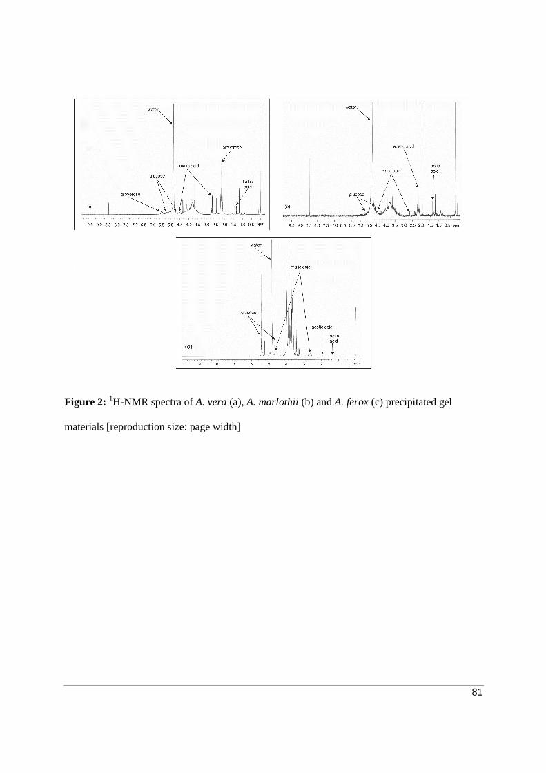
Nuclear Magnetic Resonance (1H-NMR) fingerprinting
The 1H-NMR spectra of A. vera, A. marlothii and A. ferox gel material (i.e. the precipitated
ethanol/methanol residues or polysaccharidic fraction) are given in Fig. 2a, 2b and 2c,
respectively. Aloverose (partly acetylated polymannan or acemannan), glucose and malic acid
were detected by 1H-NMR spectroscopy in the A. vera precipitated gel material. Aloverose was
not detected in the A. marlothii (Fig. 2b) and A. ferox (Fig. 2c) precipitated gel materials,
although glucose and malic acid were present.
[Insert Figure 2 here]

58
Short-term hydration study
A visual representation for the results obtained in this study with the Corneometer® and the
Visioscan® (entropy, homogeneity, energy parameters) instruments are depicted in Fig. 3a to 3d.
[Insert Figure 3 here]
As can be seen from the Corneometer® values (Fig. 3a) obtained, A. vera and A. marlothii gel
materials proved to have a larger hydrating effect than deionized water on the skin at 30 (T1), 90
(T2) and 150 (T3) min after a single application. Both these aloe materials exhibited a higher
percentage increase in skin hydration at 30 and 150 min after application and the lowest
percentage increase was observed at 90 min after application. Aloe marlothii gel material caused
a slightly higher hydration effect than A. vera gel material at 90 and 150 min after application.
Aloe ferox gel material showed a dehydrating effect (negative percentage change) over the short-
term study. This dehydrating effect of A. ferox gel material became less over time after
application. Deionized water initially dehydrated the skin at 30 and 90 min after application, but
showed a skin hydration effect at 150 min after application. This increase in skin hydration 150
min after application with deionized water was however less than that obtained with A. vera and
A. marlothii gel materials.
Fig. 3b, 3c and 3d indicate that A. marlothii gel material improved skin entropy, homogeneity
and energy to a larger extent than the A. vera and A. ferox gel materials as well as the deionized
water at 30 and 90 min after application. Aloe vera gel material improved skin entropy,
homogeneity and energy (Fig. 3b to 3d) to a larger extent than deionized water and A. ferox at 30
and 90 min after application. Aloe ferox gel material increased skin energy more than deionized
water 30 and 90 min after application.

59
The p-values revealed statistical significant effects for the treatment and the time-treatment
interaction of the Corneometer® measurements (p = 0.0001 and 0.017, respectively) and for the
homogeneity parameter (p = 0.066 and 0.084, respectively). Statistical significant effects for
treatment was found for the entropy (p = 0.036) and energy (p = 0.00001) parameter. The
significant interaction between time and treatment reveals that the effect of the treatment on the
Corneometer® measurements and the homogeneity parameter depends on time. However, it is
important to note that the significance of the interaction effect of the Corneometer®
measurements may be induced by the dominant influence of treatment given its F-value of
10.076, which is 5.6 times larger than that of time (F-value: 1.788).
Pairwise comparisons with a Bonferroni adjustment revealed no significant difference between
the levels of time for any of the skin hydration parameters investigated. Pairwise comparisons
with a Bonferroni adjustment between the different treatments revealed a statistical significant
difference between A. ferox and A. marlothii gel materials (p = 0.007), between A. ferox and A.
vera gel materials (p = 0.023) and between A. ferox and deionized water (p = 0.016) with the
Corneometer® measurements. This indicates that A. ferox gel material differed significantly
from the other aloe materials and the placebo as it showed a dehydrating effect (negative
percentage change) over the short-term study. The short-term hydrating effects of A. vera and A.
marlothii was not statistically significantly different from that of deionized water. Skin entropy
end homogeneity showed a statistical significant difference between A. ferox and A. marlothii
with p-values of 0.068 and 0.067, respectively. A statistical significant difference existed
between A. ferox and A. marlothii (p = 0.003) and between A. marlothii and deionized water (p =
0.003) for the measured energy parameter. This indicates that A. marlothii gel material
improved the general state of the skin more than deionized water.

60
Researchers previously investigated the effects of a single application and multiple applications
(1- and 2-week daily application) of formulations containing 5% w/w trilaureth-4 phosphate-
based blend supplemented with 0.10%, 0.25% or 0.50% w/w A. vera extract on the volar forearm
of volunteers. From the single application study it was seen that after an hour only the 0.50%
w/w formulation significantly increased the SC water content (measured with Corneometer® CM
825) when compared to the vehicle. At 2 and 3 h after application, the 0.25% and 0.50% w/w
formulations significantly increased the SC water content when compared to the vehicle.[5] It is
important to note that these formulations did not contain pure aloe gel materials dissolved in
water alone, but contained other excipients that may have had an interactive role in the effects
obtained on the skin.
Longer-term hydration study
Results obtained from the present multiple application study as measured by the Corneometer®
are in contrast with the results of the single application study where A. marlothii and A. vera gel
materials showed to hydrate the skin more than deionized water. Fig. 4 represents the percentage
change in skin hydration relative to the initial conditions (T0) as measured by the Corneometer®
after 1 (T1), 2 (T2), 3 (T3) and 4 (T4) weeks of treatment. From this figure it can be seen that A.
marlothii and A. vera gel materials had a predominantly dehydrating effect on the skin over the 4
week period of treatment. Aloe marlothii gel material dehydrated the skin the most of all the
aloe materials investigated from week 1 to week 4. Aloe ferox gel material showed a 1.1%
increase in skin hydration after 1 week of treatment; but thereafter also exhibited a dehydrating
effect on the skin. The dehydration effect caused by A. ferox gel material was less than caused
by the other two aloe gel materials. The placebo (i.e. deionized water), in contrast to the test
materials, increased the level of skin hydration over the 4 week time period.

61
[Insert Figure 4 here]
Investigation of the skin’s topography with the Visioscan® supported the findings obtained with
the Corneometer®. The entropy, homogeneity and energy parameters followed a similar pattern
as can be seen from Fig. 5a, 5b and 5c, respectively. The three aloe gel materials as well as the
placebo increased the skin entropy (Fig. 5a) slightly after the first week of treatment, with A.
ferox gel material showing the highest percentage increase. A decrease in skin entropy was
observed after 2, 3 and 4 weeks of treatment with the A. marlothii and A. ferox gel materials as
well as the placebo (i.e. deionized water). Aloe vera gel material showed a small percentage
increase in skin entropy after 2 weeks of treatment, thereafter also decreasing after 3 and 4 weeks
of treatment. The skin homogeneity almost followed the same pattern, except that initially A.
marlothii gel material increased this parameter the most after 1 week of treatment.
An increase in skin energy was shown with all the aloe gel materials after the first week of
treatment with A. marlothii gel material having the highest increase. Aloe vera and A. marlothii
gel materials both showed a decreasing effect on skin energy after 3 weeks of treatment, an
increase was observed after 4 weeks of treatment. Aloe ferox gel material exhibited an increase
in skin energy after 1 and 3 weeks, and a decrease in skin energy after 2 and 4 weeks of
treatment. Deionized water increased skin energy after the first week, thereafter decreasing it
after 2, 3 and 4 weeks of treatment. However, none of these differences between the treatments
were statistically significant.
[Insert Figure 5 here]
R-parameters are highly dependent on moisture content (hydration) of the skin. The R2-
parameter which indicates the gross-elasticity of the skin was found to have a negative

62
percentage change (Fig. 6a) from the baseline value for all the aloe gel materials and the placebo
indicating decrease in gross-elasticity. The lowest point in percentage decrease of the gross-
elasticity of the skin was seen after 2 weeks of treatment with the various aloe gel materials.
Aloe marlothii gel material showed the largest negative effect on the skin’s gross elasticity (R2)
when compared to the other aloes and the placebo. This correlates with the Corneometer®
values, which indicated that A. marlothii gel material had the most dehydrating effect on the
skin.
[Insert Figure 6 here]
The R6-parameter (Fig. 6b) measures the stretch capacity of the skin and negative values reflect
improved skin condition.[27] Fig. 6b shows an upward curve (positive percentage change) for all
the treatments after the first 2 weeks, thereafter the trend becomes downward. Therefore, the
R6-parameter correlates with the other R-parameters in showing that skin conditions did not
improve (positive percentage change). The only negative percentage change (indicating
improved skin conditions) is after 1 week and 4 weeks of treatment with A. vera, 1 week of
treatment with A. marlothii and after 4 weeks of treatment with A. ferox. Noteworthy is that the
placebo showed the highest percentage positive change, thus showing the least improvement of
the R6-parameter.
The R7-parameter seen in Fig. 6c showed a decrease in the elastic portion of the skin with the
highest percentage decrease after 2 weeks of treatment. Negative values in R7 reflect a decrease
in biological elasticity.[27] The complete relaxation (R8) of the skin followed almost the same
pattern as R7, thus also indicating a decrease in skin elasticity. Aloe marlothii proved to have the
most negative effect on these parameters after 2, 3 and 4 weeks of treatment. These results

63
support the findings of the Corneometer® that A. marlothii dehydrated the skin the most as it had
the most negative effect on the elasticity parameters. However, statistically the Cutometer®
results indicate that none of the treatments significantly altered the R2-, R6-, R7- and R8-
parameters.
The p-values obtained revealed statistical significant differences between the treatments when
investigated with the Corneometer® (p = 0.001). The homogeneity parameter and the stretch
capacity (R6) of the skin showed a significant difference between the times of treatment with p-
values of 0.045 and 0.091, respectively. The gross-elasticity (R2) and complete relaxation (R7)
of the skin revealed statistical significant differences for the interaction between time and
treatment with p-values of 0.009 and 0.032, respectively. Statistical significant effects for time
(p = 0.067) and interaction between time and treatment (p = 0.074) was observed for R8.
Pairwise comparisons with a Bonferroni adjustment between the levels of time revealed that the
time of treatment had no statistical significance, except for the homogeneity parameter where
statistical significant difference was seen between 1 week and 4 weeks (p = 0.037) of treatment.
Pairwise comparisons with a Bonferroni adjustment between the different treatments revealed
statistical significant differences in the Corneometer® measurements between the placebo and A.
ferox gel material (p = 0.003), A. marlothii gel material (p = 0.001) and A. vera gel material (p =
0.007) gel materials. Thus deionized water was statistically better than the aloes in improving
skin hydration.
A previous study found that A. vera extract moisturized the skin by significantly increasing the
SC water content whilst not changing the TEWL.[5] It was suggested that this could be due to the
rich composition of the A. vera extract of hygroscope mono- and polysaccharides[5, 34] and the
64
presence of the amino acids alanine, arginine, glycine, histidine, serine and threonine which
contributes to SC hydration.[5, 35] Dal’Belo et al.[5] found that relatively low A. vera extract
containing formulations (i.e. 0.10%, 0.25% and 0.50% w/w) increased skin hydration after 1 and
2 weeks of application compared to the vehicle alone. However, when the formulations were
compared with each other after 2 weeks of application the formulation containing 0.50% w/w A.
vera extract proved to be significantly better than the 0.10% and 0.25% formulations.
The findings of this longer-term, multiple application study are different from other studies
described above, which may be ascribed to differences in the composition of the formulations
tested (pure aloe gel materials dissolved in water in this study versus formulations containing
excipients in other studies), but also due to composition of the aloe gel from different species.[36]
Humectants promote the retention of water within the SC by attracting water from the outside in
(from environment to skin) and from the inside out (from dermis to epidermis/SC).[37] Although
it was not the aim of this study to determine the mechanism of skin dehydration by the aloe
materials, a possible explanation is given here that should be further investigated in future
studies. The dehydration of the skin by the aloe gel materials after multiple applications may be
attributed to attracting water not only from the dermal/epidermal layers of the skin but also from
the SC. The dehydration effect therefore potentially occurred due to absorption of moisture from
the skin into the aloe gel layer that was applied to the skin which dried by evaporation of the
water in which the gel materials were dissolved after application to the skin.
The external use of aloe gel on intact skin is not associated with adverse reactions and is
generally regarded as safe.[38] This is in accordance with the findings in other studies, although
final conclusions on general safety in one of these studies could not be made due to the rarity of
an allergy after a single application.[14] In contrast to this, case studies on the topical application

65
of aloe-derived products showed some adverse reactions which included contact urticaria,
dermatitis and acute eczema.[39] It has been suggested that the adverse effects (such as
hypersensitivity) may be due to the presence of apoptosis-inducing antrhaquinones in A. ferox.[40]
In the present study, two volunteers were withdrawn from the study due to severe allergic
reactions. From weekly questionnaires it was determined that 11 (18.64 %), 14 (23.73 %) and 2
(3.39 %) out of the 59 volunteers reported a mild allergic reaction after topical application of A.
vera, A. marlothii and A. ferox gel materials, respectively. The volunteers experienced a red
rash, especially when the aloe gel started to dry on the skin, with either a burning or itching
sensation.
Erythema study
Various tests have been used to examine the anti-inflammatory efficacy of aloe gel or its various
components and generally involve some kind of intentional wounding[4] as the first step in wound
healing is anti-inflammation.[12]
In the present study, the SLS exposure test was performed to compare the anti-erythema efficacy
of the aloe gel materials to the positive control group (hydrocortisone gel). The percentage
change in skin erythema as expressed by hemoglobin content from irritation (T1) to two time
intervals (T2 and T3) after treatment with test materials are given in Table 2.
[Insert Table 2 here]
Hydrocortisone gel showed a 13.1% decrease in erythema at T2; followed by deionized water
(7.8%) and A. ferox (7.0%), while A. vera and A. marlothii demonstrated the lowest percentage
decrease in erythema at T2. However, no statistical significant differences were obtained

66
between hydrocortisone gel, A. ferox gel material, deionized water and the untreated irritated
skin in terms of anti-erythema effect at T2. Hydrocortisone gel performed statistically
significantly better than A. vera and A. marlothii gel materials in reducing skin erythema. Aloe
ferox gel material statistically significantly reduced erythema to a larger extent than A. marlothii
gel material at T2.
At T3 (on the 7th day after 6 days of treatment), hydrocortisone gel decreased erythema by 18.8%
followed by A. vera (17.0%), A. ferox (15.2%), and A. marlothii (9.0%). The anti-erythema
effect of A. vera gel material and hydrocortisone gel was statistically significantly higher than
that of A. marlothii gel material with p-values of 0.051 and 0.046 respectively. Deionized water
and untreated irritated skin showed a similar decrease in skin erythema at T3 with a percentage
decrease of 13.0% and 13.1%, respectively.
Aloe marlothii gel material decreased erythema less than deionized water and untreated irritated
skin, although this was not statistically significant. A statistically significant difference (p =
0.0196) was observed between T2 and T3 when treated with A. vera gel material, indicating its
anti-erythema effect is time dependent. This correlates with a previous study where a lag phase
was observed and the onset of A. vera gel’s anti-inflammatory activity was found to be
delayed.[14]
The differences obtained in the anti-erythema results for the different species of aloe could be
explained by differences in their chemical compositions as confirmed with 1H-NMR.

67
CONCLUSION
The clinical significance of the Corneometer® measurements, which are regarded as the most
important indicator of skin hydration, indicates that A. vera and A. marlothii gel material did
improve the hydration of the skin after a single application, although it was not statistically
significantly different from the placebo (i.e. deionized water). Aloe ferox gel materials
dehydrated the skin after a single application when compared to the other aloe gel materials and
the placebo. After multiple applications, all the aloe gel materials had a dehydrating effect on
the skin as opposed to deionized water, which significantly improved skin hydration.
Mexameter® readings showed that A. vera and A. ferox gel materials were similar in their
erythema reducing effects after 6 days of treatment to that of hydrocortisone gel. The anti-
erythema effect of A. vera gel material was found to be dependent on time as there was a
statistical significant difference between the second day of treatment and seventh day of
treatment. Aloe marlothii gel material dehydrated the skin to the largest extend during the
longer-term study and was less effective than deionized water and untreated irritated skin in
decreasing erythema. It also caused the highest number of mild skin reactions in the volunteers.
While this study has elucidated the effects of gel materials from different aloe species on skin
hydration and erythema in human subjects, the mechanisms of action should be investigated in
future studies.

68
ACKNOWLEDGEMENTS
The authors would like to thank Organic Aloe for donation of the A. ferox material. Ms. C.
Potgieter is acknowledged for assistance with the skin measurements and Ms. M. van Reenen for
the statistical data analysis. This work was carried out with the financial support of the National
Research Foundation of South Africa, the Medical Research Council of South Africa and the
Centre of Excellence for Pharmaceutical Sciences of the North-West University, Potchefstroom
Campus, South Africa.

69
DISCLAIMER
Any opinion, findings and conclusions or recommendations expressed in this material are those
of the authors and therefore the NRF do not accept any liability in regard thereto.
70
REFERENCES
1. Tarirai C, Viljoen AM, Hamman JH. Herb-drug pharmacokinetic interactions reviewed.
Expert Opin Drug Metab Toxicol. 2010; 6: 1515-38.
2. Loots D, Van Der Westhuizen FH, Botes L. Aloe ferox leaf gel phytochemical content,
antioxidant capacity and possible health benefits. J Agric Food Chem. 2007; 55: 6891-6.
3. Morton JF. Folk uses and commercial exploitation of Aloe leaf pulp. Econ Bot. 1961;
15: 311-9.
4. Reynolds T, Dweck AC. Aloe vera leaf gel: a review update. J Ethnopharmacol. 1999;
68: 3-37.
5. Dal’Belo SE, Gaspar LR, Maia Campos PM. Moisturizing effect of cosmetic
formulations containing Aloe vera extract in different concentrations assessed by skin
bioengineering techniques. Skin Res Technol. 2006; 12: 241-46.
6. Steenkamp V, Stewart MJ. Medicinal applications and toxicological activities of Aloe
products. Pharm Biol. 2007; 45: 411-20.
7. Eshun K, He Q. Aloe vera: a valuable ingredient for the food, pharmaceutical and
cosmetic industries – a review. Crit Rev Food Sci Nutr. 2004; 44: 91-6.
8. Lee SK. Overview of Aloe study. In: Park YI, Lee SK, editors. New perspectives on
Aloe. New York: Springer; 2006. p. 1-5.
9. Choi S-W, Son B-W, Son Y-S, Park Y-I, Lee SK, Chung A-H. The wound-healing effect
of a glycoprotein fraction isolated from Aloe vera. Br J Dermatol. 2001; 145: 535-45.

71
10. Kim YS. Carbohydrates. In: Park YI, Lee SK, editors. New perspectives on Aloe. New
York: Springer; 2006. p. 57-62.
11. McDaniel HR, McAnally BH. Evaluation of poymannoacetate (Carrisyn) in the
treatment of AIDS. Clin Res. 1987; 35: 483A.
12. Jia Y, Zhao G, Jia J. Preliminary evaluation: the effects of Aloe ferox Miller and Aloe
arborescens Miller on wound healing. J Ethnopharmacol. 2008; 120: 181-9.
13. Meadows TP. Aloe as a humectants in new skin preparations. Cosmet Toiletries. 1980;
95: 51-6.
14. Reuter J, Jocher A, Stump J, Grossjohann B, Franke G, Schempp, CM. Investigation of
the anti-inflammatory potential of Aloe vera gel (97.5%) in the ultraviolet erythema test.
Skin Pharmacol Physiol. 2008; 21: 106-110.
15. Bazin R, Fanchon C. Equivalence of face and volar forearm for the testing of
moisturizing and firming effect of cosmetics in hydration and biomechanical studies. Int
J Cosmet Sci. 2006; 28: 453-60.
16. Darlenski R, Fluhr JW. Moisturizers and emollients. In: Fluhr JW, editor. Practical
aspects of cosmetic testing. Berlin Heidelberg: Springer-Verlag; 2011: p. 123-41.
17. Darlenski R, Sassning S, Tsankov N, Fluhr JW. Non-invasive in vivo methods for
investigation of the skin barrier physical properties. Eur J Pharm Biopharm. 2009; 72:
295-303.
72
18. Gu W, Song H, Wen X, Wang Y, Xia W, Fang Y. Binding interaction between aloe
polysaccharide and alizarin red by spectrophotometry and its analytical application.
Carbohyd Polym. 2010; 80: 115-22.
19. Campestrini LH, Silveira JLM, Duarte MER, Koop HS, Noseda MD. NMR and
rheological study of Aloe barbadensis partially acetylated glucomannan. Carbohydr
Polym. 2013; 94: 511-19.
20. Ramachandra CT, Rao PS. Processing of Aloe vera leaf gel: a review. Am J Agr Biol
Sci. 2008; 3: 502-510.
21. Berardesca E. Factors influencing measurements. In: Fluhr JW, editor. Practical aspects
of cosmetic testing. Berlin Heidelberg: Springer-Verlag; 2011. p. 89-99.
22. Berardesca E. EEMCO guidance for the assessment of stratum corneum hydration:
electrical methods. Skin Res Technol. 1997; 3: 126-32.
23. Courage and Khazaka electronic GmbH. Corneometer® CM825: Information and
operating instruction [homepage on the internet]. 2010 [cited 2012 Oct 15]. Available
from http://www.courage-khazaka.com/index.php/en/all-downloads/downloads-
en/file/244-anleit-cm-sonde-e
24. Courage and Khazaka electronic GmbH. Visioscan® VC 98: Information and operating
instruction [homepage on the internet]. 2009 [cited 2012 Oct 15]. Available from
http://www.courage-khazaka.com/index.php/en/all-downloads/downloads-en/file/197-
anleitvc98e

73
25. Courage and Khazaka electronic GmbH. Cutometer® dual MPA 580: Information and
operating instruction [homepage on the internet]. 2012 [cited 2012 Oct 15]. Available
from http://www.courage-khazaka.com/index.php/en/all-downloads/downloads-
en/file/239-anleit-cutometer-dual-mpa-580-e
26. Dobrev H. Use of cutometer to assess epidermal hydration. Skin Res Technol. 2000; 6:
239-44.
27. Berndt U, Elsner P. Hardware and measuring principle: the Cutometer. In: Elsner P,
Berardesca E, Wilhelm K-P, Maibach H.I, editors .Bioengineering of the skin: skin
biomechanics. Boca Raton: CRC Press; 2002. p. 91-7.
28. Tupker RA, Willis C, Berardesca, E, Lee CH, Fartasch M, Agner T, et al. Guidelines on
sodium lauryl sulfate (SLS) exposure test – a report from the Standardization Group of
the European Society of Contact Dermatitis. Contact Dermatitis. 1997; 37: 53-69.
29. Gloor M, Senger B, Langenauer M, Fluhr JW. On the course of the irritant reaction after
irritation with sodium lauryl sulfate. Skin Res Technol. 2004; 10: 144-8.
30. Arsic I, Zugic A, Tadic V Tasic-Kostov M, Misic D, Primorac M, et al. Estimation of
dermatological application of creams with St. John’s Wort oil extracts. Molecules. 2012;
17: 275-94.
31. Courage and Khazaka electronic GmbH. The Mexameter® MX 18: Information and
operating instruction [homepage on the internet]. 2012 [cited 2012 Oct 15]. Available
from http://www.courage-khazaka.com/index.php/en/all-downloads/downloads-
en/file/246-anleit-mx-sonde-e
74
32. SPSS Inc. IBM SPSS Statistics for Windows, Version 20, Release 20.0.0 [homepage on
internet]. 2011 [cited 2012 Jan 11]. Available from http://www-
01.ibm.com/software/analytics/spss/
33. Seltman, H.J. Experimental Design and Analysis [homepage on internet]. 2012 [cited
2013 Jan 16]. Available from http://www.stat.cmu.edu/_hseltman/309/Book/Book.pdf
34. Femenia A, Sanchez ES, Simal S, Rosello C. Compositional features of polysaccharides
from Aloe vera (Aloe barbadensis Miller) plant tissues. Carbohydr Polym. 1999; 39:
109-17.
35. Rieger M. Skin constituents as cosmetic ingredients. Cosm Toil. 1992; 107: 85-94.
36. Capasso F, Borrelli F, Capasso R, Di Carlo G, Izzo AA, Pinto L, et al. Aloe and its
therapeutic use. Phytother Res. 1998; 12: S124-27.
37. Rawlings AV, Canestrari DA, Dobkowski B. Moisturizer technology versus clinical
performance. Dermatol Ther. 2004; 17: 49-56.
38. Poppenga RH. Herbal medicine: potential for intoxication and interactions with
conventional drugs. Clin Tech Small An P. 2002; 17: 6-18.
39. Cosmetic Ingredient Review Expert Panel. Final report on the safety assessment of Aloe
andongensis extract, Aloe andongensis leaf juice, Aloe arborescens leaf extract, Aloe
arborescens leaf juice, Aloe arborescens leaf protoplasts, Aloe barbadensis flower
extract, Aloe barbadensis leaf, Aloe barbadensis leaf extract, Aloe barbadensis leaf juice,
Aloe barbadensis leaf polysaccharides, Aloe barbadensis leaf water, Aloe ferox leaf

75
extract, Aloe ferox leaf juice, and Aloe ferox leaf juice extract. Int J Toxicol. 2007; 26: 1-
50.
40. Chen W, Van Wyk BE, Vermaak I, Viljoen AM. Cape aloes – a review of the
phytochemistry, pharmacology and commercialization of Aloe ferox. Phytochem Lett.
2012; 5: 1-12.

76
Table legends
Table 1: Hydrocortisone gel formulation (positive control group)
Table 2: Percentage change in skin erythema (hemoglobin) from irritation (T1) to two time
intervals (T2 and T3) after treatment

77
Table 1: Hydrocortisone gel formulation (positive control group)
Components Concentration
Hydrocortisone acetate 1% w/v
Ethanol (96% v/v) 15% v/v
Polyethylene glycol 15% w/v
Carbopol Ultrez 20 1% w/v
Distilled water up to Up to 100% of preparation
Tri-ethanol amine Enough to adjust pH to approximately 6.8

78
Table 2: Percentage change in skin erythema (hemoglobin) from irritation (T1) to two time
intervals (T2 and T3) after treatment
Treatment T2 T3
Irritated -4.5 ± 21.3 -13.1 ± 18.3
Aloe vera -1.8 ± 19.4 -17.0 ± 19.1
Aloe marlothii -1.8 ± 19.3 -9.0 ± 21.2
Aloe ferox -7.0 ± 14.3 -15.2 ± 20.4
Deionized water -7.8 ± 15.7 -13.0 ± 26.6
Hydrocortisone -13.1 ± 21.6 -18.8 ± 26.0
79
Figure legends
Figure 1: A typical skin deformation curve obtained with the Cutometer®, which is similar to
previously reported curves (26, 27) [reproduction size: column width]
Figure 2: 1H-NMR spectra of A. vera (a), A. marlothii (b) and A. ferox (c) precipitated gel
materials [reproduction size: page width]
Figure 3: Percentage change measured by (a) the Corneometer®, (b) Visioscan® entropy, (c)
Visioscan® homogeneity and (d) Visioscan® energy measurements [reproduction size: page
width]
Figure 4: Percentage change in skin hydration relative to initial conditions (T0) as measured with
the Corneometer® [reproduction size: column width]
Figure 5: Percentage change relative to initial conditions (T0) as determined with the (a)
Visioscan® entropy, (b) Visioscan® homogeneity and (c) the Visioscan® energy [reproduction
size: page width]
Figure 6: Percentage change relative to initial conditions (T0) for (a) the Cutometer® R2, (b)
Cutometer® R6, (c) Cutometer® R7 and the (d) Cutometer® R8 [reproduction size: page width]

80
Figure 1: A typical skin deformation curve obtained with the Cutometer®, which is similar to
previously reported curves (26, 27) [reproduction size: column width]
81
Figure 2: 1H-NMR spectra of A. vera (a), A. marlothii (b) and A. ferox (c) precipitated gel
materials [reproduction size: page width]

82
Figure 3: Percentage change measured by (a) the Corneometer®, (b) Visioscan® entropy, (c)
Visioscan® homogeneity and (d) Visioscan® energy measurements [reproduction size: page
width]

83
Figure 4: Percentage change in skin hydration relative to initial conditions (T0) as measured with
the Corneometer® [reproduction size: column width]

84
Figure 5: Percentage change relative to initial conditions (T0) as determined with the (a)
Visioscan® entropy, (b) Visioscan® homogeneity and (c) the Visioscan® energy [reproduction
size: page width]

85
Figure 6: Percentage change relative to initial conditions (T0) for (a) the Cutometer® R2, (b)
Cutometer® R6, (c) Cutometer® R7 and the (d) Cutometer® R8 [reproduction size: page width]